Primary conjunctival amyloidosis mimicking Parinaud's syndrome
ABSTRACT
Amyloidosis is characterized by the extracellular deposition of amyloid protein fibrils in tissues, including the conjunctiva, due to their insolubility in proteolytic enzymes. Conjunctival amyloidosis, particularly involving the eyelid conjunctiva, is a rare clinical entity. We report the case of a 60-year-old Caucasian female who presented with a unilateral lesion in the inferior bulbar conjunctiva, extending to the entire inferior tarsal region and fornix. The lesion appeared as an extensive, elevated, pinkish-yellowish nodular mass with poorly defined borders. An incisional biopsy was performed, and histopathology confirmed the diagnosis of conjunctival amyloidosis. Management of conjunctival amyloidosis depends on the extent of local involvement and the patient's systemic condition, ranging from symptomatic treatment, such as the use of lubricants, to surgical excision. In most cases, complete excision is not feasible due to extensive lesion infiltration, as seen in our case, where a conservative approach was adopted.
Keywords:
Amyloidosis; Conjunctiva
RESUMO
A amiloidose é caracterizada pelo depósito extracelular de fibrilas de proteína amiloide nos tecidos, incluindo a conjuntiva, devido à sua insolubilidade em enzimas proteolíticas. A amiloidose conjuntival, particularmente quando envolve a conjuntiva palpebral, é uma entidade clínica rara. Relatamos o caso de uma paciente do sexo feminino, caucasiana, de 60 anos, que apresentou lesão unilateral na conjuntiva bulbar inferior, estendendo-se por toda a região tarsal inferior e fórnice. A lesão apresentava-se como uma massa nodular extensa, elevada, de coloração rosada-amarelada, com bordos mal definidos. Foi realizada biópsia incisional, e o exame histopatológico confirmou o diagnóstico de amiloidose conjuntival. O manejo da amiloidose conjuntival depende da extensão do envolvimento local e das condições sistêmicas do paciente, podendo variar desde o tratamento sintomático, como o uso de lubrificantes, até a excisão cirúrgica. Na maioria dos casos, a excisão completa não é viável devido à infiltração extensa da lesão, como observado no presente caso, em que se adotou uma conduta conservadora.
Descritores:
Amiloidose; Conjuntiva
INTRODUCTION
Amyloidosis describes a group of rare diseases caused by protein conformation abnormalities resulting in extracellular deposition and accumulation of insoluble fibrillary aggregates. Currently, 36 amyloid precursor proteins have been identified, each associated with a distinct disease entity.(1,2)
Amyloid can accumulate in various organs, including the liver, spleen, kidneys, heart, nerves, and blood vessels, leading to a range of clinical syndromes. These include cardiomyopathy, hepatomegaly, proteinuria, macroglossia, autonomic dysfunction, ecchymoses, neuropathy, renal failure, hypertension, and ocular abnormalities, which may affect the conjunctiva, orbit, eyelid, cornea, trabecular meshwork, retina, and vitreous.(1,3)
Conjunctival amyloidosis is a rare condition, with only a few reports of localized AL amyloidosis of the conjunctiva, typically without evidence of systemic involvement. However, conjunctival amyloidosis can sometimes indicate systemic disease early, making a thorough systemic investigation essential.(1,4)
This report describes a case of localized conjunctival amyloidosis affecting the tarsal and inferior bulbar conjunctiva, along with its diagnosis and treatment approach.
CASE REPORT
A 60-year-old Caucasian female presented to the ophthalmology clinic at Hospital de Olhos Paulista/Vision One in July 2020, reporting swelling in the lower eyelid of her right eye, which had begun four months before the consultation. The patient had no significant personal or ophthalmological history. She mentioned having four cats, one of which had an immunodeficiency.
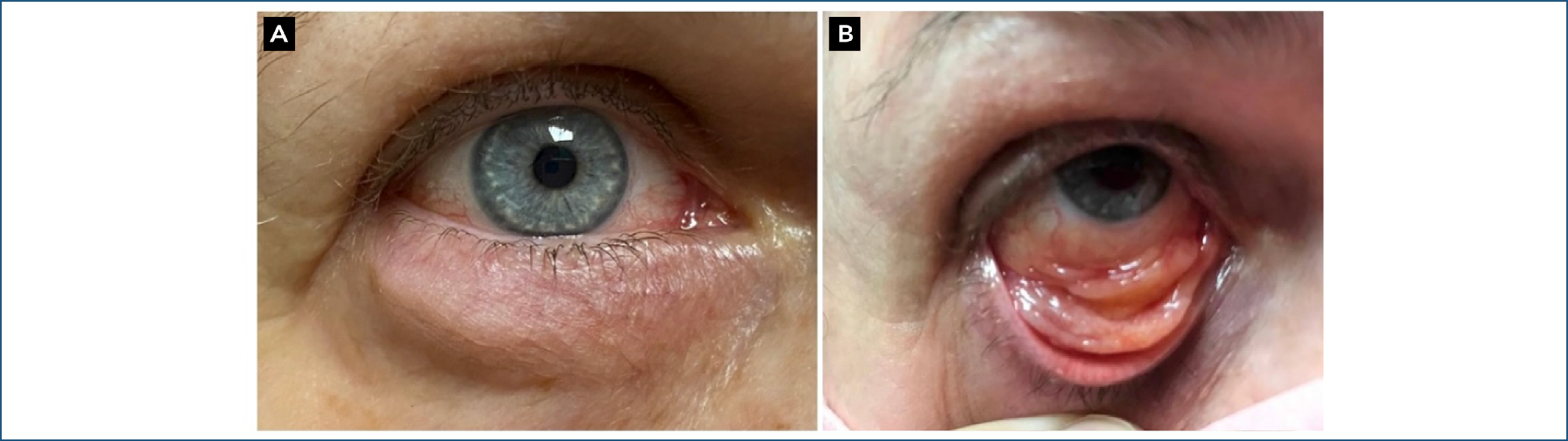
The patient reported ocular surface symptoms, including a foreign body sensation and burning. Ectoscopy revealed visible lower eyelid edema (Figure 1A). Biomicroscopy showed an extensive elevated nodular mass with a pinkish-yellow hue and poorly defined borders, affecting the entire inferior tarsal region, lower bulbar fornix, and conjunctiva (Figure 1B). The cornea was clear, with a tear film breakup time of four seconds, and the lens was transparent. The left eye presented without alterations. Visual acuity was 20/20 with correction in both eyes. Fundoscopy and extraocular motility examinations showed no abnormalities.
(A) Ectoscopy with apparent eyelid edema; (B) extensive elevated pinkish-yellowish nodular mass, with imprecise borders, affecting the entire inferior tarsal region, fornix, and inferior bulbar conjunctiva.
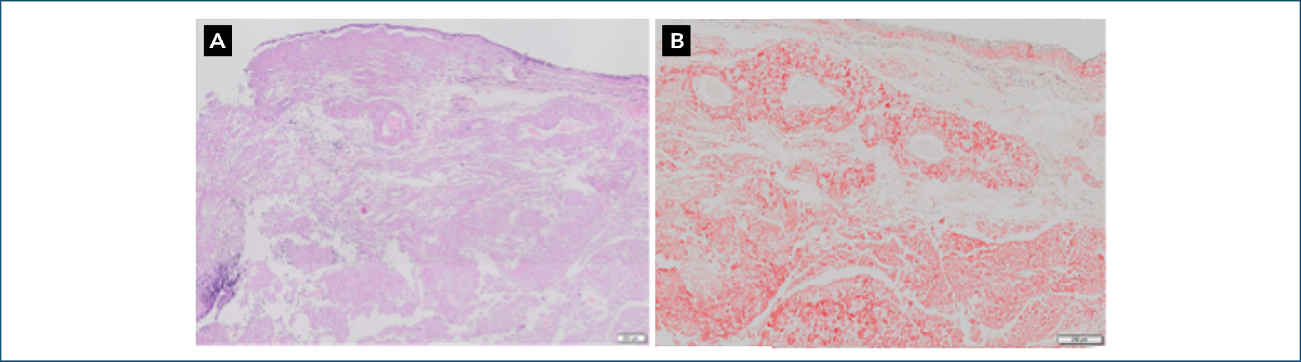
Given the appearance of the conjunctival mass, its predominant location in the tarsal region, and the patient's epidemiological history, treatment for cat-scratch disease (CSD) was initiated with doxycycline 100 mg every 12 hours, along with lubricants to address the surface symptoms. However, there was no improvement in the conjunctival mass following treatment. A conjunctival biopsy was then performed to differentiate the condition from lymphoproliferative diseases. Under local anesthesia, a 1.1 x 0.3 x 0.2 cm fragment of the tarsal conjunctiva was excised, placed in 10% formalin, and sent for histopathological analysis. Examination of the slides revealed amyloid material deposits, confirmed by Congo red staining, consistent with a diagnosis of conjunctival amyloidosis (Figures 2A and 2B).

(A) Hematoxylin-eosin – 4x. (B) -Congo red – 10x. Histological findings compatible with extensive deposition of amorphous and eosinophilic material, Congo red positive, characteristic of amyloid material, in the conjunctival mucosal chorion, with scarce focal lymphoplasmacytic infiltrate intermingled.
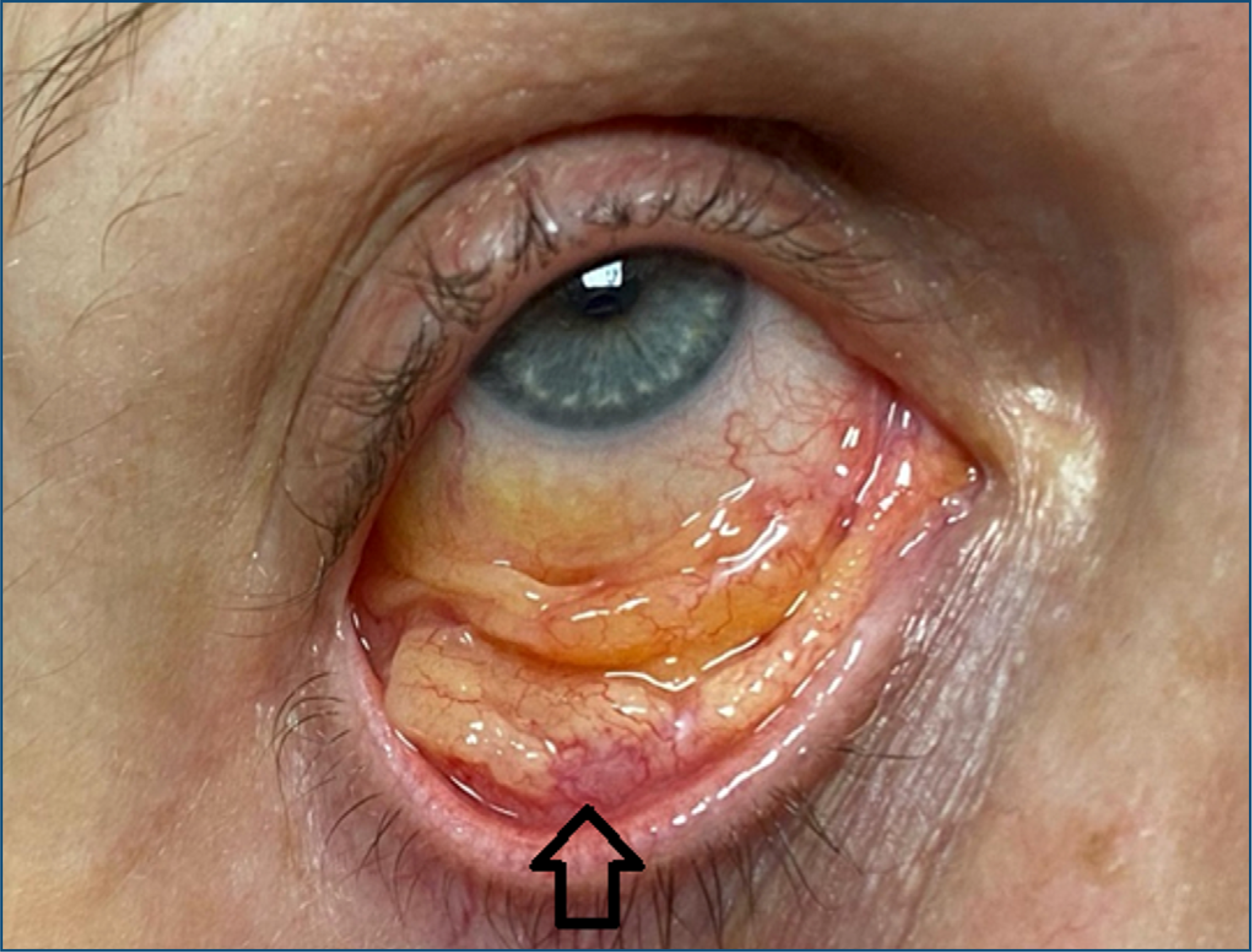
Since the patient's symptoms were related to the ocular surface, we chose expectant management with semiannual follow-ups and referred the patient to a hematologist for a systemic evaluation. The systemic assessment revealed no abnormalities, with no evidence of extraocular amyloidosis. Consequently, the diagnosis was confirmed as primary conjunctival amyloidosis. After a 4-year follow-up, the lesion retains the same appearance and size (Figure 3).
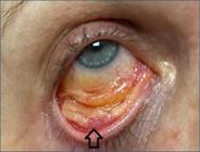
After 4 years of follow-up, the lesion shows the same appearance and size. The arrow shows the biopsy region.
DISCUSSION
This case reports a rare primary conjunctival amyloidosis in a 60-year-old female who presented with unilateral involvement of the inferior conjunctiva, with lesions affecting the bulbar, tarsal, and fornix.
Amyloidosis typically affects middle-aged individuals, and it can also occur in both children and adults. Conjunctival amyloidosis, in particular, is an uncommon condition, and its exact incidence and prevalence still need to be clarified due to its rarity and the absence of specific epidemiological studies. Clinically, conjunctival amyloidosis may present as yellowish or pink-yellow masses, often accompanied by recurrent subconjunctival hemorrhages and intrinsic vascularization. In some cases, eyelid ptosis may be associated, particularly when the palpebral conjunctiva is involved.(5)
Amyloidosis can be hereditary or acquired and may present as a primary or secondary disease, manifesting as a localized or systemic condition. The most common forms of systemic amyloidosis are immunoglobulin light chain (AL) amyloidosis, and amyloid A (AA) amyloidosis.(2) AL amyloidosis is a benign, light-chain, low-grade monoclonal gammopathy involving multiple organs and tissues. This may be associated with plasma cell dyscrasias (monoclonal gammopathy of undetermined significance, multiple myeloma, and Waldenström's macroglobulinemia.(6) By contrast, reactive systemic AA amyloidosis is commonly seen in patients with chronic inflammation, such as those with rheumatologic diseases, infections, or autoinflammatory disorders. It results from the overproduction of acute-phase serum AA protein, which can accumulate and form deposits in various tissues.(7)
Localized amyloidosis is most commonly caused by the deposition of monoclonal immunoglobulin light chains (kappa or lambda) produced by a benign B-cell clone or plasma cells, as seen in AL amyloidosis. The exact reason for localized deposition remains unclear, but it is hypothesized that local protein synthesis may be responsible rather than the deposition of light chains from distant sources. Although conjunctival amyloidosis is typically localized, ocular involvement can sometimes be the first indication of systemic disease.(6,7)
Amyloid protein deposition may or may not be symptomatic, typically presenting with unilateral eyelid edema, irritation, and subconjunctival hemorrhage. Depending on the location and extent of the involvement, patients may also experience pain, ecchymosis, ptosis, and ectropion. Subconjunctival hemorrhage may be recurrent, likely due to amyloid deposits within the walls of blood vessels. The most common site of involvement is the inferior bulbar conjunctiva; however, the eyelids, lacrimal glands, and extraocular muscles may also be affected. In the present case, the lesions were located in the bulbar conjunctiva, tarsal region, and fornix, with the patient's symptoms primarily limited to ocular surface discomfort, such as foreign body sensation and burning.(5,8)
The clinical identification of conjunctival amyloidosis can be challenging due to its rarity and varied symptoms. Diagnosis typically requires a combination of ophthalmologic examinations and histopathological confirmation of amyloid deposits via biopsy. Congo red staining is commonly employed to detect amyloid, which appears as red-stained deposits under polarized light. Additionally, amyloid protein exhibits an amorphous eosinophilic appearance on hematoxylin and eosin staining.(3,5,8)
Given the potential systemic nature of some forms of amyloidosis, it is essential to refer patients for a comprehensive systemic evaluation by a hematologist. In this case, the patient's systemic workup revealed no abnormalities or evidence of extraocular amyloidosis. Consequently, the diagnosis was confirmed as primary conjunctival amyloidosis.
Treating ocular amyloidosis depends on the severity and extent of the ocular involvement. In cases of localized amyloid deposition, the primary approach is surgical excision of the lesions. However, complete excision is often impossible, so the treatment focuses on relieving symptoms, restoring function, and maintaining ocular mobility. Extensive conjunctival infiltration can be especially challenging to manage, and supportive care is frequently the only available option. In the present case, since the patient's symptoms were primarily related to the ocular surface, we chose an expectant management with semiannual follow-up.(6)
The patient has been followed up for four years without evidence of conjunctival amyloidosis progression, so an expectant follow-up approach was recommended. The stability of the condition suggests that there is no immediate need for therapeutic intervention. Periodic monitoring is advised to assess any changes in clinical presentation and to reconsider the need for surgical treatment if necessary.(6)
Conjunctival sarcoidosis, CSD, conjunctival lymphoma, conjunctival amyloidosis, and conjunctival basal cell (CBC) carcinoma require specific diagnostic approaches. Sarcoidosis typically presents as asymptomatic conjunctival nodules, confirmed by biopsy showing non-caseating granulomas. Cat-scratch disease often manifests as Parinaud's oculoglandular syndrome, characterized by conjunctivitis, regional lymphadenopathy, and occasionally neuro retinitis or optic neuritis. Conjunctival lymphoma is a mobile, painless, pink or salmon-colored mass and may be associated with systemic symptoms if the disease has spread.(6,9)
Conjunctival amyloidosis and CBC are rare but differ in presentation and management. Amyloidosis is marked by yellowish or pink masses, often with recurrent subconjunctival hemorrhages and intrinsic vascularization. Conjunctival basal cell, however, appears as a nodular or ulcerated lesion, which may be pigmented. While amyloidosis is confirmed by Congo red staining, CBC requires surgical excision for diagnosis and treatment.(9,10)
The diagnostic process and management of these conditions depend on their systemic involvement. Sarcoidosis and amyloidosis may have systemic manifestations, requiring a broader evaluation, whereas lymphoma can also be systemic. CBC is typically localized but can be locally invasive. The need for tailored diagnostic strategies is essential, with sarcoidosis requiring systemic evaluation, amyloidosis confirmed via biopsy, CSD diagnosed through serological testing for Bartonella hanselae, lymphoma through histopathology and immunohistochemistry, and CBC treated through surgical excision.(6,9)
Conjunctival amyloidosis is a rare condition with diagnostic challenges due to its similarity to other ocular diseases. A multidisciplinary approach, including biopsy and histopathological analysis, is crucial for accurate diagnosis. Surgical excision remains the primary treatment in localized cases, but complete removal is often impossible. In this case, an expectant approach with periodic follow-up was chosen. After four years of stable disease without progression, this approach has proven to be an appropriate management strategy.(6,9)
Data Availability Statement:
The datasets generated and/or analyzed during the current study are included in the manuscript.
REFERENCES
- 1 Minnella AM, Rissotto R, Antoniazzi E, Di Girolamo M, Luigetti M, Maceroni M, et al. Ocular Involvement in Hereditary Amyloidosis. Genes (Basel). 2021;12(7):955.
- 2 Wechalekar AD, Gillmore JD, Hawkins PN. Systemic amyloidosis. Lancet. 2016;387:2641-54.
- 3 Ali Z, Fernando B. A rare case of amyloidosis of the eyelid and conjunctiva. Case Rep Ophthalmol Med. 2016;2016:2915196.
- 4 Mora-Horna ER, Rojas-Padilla R, Lopez VG, Guzman MJ, Ceriotto A, Salcedo G. Ocular adnexal and orbital amyloidosis: a case series and literature review. Int Ophthalmol. 2016;36:281-98
- 5 Prager AJ, Habib LA, Gambogi T, Busam KJ, Marra BP. Long-Term Follow-Up of 4 Patients with Conjunctival Amyloidosis. Ocul Oncol Pathol. 2018;4(5):313-7.
- 6 Leibovitch I, Selva D, Goldberg RA, Sullivan TJ, Saeed P, Davis G, et al. Periocular and orbital amyloidosis: clinical characteristics, management, and outcome. Ophthalmology. 2006;113:1657–1664.
- 7 Hazenberg BP, van Gameren II, Bijzet J, Jager PL, van Rijswijk MH. Diagnostic and therapeutic approach of systemic amyloidosis. Neth J Med. 2004;62:121-8.
- 8 Demirci H, Shields CL, Eagle Júnior RC, Shields JA. Conjunctival Amyloidosis: Report of Six Cases and Review of the Literature. Survey of Ophthalmology. 51(4):419-33.
- 9 Shields CL, Shields JA, Carvalho C, Rundle P, Smith AF. Conjunctival lymphoid tumors: clinical analysis of 117 cases and relationship to systemic lymphoma. Ophthalmology. 2001;108(5):979-84.
- 10 Meduri A, Rechichi M, Mazzotta C, Scalinci SZ, Jaroudi MO. Ectropion and conjunctival mass in a patient with primary bilateral conjunctival amyloidosis. Case Rep Ophthalmol Med. 2016;2016:5610753.
Edited by
-
Associate editor:
Ricardo Augusto Paletta Guedes Universidade Federal de Juiz de Fora, Juiz de Fora, MG, Brazil https://orcid.org/0000-0002-9451-738X