 Insights about use of p57 in differentiation between complete and partial hydatidiform mole: a scoping review
Abstract
Objective To assess the utility of p57 in differentiating histological subtypes of hydatidiform mole and its applicability in clinical practice.
Methods A scoping review was conducted following the Joanna Briggs Institute methodology and PRISMA-ScR guidelines. The article search was conducted in the PubMed, EMBASE, BVS, Cochrane, Web of Science, and Scopus databases. Studies published between 2015 and 2025 that evaluated the use of p57 in suspected cases of hydatidiform mole were included.
Results Nine studies met the inclusion criteria. Most demonstrated high specificity of p57 for CHM diagnosis and a significant reduction in diagnostic discordance compared to morphology alone. However, p57 does not distinguish PHM from non-molar abortions. In addition, rare cases of divergent p57 expression were reported, associated with genetic alterations that may require complementary evaluation using molecular techniques.
Conclusion IHC for p57 is a widely accessible, low-cost, and effective tool in clinical practice, particularly useful in resource-limited settings. Its routine use, in association with morphology and molecular methods when available, contributes to more accurate diagnoses and safer clinical management of GTDs.
Keywords
Hydatidiform mole; Gestational trophoblastic disease; P57 protein; Cyclin dependent kinase inhibitor P57; Immunohistochemistry
Introduction
Hydatidiform mole (HM) is an abnormal product of conception with the potential for malignant transformation into gestational trophoblastic neoplasia (GTN). Its incidence is estimated at 1 to 2 per 1,000 pregnancies worldwide, and it can be even more frequent in some regions of the world.(1,2) Gestational trophoblastic disease can be subclassified into complete hydatidiform mole (CHM) and partial hydatidiform mole (PHM). Complete moles typically have an androgenetic diploid genome entirely derived from paternal DNA, while partial moles have a diandric triploid genome, continuing both maternal and duplicated paternal inheritance genetic material.(3) Differentiating between these two histological types is crucial, as CHM presents a higher risk of progressing into GTN – occurring in about 20% of cases – opposed to 5% in PHM.(4)
Post-molar follow-up is typically performed with weekly beta-hCG measurements (used as a tumor marker for the disease) until the results are negative, followed by monthly testing. In CHM cases, a six-month surveillance after a negative result is required before monitoring can be safely discontinued. Conversely, in PHM cases, post-molar GTN rarely occurs after beta-hCG normalization, requiring only one month re-evaluation after negative test.(4,5) When the histological subtype cannot be safely determined, follow-up is conducted as in CHM, leading to higher costs to the healthcare system. In the other hand, HM is frequently erroneously classified as abortion resulting in diagnostic delay, which can potentially impact prognosis leading to poorer outcomes in cases of malignancy.(6)
The differentiation between the two histological types can be challenging, as both histological types and other products of conception, such as abortions, can share similar histopathological characteristics.(7) This overlap contributes to significant diagnostic variability among different observers, found even in experienced pathologists.(7,8) Genotyping is considered the gold standard method for diagnosis, as it enables the assessment of both ploidy and genetic origin. However, its clinical use is still limited due to high cost and low availability.(3,9) In this context, the use of immunohistochemistry for evaluating the cyclin-dependent kinase inhibitor p57 is helpful in differentiating the histological types of HM.(10) p57 is a protein encoded by the CDKN1C gene, located on chromosome 11, with exclusive maternal allele expression and paternal allele imprinting (silenced). Thus, CHM does not express p57, since exclusive paternal genetic material is present; while expression is found in PHM.(3,11) This difference in expression makes p57 detection a valuable diagnostic tool, especially in laboratories with limited access to molecular techniques. However, discordant cases between the expected pattern of p57 expression and the final diagnosis have been reported, either due to the presence of mutations, mosaicism, or technical limitations.(10,12) Additionally, expression of p57 sensitivity and specificity as a diagnostic tool vary among studies.(7,13)
The objective of this scoping review is to evaluate the utility of p57 in differentiating the histological types of HM and its applicability in clinical practice. Secondary objectives include comparing the use of p57 with other methods and defining its advantages and limitations.
Methods
This study was conducted following the Joanna Briggs Institute methodology for scoping reviews.(14) The following question was formulated: "What is the utility of p57 in differentiating the histological types of hydatidiform mole?". Based on this question, the search was guided by the "Population, Concept and Context" strategy, structured as follows: Population – women diagnosed with HM; Concept – evaluation of p57 protein expression as a diagnostic tool in differentiating between histological subtypes of HM; Context – contribution of the p57 test to clinical management.
This study followed the PRISMA-ScR guidelines, in accordance with the EQUATOR network.(15)
Search strategy
The article search was conducted in the PubMed, EMBASE, BVS, Cochrane, Web of Science, and Scopus databases in June 2025. The descriptors used were "gestational trophoblastic disease", "hydatidiform mole", "molar pregnancy", "p57 protein", "p57kip2", "cyclin-dependent kinase inhibitor p57", combined with the aid of Boolean operators "AND" and "OR". A temporal filter was applied to select articles published in the last 10 years (from 2015 to 2025). (Supplementary Materials - Chart 1S).
Selection process and analysis
Two independent researchers evaluated the titles and abstracts for article selection. Any disagreements were resolved by a third researcher. Original studies addressing the role of p57 in the differential diagnosis between CHM and PHM and its applicability in clinical practice were included. Case reports, literature reviews, qualitative studies, animal model research, or studies in foreign languages other than English or Portuguese were excluded. Studies that did not directly evaluate p57 expression or its application in the context of gestational trophoblastic disease were also excluded.
Quality assessment
The studies chosen for full-text reading had their methodology evaluated using the Quality Assessment of Diagnostic Accuracy Studies (QUADAS-2) tool.(16) The study was registered on the OSF platform with registration https://doi.org/10.17605/OSF.IO/4BW7G.
Results
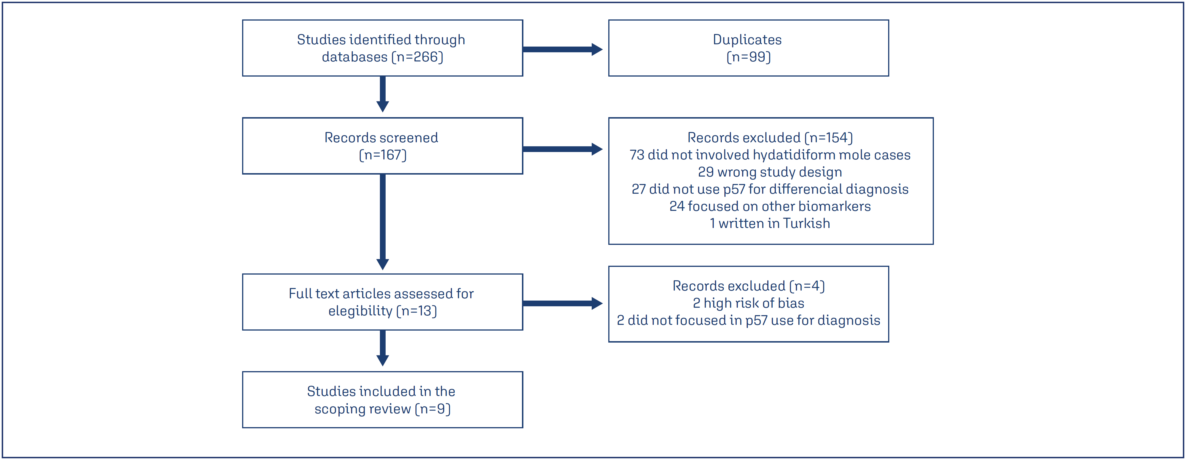
Initially, 266 articles were identified, as per the flowchart (Figure 1). After applying the criteria, 13 selected studies were read in full. Of these, two were excluded because they only mentioned the use of p57 with an objective other than method evaluation, and another two were excluded due to a high risk of methodological bias after evaluation with QUADAS-2. Ultimately, nine articles were eligible for this scoping review.
Of the nine articles, seven were retrospective cohort studies, one was a prospective study, and one was a cross-sectional study. Molecular methods were employed in five studies, with two utilizing fluorescence in situ hybridization (FISH) and two using Short Tandem Repeats (STR) genotyping in all cases. One of them used molecular methods only in inconclusive cases. In the study by Usui et al.,(17) immunohistochemistry was evaluated in conjunction with FISH. The main findings of each article are summarized in chart 1. The QUADAS-2 assessment of the included studies is presented on Supplementary Materials (Figure 1S).
Discussion
In this review, a consensus was observed among the included authors that the isolated use of the morphological method with hematoxilin and eosin presents high rates of diagnostic error. Discordance between morphological diagnosis and that performed by immunohistochemistry and genotyping was observed in 9% of cases in Triratanachat et al.'s(19) study, 26% in López et al.'s(21) study, and an alarming 33% in Zainal et al.'s(24) study, proving the necessity of using ancillary techniques in the evaluation of suspected HM cases.(19,21,24) Furthermore, performing immunohistochemistry with identification of the p57 protein is associated with better accuracy in the differential diagnosis of HM.(26) Immunohistochemistry for p57 is often the first complementary technique used in cases of diagnostic question due to its easy accessibility.(27) However, most gestational trophoblastic disease guidelines – both international and Brazilian – do not include its routine use in pathological diagnosis in cases of gestational loss or suspected HM.(1,5,28)
Most studies have shown the high accuracy of p57 for diagnosing CHM cases,(18,20,22,23,25) with a negative p57 immunohistochemistry result being highly predictive of this type of mole. Such findings underscore the importance of routine p57 immunohistochemistry use, highlighting that complete moles have a higher risk of malignant evolution and poorer outcomes, and non-diagnosis can lead to delays in initiating appropriate treatment.(6,24,25) On the other hand, cases of over-diagnosis of CHM are also detrimental, as they unnecessarily increase follow-up time and costs for the healthcare service; in addition to impacting patients’ emotional aspects, increasing anxiety and depression rates associated with diagnosis.(24,29)
Beyond its diagnostic importance, some authors demonstrated the utility of p57 as a prognostic marker, showing that cases presenting p57 negativity were associated with a higher rate of persistent disease and progression to GTN.(19) Such a correlation can be attributed to p57's function as a cyclin-dependent cell cycle inhibitor, acting in the G1 to S phase transition.(30) Thus, its absence, characteristic of androgenetic complete moles, results in the loss of cell proliferation control, favoring a more aggressive biological behavior and higher potential for malignancy.(31) Similarly, Erol et al.(18) reported that p57 acts as a tumor suppressor and its absence, combined with overexpression of epidermal growth factor receptor type 2 (c-erbB-2) and alterations in Bcl-2 and CD117 expression, was associated with higher proliferative potential and risk of progression to persistent disease. López et al.(21) also identified progression to invasive mole in a case with p57 absence and c-erbB-2 amplification, reinforcing the hypothesis that alterations in cell proliferation control are associated with worse prognosis. Xing et al.(23) observed that heterozygous CHM have a greater propensity for progression to GTN than homozygous ones, attributing this behavior to the duplicated genetic load and increased trophoblastic proliferative activity.
Although Usui et al.'s study(17) evaluate the use of p57 in combination with the FISH technique, it was included in this review for significantly contributing to the understanding of p57's diagnostic limitations, particularly in distinguishing between PHM and hydropic abortion, conditions that share positivity for this marker. However, similar to what was observed in other studies, the authors also reported high specificity of p57 in identifying CHM cases.(17) Generally, articles widely recommend performing p57 immunohistochemistry as a screening method in all suspected cases of molar pregnancy, reserving molecular analyses for cases with positive p57 expression where diagnostic doubt between PHM and hydropic abortion persists.(19,22,24)
Rare cases of divergent or unexpected p57 expression were identified. Samadder et al. (20) described a case of CHM with positive p57 expression, raising the hypothesis of maternal chromosome 11 retention in an androgenetic conceptus – a rare phenomenon in which residual maternal genetic material is inherited at the time of conception, resulting in positive p57 expression in complete moles.(25,32) This alteration was confirmed in a case described by Wong et al.,(25) who demonstrated 46,XX diploidy associated with trisomy of chromosome 11 using the FISH technique. Cases of chromosome 11 loss were also reported, leading to the occurrence of PHM with negative p57 expression, as described by Xing et al.,(23) who diagnosed the alteration by STR genotyping, revealing triploidy. This condition can generate false-positive diagnoses of CHM and was also suspected in two cases of erroneous p57 expression reported by Erol et al.(18) Additionally, Xing et al.(23) reported five cases of CHM with biparental genotype and negative p57 expression, suggesting the presence of a possible familial syndrome associated with mutations in the NLRP7 gene. López et al.(21) also reported similar cases of CHM with triploidy and negative p57. Such cases are extremely rare, frequently associated with recurrent HM, and their correct diagnosis is only possible through genotyping, highlighting the limitations of isolated p57 immunohistochemistry use.(21,23)
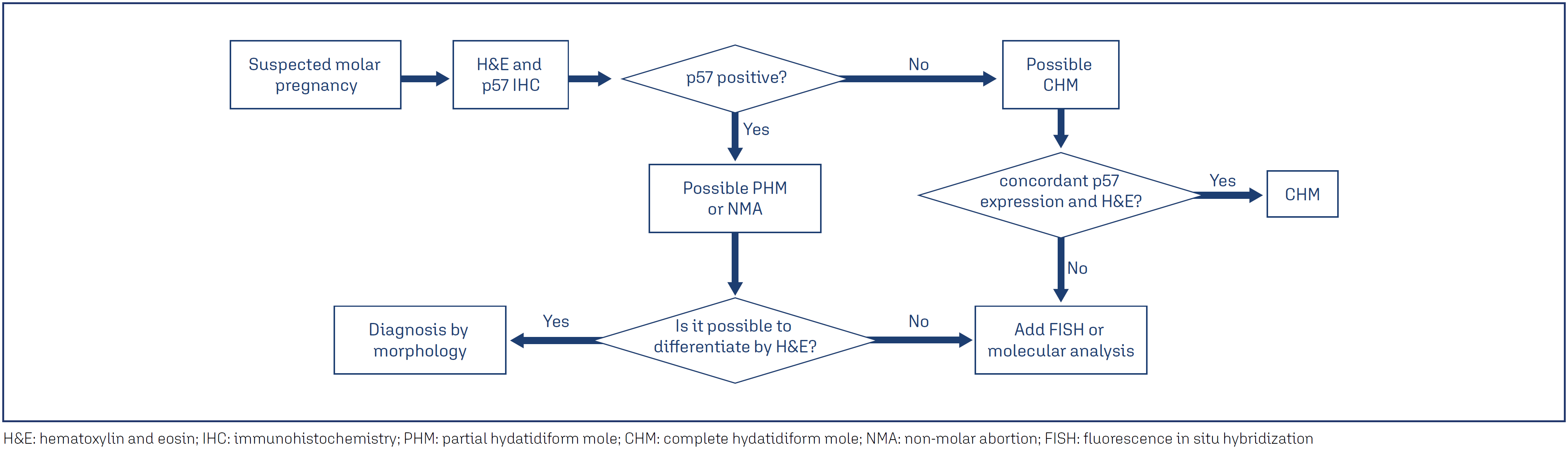
The integration between morphology, p57 immunohistochemistry, and molecular methods allows for the construction of more precise diagnostic algorithms, as suggested by some authors, and should be implemented whenever possible.(19,23,25) Through the study of the included articles, we suggest a flowchart (Figure 2) for the differential diagnosis of the different histological subtypes, as a way to optimize resources and improve the diagnosis of gestational trophoblastic diseases.
It is worth noting that genotyping and more refined molecular methods may not yet be a reality in many places, especially in developing countries. On the other hand, the use of p57 immunohistochemistry in the routine of suspected molar pregnancy cases can be considered a more accessible, relatively low-cost, and highly reliable method for clinical practice, being much more accurate than the isolated use of the morphological method.(17,22)
Conclusion
The use of the p57 marker via immunohistochemistry enhances the diagnostic accuracy of HM, particularly in distinguishing between CHM and PHM or hydropic abortion. Although it presents limitations in rare situations, its use combined with morphology and, when necessary, molecular methods, contributes to more precise diagnoses and safer clinical management, being routinely recommended in suspected cases of molar pregnancy.
Data availability
The research data are described in the article presented.
References
-
1 Horowitz NS, Eskander RN, Adelman MR, Burke W. Epidemiology, diagnosis, and treatment of gestational trophoblastic disease: a Society of Gynecologic Oncology evidenced-based review and recommendation. Gynecol Oncol. 2021;163(3):605-13. doi: 10.1016/j.ygyno.2021.10.003
» https://doi.org/10.1016/j.ygyno.2021.10.003 -
2 Joneborg U. Epidemiology of gestational trophoblastic disease. Hematol Oncol Clin North Am. 2024;38(6):1199-210. doi: 10.1016/j.hoc.2024.07.003
» https://doi.org/10.1016/j.hoc.2024.07.003 -
3 Fisher RA, Maher GJ. Genetics of gestational trophoblastic disease. Best Pract Res Clin Obstet Gynaecol. 2021;74:29-41. doi: 10.1016/j.bpobgyn.2021.01.004
» https://doi.org/10.1016/j.bpobgyn.2021.01.004 -
4 Ngan HY, Seckl MJ, Berkowitz RS, Xiang Y, Golfier F, Sekharan PK, et al. Diagnosis and management of gestational trophoblastic disease: 2021 update. Int J Gynaecol Obstet. 2021;155 (Suppl 1):86-93. doi: 10.1002/ijgo.13877
» https://doi.org/10.1002/ijgo.13877 -
5 Ministério da Saúde. Secretaria de Atenção Primária à Saúde. Departamento de Ações Programáticas e Estratégicas. Linha de cuidados para doença trofoblástica gestacional. Brasília (DF): Ministério da Saúde; 2022 [citado 2025 Ago 22]. Disponível em: https://portaldeboaspraticas.iff.fiocruz.br/wp-content/uploads/2022/01/linha_cuidado_trofoblasticas.pdf
» https://portaldeboaspraticas.iff.fiocruz.br/wp-content/uploads/2022/01/linha_cuidado_trofoblasticas.pdf -
6 Lelic M, Fatusic Z, Iljazovic E, Ramic S, Markovic S, Alicelebic S. Challenges in the routine praxis diagnosis of hydatidiform mole: a tertiary health center experience. Med Arch. 2017;71(4):256-60. doi: 10.5455/medarh.2017.71.256-260
» https://doi.org/10.5455/medarh.2017.71.256-260 -
7 Madi JM, Braga A, Paganella MP, Litvin IE, Wendland EM. Accuracy of p57 KIP2 compared with genotyping to diagnose complete hydatidiform mole: a systematic review and meta-analysis. BJOG. 2018;125(10):1226-33. doi: 10.1111/1471-0528.15289
» https://doi.org/10.1111/1471-0528.15289 -
8 Schoenen S, Delbecque K, Van Rompuy AS, Marbaix E, Noel JC, Delvenne P, et al. Importance of pathological review of gestational trophoblastic diseases: results of the Belgian Gestational Trophoblastic Diseases Registry. Int J Gynecol Cancer. 2022;32(6):740-5. doi: 10.1136/ijgc-2021-003312
» https://doi.org/10.1136/ijgc-2021-003312 -
9 McConnell TG, Murphy KM, Hafez M, Vang R, Ronnett BM. Diagnosis and subclassification of hydatidiform moles using p57 immunohistochemistry and molecular genotyping: validation and prospective analysis in routine and consultation practice settings with development of an algorithmic approach. Am J Surg Pathol. 2009;33(6):805-17. doi: 10.1097/PAS.0b013e318191f309
» https://doi.org/10.1097/PAS.0b013e318191f309 -
10 Ronnett BM. Hydatidiform moles: ancillary techniques to refine diagnosis. Arch Pathol Lab Med. 2018;142(12):1485-502. doi: 10.5858/arpa.2018-0226-RA
» https://doi.org/10.5858/arpa.2018-0226-RA -
11 Hui P, Buza N, Murphy KM, Ronnett BM. Hydatidiform moles: genetic basis and precision diagnosis. Annu Rev Pathol. 2017;12:449-85. doi: 10.1146/annurev-pathol-052016-100237
» https://doi.org/10.1146/annurev-pathol-052016-100237 -
12 Xing D, Miller K, Beierl K, Ronnett BM. Loss of p57 expression in conceptions other than complete hydatidiform mole: a case series with emphasis on the etiology, genetics, and clinical significance. Am J Surg Pathol. 2022;46(1):18-32. doi: 10.1097/PAS.0000000000001749
» https://doi.org/10.1097/PAS.0000000000001749 -
13 Gaillot-Durand L, Patrier S, Aziza J, Devisme L, Riera AC, Marcorelles P, et al. p57-discordant villi in hydropic products of conception: a clinicopathological study of 70 cases. Hum Pathol. 2020;101:18-30. doi: 10.1016/j.humpath.2020.04.011
» https://doi.org/10.1016/j.humpath.2020.04.011 -
14 Peters MD, Godfrey CM, Khalil H, McInerney P, Parker D, Soares CB. Guidance for conducting systematic scoping reviews. Int J Evid Based Healthc. 2015;13(3):141-6. doi: 10.1097/XEB.0000000000000050
» https://doi.org/10.1097/XEB.0000000000000050 -
15 Tricco AC, Lillie E, Zarin W, O’Brien KK, Colquhoun H, Levac D, et al. PRISMA extension for scoping reviews (PRISMA-ScR): checklist and explanation. Ann Intern Med. 2018;169(7):467-73. doi: 10.7326/M18-0850
» https://doi.org/10.7326/M18-0850 -
16 Whiting PF, Rutjes AW, Westwood ME, Mallett S, Deeks JJ, Reitsma JB, et al. QUADAS-2: a revised tool for the quality assessment of diagnostic accuracy studies. Ann Intern Med. 2011;155(8):529-36. doi: 10.7326/0003-4819-155-8-201110180-00009
» https://doi.org/10.7326/0003-4819-155-8-201110180-00009 -
17 Usui H, Hoshimoto K, Sato A, Kano M, Fukusato T, Nakatani Y, et al. Evaluation of combined p57KIP2 immunohistochemistry and fluorescent in situ hybridization analysis for hydatidiform moles compared with genotyping diagnosis. Int J Gynecol Pathol. 2024;43(5):474-86. doi: 10.1097/PGP.0000000000001000
» https://doi.org/10.1097/PGP.0000000000001000 -
18 Erol O, Suren D, Tutus B, Yararbas K, Sayiner A, Orel MK, et al. Comparison of p57, c-ERBB-2, CD 117, and Bcl-2 expression in the differential diagnosis of hydatidiform mole and hydropic abortion. Eur J Gynaecol Oncol. 2016;37(4):522-9. doi: 10.12892/ejgo3139.2016
» https://doi.org/10.12892/ejgo3139.2016 -
19 Triratanachat S, Nakaporntham P, Tantbirojn P, Shuangshoti S, Lertkhachonsuk R. Role of P57KIP2 immunohistochemical expression in histological diagnosis of hydatidiform moles. Asian Pac J Cancer Prev. 2016;17(4):2061-6. doi: 10.7314/APJCP.2016.17.4.2061
» https://doi.org/10.7314/APJCP.2016.17.4.2061 -
20 Samadder A, Kar R. Utility of p57 immunohistochemistry in differentiating between complete mole, partial mole and non-molar or hydropic abortus. Indian J Med Res. 2017;145(1):133-7. doi: 10.4103/ijmr.IJMR_982_15
» https://doi.org/10.4103/ijmr.IJMR_982_15 -
21 López CL, Figueira Gouvêa AL, Rodrigues FR, Braga A, Valente Machado MD, Lopes VS. HER2 FISH and P57KIP2 immunohistochemistry using tissue microarray: improving histopathological subtyping of hydatidiform mole. Placenta. 2020;99:166-72. doi: 10.1016/j.placenta.2020.07.027
» https://doi.org/10.1016/j.placenta.2020.07.027 -
22 Awosusi BL, Ajani MA, Adegoke OO, Salami AA, Okolo CA. P57kip2 immunohistochemical marker as a diagnostic tool for cases of hydatidiform moles in a tertiary health facility in Southwestern Nigeria. Niger Postgrad Med J. 2020;27(4):331-5. doi: 10.4103/npmj.npmj_231_20
» https://doi.org/10.4103/npmj.npmj_231_20 -
23 Xing D, Adams E, Huang J, Ronnett BM. Refined diagnosis of hydatidiform moles with p57 immunohistochemistry and molecular genotyping: updated analysis of a prospective series of 2217 cases. Mod Pathol. 2021;34(5):961-82. doi: 10.1038/s41379-020-00691-9
» https://doi.org/10.1038/s41379-020-00691-9 -
24 Zainal N, Kampan NC, Rose IM, Ghazali R, Shafiee MN, Yussoff NH, et al. Complementary role of p57kip2 immunostaining in diagnosing hydatidiform mole subtypes. Horm Mol Biol Clin Investig. 2021;42(3):311-6. doi: 10.1515/hmbci-2020-0086
» https://doi.org/10.1515/hmbci-2020-0086 - 25 Wong PY, Chia WK, Selimin A, Chia PY, Mustangin M, Shuib S, et al. Diagnostic utility of p57 immunohistochemistry and DNA ploidy analysis by fluorescence in situ hybridisation in hydatidiform moles. Malays J Pathol. 2021;43(3):341-51.
-
26 Colgan TJ, Noor A, Nanji S, Chang MC, Kolomietz E. Molecular diagnosis of placental hydatidiform mole: innovation and outcomes. J Obstet Gynaecol Can. 2017;39(11):1049-52. doi: 10.1016/j.jogc.2017.05.023
» https://doi.org/10.1016/j.jogc.2017.05.023 -
27 Laviola GM, Fortini AS, Salles D, Lourenço CS, Ribeiro DA, Sun SY, et al. Complementary tool in diagnosis of hydatidiform mole: review. Pathol Res Pract. 2022;237:154041. doi: 10.1016/j.prp.2022.154041
» https://doi.org/10.1016/j.prp.2022.154041 -
28 Lok C, van Trommel N, Braicu EI, Planchamp F, Berkowitz R, Seckl M, et al. Practical guidelines for the treatment of gestational trophoblastic disease: collaboration of the European Organisation for the Treatment of Trophoblastic Disease (EOTTD), European Society of Gynaecologic Oncology (ESGO), Gynecologic Cancer InterGroup (GCIG) and International Society for the Study of Trophoblastic Diseases (ISSTD). J Clin Oncol. 2025;43(18):2119-28. doi: 10.1200/JCO-24-02326
» https://doi.org/10.1200/JCO-24-02326 -
29 França AC, Uberti EM, Muller KP, Cardoso RB, Giguer F, El Beitune P, et al. Emotional and clinical aspects observed in women with gestational trophoblastic disease: a multidisciplinary action. Rev Bras Ginecol Obstet. 2022;44(4):343-51. doi: 10.1055/s-0042-1742681
» https://doi.org/10.1055/s-0042-1742681 -
30 Song HL, Liu TH, Wang YH, Li FF, Ruan LL, Adu-Gyamfi, et al. Appropriate expression of P57kip2 drives trophoblast fusion via cell cycle arrest. Reproduction. 2021;161(6):633-44. doi: 10.1530/REP-20-0638
» https://doi.org/10.1530/REP-20-0638 - 31 Suganthi C, Suganya U, Manibarathi R. The role of immunohistochemical markers in evaluating the malignant potential of molar pregnancies. Int J Life Sci. 2024;13(3):31-6.
-
32 Slim R. Genetics and genomics of gestational trophoblastic disease. Hematol Oncol Clin North Am. 2024;38(6):1219-32. doi: 10.1016/j.hoc.2024.07.004
» https://doi.org/10.1016/j.hoc.2024.07.004
Edited by
-
Associate Editor
Sue Yazaki Sun (https://orcid.org/0000-0001-6573-7386) Universidade Federal de São Paulo, São Paulo, SP, Brazil