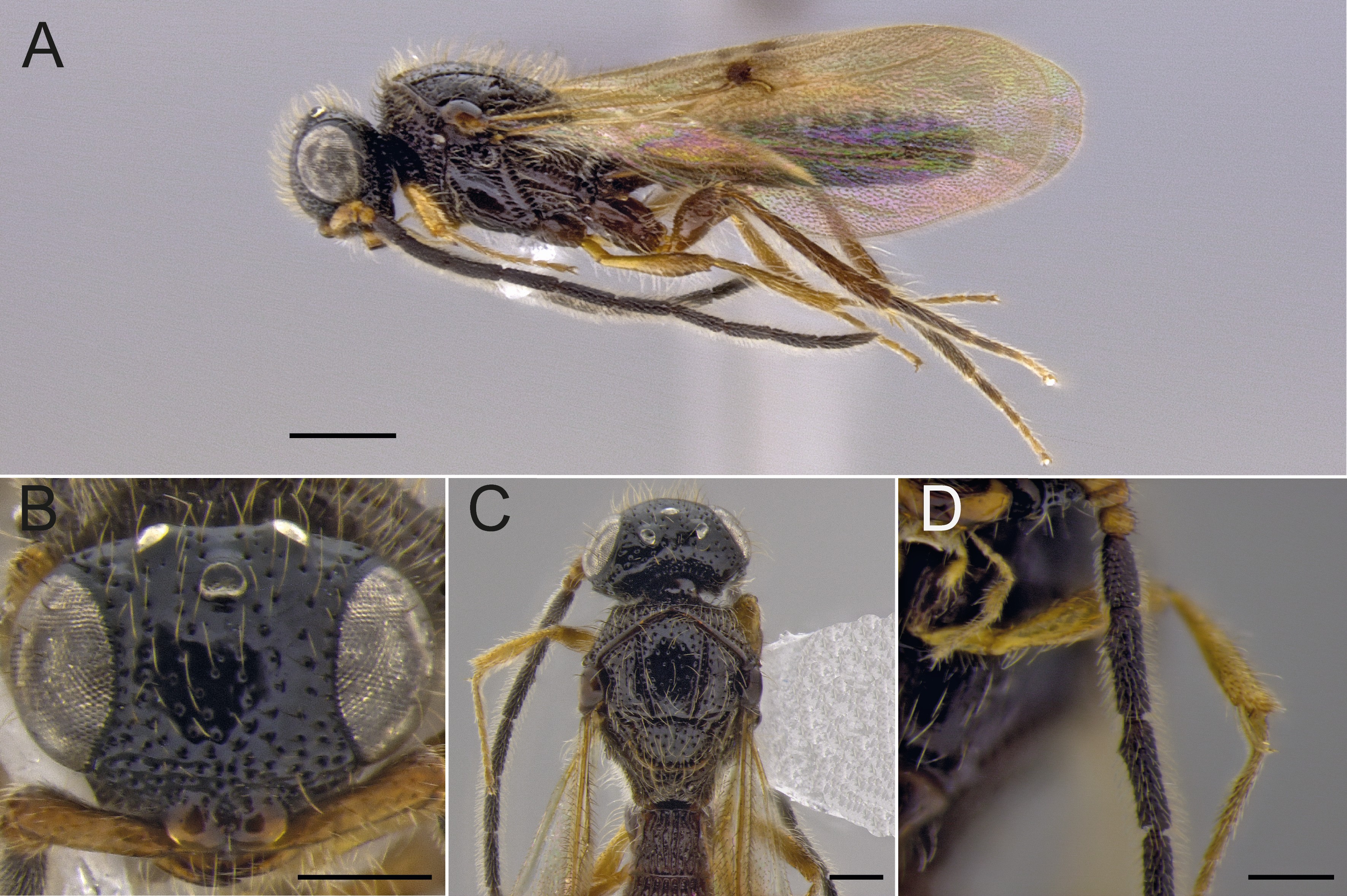
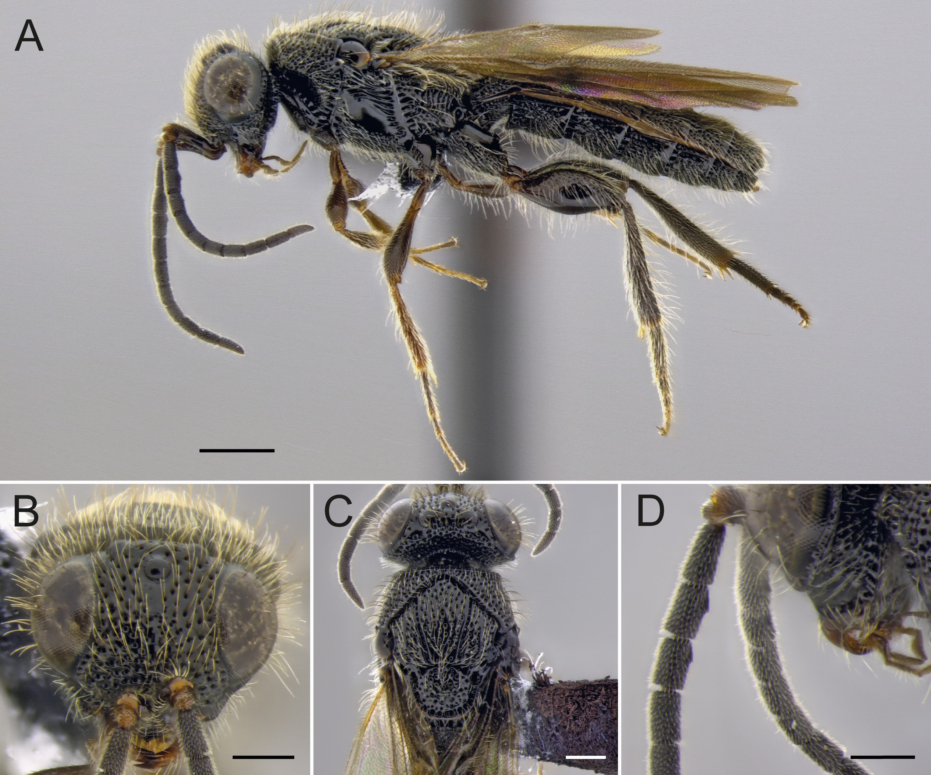
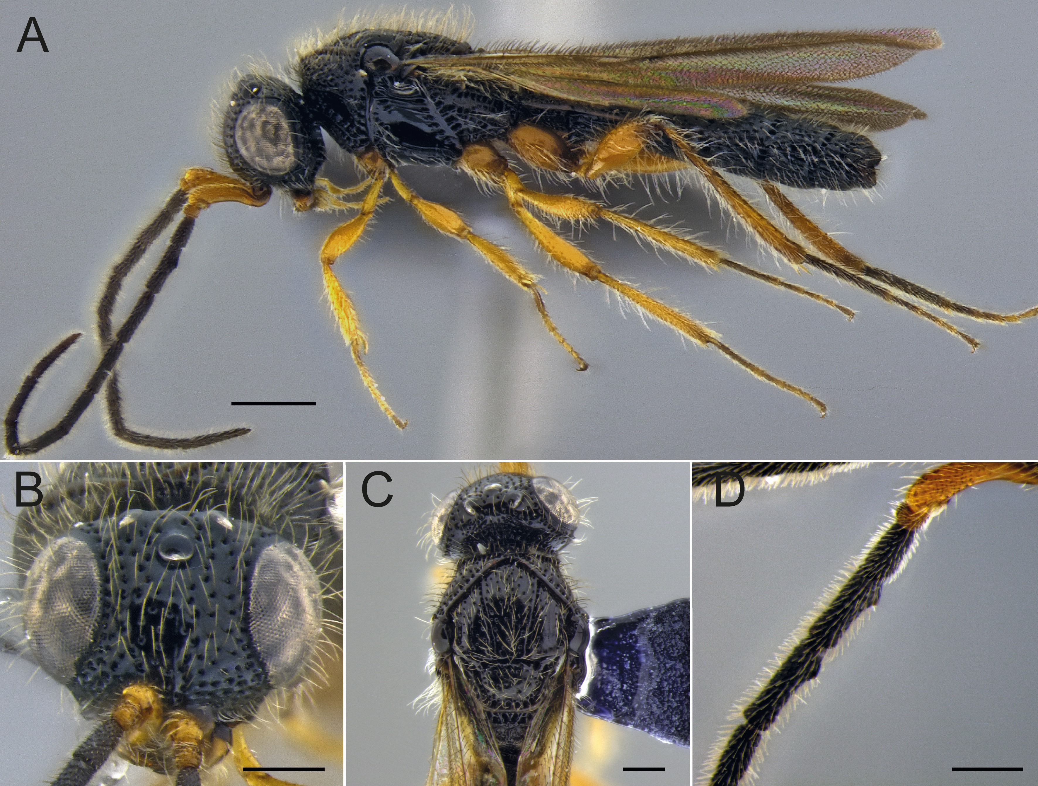
Figure 1
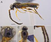
Sceliomorpha brunnipessp. nov. (holotype, male). A, habitus, lateral view; B, head, frontal view; C, head and mesosoma, dorsal view; D, flagellomeres 1-4 focusing on the toothlike projections. Scale bars: A, 1 mm; B–C, 0.5 mm; D, 0.4 mm.
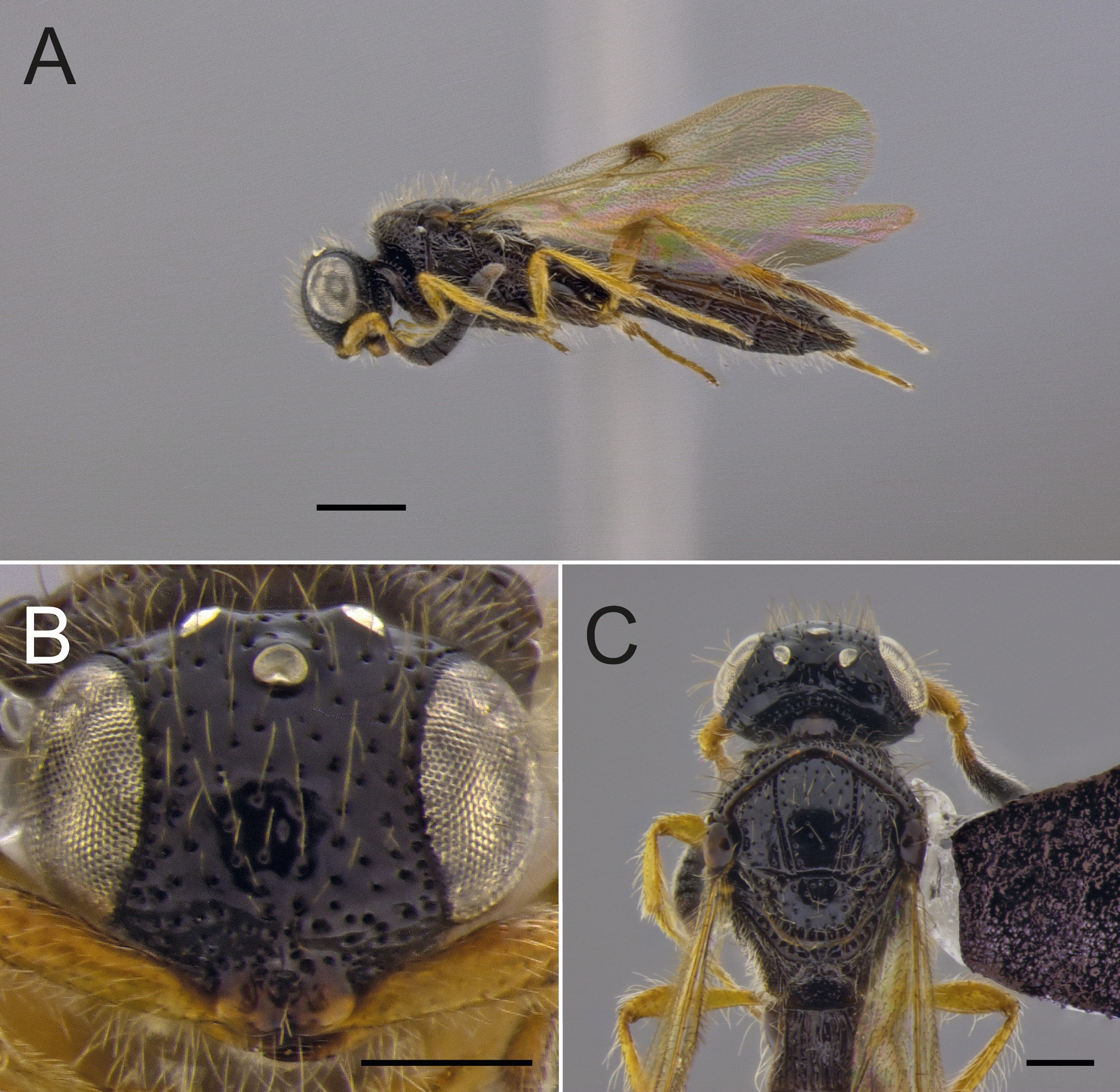
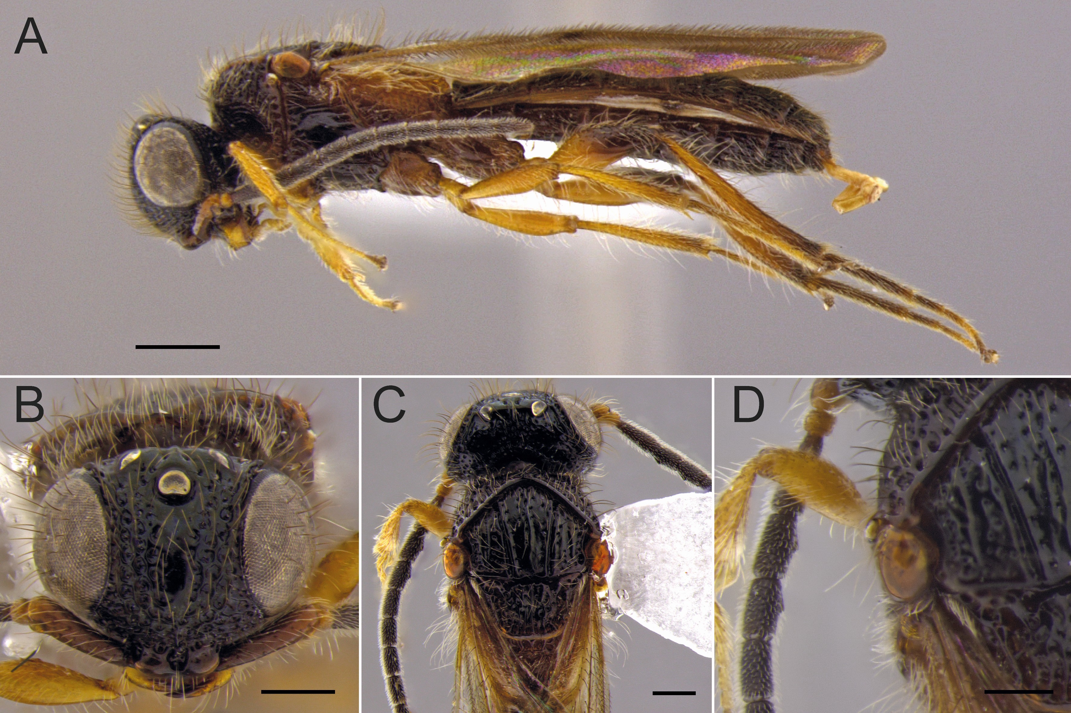
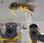
Figure 2
Sceliomorpha brunnipessp. nov. (paratype, female). A, habitus, lateral view; B, head, frontal view; C, head and mesosoma, dorsal view. Scale bars: A, 1 mm; B–C, 0.5 mm.
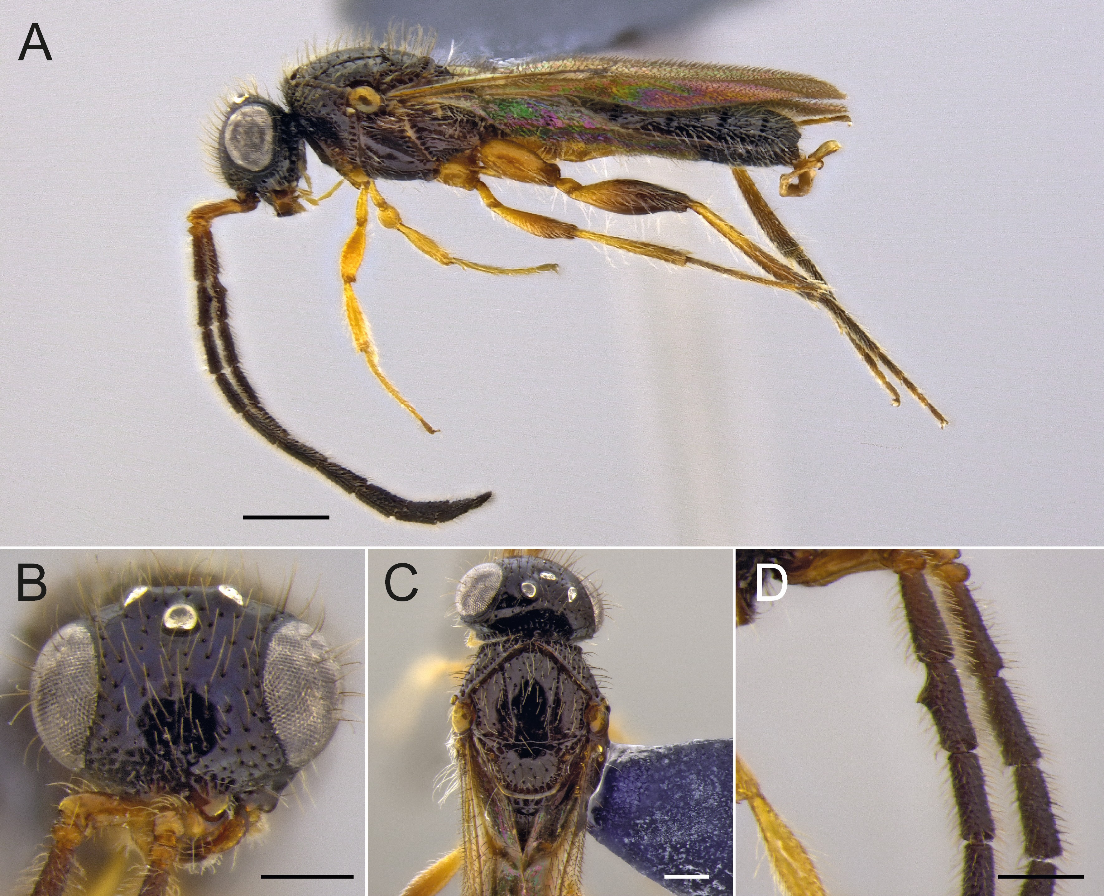
Figure 3
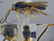
Sceliomorpha dentatasp. nov. (holotype, male). A, habitus, lateral view; B, head, frontal view; C, head and mesosoma, dorsal view; D, flagellomeres 1-4 focusing on the toothlike projections. Scale bars: A, 1 mm; B–C, 0.5 mm; D, 0.4 mm.
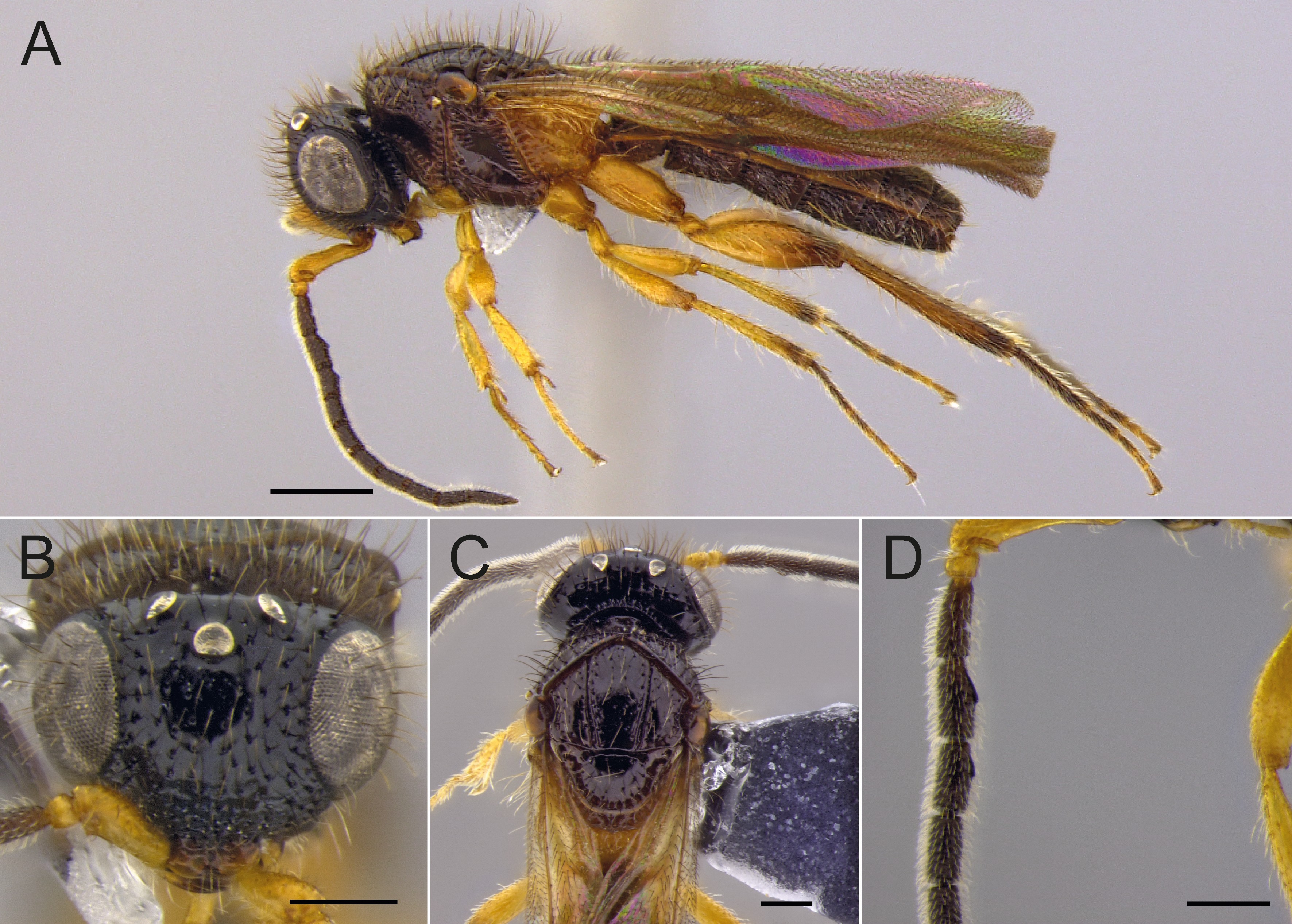
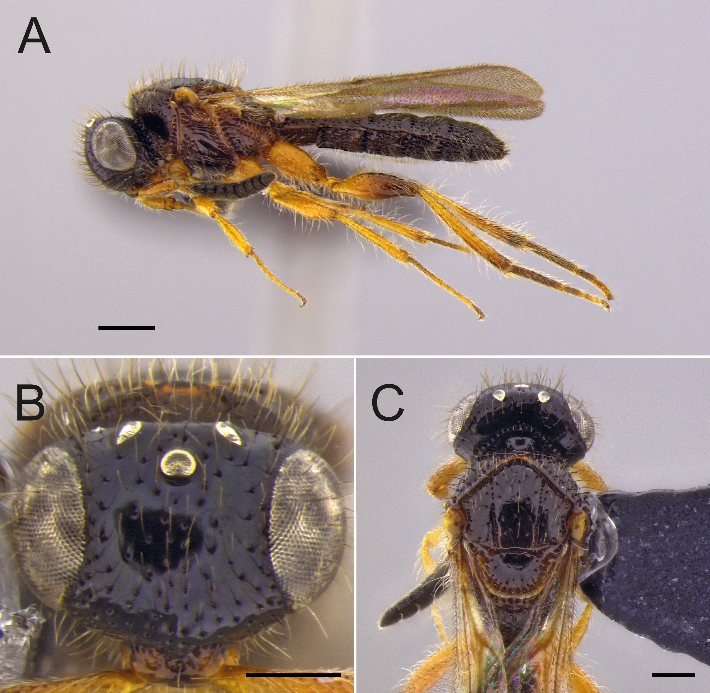
Figure 4
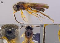
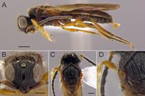
Sceliomorpha fulgidasp. nov. (holotype, male). A, habitus, lateral view; B, head, frontal view; C, head and mesosoma, dorsal view; D, flagellomeres 1-4 focusing on the toothlike projections. Scale bars: A, 1 mm; B–C, 0.5 mm; D, 0.4 mm.
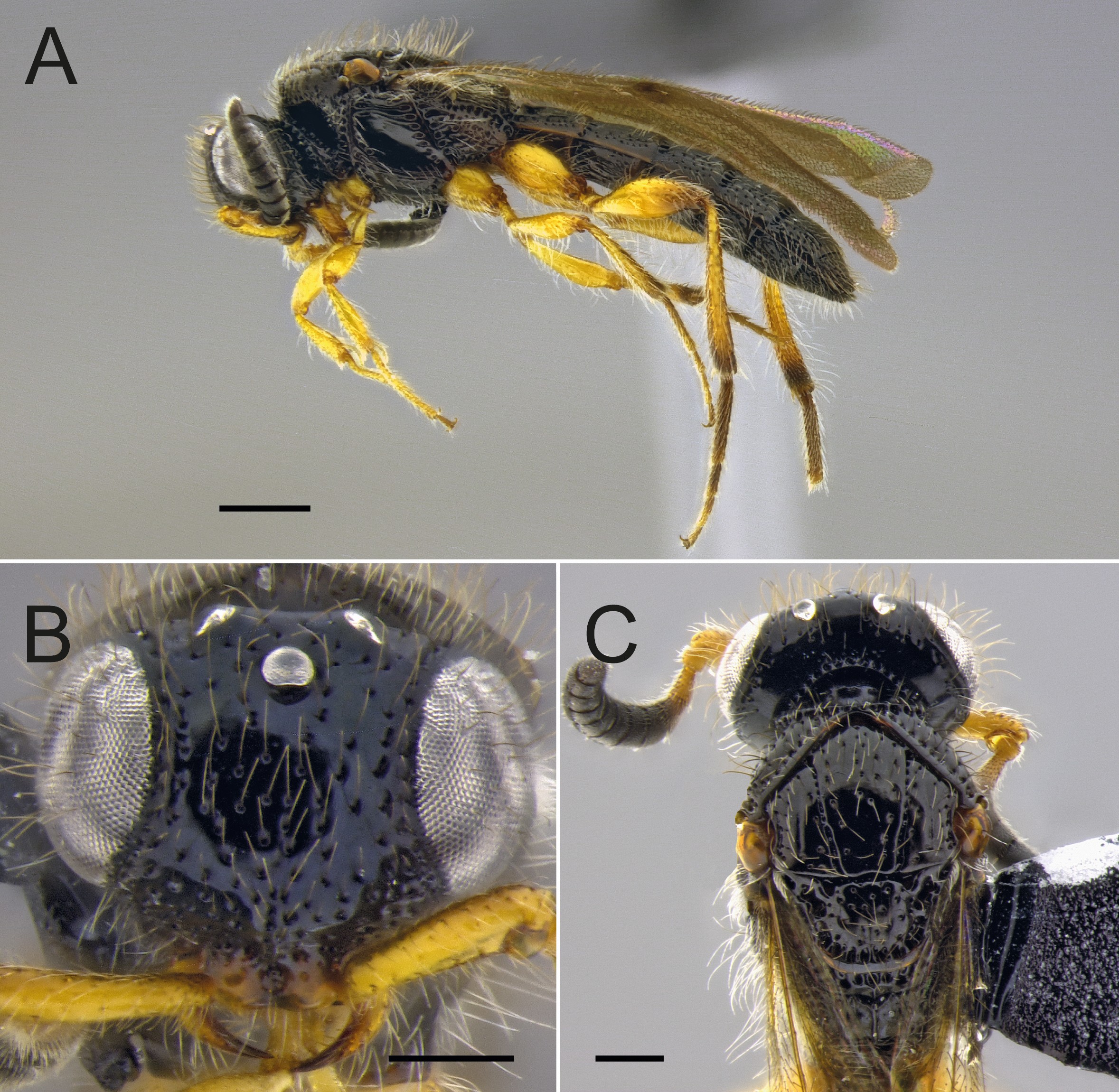
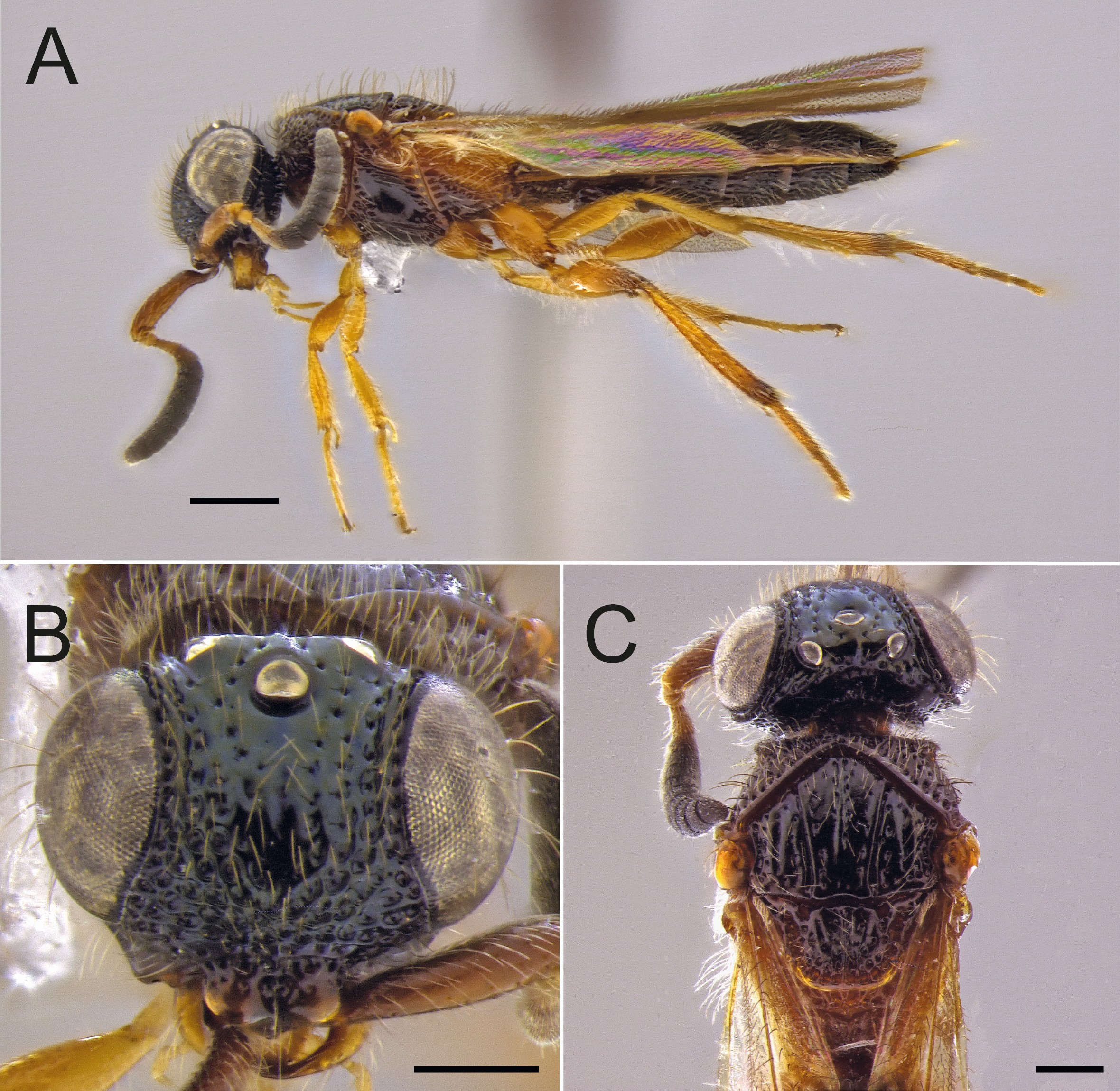
Figure 5
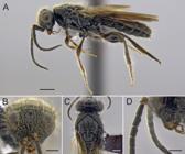
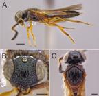
Sceliomorpha fulgidasp. nov. (paratype, female). A, habitus, lateral view; B, head, frontal view; C, head and mesosoma, dorsal view. Scale bars: A, 1 mm; B–C, 0.5 mm.
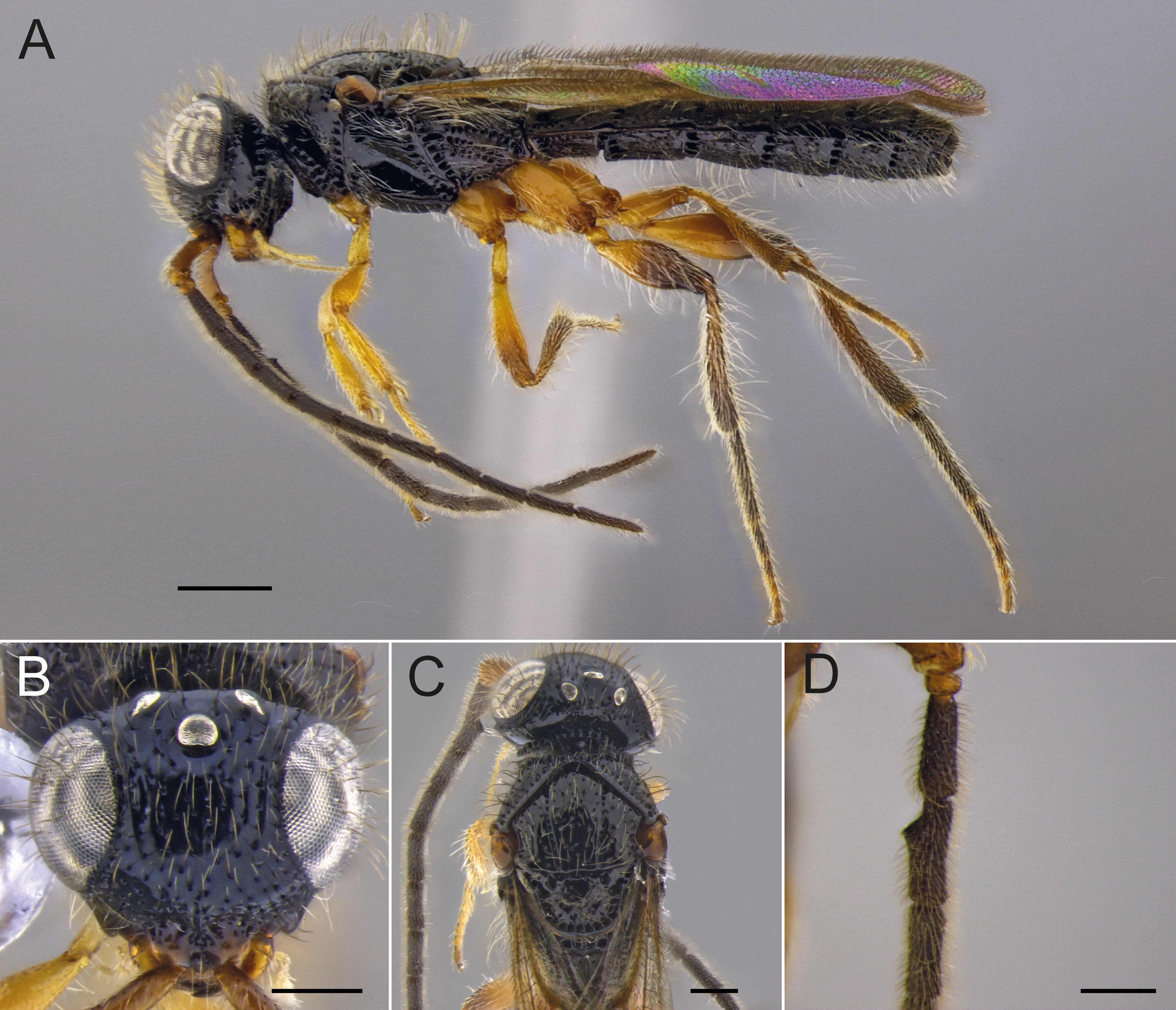
Figure 6
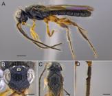
Sceliomorpha nigripessp. nov. (holotype, male). A, habitus, lateral view; B, head, frontal view; C, head and mesosoma, dorsal view; D, flagellomeres 1-4 focusing on the toothlike projections. Scale bars: A, 1 mm; B–C, 0.5 mm; D, 0.4 mm.
Figure 7
Sceliomorpha orbiculatasp. nov. (holotype, male). A, habitus, lateral view; B, head, frontal view; C, head and mesosoma, dorsal view; D, flagellomeres 1-4 focusing on the toothlike projections. Scale bars: A, 1 mm; B–C, 0.5 mm; D, 0.4 mm.
Figure 8
Sceliomorpha orbiculatasp. nov. (paratype, female). A, habitus, lateral view; B, head, frontal view; C, head and mesosoma, dorsal view. Scale bars: A, 1 mm; B–C, 0.5 mm.
Figure 9
Sceliomorpha porosasp. nov. (holotype, male). A, habitus, lateral view; B, head, frontal view; C, head and mesosoma, dorsal view; D, flagellomeres 1-4 focusing on the toothlike projections. Scale bars: A, 1 mm; B–C, 0.5 mm; D, 0.4 mm.
Figure 10
Sceliomorpha signatasp. nov. (holotype, male). A, habitus, lateral view; B, head, frontal view; C, head and mesosoma, dorsal view; D, flagellomeres 1-4 focusing on the toothlike projections. Scale bars: A, 1 mm; B–C, 0.5 mm; D, 0.4 mm.
Figure 11
Sceliomorpha signatasp. nov. (paratype, female). A, habitus, lateral view; B, head, frontal view; C, head and mesosoma, dorsal view. Scale bars: A, 1 mm; B–C, 0.5 mm.
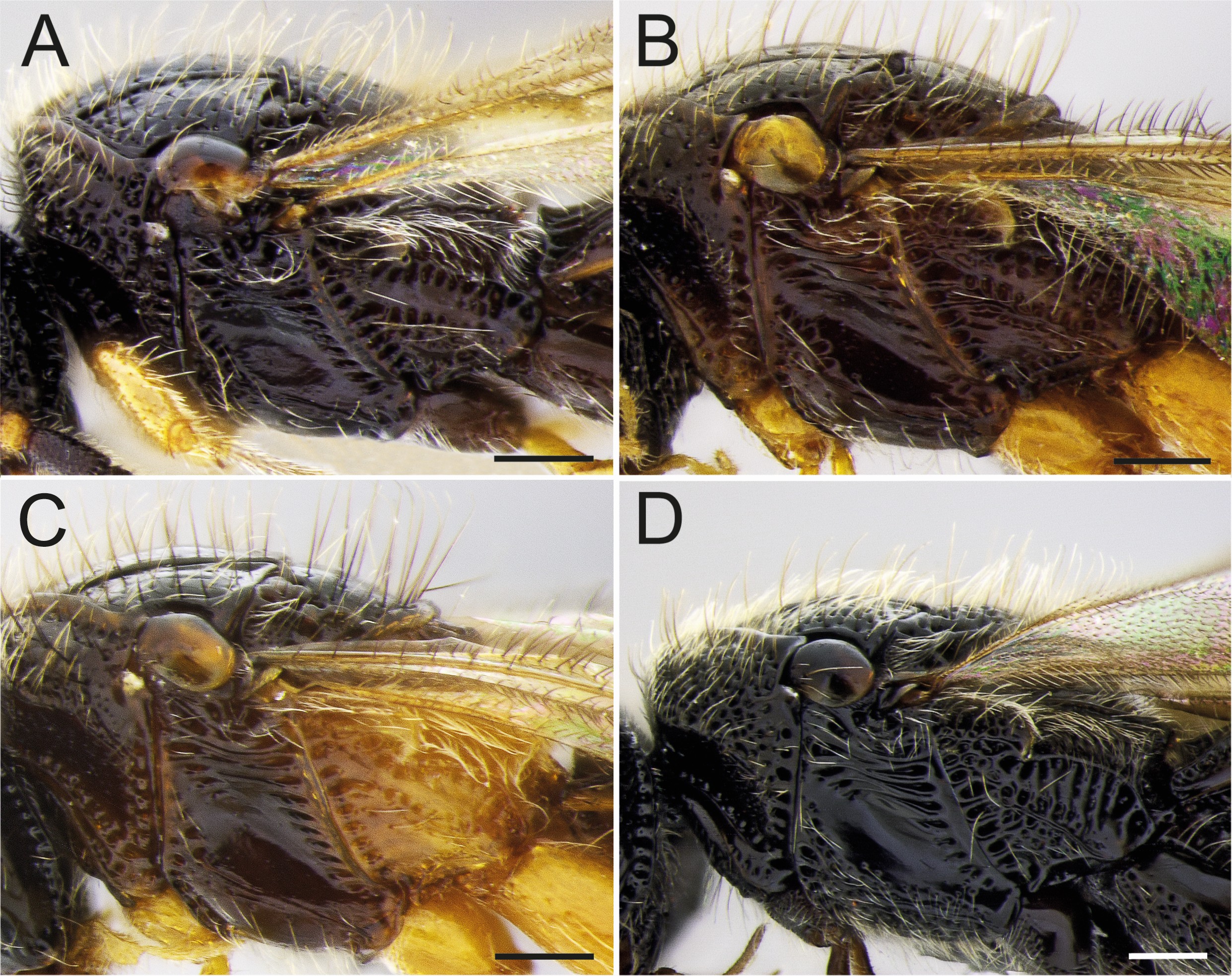
Figure 12
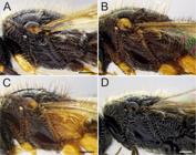
Mesosoma in lateral view. A, Sceliomorpha brunnipes sp. nov. (holotype, male); B, Sceliomorpha dentata sp. nov. (holotype, male); C, Sceliomorpha fulgida sp. nov. (holotype, male); D, Sceliomorpha nigripes sp. nov. (holotype, male). Scale bars: 0.2 mm.
Figure 13
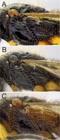
Mesosoma in lateral view. A, Sceliomorpha orbiculata sp. nov. (holotype, male); B, Sceliomorpha porosa sp. nov. (holotype, male); C, Sceliomorpha signata sp. nov. (holotype, male). Scale bars: 0.2 mm.
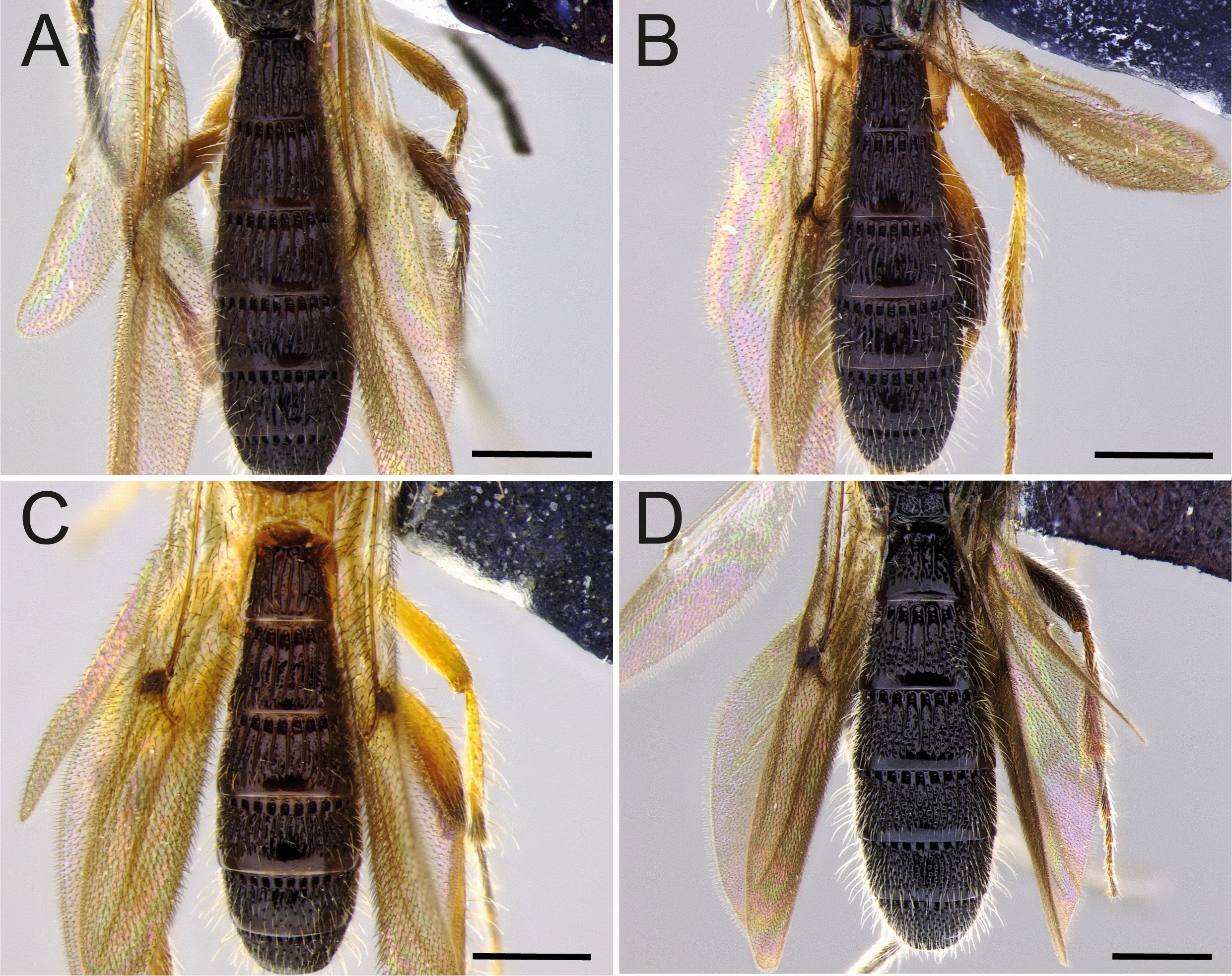
Figure 14
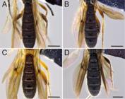
Metasoma in dorsal view. A, Sceliomorpha brunnipes sp. nov. (holotype, male); B, Sceliomorpha dentata sp. nov. (holotype, male); C, Sceliomorpha fulgida sp. nov. (holotype, male); D, Sceliomorpha nigripes sp. nov. (holotype, male). Scale bars: 0.5 mm.
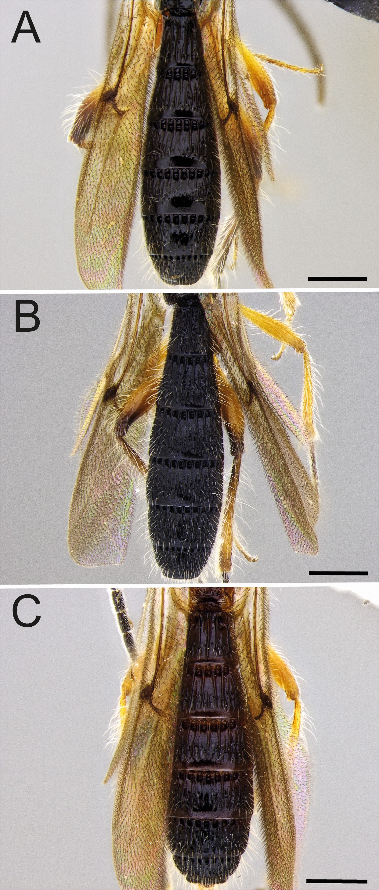
Figure 15
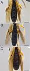
Metasoma in dorsal view. A, Sceliomorpha orbiculata sp. nov. (holotype, male); B, Sceliomorpha porosa sp. nov. (holotype, male); C, Sceliomorpha signata sp. nov. (holotype, male). Scale bars: 0.5 mm.
 Seven new species of Sceliomorpha Ashmead (Hymenoptera: Platygastroidea: Sparasionidae) from Brazil
 Thumbnail
 Thumbnail
 Thumbnail
 Thumbnail
 Thumbnail
 Thumbnail
 Thumbnail
 Thumbnail
 Thumbnail
 Thumbnail
 Thumbnail
 Thumbnail
 Thumbnail
 Thumbnail
 Thumbnail