 Physiotherapeutic Intervention on Internal Lymphedema Post-Treatment of HPV-Positive Tonsillar Carcinoma: Case Report
ABSTRACT
Introduction: Internal lymphedema is a frequent and underdiagnosed complication after treatment of oropharyngeal squamous cell carcinoma (SCC), particularly in HPV-positive cases. The lack of specific protocols hinders early detection and proper management. This study aims to report the physiotherapeutic intervention in a patient with bilateral internal lymphedema resulting from the treatment of HPV-positive tonsillar SCC.
Case report: A 49-year-old male patient underwent bilateral surgery and radiotherapy, developing internal lymphedema with significant obstructive and dysphagic symptoms. The physiotherapeutic approach included manual lymphatic drainage, therapeutic exercises, intra- and extraoral photobiomodulation, shockwave therapy, and cervical fibrosis release. A custom-made flat compression garment was also applied to the cervicofacial region. Subjective and functional improvements were observed, allowing the patient to complete radiotherapy without interruption.
Conclusion: The integrated physiotherapeutic intervention was effective in reducing internal lymphedema symptoms in a patient with oropharyngeal cancer. This case highlights the importance of early detection and the need for specific therapeutic strategies based on lymphatic physiology.
Key words:
Lymphedema/complications; Physical Therapy Services; Head and Neck Neoplasms/surgery; Human Papillomavirus Viruses; Rehabilitation
RESUMO
Introdução: O linfedema interno é uma complicação frequente e subdiagnosticada após o tratamento do carcinoma espinocelular (CEC) de orofaringe, especialmente em casos HPV-positivos. A falta de protocolos específicos dificulta a identificação precoce e o manejo terapêutico adequado. Este estudo tem como objetivo relatar a intervenção fisioterapêutica em um paciente com linfedema interno bilateral decorrente do tratamento de CEC de amígdala HPV-positivo.
Relato do caso: Paciente masculino de 49 anos, submetido a tratamento cirúrgico e radioterápico bilateral. Desenvolveu linfedema interno com sintomas obstrutivos e disfágicos relevantes. A intervenção fisioterapêutica incluiu drenagem linfática manual, cinesioterapia, fotobiomodulação intra e extraoral, terapia por ondas de choque e liberação de fibroses cervicais. Também foi utilizada malha compressiva plana sob medida na região cervicofacial. Houve melhora subjetiva e funcional dos sintomas, sem interrupção do plano radioterápico.
Conclusão: A intervenção fisioterapêutica integrada mostrou-se eficaz na redução dos sintomas do linfedema interno em paciente com câncer de orofaringe. O caso ressalta a importância do reconhecimento precoce da condição e a necessidade de estratégias terapêuticas específicas baseadas em fisiologia linfática.
Palavras-chave:
Linfedema/complicações; Serviços de Fisioterapia; Neoplasias de Cabeça e Pescoço/cirurgia; Papilomavírus Humanos; Reabilitação
RESUMEN
Introducción: El linfedema interno es una complicación frecuente y subdiagnosticada tras el tratamiento del carcinoma escamocelular (CEC) de orofaringe, especialmente en casos positivos para VPH. La ausencia de protocolos específicos dificulta su detección temprana y manejo terapéutico. Este estudio tiene como objetivo describir la intervención fisioterapéutica en un paciente con linfedema interno bilateral derivado del tratamiento del CEC de amígdala positivo para VPH.
Informe del caso: Paciente masculino de 49 años, sometido a cirugía y radioterapia bilateral. Desarrolló linfedema interno con síntomas obstructivos y disfágicos significativos. La intervención fisioterapéutica incluyó drenaje linfático manual, kinesioterapia, fotobiomodulación intra y extraoral, terapia de ondas de choque y liberación de fibrosis cervicales. Se utilizó una prenda de compresión plana hecha a medida para la región cervicofacial. Se observó mejoría subjetiva y funcional de los síntomas, sin necesidad de interrumpir la radioterapia.
Conclusión: La intervención fisioterapéutica integrada fue eficaz para reducir los síntomas del linfedema interno en un paciente con cáncer de orofaringe. El caso refuerza la importancia del diagnóstico temprano y la necesidad de estrategias terapéuticas específicas basadas en la fisiología linfática.
Palabras clave:
Linfedema/complicaciones; Servicios de Fisioterapia; Neoplasias de Cabeza y Cuello/cirugía; Virus del Papiloma Humano; Rehabilitación
INTRODUCTION
Oropharyngeal squamous cell carcinoma (OPSCC), especially in tonsils, has been increasingly associated with human papilloma virus (HPV) infection, particularly subtype 16. This viral subtype is related to a distinct clinical profile: younger patients, low association with smoking and alcohol use and better oncologic prognosis1.
Prolonged survival does not eliminate the adverse effects of the treatment. The lymphedema of head and neck is a prevalent condition and clinically relevant2. External lymphedema is easily identified by visible and palpable alterations, but internal lymphedema, on its turn, affects deep structures as pharynx, larynx and base of the tongue and is associated with dysphagia, sensation of obstruction, pain and vocal changes3. For patients with head and neck cancer treated with primary (chemo) radiotherapy, the mean prevalence of internal lymphedema is 78%. For those submitted to unilateral radiotherapy, the prevalence of internal lymphedema was 73.9%, while for those treated with bilateral radiotherapy, the prevalence was 83.3%4.
Despite being more frequent than external lymphedema with significant functional impact, internal lymphedema is often underdiagnosed. Although there are descriptions that internal lymphedema tends to diminish over time post treatment, recent evidences showed that it persists frequently and can become a chronic condition if not properly treated5. Affecting 86% of the patients, internal lymphedema reaches its peak of prevalence three months after the treatment. Although there is gradual improvement between 12 and 18 months, 80% of the patients still present internal lymphedema6.
This article reports a case of a patient with HPV-positive tonsillar OPSCC who developed internal lymphedema during treatment. The objective is to contribute to the clinical recognition of this condition and stimulate the discussion about the necessity of specific diagnostic and therapeutic approaches.
The Ethics Committee of "Centro Universitário das Faculdades Associadas de Ensino (Unifae)" approved the study, report number 8237272 (CAAE (submission for ethical review): 94869826.9.0000.5382) in compliance with Directive 466/20127 of the National Health Council.
CASE REPORT
Male patient, 49 years old, diagnosed on November 11, 2024 with HPV-positive (subtype 16) tonsillar OPSCC. He was submitted to amygdalotomy with free margins and left cervical lymphadenectomy, encompassing levels I to VII, with removal of left internal jugular vein. Two lymph nodes were positive, the largest with 3.8 cm. Adjuvant treatment consisted in 30 sessions of radiotherapy, total dose of 60Gy on the surgical bed and 54 Gy in the cervical regions, including the complete extension of the surgical scar.
During radiotherapy, the patient presented important acute toxicities, mainly oral mucositis and fatigue, being followed-up with photobiomodulation (PBM)-based physiotherapeutic protocol, extracorporeal shockwave therapy (ESWT), manual lymphatic drainage (MLD), cervical mobilization and skincare. In this period, the initial complaint was compatible with internal lymphedema, including sensation of tightness, changes in deglutition and mild respiratory difficulty. External lymphedema was mild, limited to the left mentonian region.
In August 2025, a PET-CT identified hypermetabolic right cervical chain lymph node. Right cervical lymphadenectomy was performed from level I to IV. Four less than 2 cm lymph nodes were metastatic. Due to location and risk of regional relapse, the patient was submitted to a new cycle of 30 sessions of radiotherapy with total dose of 54 Gy on the right cervical region.
The symptoms of internal lymphedema intensified progressively after the sixth radiotherapy session on the right cervical region. The patient reported nocturnal snoring, increased difficulty to sleep in supine, persistent sensation of pharyngeal obstruction, aural fullness, dysphagia and a permanent sensation of strangling.
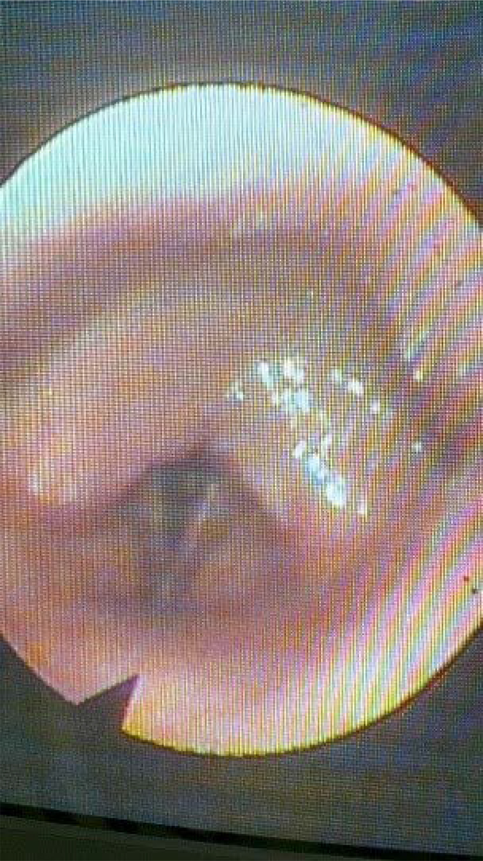
Nasofibrolaryngoscopy revealed hyperemia and edema of the mucosa of soft palate and remaining tonsillar region associated with lymphatic stasis, diffuse edema of the base of the tongue and thickening of the posterior wall of the oropharynx with reduction of the airway and blurring of the anatomic contours, in addition to bilateral thickening of the aryepiglottic folds and reduction of the laryngeal mobility with deep lymphatic accumulation on the supraglottic region (Figure 1).
In view of this scenario, the physiotherapy conduct was adapted to the anatomic complexity imposed by earlier cervical lymphadenectomies. MLD maneuvers were applied with soft, slow and rhythmic movements and targeted to alternative routes, prioritizing areas still functional as the suboccipital, supraclavicular and axillary regions.
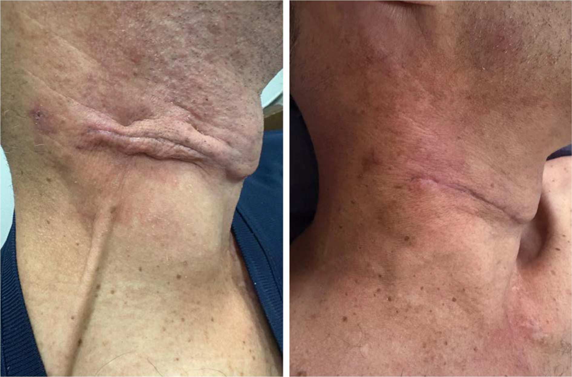
MLD and kinesiotherapy (active exercises and stretching) were associated with intra and extraoral PBM ((3J/point, 850 nm, 100 mW, in-point therapy, 1 cm2 space between points in the entire area of lymphadenectomy) aimed at inflammatory modulation and stimulation of tissue regeneration of areas submitted to radiotherapy. ESWT (piezoelectric, 6 mJ/mm2) and manual therapy applied on the cervical scar and adjacent tissues contributed for the myofascial release and reduction of adherences, optimizing the deep and superficial lymphatic drainage (figure 2). The patient reported immediate relief post sessions with reduction of the pressure and improvement of breathing.
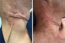
Initial aspect with retracted scar, thickening of adjacent tissues and slight submental bulging; after seven days of daily physiotherapy sessions
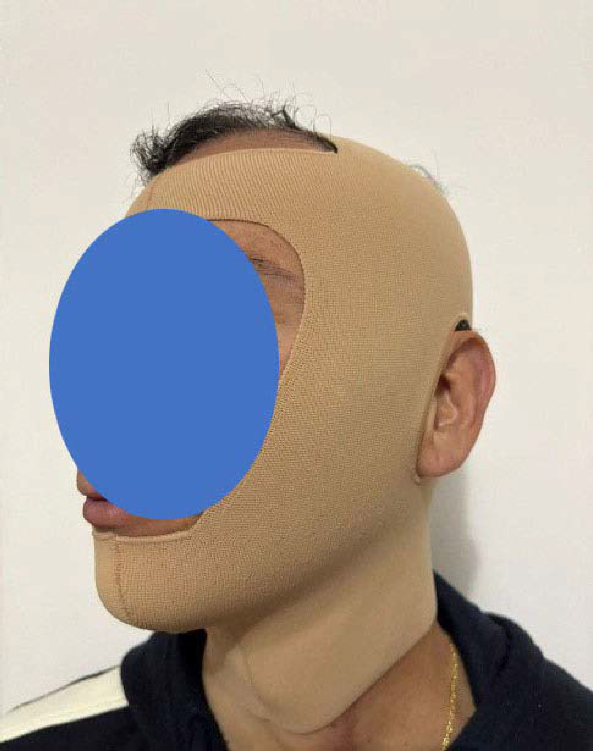
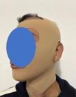
A custom-made flat compression garment produced by Juzo® (figure 3) was introduced. The compression was applied on the face and cervical region. The patient reported breathing improvement after 15 minutes and continued effects. He woke up only once at night, reduced the use of elevated head of the bed, improvement of the feeling of aural fullness, and cessation of snoring episodes. The sensation of suffocation then continuous turned occasional. The combination of compression garment with physiotherapy allowed the patient to continue the radiotherapy plan uninterrupted.
DISCUSSION
Internal lymphedema symptoms as dysphagia, obstructive sensation and snoring are reported by great part of irradiated patients, nevertheless, most of the health professionals do not admit the possibility of effective physiotherapy intervention. Common compression strategies are targeted to the treatment of external lymphedema on the face or limbs, for instance and they are not designed to directly influence deep structures of aerodigestive route5.
MLD is a widely recommended therapeutic intervention to manage lymphedema, especially in the initial phases of the condition. It is a soft, rhythmic and targeted maneuver technique aimed at stimulating the reabsorption and motility of the lymph through superficial and alternative pathways favoring tissue decongestion.
The technique does not only contribute to improve the lymphatic return but also plays a key role in preventing the progression of the lymphedema because it reduces the interstitial stasis, minimizes the risk of fibrosis and optimized the conditions of the tissue to maintain the functional drainage.
Whether earlier applied, the technique can preserve the integrity of the remaining lymphatic pathways, potentializing compensatory mechanisms and avoiding the aggravation of the clinical status8.
The use of compressive flat-knit garment on the cervicofacial region is a promising strategy. While applying soft and sustained compression over the cervical region it was possible to obtain immediate subjective improvement of the respiratory and suffocation symptoms with direct impact on functionality. The flat-knit garment allows uniform and controlled compression on uneven anatomic areas. Because its construction uses rigid walls and less elastic materials in comparison to circular-knit garment, it provided better structural stability, preventing the formation of a tourniquet effect and promoting better contention of the edema, especially in regions of irregular contours. The thicker and least flexible tissue ensures the garment a more effective pressure and the effect of localized massage in-between the points, favoring the redistribution of the lymphatic fluid during the movement. The custom-made garment was essential to ensure accurate anatomic adjustment and avoiding constrictions to the patient9.
The lymphangiogenesis is a core mechanism for the functional recovery of the lymphatic system post-lesion or oncologic treatment-induced dysfunction. In the context of head and neck lymphedema, evidences indicate the potential role of PBM in the stimulation of lymphangiogenesis, which can modulate the local inflammatory response, reduce the presence of macrophages and promote the required biological environment for the regeneration of lymphatic vessels, suggesting a pro-regenerative action in tissues affected by lymphatic stasis10.
The utilization of ESWT expands the expression of pro-lymphangiogenic factors as VEGFC, VEGFR3 and bFGF, and is associated with the increase of the density of lymphatic vessels on the areas treated, indicating the formation of new lymphatic vessels. The functional improvement of lymphedematous tissues subsequently to these molecular and histologic alterations suggested that ESWT can induce therapeutic lymphangiogenesis and contribute to restore the lymphatic drainage11.
The presence of dense and adhered cicatricial tissue can significantly compromise the lymphatic function, acting as a physical barrier to the motility of the lymph and local vascular regeneration. Tissue fibrosis post lymphatic lesion is associated with worsening of the lymphedema due to collagen deposition and disorganization of the extracellular matrix that increase the resistance to the lymphatic flow and hinder lymphangiogenesis. The accumulation of fibrotic tissue in the tissues affected reduced the ability of the functional lymphatic drainage and damaged the formation of new lymphatic vessels, perpetuating the stasis and the edema.
Adhered scars may not only represent a mechanic obstacle but also an active factor in the maintenance of lymphatic dysfunction, which reinforces the importance of therapeutic strategies focused to mobilization and early treatment of cicatricial areas12.
The association of PBM, ESWT and manual therapy over the cicatricial tissue contributed to the myofascial release, essential to partially restore the lymphatic flows in deep planes.
Despite the positive clinical results observed, some limitations should be considered as the cost and availability of the custom-made flat-knit garment. An additional challenge is over-relying on professionals with specific skills for oncologic lymphatic rehabilitation.
CONCLUSION
The clinical experience reported reinforces the early recognition of internal lymphedema, a condition often neglected and demonstrates that individualized physiotherapeutic interventions can grant symptomatic relief, preserved functionality and therapeutic continuity. This report contributed to the expansion of the clinical knowledge on internal lymphedema and highlights the necessity of specific therapeutic guidelines in the context of oncologic rehabilitation.
DATA AVAILABILITY STATEMENT
All the content underlying the text is contained in the manuscript.
References
-
1 Zupancic M, Kostopoulou ON, Marklund L, et al. Therapeutic options for human papillomavirus-positive tonsil and base of tongue cancer. J Intern Med. 2025;297(6):608-29. doi: https://doi.org/10.1111/joim.20088
» https://doi.org/10.1111/joim.20088 -
2 Cheng JT, Leite VF, Tennison JM, et al. Rehabilitation interventions for head and neck cancer-associated lymphedema: a systematic review. JAMA Otolaryngol Head Neck Surg. 2023;149(8):743-53. doi: https://doi.org/10.1001/jamaoto.2023.1473
» https://doi.org/10.1001/jamaoto.2023.1473 -
3 Taylor RR, Pandey SK, Smartz T, et al. Lymphedema of the head and neck-where do we stand and where we are headed. J Craniofac Surg. 2024;35(7):2045-8. doi: https://doi.org/10.1097/SCS.0000000000010505
» https://doi.org/10.1097/SCS.0000000000010505 -
4 Arends C, Veij Mestdagh P, Al-Mamgani A, et al. Severity of internal lymphedema in unilateral or bilateral radiotherapy patients: an exploratory study. Radiother Oncol. 2025;206:110834. doi: https://doi.org/10.1016/j.radonc.2025.110834
» https://doi.org/10.1016/j.radonc.2025.110834 -
5 Jeans C, Brown B, Ward EC, et al. A prospective, longitudinal and exploratory study of head and neck lymphoedema and dysphagia following chemoradiotherapy for head and neck cancer. Dysphagia. 2023;38(4):1059-71. doi: https://doi.org/10.1007/s00455-022-10526-1
» https://doi.org/10.1007/s00455-022-10526-1 -
6 Ridner SH, Dietrich MS, Niermann K, et al. A prospective study of the lymphedema and fibrosis continuum in patients with head and neck cancer. Lymphat Res Biol. 2016;14(4):198-205. doi: https://doi.org/10.1089/lrb.2016.0001
» https://doi.org/10.1089/lrb.2016.0001 - 7 Conselho Nacional de Saúde (BR). Resolução n° 466 de 12 de dezembro de 2012. Aprova diretrizes e normas regulamentadoras de pesquisas envolvendo seres humanos. Diário Oficial da União, Brasília, DF. 2013 jun 13; Edição 112; Seção 1:59.
-
8 Palmer SJ. An overview of manual lymphatic drainage. Br J Community Nurs. 2024;29(9):438-40. doi: https://doi.org/10.12968/bjcn.2024.0101
» https://doi.org/10.12968/bjcn.2024.0101 -
9 Reich-Schupke S, Stücker M. Round-knit or flat-knit compression garments for maintenance therapy of lymphedema of the leg? - Review of the literature and technical data. J Dtsch Dermatol Ges. 2019;17(8):775-84. doi: https://doi.org/10.1111/ddg.13895
» https://doi.org/10.1111/ddg.13895 -
10 Deng J, Lukens JN, Swisher-McClure S, et al. Photobiomodulation therapy in head and neck cancer-related lymphedema: a pilot feasibility study. Integr Cancer Ther. 2021;20:15347354211037938. doi: https://doi.org/10.1177/15347354211037938
» https://doi.org/10.1177/15347354211037938 -
11 Serizawa F, Ito K, Matsubara M, et al. Extracorporeal shock wave therapy induces therapeutic lymphangiogenesis in a rat model of secondary lymphoedema. Eur J Vasc Endovasc Surg. 2011;42(2):254-60. doi: https://doi.org/10.1016/j.ejvs.2011.02.029
» https://doi.org/10.1016/j.ejvs.2011.02.029 -
12 Tsai KY, Liao SF, Chen KL, et al. Effect of early interventions with manual lymphatic drainage and rehabilitation exercise on morbidity and lymphedema in patients with oral cavity cancer. Medicine (Baltimore). 2022;101(42):e30910. doi: https://doi.org/10.1097/MD.0000000000030910
» https://doi.org/10.1097/MD.0000000000030910
Edited by
-
Associate editor:
Adriana Tavares de Moraes Atty. Orcid iD: https://orcid.org/0000-0003-2271-746X
-
Scientific-editor:
Anke Bergmann. Orcid iD: https://orcid.org/0000-0002-1972-8777