| PD |
- Thinning of the substantia nigra pars compacta, best visualized in axial T2-weighted sequences and susceptibility-weighted imaging(12)
|
- Quite high specificity, although quite low sensitivity, found only in the more advanced stages(12)
|
|
- Reduced FA in the substantia nigra, more evident in the caudal portion(8,10,11,21-25)
|
- High specificity and sensitivity, even in the early stages of the disease, data more established in the literature(8,10,11,21-25)
|
| - Reduced FA in the frontal lobes (motor, premotor and supplemental motor cortices)(18,21,27)
|
- Low specificity and sensitivity(18,21,27)
|
| - Reduced FA in the striatum and cerebellar hemispheres(28,29)
|
- Rarely studied but could be associated with specific signs of the disease(28,29)
|
| - Diffuse cortical atrophy, mainly in the frontal lobes(12)
|
- Low sensitivity and specificity, occurs in the terminal phase(12)
|
- Reduced FA in the genu of the corpus callosum, cingulate gyrus, hip-pocampus, superior/inferior longitudinal fasciculi and inferior/uncinate fronto-occipital fasciculi(30-36)
|
- Appears to be related to dementia in PD but warrants further study(30-36)
|
| - Reduced FA in the dorsomedial thalamus and anterior cingulate gyrus(11,31,37)
|
- Could be related to depression, although it has not been extensively studied(11,31,37)
|
| - Reduced FA in the olfactory bulbs, white matter adjacent to the primary olfactory cortex, and gyrus rectus(38,40,41)
|
- Can be present in individuals with altered olfactory tests, with the potential to identify patients in the premotor phase of PD(38,40,41)
|
| MSA |
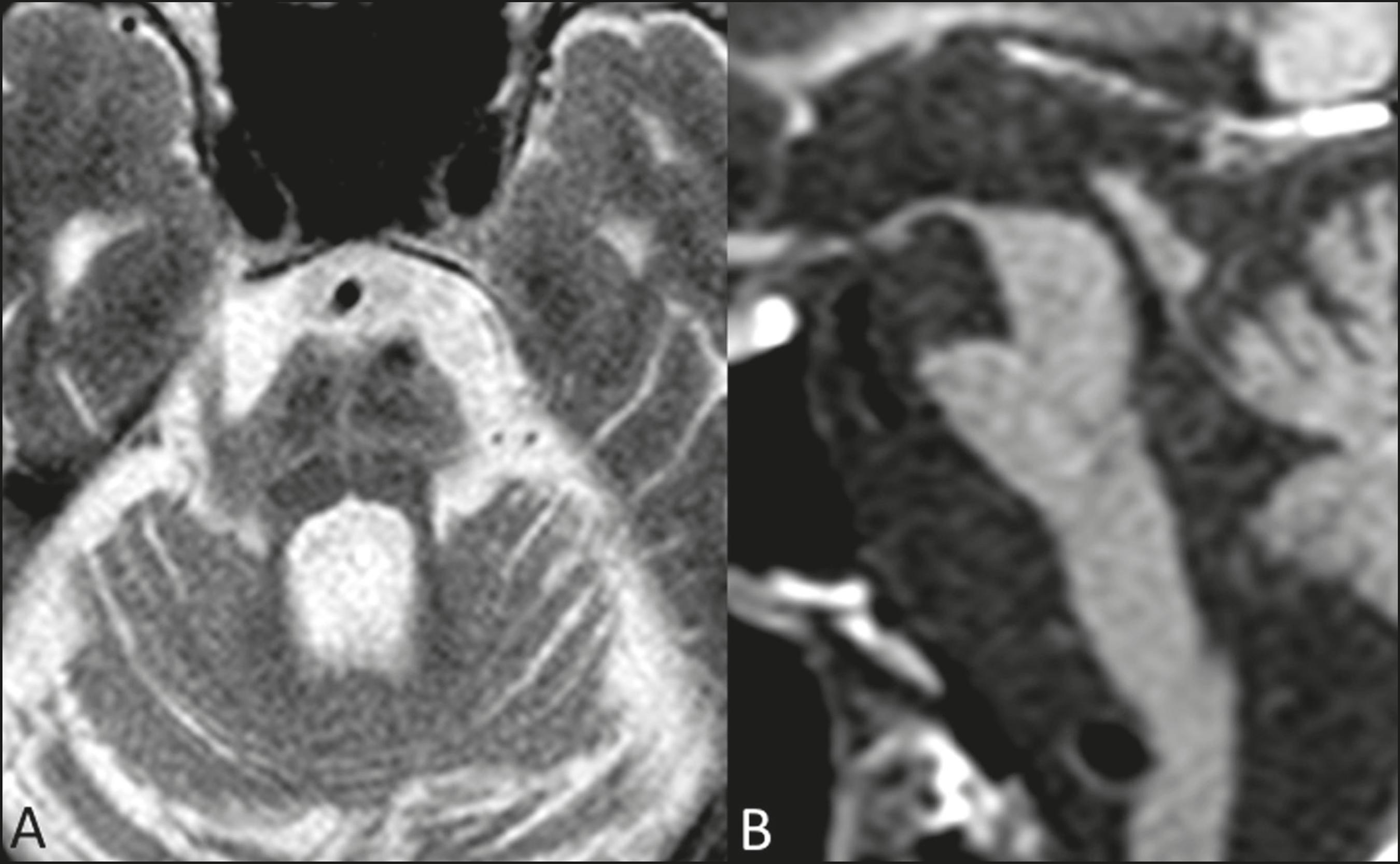
- Hot cross bun sign in axial T2-weighted sequences, with atrophy of the pons(13-15)
|
- All have good specificity but can be seen in other conditions(13-15)
|
|
- Reduced FA and increased ADC in the putamen(43-48)
|
- High sensitivity and specificity, especially in MSA-P, but can also occur in PSP(43-48)
|
| - Hyperintense signal in axial T2-weighted sequences, with atrophy of the putamen(13-15)
|
- Moderate sensitivity, because they are not normally seen in the early stages of the disease(13-15)
|
- Reduced FA and increased ADC in the pons, cerebellum, and middle cerebellar peduncles(43,50-52)
|
- High sensitivity and specificity, especially in MSA-C. In conjunction with changes in the putamen, they increase the specificity consider-ably43,50-52)
|
| - Atrophy of the cerebellar hemispheres and the middle cerebellar peduncles(13-15)
|
- Diffusion measures, taken together with conventional MRI measures, present high accuracy in the differential diagnosis with PD(53)
|
| PSP |
- Hummingbird or penguin sign in the sagittal plane(13-15)
|
- All have very high specificity, al-though they might not be seen in the early stages(13-15)
|
|
- Reduced FA in the midbrain and superior cerebellar peduncles(16,49,52)
|
- High sensitivity and specificity(16,49,52)
|
| - Mickey Mouse sign in the axial plane(13-15)
|
- Reduced FA in the globus pallidus and caudate nucleus(47,49,50,54)
|
- Can facilitate the differential diag-nosis but have not been widely studied(47,49,50,54)
|
| - Atrophy of midbrain and superior cerebellar peduncles(13-15)
|
| CBD |
- Asymmetric atrophy of the parietal lobes(16)
|
- Low specificity and sensitivity(16)
|
|
- Reduced FA and increased ADC in the corpus callosum and frontal lobes, mainly in the corticospinal tract contralateral to the symptomatic side(16,55)
|
- Low sensitivity and specificity but can be seen in the early stages, even in the absence of atrophy(16,55)
|
| - Thinning of the corpus callosum(16)
|
- Increased ADC in the motor thala-mus, precentral gyrus, and postcentral gyrus(56)
|
- Rarely studied but can facilitate the differential diagnosis with PSP, especially if asymmetric(56)
|

 The role of diffusion magnetic resonance imaging in Parkinson's disease and in the differential diagnosis with atypical parkinsonism