 GREEN SYNTHESIS AND PHOTOTHERMAL EFFECT OF SILVER NANOPARTICLES USING ADENANTHERA PAVONINA SEED EXTRACT
Abstract
This is the first study to investigate the photothermal effect of silver nanoparticles (AgNPs) encapsulated with the polysaccharide galactomannan (GAP), synthesized from the aqueous extract of Adenanthera pavonina seeds. The synthesized nanoparticles were characterized by X-ray diffraction (XRD), Fourier transform infrared spectroscopy (FTIR), transmission electron microscopy (TEM), energy-dispersive X-ray spectroscopy (EDS), and ultraviolet-visible (UV-Vis) absorption spectroscopy. The UV-Vis absorption spectra showed an increase in the absorption band around 425 nm, indicating the presence of AgNPs. The average particle size was 9.61 ± 5.0 nm. The photothermal performance was evaluated under infrared radiation using a near-infrared (NIR) laser with a wavelength of 850 nm. It was observed that AgNPs encapsulated with a higher concentration of GAP polysaccharides exhibited a significant temperature increase, reaching a temperature variation (∆T) of approximately 40 °C. Furthermore, they demonstrated excellent photothermal stability over repeated cycles, indicating their potential as photothermal agents.
Keywords:
green synthesis; biopolymer; silver nanoparticles; photothermal effect.
INTRODUCTION
Nanotechnology is an interdisciplinary field focused on the manipulation and control of matter at the nanometric scale, typically between 1 and 100 nm, where materials exhibit physicochemical properties that differ significantly from their bulk counterparts.1 These size-dependent properties have enabled advances across multiple areas, including medicine, electronics, catalysis, and energy applications. Central to this field is the development of reliable synthesis methods that allow precise control over nanoparticle size, shape, and composition.2 Traditional top-down and bottom-up chemical synthesis routes for metallic nanoparticles often rely on hazardous reducing and stabilizing agents, such as sodium borohydride or hydrazine, which raise environmental and biological concerns due to their toxicity and poor biodegradability.3 In addition, the high surface energy of nanoparticles promotes aggregation, compromising colloidal stability and functional performance.
These limitations have driven increasing interest in sustainable and environmentally benign synthesis strategies.4 Green synthesis has emerged as a promising alternative, employing biological systems such as plants, microorganisms, or biopolymers as reducing and stabilizing agents.5,6 Among these approaches, plant-mediated synthesis has gained particular attention due to its simplicity, cost-effectiveness, and scalability.7 Plant extracts contain a diverse range of biomolecules, including polyphenols, flavonoids, sugars, polysaccharides, amino acids, and proteins, which can act synergistically during nanoparticle formation, promoting both reduction of metal ions and stabilization of the resulting nanostructures.8 Silver nanoparticles (AgNPs) are among the most extensively studied metallic nanomaterials due to their unique optical, electronic, and biological properties.9,10
These properties are largely governed by localized surface plasmon resonance (LSPR), which arises from the collective oscillation of conduction electrons at the nanoparticle surface upon interaction with electromagnetic radiation.10,11 LSPR leads to enhanced light absorption and scattering and enables efficient non-radiative relaxation processes that convert absorbed optical energy into heat, giving rise to the photothermal effect.11,12 The photothermal effect has been widely explored for biomedical applications, particularly in photothermal therapy (PTT), where plasmonic nanoparticles act as nano-transducers that generate localized hyperthermia upon laser irradiation.13
When irradiated in the near-infrared (NIR) region, where biological tissues exhibit reduced absorption and increased penetration depth, AgNPs can induce controlled temperature increases capable of damaging tumor cells or pathogenic microorganisms while minimizing effects on surrounding healthy tissues.14 The efficiency of this process is strongly influenced by nanoparticle size, morphology, surface chemistry, and the surrounding dielectric environment, all of which are directly linked to the synthesis route.15
While photothermal effects have been demonstrated for AgNPs synthesized using different green approaches, such as biopolymer-based systems, the number of studies specifically addressing plant extract-mediated AgNPs and their photothermal performance remains limited. In particular, the role of plant-derived reducing and encapsulating agents in modulating photothermal efficiency, thermal stability, and light-to-heat conversion has not been systematically explored.16 Adenanthera pavonina is a plant species widely distributed in tropical regions, including Brazil. Its seed extract, rich in galactomannan polysaccharides and phenolic compounds, has been reported17 to exhibit antioxidant, anti-inflammatory, and low-toxicity properties.
The extract obtained from A. pavonina seeds constitutes a complex bio-organic system, composed predominantly of galactomannan-type polysaccharides, consisting of a main mannose backbone with β-(1→4) linkages and galactose side units linked through α-(1→6) bonds. In addition to this polysaccharide fraction, the extract contains triterpenes (such as nonacosane and hentriacontane), terpenoids, tannins, and flavonoids,18 with gallic acid being particularly noteworthy, as well as phenolic compounds and other secondary metabolites.17-20 Phytochemical studies18 also report the presence of glycosides, saponins, and steroids. Although the exact quantitative composition may vary depending on environmental and extraction conditions, these components are known to exhibit reducing and stabilizing functionalities, making the extract suitable for green synthesis of metallic nanoparticles.19,20
These components play a crucial role in plant-mediated nanoparticle synthesis by acting simultaneously as reducing agents for Ag+ ions and as stabilizing agents for the resulting nanoparticles. Despite these characteristics, only a few studies21,22 have explored the use of A. pavonina extracts in the synthesis of metallic nanoparticles, and the photothermal behavior of AgNPs derived from this plant-based route remains largely unexplored. In this context, the present work aims to synthesize silver nanoparticles via a plant-based green synthesis route using A. pavonina seed extract as both reducing and stabilizing agent. Furthermore, this study investigates the photothermal performance of the resulting AgNPs under NIR laser irradiation, with particular emphasis on the influence of extract volume on nanoparticle formation, stability, and photothermal efficiency. By focusing specifically on plant-mediated synthesis, this work seeks to clarify the relationship between bio-organic components and the photothermal behavior of AgNPs, contributing to their rational design for biomedical applications.
EXPERIMENTAL
Extract preparation
Adenanthera pavonina seeds were collected from the streets of the Historic Center of São Luís, MA, Brazil. To obtain the extract, the seeds were washed with distilled water at ambient temperature and air-dried at room temperature for 24 h. The dried seeds were then fragmented, and the inner extract was carefully removed and sieved through a 75 μm mesh. Subsequently, 2.0 g of the sieved extract was added to a beaker containing 100 mL of distilled water at ambient temperature and stirred for 1 h. The mixture was then filtered, and the resulting extract was stored in a hermetically sealed container at 4 °C. The extract was used without further purification in order to preserve its natural bio-organic composition, which is consistent with plant-based green synthesis approaches reported in the literature.20
Synthesis of AgNPs
For the synthesis of AgNPs, three different volumes of the extract: 10, 15, and 30 mL were used. Each volume was added to an aqueous solution of silver nitrate (1 mM AgNO3, 99% purity, Isofar) with corresponding volumes of 90, 85, and 70 mL, respectively. The mixtures were stirred at room temperature for 24 h. A color change was observed after approximately 30 min, indicating the formation of nanoparticles. The suspensions were then washed with deionized water, centrifuged at 12,000 rpm, and dried in an oven at 60 °C for 24 h. See Figure 1 for a summary of the process to obtain the AgNPs.

Characterization of AgNPs
The synthesized AgNPs were characterized using dispersions prepared at a concentration of 1 mg mL-1. The solutions were sonicated for 30 min in an ultrasonic bath to ensure homogeneous nanoparticle dispersion and prevent aggregation. A small aliquot was used to measure the absorption spectrum with a single-beam ultraviolet-visible (UV-Vis) spectrophotometer (Aquamate 8000, Thermo Orion) in the wavelength range of 300 to 700 nm. X-ray diffraction (XRD) analysis was performed using a Bruker D8 Advance diffractometer with Cu Kα radiation (λ = 1.548 Å). Fourier transform infrared (FTIR) spectroscopy was performed to identify the functional groups present in the A. pavonina extract and the biosynthesized AgNPs. Spectra were acquired in attenuated total reflectance (ATR) mode using a Shimadzu IR Prestige 21 spectrophotometer over a wavenumber range of 400 to 4000 cm-1.
High-resolution transmission electron microscopy (HR-TEM) and selected area electron diffraction (SAED) analyses were performed using a JEM-2100 microscope. For this, a small aliquot of the purified AgNPs was dispersed and sonicated to ensure a uniform deposit on the grid. HR-TEM allowed for detailed observation of nanoparticle morphology and size, while SAED provided information about the crystalline structure, confirming crystallinity and possible preferential orientations.
Photothermal tests
The photothermal performance was evaluated using a near infrared (NIR) laser source (Elubor) with a wavelength of 85 nm, operating in continuous wave (CW) mode at a power of 0.660 W. The extract and the AgNP samples (AgNPs10, AgNPs15, and AgNPs30) were dispersed in deionized water under vigorous sonication to achieve a concentration of 1 mg mL-1. A 1 mL aliquot of each dispersion was placed in an Eppendorf tube (made of pure polypropylene) and irradiated for 20 min, followed by a 10-min cooling period without irradiation. The temperature change was monitored using a professional thermal camera (UNI-T Uti260b), and the data were analyzed using computer software OriginPro 2018 (OriginLab Corporation, Northampton, MA, USA). The photothermal conversion efficiency (η) was calculated using the following equation:
where h is the heat transfer coefficient, S is the surface area of the container, Tmax is the maximum equilibrium temperature, Tsol is the initial solution temperature before the first cycle, QDis is the heat dissipated by the solution and the container, I is the laser power (measured in Watts), and A850nm is the absorbance of the solution at 850 nm.
RESULTS AND DISCUSSION
UV-Vis spectroscopy
The formation and stability of silver nanoparticles (AgNPs) were primarily confirmed through UV-Vis spectroscopy. The green-synthesized silver nanoparticles in aqueous solution are shown in Figure 2. The extract solution from A. pavonina seeds, initially nearly colorless, exhibited distinct visual changes following the addition of AgNO3. The formation of silver nanoparticles could be visually confirmed by the color changes observed in the reaction mixture (see inset in Figure 2), a characteristic phenomenon indicative of silver nanoparticle synthesis. A noticeable color shift occurred within the first 15 min after the addition of silver nitrate (AgNO3), however, after 24 h, distinct shades were observed depending on the volume of extract used: 10 mL (yellow), 15 mL (orange), and 30 mL (brown), indicating the nucleation and growth of AgNPs. These color variations are associated with the excitation of localized surface plasmon resonance (LSPR) vibrations of the silver nanoparticles formed in the reaction medium.23 These observations are consistent with previous studies24,25 on the biosynthesis of AgNPs using various plant extracts. Furthermore, the different ratios between plant extract and AgNO3 in the synthesis process resulted in significant variations in both the intensity and width of the absorption bands, as clearly observed in Figure 2. The sample synthesized with the highest extract-to-AgNO3 ratio (AgNPs10: 90 mL extract / 10 mL AgNO3) exhibited the most intense and sharpest LSPR band.26

This high intensity and narrow profile suggest an efficient reduction of the available silver ions, leading to a high yield of nanoparticles with a relatively monodisperse size distribution, as the abundant biomolecules effectively cap and stabilize the newly formed nuclei.27 Conversely, the sample with the lowest extract-to-AgNO3 ratio (AgNPs30: 70 mL extract / 30 mL AgNO3) showed a lower absorption intensity and a broader SPR band. The reduced intensity indicates a lower yield of formed nanoparticles, as the amount of reducing agents in the extract may have been insufficient to completely reduce the higher concentration of Ag+ ions.26,28
The band broadening suggests an increase in polydispersity, which can result from Ostwald ripening or less effective stabilization under these conditions. The AgNPs15 sample exhibited an intermediate profile. Despite these differences, all samples exhibited characteristic UV-Vis absorption bands with maximum absorbance around 420 nm, in agreement with the literature.25 Similar studies29 on the synthesis of AgNPs have demonstrated that high concentrations of Moringa oleifera leaf extract hinder the detection of characteristic peaks in the UV-Vis spectra. No absorbance bands were observed in the spectrum of the A. pavonina seed extract. Moreover, after 24 h, the stability of the AgNPs was confirmed, attributed to the encapsulation of biomolecules that act as stabilizing agents in the solution.24,30
Water-soluble phytochemicals such as organic acids, quinones, and flavones compounds present in A. pavonina seeds are mainly responsible for the immediate reduction of Ag+ ions to Ag0.31 The UV-Vis spectra also provide information about the morphology of the biosynthesized silver nanoparticles. The synthesized AgNPs in this study are isotropic and spherical, as indicated by single, clear surface plasmon peak observed in Figure 2.32
X-ray diffraction (XRD)
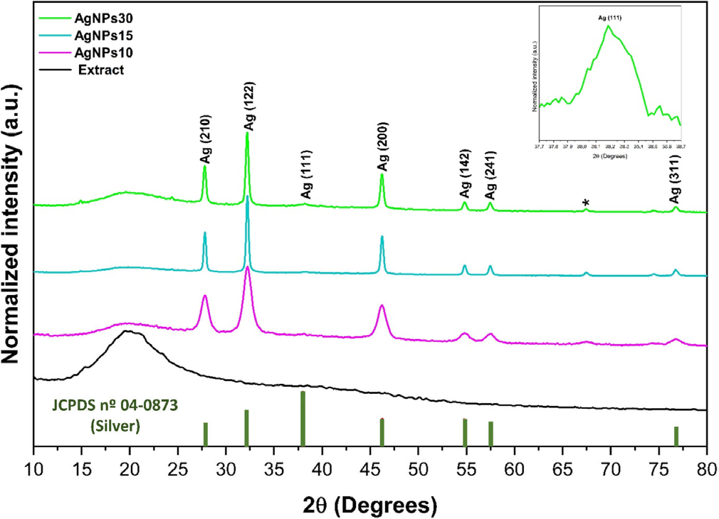
The XRD patterns of the AgNPs synthesized using different volumes of A. pavonina seed extract, together with the reference pattern of metallic silver (Joint Committee on Powder Diffraction Standards (JCPDS) file No. 04-0783), are presented in Figure 3. The diffraction profiles of the AgNPs15 and AgNPs30 samples exhibit distinct peaks at 2θ values of approximately 27.8, 32.2, 38.1, 46.2, 54.8, 57.4, and 76.7°, which can be indexed to the (210), (122), (111), (200), (142), (241), and (311) crystallographic planes, respectively. These reflections are characteristic of face-centered cubic (FCC) metallic silver, confirming the successful reduction of Ag+ ions into crystalline Ag0 nanoparticles.33 The average crystallite size of the AgNPs was estimated using Scherrer’s equation applied to the (111) diffraction peak, yielding a value of approximately 2 nm.

The presence of intense and well-defined Ag diffraction peaks indicates that metallic silver is the dominant crystalline phase in the synthesized materials. This observation is consistent with previous reports33,34 on green-synthesized AgNPs, in which the crystalline structure is primarily governed by metallic silver, while organic components derived from plant extracts act mainly as reducing and stabilizing agents rather than forming crystalline domains detectable by XRD. In addition to the characteristic Ag reflections, a low-intensity additional diffraction peak was observed at 2θ = 67.39°, as indicated by an asterisk in Figure 3. This peak does not correspond to any of the standard reflections of FCC metallic silver listed in the JCPDS database, nor can it be unambiguously assigned to a known crystalline silver oxide phase.
Similar secondary diffraction features have been reported33,35 in plant-mediated syntheses of AgNPs and are often attributed to the presence of residual bio-organic components, interfacial phases, or weakly ordered structures originating from the biological matrix. However, it is important to note that the specific peak position observed in the present study does not show a direct correspondence with the additional peaks reported in those works, which were detected at different 2θ values and under different synthesis conditions.
Plant extracts are complex mixtures predominantly composed of amorphous or poorly crystalline organic compounds, such as polysaccharides, polyphenols, and secondary metabolites. As a result, XRD is not a definitive technique for identifying specific organic phases within such systems. Therefore, the additional diffraction feature observed at 2θ = 67.39° should be interpreted cautiously and regarded as a minor contribution arising from the bio-organic environment or possible interfacial interactions between silver nanoparticles and residual extract-derived compounds, rather than as evidence of a distinct crystalline organic phase. The relatively low intensity of this peak, when compared to the dominant Ag reflections, further indicates that it does not significantly affect the overall crystalline structure of the material. Instead, the crystallinity of the synthesized nanoparticles is overwhelmingly dictated by metallic silver.
Complementary characterization techniques, particularly FTIR spectroscopy, provide more reliable evidence for the presence and role of organic functional groups associated with nanoparticle stabilization and surface chemistry. Overall, the XRD results confirm the formation of crystalline FCC silver nanoparticles,36,37 while the minor additional diffraction feature highlights the complex nature of green-synthesized nanomaterials and the inherent limitations of XRD in resolving weakly ordered bio-organic contributions.
Fourier transform infrared spectroscopy (FTIR)
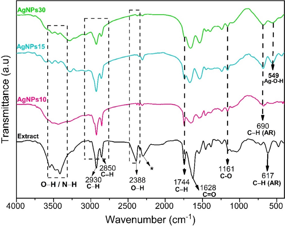
Figure 4 presents the FTIR spectra of the AgNPs synthesized using A. pavonina seed extract. FTIR analysis was employed to qualitatively identify the main functional groups present in the extract and to investigate their possible interactions with silver nanoparticles. It should be emphasized that plant extracts are complex bio-organic systems and that FTIR provides indicative, rather than definitive, chemical assignments.
FTIR spectra of A. pavonina seed extract and the green-synthesized AgNPs. The asterisk (*) denotes an absorption band attributed to atmospheric CO2
For the AgNPs10 sample, a broad and intense band centered at approximately 3421 cm-1 was observed, mainly attributed to O-H stretching vibrations associated with hydroxyl groups from polysaccharides and phenolic compounds, as well as contributions from adsorbed water molecules on the nanoparticle surface.38
In the other samples, a narrower and lower-intensity band appears around 3440 cm-1, which can be associated with O-H stretching vibrations of alcohol groups and, to a lesser extent, N-H stretching vibrations of amine-containing compounds, as reported in similar plant-mediated synthesis systems.39 Bands observed in the regions around 2930 and 2850 cm-1 correspond to asymmetric and symmetric C-H stretching vibrations of aliphatic -CH2 and -CH3 groups, commonly associated with the organic backbone of polysaccharides and other extract-derived constituents.40 The weak band detected at approximately 2388 cm-1 is not characteristic of O-H vibrations of carboxylic acids. Instead, it is more plausible to attributed it to atmospheric CO2 or carbonate-related absorptions, which are frequently observed as background features in FTIR measurements.41 Consequently, this band is not directly associated with the chemical composition of the extract or with the nanoparticle synthesis process. A related CO2 absorption feature is also observed near 2297 cm-1, supporting this interpretation. The bands located at 1744 and 1628 cm-1 are attributed to carbonyl-related vibrations.
The band at 1744 cm-1 is associated with C=O stretching vibrations of ester or carbonyl groups, while the band at 1628 cm-1 likely arises from overlapping contributions, including C=O stretching vibrations of amide groups or conjugated carbonyls, C=C stretching of aromatic structures, and the bending mode of adsorbed water molecules (δ H-O-H).42 Given the hydrophilic nature of the extract and the absence of high-temperature dehydration prior to FTIR analysis, the contribution of adsorbed moisture to this band cannot be excluded. The band observed at 1161 cm-1 is attributed to C-O stretching vibrations, characteristic of polysaccharides and phenolic compounds, which are known to play a stabilizing role in plant-mediated nanoparticle synthesis.8,42 Low-wavenumber bands detected at approximately 690 and 617 cm-1 are associated with out-of-plane C-H bending vibrations of aromatic rings, indicating the presence of phenolic structures within the extract-derived organic matrix.42
A weak band at around 549 cm-1 may be attributed to Ag-O related vibrations, possibly arising from surface interactions between silver species and oxygen-containing functional groups from the extract. However, this band should be interpreted cautiously and regarded as indicative of surface interactions rather than as direct confirmation of AgNP formation, which is more reliably established by XRD and UV-Vis analyses.17,42 Overall, the observed shifts and changes in band intensities between the extract and AgNP spectra suggest interactions between silver nanoparticles and extract-derived functional groups, particularly hydroxyl and carbonyl moieties. These interactions are commonly reported in plant-mediated synthesis and are generally attributed to the coordination of Ag+/Ag0 species with oxygen-containing functional groups, which can facilitate reduction and provide colloidal stabilization through surface capping effects.43,44 To summarize, the main bands and their assignments are presented in Table 1.
Main FTIR bands, functional group assignments, and their proposed role in the synthesis of AgNPs
When compared to AgNPs synthesized using A. pavonina leaf extract,21 noticeable differences in FTIR profiles are observed. These differences can be attributed to variations in biochemical composition between seeds and leaves of the same plant species. Leaf-mediated synthesis typically exhibits spectral features associated with flavonoids, terpenoids, and protein-related functional groups, whereas seed-derived AgNPs show more pronounced contributions from polysaccharideand carbonyl-related functional groups. Such differences reflect the distinct roles played by biomolecules inherent to different plant parts in the reduction and stabilization of silver nanoparticles.
Comparisons with AgNPs synthesized using extracts from other plant species were not further considered, as the chemical constituents governing FTIR spectra vary substantially among different botanical systems. Therefore, cross-species comparisons may not provide meaningful insights into nanoparticle-biomolecule interactions within a specific plant-mediated synthesis route. In contrast, chemically synthesized AgNPs obtained using conventional reducing agents such as sodium borohydride or trisodium citrate typically exhibit minimal organic signatures in FTIR spectra, often limited to weak bands associated with adsorbed water or simple carboxylate groups.45 This distinction further emphasizes the unique bio-organic environment associated with plant-mediated AgNP synthesis.
TEM and HR-TEM
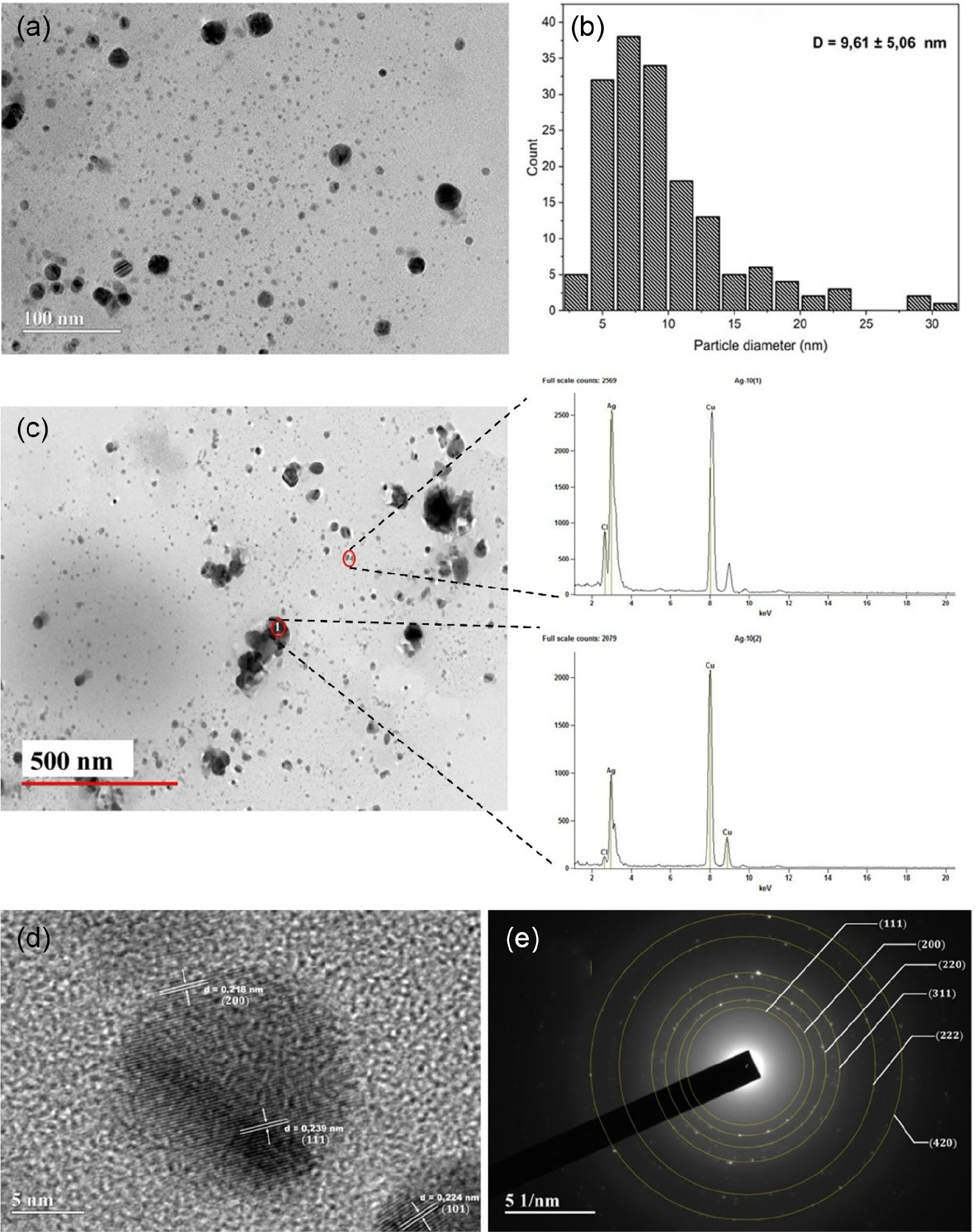
Figure 5 presents the TEM and HR-TEM characterization of the AgNPs10 sample. This sample was synthesized using 10 mL of A. pavonina seed extract and 90 mL of AgNO3 solution. The TEM micrographs shown in Figure 5a indicate that the synthesized nanoparticles exhibit a predominantly quasi-spherical morphology, characterized by rounded contours and moderate shape dispersion. Due to the limited number of TEM images and the absence of a large statistical dataset, a fully isotropic spherical morphology cannot be conclusively established. Nevertheless, the observed particle shapes suggest a clear tendency toward quasi-spherical nanoparticles, which is commonly reported in plant-mediated synthesis routes.

AgNPs10: (a) TEM analysis; (b) particle size distribution; (c) EDAX spectrum; (d,e) SAED patterns by HR-TEM
The particle size distribution (Figure 5b) reveals an average diameter of 9.61 ± 5.06 nm, indicating a polydisperse system. Such polydispersity is consistent with green synthesis approaches and may contribute to the broadening and increased intensity of the surface plasmon resonance (SPR) band observed in the UV-Vis spectra (Figure 4), as variations in particle size and shape influence plasmonic behavior. The EDAX spectrum presented in Figure 5c confirms the presence of silver (Ag) as the main constituent of the nanoparticles. Additional peaks corresponding to copper (Cu) originate from the copper grid used during TEM analysis. Although EDAX does not allow for precise quantitative analysis of individual nanoparticles, the strong Ag signal corroborates the successful formation of silver-based nanostructures. Future studies employing elemental mapping techniques, such as STEM-EDS, may provide further insights into possible variations in silver distribution as a function of particle size.
The HR-TEM image shown in Figure 5d reveals clear lattice fringes, indicating the crystalline nature of the nanoparticles. The measured interplanar spacings of approximately 0.239, 0.218, and 0.224 nm correspond to the (111), (200), and (101) planes of metallic silver, respectively, in agreement with literature data.30 The SAED pattern displayed in Figure 5e consists of well-defined diffraction rings that can be indexed to the (111), (200), (220), and (311) planes, confirming the face-centered cubic (FCC) crystalline structure of silver (JCPDS card No. 04-0783). Notably, three crystallographic planes, (111), (200), and (311) are consistently identified by both SAED and XRD analyses, while the (220) reflection appears exclusively in the SAED pattern, as expected for a local probe technique.
This correspondence demonstrates strong agreement between XRD, which provides bulk-averaged structural information, and SAED, which probes individual nanoparticles. Together, these results confirm that the synthesized AgNPs possess an FCC crystalline structure at both the macroscopic and nanoscopic levels.
Photothermal analysis
The photothermal performance of the AgNPs synthesized using different volumes of A. pavonina seed extract was systematically evaluated under NIR laser irradiation (850 nm). The results demonstrate a pronounced and reproducible light-to-heat conversion capability for all AgNP-containing samples, as well as good thermal stability under prolonged and cyclic irradiation. Figures 6a and 6b illustrate a representative thermal response of the AgNPs30 sample before and during NIR irradiation. The initial temperature of 28.9 °C corresponds to the thermal equilibrium of the system prior to laser exposure. Upon irradiation, the temperature rapidly increased, reaching approximately 70.0 °C within a few minutes and remaining stable thereafter. This temperature plateau indicates the establishment of a steady state, in which the rate of photothermal heat generation is balanced by heat dissipation to the surrounding environment. Such behavior is characteristic of efficient photothermal agents.

Thermal images of AgNPs30 sample (a) before and (b) during NIR irradiation (steady state); (c) heating and cooling curves of the samples under continuous NIR laser irradiation; (d) temperature variation (∆T) as a function of time for AgNPs synthesized with different extract volumes; (e) heating-cooling cycles and (f) conversion factor efficiency for AgNPs30
Figure 6c compares the heating profiles of all samples at a fixed concentration of 1 mg mL-1. Among the AgNP suspensions, AgNPs30 exhibited the highest maximum temperature (ca. 70 °C), followed by AgNPs15 (ca. 57.3 °C) and AgNPs10 (ca. 46.1 °C). These temperature values are within the range commonly reported as suitable for photothermal therapeutic applications under NIR irradiation.46 In contrast, the aqueous suspension containing only the A. pavonina extract showed a minimal temperature increase, reaching approximately 31.8 °C. This result indicates that the extract alone has a negligible photothermal effect under the experimental conditions employed.
The pronounced heating observed for the AgNP-containing samples is primarily attributed to the LSPR of metallic silver nanoparticles. Upon NIR irradiation, collective oscillations of conduction-band electrons are excited on the nanoparticle surface, leading to efficient absorption of photon energy. This energy is predominantly dissipated through non-radiative relaxation pathways, resulting in rapid conversion of optical energy into heat. The significantly lower temperature increase observed for the extract-only sample confirms that plasmonic heating from AgNPs is the dominant photothermal mechanism in this system.
Nevertheless, the A. pavonina seed extract contains conjugated organic compounds, such as polyphenols and flavonoids, which possess delocalized π-electron systems capable of absorbing incident photons.47 Excitation and subsequent non-radiative relaxation of these molecular systems may contribute marginally to heat generation, as suggested by the slight temperature increase observed in the extract-only control.47 However, this contribution is minor when compared to the plasmon-mediated heating induced by AgNPs.
Figure 6d presents the temperature increase (∆T) as a function of irradiation time. All AgNP samples exhibit a rapid temperature rise followed by stabilization within approximately 160 s, highlighting their fast thermal response and efficient energy dissipation. AgNPs30 showed the highest ∆T (ca. 40 °C), whereas AgNPs15 and AgNPs10 displayed intermediate values. This trend indicates that synthesis conditions significantly influence photothermal efficiency. Importantly, the dependence of photothermal performance on the volume of extract used during synthesis should be interpreted as an indirect effect.
While the extract is not the primary photothermal agent, it plays a crucial role in modulating nanoparticle properties, such as particle size distribution, surface chemistry, colloidal stability, and the local dielectric environment surrounding the AgNPs. These factors directly affect plasmonic absorption and non-radiative relaxation efficiency, thereby influencing overall photothermal behavior. Thermal cycling experiments (Figure 6e) further demonstrate the robustness of the synthesized AgNPs. All samples exhibited stable and reproducible heating and cooling profiles over three consecutive irradiation cycles, with no significant loss of performance. This behavior indicates excellent photothermal stability and reversibility, particularly for the AgNPs30 sample.
The calculated photothermal conversion efficiencies (Figure 6f) are consistent with the observed temperature profiles. The extract-only suspension showed a low efficiency of approximately 1%, whereas AgNPs10, AgNPs15, and AgNPs30 exhibited substantially higher efficiencies of ca. 36, 49, and 77%, respectively. These results further support the conclusion that plasmonic AgNPs are responsible for the dominant photothermal effect, while the extract volume influences efficiency by governing nanoparticle formation and surface characteristics, rather than acting as an independent photothermal converter.48
Overall, the results indicate that the enhanced photothermal performance arises from plasmon-mediated heating of AgNPs, with the plant extract exerting a secondary and modulating role through its influence on the synthesis and stabilization of the nanoparticles. Further spectroscopic and time-resolved studies would be required to quantitatively decouple the individual contributions of metallic nanoparticles and organic components.
CONCLUSIONS
This study successfully demonstrated the green synthesis of AgNPs using A. pavonina seed extract as both a reducing and encapsulating agent, highlighting an eco-friendly and sustainable approach. The synthesized AgNPs exhibited notable physicochemical properties, as confirmed by characterization techniques such as XRD, FTIR, UV-Vis, TEM, and EDS, which revealed a face-centered cubic crystalline structure, an average particle size of 9.61 ± 5.0 nm, and the presence of organic functional groups derived from the plant extract. Photothermal analysis showed that AgNPs encapsulated with higher concentrations of galactomannan (GAP) achieved a significant temperature increase (∆T ca. 40 °C) under NIR irradiation, along with excellent stability over repeated cycles, reaching a photothermal conversion efficiency of up to 77%. These findings underscore the potential of AgNPs as efficient photothermal agents, particularly for biomedical applications such as hyperthermia therapy and antibacterial treatments.
ACKNOWLEDGMENTS
This study was funded by FAPEMA. The authors thank the Colloidal Systems Laboratory (UFMA) for infrastructure and equipment, and LabMic/UFG for technical assistance.
DATA AVAILABILITY STATEMENT
The authors confirm that the data supporting the findings of this study are available within the article.
REFERENCES
-
1 Chugh, D.; Viswamalya, V. S.; Das, B.; J. Genet. Eng. Biotechnol. 2021, 19, 126. [Crossref]
» Crossref -
2 Ansar, S.; Tabassum, H.; Aladwan, N. S. M.; Ali, M. N.; Almaarik, B.; AlMahrouqi, S.; Abudawood, M.; Banu, N.; Alsubki, R.; Sci. Rep. 2020, 10, 18564. [Crossref]
» Crossref -
3 Singh, J.; Dutta, T.; Kim, K. H.; Rawat, M.; Samddar, P.; Kumar, P.; J. Nanobiotechnol. 2018, 16, 84. [Crossref]
» Crossref -
4 Arshad, F.; Naikoo, G. A.; Hassan, I. U.; Chava, S. R.; El-Tanani, M.; Aljabali, A. A.; Tambuwala, M. M.; Appl. Biochem. Biotechnol. 2024, 196, 3636. [Crossref]
» Crossref -
5 Alam, M.; J. King Saud Univ., Sci. 2022, 34, 102327. [Crossref]
» Crossref -
6 Mallikarju, K.; Dillip, G. R.; Narasimha, G.; Sushma, N. J.; Prasad Raj, B. D.; Research Journal of Nanoscience and Nanotechnology 2012, 2, 17. [Crossref]
» Crossref -
7 Mallik, K.; Mandal, M.; Pradhan, N.; Pal, T.; Nano Lett. 2001, 1, 319. [Crossref]
» Crossref -
8 Mustapha, T.; Misni, N.; Ithnin, N. R.; Daskum, A. M.; Unyah, N. Z.; Int. J. Environ. Res. Public Health 2022, 19, 674. [Crossref]
» Crossref -
9 Tariq, M.; Mohammad, K. N.; Ahmed, B.; Siddiqui, M. A.; Lee, J.; Molecules 2022, 27, 4754. [Crossref]
» Crossref -
10 Lee, J. S.; Kim, J.; Ye, Y.; Kim, T.; Adv. Drug Delivery Rev. 2022, 186, 114339. [Crossref]
» Crossref -
11 Li, W. R.; Xie, X. B.; Shi, Q. S.; Zeng, H. Y.; Yang, Y. S. O.; Chen, Y. B.; Appl. Microbiol. Biotechnol. 2010, 85, 1115. [Crossref]
» Crossref -
12 Zhang, J.; Tang, T.; Yang, R.; Wang, G.; Ye, K. H.; Shi, J.; Microstructures 2024, 4, 2024008. [Crossref]
» Crossref -
13 Ahmad, S.; Ahmad, S.; Ali, S.; Esa, M.; Khan, A.; Yan, H.; Int. J. Nanomed. 2024, 19, 3187. [Crossref]
» Crossref -
14 Kah, G.; Chandran, R.; Abrahamse, H.; Cells 2023, 12, 2012. [Crossref]
» Crossref -
15 El Mouzahim, M.; Eddarai, E. M.; Eladaoui, S.; Guenbour, A.; Bellaouchou, A.; Zarrouk, A.; Boussen, R.; Food Chem. 2023, 410, 135470. [Crossref]
» Crossref -
16 Alexeree, S. M. I.; Abou-Seri, H. M.; EL-Din, H. E. S.; Youssef, D.; Ramadan, M. A.; Lasers in Medical Science 2024, 39, 43. [Crossref]
» Crossref -
17 Ara, A.; Arifuzzaman, M.; Ghosh, C. K.; Hashem, M. A.; Ahmad, M. U.; Bachar, S. C.; Nahar, L.; Sarker, S. D.; Rev. Bras. Farmacogn. 2010, 20, 929. [Crossref]
» Crossref -
18 Gawande, R.; Int. J. Pharm. Sci. 2025, 3, 2479. [Crossref]
» Crossref -
19 Koodalingam, A.; Manikandan, R.; Indhumathi, M.; Kaviya, E. S.; Asian Pac. J. Trop. Med. 2015, 8, 112. [Crossref]
» Crossref -
20 Nobre, K. A.; Soares, C. E. A.; Vieira, I. G. P.; de Almeida, R. R.; Moreira, R. A.; de Araújo, T. G.; Ribeiro, M. E. N. P.; Ricardo, N. M. P. S.; Quim. Nova 2018, 41, 607. [Crossref]
» Crossref -
21 Ravindran, R. S. E.; Shanmugam, K.; Veeramani, S.; Ilangovan, R.; Ali, D.; Almutairi, B. O.; Palanivel, H.; Goel, M.; J. Nanomater. 2022, 2022, 760829. [Crossref]
» Crossref -
22 Barkat, A. M.; Harshita; Beg, S.; Naim, M. J.; Pottoo, F. H.; Singh, S. P.; Ahmad, F. J.; Recent Pat. Anti-Infect. Drug Discovery 2018, 13, 53. [Crossref]
» Crossref -
23 Asif, M.; Yasmin, R.; Asif, R.; Ambreen, A.; Mustafa, M.; Umbreen, S.; Dose-Response 2022, 20, 2. [Crossref]
» Crossref -
24 Ahmed, M. J.; Murtaza, G.; Mehmood, A.; Bhatti, T. M.; Mater. Lett. 2015, 153, 10. [Crossref]
» Crossref -
25 Ajitha, B.; Reddy, Y. A. K.; Reddy, P. S.; Spectrochim. Acta, Part A 2014, 121, 164. [Crossref]
» Crossref -
26 Maduraimuthu, V.; Ranishree, J. K.; Gopalakrishnan, R. M.; Ayyadurai, B.; Raja, R.; Heese, K.; Antioxidants 2023, 12, 1298. [Crossref]
» Crossref -
27 Ali, M.; Kim, B. D.; Belfield, K.; Norman, D.; Brennan, M.; Ali, G. S.; Mater. Sci. Eng., C 2016, 58, 359. [Crossref]
» Crossref -
28 Ulusoy, E.; Bozkurt, A.; Durmaz, S.; Servi, H.; Vardar, F.; Erisen, S.; BMC Plant Biol. 2024, 24, 362. [Crossref]
» Crossref -
29 Mohammed, G. M.; Hawar, S. N.; Int. J. Biomater. 2022, 2022, 136641. [Crossref]
» Crossref -
30 Aboelfetoh, E. F.; El-Shenody, R. A.; Ghobara, M. M.; Environ. Monit. Assess. 2017, 189, 349. [Crossref]
» Crossref -
31 Das, D.; Ghosh, R.; Mandal, P.; SN Appl. Sci. 2019, 1, 498. [Crossref]
» Crossref -
32 Liknaw, T.; Belay, Y.; Ramesh, R.; Prassad, R. D. M.; Sci. Rep. 2025, 15, 22481. [Crossref]
» Crossref -
33 Karthik, L.; Kumar, G.; Kirthi, A. V.; Rahuman, A. A.; Rao, K. V. B.; Bioprocess Biosyst. Eng. 2014, 37, 261. [Crossref]
» Crossref -
34 Halawani, E. M.; J. Biomater. Nanobiotechnol. 2017, 08, 22. [Crossref]
» Crossref -
35 Anandalakshmi, K.; Venugobal, J.; Ramasamy, V.; Appl. Nanosci. 2016, 6, 399. [Crossref]
» Crossref -
36 Meng, Y.; Nanomaterials 2015, 5, 1124. [Crossref]
» Crossref -
37 Priyadharshini, R. I.; Prasannaraj, G.; Geetha, N.; Venkatachalam, P.; Appl. Biochem. Biotechnol. 2014, 174, 2777. [Crossref]
» Crossref -
38 Ali, Z. H.; Al-Fatlawi, A. H.; Results Eng. 2023, 17, 100988. [Crossref]
» Crossref -
39 Liu, Y.; Kang, S.; Li, K.; Chen, J.; Bae, B.; Hwang, I.; Ahn, E. Y.; Park, Y.; Chun, K. H.; Lee, J.; J. Cleaner Prod. 2022, 379, 134655. [Crossref]
» Crossref -
40 Barabadi, H.; Mojab, F.; Vahidi, H.; Marashi, B.; Talank, N.; Hosseini, O.; Saravanan, M.; Inorg. Chem. Commun. 2021, 129, 108647. [Crossref]
» Crossref -
41 Kanna, A. V.; Amirthavarshini, R.; Sankari, T. U.; International Journal of Advances in Scientific Research 2017, 3, 12. [Crossref]
» Crossref -
42 Dua, T. K.; Giri, S.; Nandi, G.; Sahu, R.; Shaw, T. K.; Paul, P.; Chem. Pap. 2023, 77, 2947. [Crossref]
» Crossref -
43 Babu, S. A.; Prabu, H. G.; Mater. Lett. 2011, 65, 1675. [Crossref]
» Crossref -
44 Sreelekha, E.; George, B.; Shyam, A.; Sajina, N.; Mathew, B.; Bionanoscience 2021, 11, 489. [Crossref]
» Crossref -
45 Khatoon, U. T.; Velidandi, A.; Rao, G. V. S. N.; Mater. Chem. Phys. 2023, 294, 126997. [Crossref]
» Crossref -
46 Sheng, J.; Zu, Z.; Qi, J.; Zhang, Y.; Wu, H.; Wang, Z.; Miao, Y.; Zheng, T.; Wang, S.; Zhang, L.; Lu, G.; Zhang, L.; Chem. Eng. J. 2024, 485, 149882. [Crossref]
» Crossref -
47 Wang, C.; Wang, X.; Chen, Y.; Fang, Z.; J. Photochem. Photobiol., B 2020, 204, 111587. [Crossref]
» Crossref -
48 Melo, R. M.; Albuquerque, G. M.; Monte, J. P.; Pereira, G. A. L.; Pereira, G.; Pharmaceuticals 2025, 18, 970. [Crossref]
» Crossref
Edited by
-
Executive Editor handled this article:
Júlio S. Rebouças