 Biodegradable chitosan membranes incorporating Jatropha mollissima for the treatment of varicose ulcers
Abstract
Varicose ulcers represent a severe manifestation of chronic venous insufficiency and remain a major public health challenge. In this study, chitosan-based dressings incorporating Jatropha mollissima extract (1-5% m/m) were developed via lyophilization to enhance healing performance. The membranes exhibited a predominantly fibrous morphology with high swelling capacity, approximately 1600% for pure chitosan, with a progressive reduction upon extract incorporation, indicating a decrease in hydrophilicity and increase in network density. Fourier transform infrared spectroscopy (FTIR) analysis revealed intensified bands associated with O-H and N-H groups, suggesting hydrogen bonding interactions between chitosan and phenolic compounds of the extract. Thermogravimetric analysis showed improved thermal stability, with reduced mass loss in extract-loaded membranes (ca. 50-60%) compared to pure chitosan. Biodegradation assays demonstrated a decrease in mass loss from 73.43 (M) to 65.00% (MJ5) after 21 days in phosphate salt (PBS), indicating enhanced structural stability. All samples exhibited no cytotoxicity toward L929 fibroblasts. These results demonstrate that the incorporation of J. mollissima modulates physicochemical properties through intermolecular interactions, leading to reduced swelling, controlled degradation, and improved stability. The study highlights the novelty of combining chitosan with a Caatinga-derived bioactive extract to produce multifunctional membranes with tunable physicochemical and biological properties, although further studies are required to confirm their suitability for biomedical applications.
Keywords:
biomaterials; venous ulcer; tissue regeneration.
INTRODUCTION
The skin is the largest organ in the human body, responsible for protection, thermoregulation, tactile sensation, and immunological functions. When injured, it loses its protective role, resulting in wounds that may become chronic. Among these, varicose ulcers stand out due to chronic venous insufficiency and represent a significant public health problem with high therapeutic costs and recurrence rates.1 These lesions severely impact the quality of life of the patients, causing pain, reduced mobility, and social and professional limitations, in addition to imposing a considerable economic burden on healthcare systems.2
In this context, the development of advanced biomaterials has been widely explored to improve the treatment of chronic wounds. Biodegradable materials for wound treatment, particularly in membrane form, have shown superior performance compared to conventional therapies, as they are able to maintain a moist environment, control exudate, reduce infection, and promote tissue regeneration.3
The effectiveness of these materials is strongly dependent on their physicochemical properties, which are directly related to their chemical structure and intermolecular interactions.
Among the available biomaterials, chitosan has attracted considerable attention due to its biocompatibility, biodegradability, antimicrobial activity, and regenerative potential.4 Structurally, chitosan is a cationic polysaccharide composed of D-glucosamine and N-acetyl-D-glucosamine units, containing amino (-NH2) and hydroxyl (-OH) groups. These functional groups enable intermolecular interactions such as hydrogen bonding and electrostatic interactions, which directly influence swelling behavior, degradation rate, and mechanical stability.4 Due to its high reactivity, chitosan can be combined with bioactive compounds, including plant extracts rich in phenolic molecules, enhancing its therapeutic performance.5
The incorporation of plant-derived extracts into polymeric matrices has emerged as a promising strategy to modulate material properties and introduce biological functionality. Phenolic compounds, flavonoids, and tannins can interact with polymer chains through hydrogen bonding and secondary interactions, potentially altering the organization of the polymer network, reducing hydrophilicity, and modifying swelling and degradation behavior.5 However, despite advances in chitosan-based systems, there is still a limited understanding of how specific plant extracts influence the physicochemical structure of the polymer network, particularly those derived from underexplored biomes such as the Caatinga.
Jatropha mollissima is a medicinal plant endemic to the Caatinga biome, known for its anti-inflammatory, antimicrobial, and healing properties.6 Its ethanolic extract contains bioactive compounds such as flavonoids, tannins, diterpenes, and triterpenes, with functional groups including hydroxyl (-OH), carbonyl (C=O), and carboxyl (-COOH), which are capable of interacting with polymeric matrices.7
These chemical groups are expected to promote intermolecular interactions with chitosan chains, potentially increasing network density and influencing properties such as hydrophilicity, swelling capacity, thermal stability, and biodegradation. Nevertheless, the effect of these interactions on the structure-property relationship of chitosan-based membranes remains insufficiently explored.
Based on this, we hypothesize that the incorporation of J. mollissima extract into chitosan membranes promotes hydrogen bonding and secondary interactions between the extract components and the polymer chains, leading to reduced hydrophilicity, controlled swelling, enhanced thermal stability, and slower biodegradation, without compromising biocompatibility.
Therefore, this study aims to develop and characterize chitosan-based membranes containing J. mollissima extract, focusing on understanding how extract-polymer interactions modulate the physicochemical and biological properties of the material for application in venous ulcer treatment.
EXPERIMENTAL
Materials
Low molecular weight chitosan (less than 150 kDa), medical grade, supplied from the Northeast Biomaterials Evaluation and Development Laboratory (CERTBIO), in powder form and with a degree of deacetylation of 85 to 90% according to supplier information (National System for the Management of Genetic Heritage and Associated Traditional Knowledge (SisGen) Registration: AEBD12D), and J. mollissima sap extracted in situ (SisGen Registration AD5B98B) were used. The reagents acetic acid (CH3COOH) and sodium hydroxide (NaOH) were acquired from Vetec®; phosphate salt (PBS) pH 7.2 and lysozyme were acquired from Sigma-Aldrich®.
Methods
Preparation of the ethanolic extract
The procedures for collecting and identifying the plant, as well as for extracting the plant sample, were carried out based on the methodology of Ribeiro et al.8 The sample was collected in the city of Boa Vista, Paraíba. Botanical identification was performed by comparing the collected plant with the exsiccata, and a duplicate of the species was deposited for registration at the EAN Herbarium - CCEN (Center for Exact and Natural Sciences) Biodiversity Museum located at the Federal University of Paraíba (UFPB), under registration No. 29.459. Subsequently, the samples were transported to the Laboratory for Evaluation and Development of Biomaterials of the Northeast (CERTBIO).
There, the samples were transferred to Falcon tubes, rigorously identified, and stored in a freezer at 4 °C. The mixture extracted from the plant remained at rest for 72 h at room temperature (ca. 25 °C), protected from light, to allow the gradual extraction of bioactive metabolites present in the sap. The ethanolic extract of J. mollissima (EEJM) was prepared from 1 L of fresh sap in a 1:1 ratio with absolute ethanol. After the extraction period, the material was subjected to vacuum filtration to remove suspended solids and then concentrated in an SL-126 rotary evaporator (Solab, São Paulo, Brazil), ensuring the complete elimination of the ethanolic solvent. The concentrated extract was transferred to sterile Petri dishes, frozen in an ultra-freezer (-86 °C) for 48 h, and subsequently lyophilized, resulting in a fine, stable, and easily handled powder.
Preparation of the chitosan solution
This solution was obtained by dissolving 2 g of chitosan in 100 mL of a 1% (v/v) aqueous acetic acid solution, under constant mechanical stirring for 2 h at room temperature (ca. 25 °C).
Preparation of chitosan membranes with EEJM
The composites were prepared by incorporating J. mollissima extract (1, 3, and 5% m/m relative to chitosan) into the chitosan solution under mechanical stirring (200 rpm) for 1 h at 24 °C to ensure homogeneous dispersion. Aliquots of 10 mL were cast into Petri dishes (5.5 cm diameter), resulting in membranes with an approximate thickness of 41 µm.
The samples were frozen at -80 °C for 48 h and lyophilized for 72 h. Afterward, the membranes were neutralized in 1 mol L-1 NaOH solution for 10 min, washed with distilled water until pH ca. 7.4, and subjected to a second freeze-drying cycle under the same conditions to ensure reproducibility. The membranes were coded as M (pure chitosan), MJ1 (1%), MJ3 (3%), and MJ5 (5%).
Membrane characterization
Degree of swelling
The swelling degree (S) was determined by the gravimetric method using samples in triplicate (n = 3). Membranes were dried in a desiccator for 24 h to obtain the dry mass (Wd), then immersed in water at 37 °C. At predetermined times (5, 40, 80, 120, and 160 min), samples were removed, gently blotted to remove excess surface water, and weighed to obtain the wet mass (Ww). The results are presented as mean ± standard deviation, the value of the degree of swelling was determined using Equation 1, as follows:
where: S is the degree of swelling (%), Wd is the dry mass, and Ww is the wet mass.
FTIR and optical microscopy tests
Fourier transform infrared spectroscopy (FTIR) was performed on a Perkin Elmer 400 FT Mid-IR spectrometer (Perkin Elmer, Beaconsfield, UK), equipped with an attenuated total reflectance (ATR) accessory, with a scan range of 4000 to 600 cm-1. The analysis was carried out in accordance with ASTM F2778-09.9
Optical microscopy (OM) analysis was performed using a Hirox reflection and transmission optical microscope, which was coupled to an analysis station for the images produced. Images were obtained at 500× magnification.
Thermogravimetric analysis
Thermogravimetric analysis (TGA) was performed using a Perkin Elmer Pyris 1 TGA instrument. Approximately 5.0 mg of each sample was heated at a rate of 10 °C min-1 under a synthetic air atmosphere (50 mL min-1) using an alumina crucible. TG curves were recorded from 20 to 600 °C.
Enzymatic biodegradation test
Samples (1 cm2) were prepared in triplicate (n = 3) and incubated in PBS (pH ca. 7.4) and PBS containing lysozyme at 37 °C for 1, 7, 14, 21, and 28 days. At each time point, samples were removed, washed, dried to constant weight, and weighed. The results were expressed as mean ± standard deviation. The percentage differences in the masses of the samples before and after the test correspond to the biodegradation results, according to Equation 2:
where: mi is the initial mass and mt is the final mass.
Cytotoxicity assay
The cytotoxic activity of the samples was evaluated using the agar diffusion method, in accordance with ISO 10993-5.10 For this, L929 cell line cells (mouse fibroblasts) were cultured in Roswell Park Memorial Institute Medium (RPMI)-1640 medium until they reached maximum confluence, then trypsinized with 0.25% trypsin/ethylenediamine tetraacetic acid (EDTA) and adjusted to a density of 1.0 to 3.0 × 10⁵ cells mL-1. The cells were then distributed into 6-well plates (4 mL per well) and incubated for 48 h, forming monolayers with confluence greater than 80%. A 1.8% agar medium containing 0.01% neutral red was then added to the wells, followed by an equal proportion of Eagle’s minimum essential medium (MEM, 2× concentration) at a ratio of 1 mL per well. After this, the plates were left to rest for 10 min to enable the medium to solidify. After this time, duplicate test samples, consisting of squares (1 × 1 cm2), were placed in the center of each well.
In a separate plate, also containing the solidifying medium, positive controls (latex sheet) and negative controls (non-toxic filter paper) were added, also in duplicate. The plates were incubated for 24 h at 37 °C with 5% CO2. After this incubation period, cytotoxic analysis was performed by qualitative observation, using a digital inverted microscope (Nikon Eclipse TS100), based on the presence of discoloration zones and quantified by measuring the halo by visual observation. All assays were performed in duplicate, and the results were qualitatively evaluated according to ISO 10993-5.10
RESULTS AND DISCUSSION
Analysis of the degree of swelling
Figure 1 illustrates the degree of swelling of the membranes (M, MJ1, MJ3, and MJ5). The pure chitosan membrane (M) exhibited a swelling degree of approximately 1600 ± 66.93%, indicating a highly hydrophilic polymer network. With the incorporation of J. mollissima extract, a progressive reduction in swelling was observed, being more pronounced for sample MJ5.

Swelling degree of chitosan membranes (M, MJ1, MJ3, and MJ5) measured at 37 °C over time. Data are expressed as mean ± standard deviation (n = 3)
The swelling behavior of polymeric systems is strongly dependent on the network structure and the intermolecular interactions within the matrix, which govern the ability of the material to absorb and retain water.11
In this study, the reduction in swelling with increasing extract concentration suggests an increase in the effective network density resulting from these interactions. This effect is consistent with the presence of phenolic compounds in the extract, which can interact with hydrophilic groups of chitosan, reducing their availability for water interaction.
This behavior can be attributed to intermolecular interactions between chitosan chains and phenolic compounds present in the extract. Functional groups such as hydroxyl (-OH) and carbonyl (C=O) from the extract are likely to form hydrogen bonds with the amino (-NH2) and hydroxyl groups of chitosan, promoting a more compact polymer network.
As a result, the mobility of polymer chains is restricted, reducing free volume and limiting water diffusion into the matrix. Additionally, part of the hydrophilic groups becomes involved in intermolecular interactions rather than interacting with water molecules, leading to a decrease in the overall hydrophilicity of the system. This interpretation is consistent with previous reports11 indicating that increased intermolecular interactions and effective network density reduce swelling capacity in chitosan-based systems.
Morphological analysis
Optical microscopy images (Figure 2) of membranes without (M) and with different concentrations of J. mollissima extract (MJ1, MJ3, MJ5) showed that the extract content did not alter the membrane color, although insoluble particles were observed in the samples containing the extract. All samples presented a porous structure characteristic of the lyophilization method. Pores play an important role in wound dressings, allowing drainage of exudate and maintaining a moist environment favorable to healing.12

Optical microscopy images of chitosan membranes (M, MJ1, MJ3, and MJ5) showing morphological differences as a function of extract concentration. Magnification: 500×
The control membrane (M), composed only of 2% chitosan, presents a relatively homogeneous structure, with fibrous bundles and small, fibrous and irregular morphology, with limited and non-uniform pore distribution with predominant elongated fibrous structures and occasional voids. This morphology is typical of materials obtained by freeze-drying, in which the freezing step promotes the formation of ice crystals that act as porogens, followed by sublimation during lyophilization, resulting in an interconnected porous network suitable for gas exchange and nutrient diffusion in biomedical applications.13
With the incorporation of the plant extract (MJ1, MJ3, and MJ5), changes in surface texture and matrix organization were observed. Sample MJ1 still maintains a relatively organized fibrous structure; however, the appearance of irregular regions and dark particles suggests the presence of dispersed extract components. In sample MJ3, the matrix becomes more compact and heterogeneous, indicating that increasing J. mollissima content interferes with ice crystal formation during freezing, leading to modifications in pore size, distribution, and overall porosity.14
In sample MJ5, a denser structure with a greater number of inclusions was observed, suggesting stronger intermolecular interactions and possible agglomeration of extract components within the polymeric matrix. This increase in structural compactness is consistent with the reduction in swelling capacity observed for higher extract concentrations, indicating a direct relationship between processing conditions, microstructure, and functional properties.
Additionally, the presence of dispersed extract domains may contribute to a controlled release of bioactive compounds, which is desirable in wound dressing applications. Overall, the results demonstrate that the freeze-drying process governs pore formation, while the incorporation of J. mollissima modulates the microstructure of the membranes, directly influencing porosity, swelling behavior, and potential therapeutic performance.
An increase in whitish regions was also observed, which may be associated with agglomeration of fibrous bundles, indicating higher structural densification. This behavior correlates with the reduced swelling observed in extract-containing membranes.
Figure 3 shows the FTIR spectra of chitosan membranes without extract (M) and with J. mollissima extract (MJ1, MJ3, and MJ5). The spectrum of pure chitosan (M) exhibited characteristic bands at 3400-3200 cm-1 (O-H/N-H stretching), 2870 cm-1 (aliphatic C-H), 1630 and 1580 cm-1 (amide I and NH2 deformation), 1420 cm-1 (CH2), 1370 cm-1 (CH3), and 1030 cm-1 (C-O-C), confirming the typical structure of the polymer matrix.15

FTIR spectra of chitosan membranes with and without J. mollissima extract (M, MJ1, MJ3, MJ5), highlighting changes in band intensity and position associated with intermolecular interactions
Upon incorporation of the extract, changes in both band intensity and position were observed. A slight shift and broadening of the band in the 3400-3200 cm-1 region toward lower wavenumbers (around 3300 cm-1), along with increased intensity, were detected, especially in sample MJ5. This behavior suggests the formation of hydrogen bonds between the hydroxyl (-OH) and carbonyl (C=O) groups of phenolic compounds present in the extract and the amino (-NH2) and hydroxyl groups of chitosan.
Additionally, the bands in the region of 2883-2895 cm-1 showed increased intensity compared to pure chitosan, indicating the contribution of aliphatic groups from the extract and possible changes in the local chemical environment of the polymer chains. Minor variations in the amide region (around 1630-1580 cm-1) also suggest interactions affecting the chitosan backbone.
These spectral modifications indicate that the incorporation of J. mollissima extract does not merely result in physical dispersion, but promotes intermolecular interactions that lead to structural reorganization of the polymer network. Such interactions contribute to increased network cohesion, which is consistent with the reduced swelling capacity and slower biodegradation observed for extract-containing membranes. These findings are in agreement with previous studies15-17 reporting interactions between chitosan and plant-derived phenolic compounds.
Thermogravimetric analysis
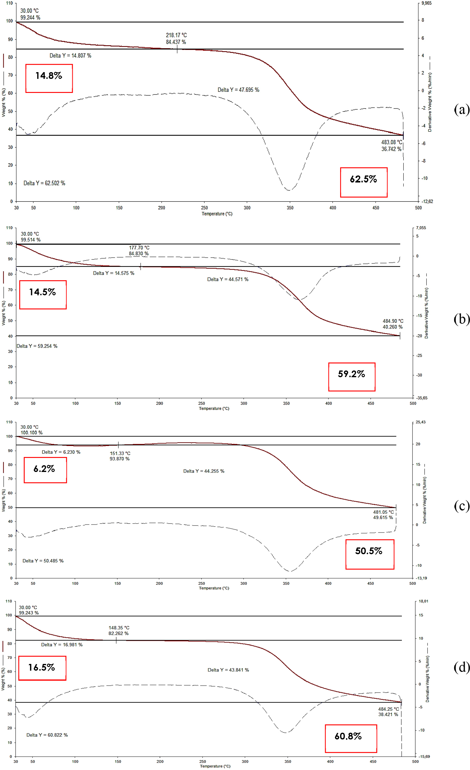
TGA of the membranes revealed two main stages of mass loss, as shown in Figure 4. The first stage occurred approximately between 40 150 °C, is associated with the evaporation of physically adsorbed water, with mass losses ranging from approximately 6.2 to 16.5%. Notably, membranes containing higher concentrations of J. mollissima extract, particularly MJ5, exhibited lower water loss, suggesting reduced hydrophilicity of the system. The second stage, corresponding to the thermal degradation of the polymeric matrix, was observed between 200-350 °C, with total mass losses ranging from approximately 50.5 to 62.5%, which is characteristic of chitosan decomposition processes.18

Thermogravimetric (TG) and derivative (DTG) curves of chitosan membranes with different concentrations of J. mollissima extract: (a) M, (b) MJ1 (1%), (c) MJ3 (3%), and (d) MJ5 (5%). Analyses were performed under synthetic air atmosphere at a heating rate of 10 °C min-1
A quantitative evaluation of thermal stability was performed by extracting the onset degradation temperature (Tonset) and the maximum degradation temperature (Tmax) from the TGA/DTG curves, as summarized in Table 1.
Thermal parameters obtained from TGA analysis of chitosan membranes with and without J. mollissima extract
The pure chitosan membrane (M) presented a Tonset of approximately 250 °C and a Tmax of approximately 355 °C. With the incorporation of J. mollissima extract, a progressive increase in these parameters was observed, reaching approximately 265 (Tonset) and 365 °C (Tmax) for sample MJ5. Additionally, the total mass loss decreased from 62.5 (M) to 50.5% (MJ5), indicating enhanced thermal stability.
This behavior is consistent with literature reports19 for chitosan-based systems, in which thermal degradation typically occurs between 200 and 350 °C depending on composition and structural organization. The increase in Tonset and Tmax, along with the reduction in mass loss, suggests that the incorporation of the extract enhances the thermal resistance of the material.
This improvement can be attributed to the increased intermolecular interactions between the chitosan chains and the phenolic compounds present in the extract, particularly hydrogen bonding. These interactions promote a more cohesive polymer network, restrict chain mobility, and require higher energy for thermal degradation. Furthermore, the reduced water loss observed for higher extract concentrations indicates a decrease in hydrophilicity, which is consistent with the swelling results.
Overall, the TGA results demonstrate that the incorporation of J. mollissima extract improves the structural and thermal stability of the membranes, reinforcing the structure-property relationship observed throughout the study.
Biodegradability analysis
The biodegradation assay of the membranes (Table 2), performed in PBS and in lysozyme/PBS solution, revealed that all samples underwent more pronounced degradation in the presence of the enzyme, confirming the efficiency of lysozyme in breaking down the chitosan matrix. Over the periods of 7, 14, and 21 days, it was observed that the pure membranes (M) showed greater mass loss compared to the membranes containing J. mollissima extract (MJ1, MJ3, and MJ5), indicating that the incorporation of the extract conferred greater stability to the polymeric matrix. During the degradation process, the released oligomers, such as N-glucosamine, demonstrate bioactivity, potentially favoring positive physiological responses, including probiotic action and promotion of tissue regeneration, which highlights the efficiency of this enzyme in the biodegradation of the chitosan matrix and corroborates the results found.20
Percentage mass loss of membranes (M, MJ1, MJ3 and MJ3) at intervals of 7, 14 and 21 days subjected to the biodegradation test
From a mechanistic perspective, the reduced degradation observed in membranes containing J. mollissima extract can be attributed to the increase in intermolecular interactions within the polymer network. Phenolic compounds present in the extract, containing hydroxyl (-OH) and carbonyl (C=O) groups, are able to form hydrogen bonds with the amino (-NH2) and hydroxyl groups of chitosan, resulting in a more cohesive and compact structure.
This structural reinforcement reduces the accessibility of glycosidic bonds to enzymatic attack by lysozyme, thereby slowing down the degradation process. In addition, the increased network density limits water diffusion into the matrix, further restricting hydrolytic degradation.
From a kinetic perspective, the results in Table 2 indicate a progressive reduction in the degradation rate with increasing extract concentration, as evidenced by the consistently lower mass loss values for MJ1, MJ3, and MJ5 over time. This suggests that the extract acts as a stabilizing agent, modulating the degradation profile and promoting a more controlled and sustained degradation behavior.
Such controlled degradation is particularly advantageous for wound dressing applications, as it allows the material to maintain structural integrity for longer periods while supporting gradual tissue regeneration.
Cytotoxicity analysis
In the cytotoxicity assay (Table 3), membranes M, MJ1, MJ3, and MJ5 did not show the formation of cell lysis halos, indicating an absence of cytotoxicity to L929 fibroblasts under the tested conditions. The positive control (latex) showed severe cytotoxicity (grade 4), while the negative control (filter paper) showed no effect, confirming the validity of the assay. These results reinforce the safety of the membranes and their potential as biomaterials for applications in tissue engineering and treatment of chronic wounds, combined with the bioactive effect of J. mollissima extract.21
Chitosan is widely recognized in the literature for producing scaffolds with excellent biological compatibility and absence of cytotoxicity. However, the combination of this biopolymer with J. mollissima extract is still poorly studied, making it necessary to carry out specific cytotoxicity assessments to verify its safety in biomedical applications.22
Phytochemical investigations indicate that J. mollissima extract contains bioactive compounds, including phenols, saponins, condensed tannins, and flavonoids, which can influence cellular processes such as proliferation and viability. Among these components, rutin stands out; previously identified in extracts of the species, which has shown potential to increase the viability of fibroblasts, keratinocytes, and HaCaT cells in different studies.16
Figure 5 illustrates the optical microscopies of all the samples studied. After analyzing these figures, it was observed that the negative control did not promote cell lysis (Figure 5a), while the positive control showed cell lysis with the formation of a discoloration halo (Figure 5b), reaching a cytotoxicity level of 4 (severely cytotoxic). No discoloration halos (cell lysis) were formed for any of the samples, as determined by the in vitro cytotoxicity assay following the ISO 10993-510 standards.

Optical microscopy images (100× magnification) of cytotoxicity assay using the agar diffusion method, showing negative control (a), positive control (b), and membranes M (c), MJ1 (d), MJ3 (e) and MJ5 (f)
Although the results indicate the absence of cytotoxic effects, it is important to emphasize that the agar diffusion method provides a qualitative assessment of cytocompatibility, which may limit the detection of subtle cellular responses. Therefore, while the materials can be considered non-cytotoxic under the tested conditions, this method does not provide quantitative information on cell viability or proliferation.
In this context, further studies using quantitative assays, such as 3-(4, 5-dimethylthiazolyl-2)-2, 5-diphenyltetrazolium bromide (MTT), resazurin, or live/dead analysis, are recommended to more comprehensively evaluate the biological performance of the membranes and confirm their cytocompatibility.
These results suggest that the membranes have potential for applications in tissue engineering and bioactive dressings, preserving cell viability. The presence of J. mollissima extract did not compromise cell compatibility, demonstrating that the material can be used in contact with biological tissues. These findings are consistent with previous reports23 on chitosan-based systems and bioactive composites, which exhibit favorable cytocompatibility and support for cellular activity.
CONCLUSIONS
Chitosan membranes with predominantly fibrous morphology containing J. mollissima extract were successfully developed using the freeze-drying method. The incorporation of the extract significantly influenced the physicochemical properties of the material. The pure chitosan membrane exhibited a swelling degree of approximately 1600%, while extract-containing membranes showed a progressive reduction in swelling. Thermogravimetric analysis revealed an increase in thermal stability, with Tonset increasing from ca. 250 to ca. 265 °C and Tmax from ca. 355 to ca. 365 °C, accompanied by a reduction in total mass loss from 62.5 to 50.5%. The initial mass loss observed at low temperatures is associated with moisture evaporation, preceding the main degradation stage. Biodegradation assays demonstrated a decrease in mass loss from 73.43 (M) to 65.00% (MJ5) in PBS after 21 days, indicating improved structural stability.
These changes are attributed to intermolecular interactions, particularly hydrogen bonding between the chitosan chains and the phenolic compounds present in the extract, leading to an increase in the effective network density, a reduction in hydrophilicity, and a restriction in the mobility of the polymer chain. This structural reorganization was consistently supported by FTIR, swelling, TGA, and biodegradation results, demonstrating a clear structure-property relationship.
The membranes showed no cytotoxic effects in qualitative assays, indicating preliminary biocompatibility. However, further quantitative biological evaluations are necessary to fully assess their performance. Overall, the results suggest that the incorporation of J. mollissima extract is an effective strategy to modulate the properties of chitosan-based materials, although additional in vitro and in vivo studies are required before considering biomedical applications.
ACKNOWLEDGMENTS
The authors thank CNPq-Brazil and CAPES-Brazil for financial support, and UFCG and CERTBIO for support in this work.
DATA AVAILABILITY STATEMENT
All data used and analyzed in this study are fully presented within the body of the manuscript.
REFERENCES
-
1 Barzegar, S.; Zare, M. R.; Shojaei, F.; Zareshahrabadi, Z.; Koohi Hosseinabadi, O.; Saharkhiz, M. J.; Iraji, A.; Zomorodian, K.; Khorram, M.; Int. J. Pharm. 2021, 597, 120288. [Crossref]
» Crossref -
2 Silva, M. H.; Jesus, M. C. P.; Tavares, R. E.; Caldeira, E. A. C.; Oliveira, D. M.; Merighi, M. A. B.; Revista Gaúcha de Enfermagem 2019, 40, e20180024. [Crossref]
» Crossref -
3 Dwivedi, R.; Kumar, S.; Pandey, R.; Mahajan, A.; Nandana, D.; Katti, D. S.; Mehrotra, D.; Journal of Oral Biology and Craniofacial Research 2020, 10, 381. [Crossref]
» Crossref -
4 Ul-Islam, M.; Alabbosh, K. F.; Manan, S.; Khan, S.; Ahmad, F.; Ullah, M. W.; Adv. Ind. Eng. Polym. Res. 2024, 7, 79. [Crossref]
» Crossref -
5 Mawazi, S. M.; Kumar, M.; Ahmad, N.; Ge, Y.; Mahmood, S.; Polymers 2024, 16, 1351. [Crossref]
» Crossref -
6 Souza, T. A.; Pereira, L. H. A.; Alves, A. F.; Dourado, D.; Lins, J. S.; Scotti, M. T.; Scotti, L.; Abreu, L. S.; Tavares, J. F.; Silva, M. S.; Pharmaceuticals 2024, 17, 1399. [Crossref]
» Crossref -
7 Passos, J. G. R.; Gomes, J. A. S.; Xavier-Santos, J. B.; Yamashita, F. O.; Cavalcanti-Cruz, J. V.; Siqueira, E. M. S.; Garcia, V. B.; Zucolotto, S. M.; de Araujo Junior, R. F.; Ferreira, L. S.; Biomed. Pharmacother. 2024, 173, 116290. [Crossref]
» Crossref -
8 Ribeiro, A. R. C.; Andrade, F. D.; Medeiros, M. C.; Camboim, A. S.; Pereira Junior, F. A.; Athayde, A. C. R.; Rodrigues, O. G.; Silva, W. W.; Pesquisa Veterinária Brasileira 2014, 34, 1051. [Crossref]
» Crossref - 9 ASTM F2778-09: Standard Test Method for Measurement of Percent Crystallinity of Polyetheretherketone (PEEK) Polymers by Means of Specular Reflectance Fourier Transform Infrared Spectroscopy (R-FTIR), West Conshohocken, 2020.
- 10 ISO 10993-5: Biological Evaluation of Medical Devices. Part 5: Tests for in Vitro Cytotoxicity, ISO: Geneva, 2009.
-
11 Ma, Y.; Xin, L.; Tan, H.; Fan, M.; Li, J.; Jia, Y.; Ling, Z.; Chen, Y.; Hu, X.; Mater. Sci. Eng., C 2017, 81, 522. [Crossref]
» Crossref -
12 Wang, Y.; Wang, H.; Lu, B.; Yu, K.; Xie, R.; Lan, G.; Xie, J.; Hu, E.; Lu, F.; Int. J. Biol. Macromol. 2023, 253, 127000. [Crossref]
» Crossref -
13 Nokoorani, Y. D.; Shamloo, A.; Bahadoran, M.; Moravvej, H.; Sci. Rep. 2021, 11, 16164. [Crossref]
» Crossref -
14 Grenier, J.; Duval, H.; Barou, F.; Lv, P.; David, B.; Letourneur, D.; Acta Biomater. 2019, 94, 195. [Crossref]
» Crossref -
15 Rodrigues, J. F. B.; Queiroz, J. V. S. A.; Medeiros, R. P.; Santos, R. O.; Fialho, D. A.; Neto, J. E. S.; Santos, R. L.; Barbosa, R. C.; Sousa, W. J. B.; Torres, M. C. M.; Pharmaceuticals 2023, 16, 1399. [Crossref]
» Crossref -
16 de Souza, M. F.; da Silva, H. N.; Rodrigues, J. F. B.; Macêdo, M. D. M.; de Sousa, W. J. B.; Barbosa, R. C.; Fook, M. V. L.; Polymers 2023, 15, 603. [Crossref]
» Crossref -
17 Hua, Y.; Ma, C.; Wei, T.; Zhang, L.; Shen, J.; Int. J. Mol. Sci. 2020, 21, 313. [Crossref]
» Crossref -
18 Vrabič-Brodnjak, U.; Yavorov, N.; Lasheva, V.; Todorova, D.; Coatings 2023, 13, 828. [Crossref]
» Crossref -
19 Alehosseini, E.; Tabarestani, H. S.; Kharazmi, M. S.; Jafari, S. M.; Foods 2022, 11, 3841. [Crossref]
» Crossref -
20 Macêdo, M. D. M.; Lucena, B. M.; de Cerqueira, G. R. C.; de Sousa, W. J. B.; Pedrosa, T. C.; Barbosa, R. C.; de Azevedo, A. C. S.; de Souza, M. F.; de Oliveira, D. K. M.; Fook, M. V. L.; Research, Society and Development 2021, 10, e25610414031. [Crossref]
» Crossref -
21 de Jesus, R. A.; Silva, S. M. L.; Anais do XX Congresso de Iniciação Científica, Desenvolvimento Tecnológico e Inovação da Universidade Federal de Campina Grande; Campina Grande, Brasil, 2023. [Link] accessed in April 2026
» Link -
22 Kim, Y.; Zharkinbekov, Z.; Raziyeva, K.; Tabyldiyeva, L.; Berikova, K.; Zhumagul, D.; Temirkhanova, K.; Saparov, A.; Pharmaceutics 2023, 15, 807. [Crossref]
» Crossref -
23 Yang, Y.; Ritchie, A. C.; Everitt, N. M.; Mater. Sci. Eng., C 2021, 121, 111846. [Crossref]
» Crossref
Edited by
-
Associate Editor handled this article:
Fernanda G. Finelli