Rabies in a free-ranging jaguarundi (Herpailurus yagouaroundi) in northeastern Brazil
ABSTRACT:
Rabies is a fatal viral disease that affects animals and humans and is widespread among national ecological systems. We describe the anatomopathological and immunohistochemical findings of a case of rabies in a jaguarundi (Herpailurus yagouaroundi) found in a rural peri-domestic area after showing aggression and attacking two animals. Histopathology of brain tissue showed lymphoplasmacytic meningoencephalitis associated with intracytoplasmic eosinophilic inclusion bodies in neurons, gliosis and neuronophagia. The diagnosis of rabies was based on the lesions and confirmed by immunohistochemistry and qPCR. In Brazil, rabies in wild felines of the genus Puma has only been described in animals through serological examinations. The pathological findings are essential for the diagnosis and elucidation of the participation of this species in the wild cycle of the disease, as well as in the maintenance and conservation of the balance of wild fauna.
INDEX TERMS:
Viral encephalitis; Lyssavirus; zoonosis; Herpailurus yagouaroundi; jaguarundi; wild felids
RESUMO:
A raiva é uma doença viral fatal que afeta animais e o homem, estando disseminada entre os sistemas ecológicos nacionais. Descrevem-se os achados anatomopatológicos e imuno-histoquímicos de um caso de raiva em um jaguarundi (Herpailurus yagouaroundi), encontrado em área peridomiciliar rural, após apresentar agressividade e ataque a dois animais. Microscopicamente, observou-se meningoencefalite linfoplasmocítica associada a corpúsculos de inclusão intracitoplasmáticos eosinofílicos em neurônios, gliose e neuronofagia. O diagnóstico de raiva foi estabelecido pelos achados patológicos e confirmado por imuno-histoquímica e qPCR. A ocorrência de raiva em felinos silvestres do gênero Puma só foi descrita por meio de exames sorológicos. Os achados patológicos são essenciais para o diagnóstico e elucidação da participação desta espécie no ciclo silvestre da doença, bem como na manutenção e conservação do equilíbrio da fauna silvestre.
TERMOS DE INDEXAÇÃO:
Encefalite viral; Lyssavirus; zoonoses; Herpailurus yagouaroundi; jaguarundi; felídeos silvestres
Introduction
The genus Herpailurus was proposed by Nikolai Severtzov in 1858, and further, many authors classified the jaguarundi into the genus Felis and Puma. Afterward, the phylogeographic analysis of jaguarundi samples evidenced no subspecies and Herpailurus genus is recommended (Kitchener et al. 2017).
Herpailurus yagouaroundi is the second most widely distributed carnivore in the Americas; however, it is considered vulnerable to extinction in Brazil, according to the Red Book of Brazilian Fauna Threatened with Extinction (ICMBio 2018). Wild feline species have gradually increased contact with humans. This interaction can result in possible spillovers or sharing of various pathogens, participating in the cycle of various zoonoses (Celeste & Bezerra 2020).
Rabies is a zoonotic, acute infectious and fatal disease caused by Lyssavirus rabies, an enveloped RNA virus of the order Mononegavirales, family Rhabdoviridae and genus Lyssavirus (Lemos & Riet-Correa 2023). All mammal species can be affected by this virus, with a variety of clinical signs that generally progress acutely and progress to death (Batista et al. 2007, Brasil 2020b).
In the Northeastern region of Brazil, among wild canids, crab-eating fox (Cerdocyon thous) is considered the main reservoir of the rabies virus (Brasil 2021b). However, rabies has been described as affecting vampires (Desmodus rotundus) (Carneiro et al. 2010) and insectivorous (Molossus molossus) bats (Araújo et al. 2014b), marmosets (Callithrix jacchus) (Favoretto el al. 2001), as well as several domestic animal species (Lemos & Riet-Correa 2023).
Although serological studies have already demonstrated exposure to the rabies virus in wild felines in Brazil (Onuma et al. 2016), to date, there is no description of the disease with clinical manifestation, anatomopathological and immunohistochemical findings in H. yagouaroundi. We describe a case of rabies in a jaguarundi in the semi-arid region of Paraíba, northeastern Brazil.
Case Report
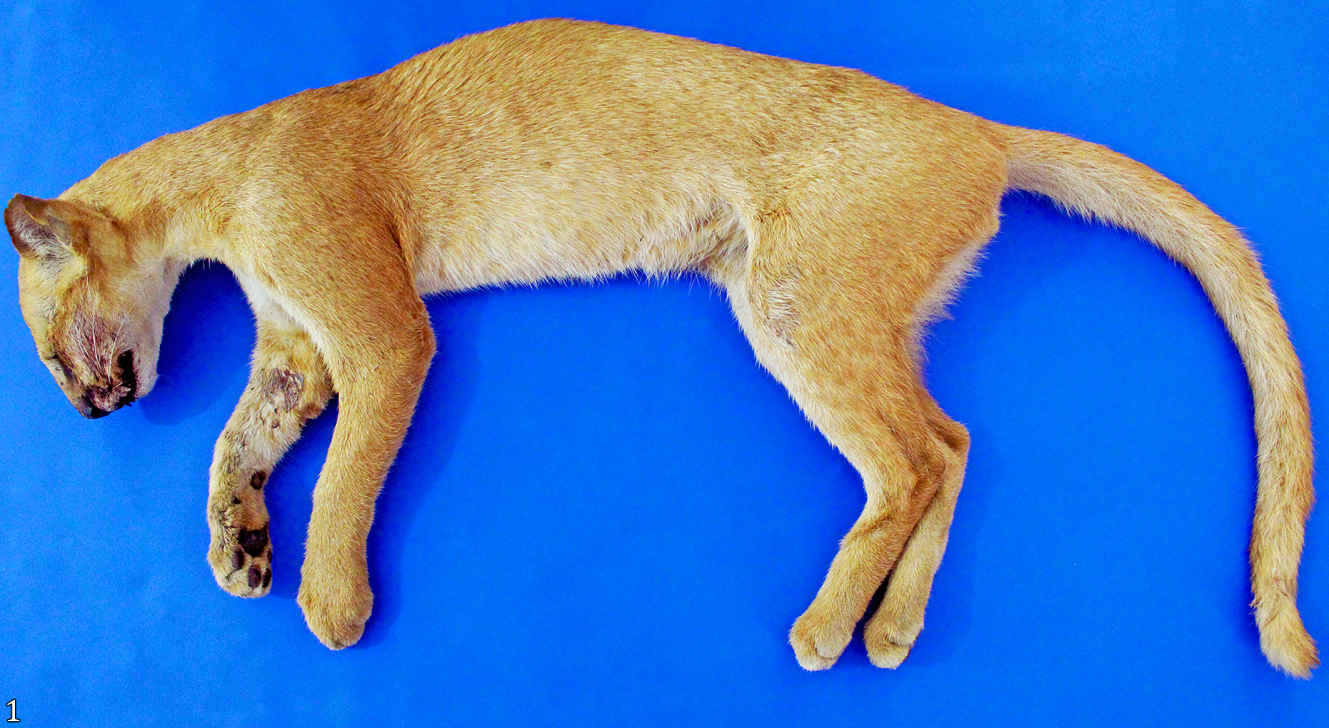
A rural producer reported a male jaguarundi (Herpailurus yagouaroundi) with neurological symptoms as aggressiveness and attempted attack on two horses on a rural property in the municipality of São Mamede, Paraíba, Brazil (06o55’36” S 37o05’44” W). The jaguarundi (Fig.1) died after suffering head trauma caused by the attacked horses.

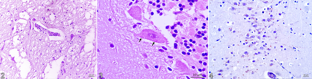
At necropsy, focally extensive areas of hemorrhage were seen in the submandibular and cranial regions, associated with parietal bone fracture. The brain was diffusely red, with a subarachnoid hemorrhage in the left hemisphere. Samples of organs from the thoracic and abdominal cavities and the central nervous system were fixed in 10% buffered formalin, routinely processed, embedded in paraffin and cut into 3µm sections. Sections were stained with hematoxylin and eosin (HE). Histopathological evaluation of the brain revealed lymphoplasmacytic meningoencephalitis, characterized by multifocal areas of discrete mononuclear inflammatory infiltrate perivascular composed of plasma cells and lymphocytes (perivascular cuffing) (Fig.2), associated with mild gliosis and neuronophagia. The inflammation affected the hippocampus, thalamus, fourth ventricle and rostral colliculus. In the occipital lobe, thalamus, rostral and caudal colliculus, fourth ventricle and Purkinje neurons of the cerebellum revealed single or multiple ovoid, eosinophilic intracytoplasmic inclusions in neurons (Negri bodies) (Fig.3). The leptomeninges had discrete mononuclear inflammatory infiltrate.

Rabies in Herpailurus yagouaroundi. Brain, multifocal areas of discrete mononuclear inflammatory infiltrate consisting of plasma cells and lymphocytes (perivascular cuffs) are observed in the fourth ventricle. HE, bar = 20µm. Fig.3. Cerebellum, ovoid and eosinophilic intracytoplasmic inclusion are observed in neurons (Negri bodies) in Purkinje neurons (arrows). HE, bar = 10µm. Fig.4. Hippocampus marked immunostaining with large, single or multiple corpuscles in the cytoplasm of neurons. IHQ-DAB, bar = 20µm.
For immunohistochemistry (IHC), hippocampal fragments were used in paraffin blocks, cut in positive charge slides. After deparaffinization and rehydration of the tissues, antigen retrieval was performed with a citrate solution (pH 6.0) in a microwave oven, at maximum power, for 10 minutes. The primary antibody used was polyclonal for rabies produced in goats labeled with FITC (fluorescence isothiocyanate conjugate antibody - Chemicon #5199)8, diluted 1:1000 in phosphate-buffered saline (PBST) with Tween® 20 (Sigma P2287)9, and incubated for 60 min at 37Co. The biotinylated secondary antibody and the streptavidin-biotin-peroxidase complex (LSAB+System HRP)3 were used consecutively, incubated at room temperature for 30 min and labeled by adding DAB + Substrate - Chromogen System10 and counterstained with Harris hematoxylin. As a positive control, histological sections from confirmed cases of rabies in cattle were used. The same sections were used as a negative control, replacing the primary antibody with PBST. IHC with the polyclonal antibody for rabies revealed marked immunostaining demonstrating multiple aggregates of brownish granules in the form of large, single or multiple corpuscles in the cytoplasm of hippocampal neurons (Fig.4). The horses attacked by the jaguarundi did not show clinical neurological manifestations 30 days after the attack. Quantitative real-time PCR, as per Hayman et al. (2011), resulted positive for a pool of central nervous system, but attempts to produce Sanger sequences from this sample failed.
Discussion and Conclusion
The diagnosis of rabies was established according to anatomopathological findings and confirmed by immunohistochemistry. In Brazil, rabies in acute, fatal and classic forms or even serological diagnosis has already been reported in different wild species such as vampire (Desmodus rotundus) (Carneiro et al. 2010) and insectivores (Molossus molossus) bats (Araújo et al. 2014b), crab-eating fox (Cerdocyon thous) (Araújo et al. 2014a), common marmoset (Callithrix jacchus), raccoon (Procyon cancrivorus), opossum (Didelphis aurita), capuchin monkey (Cebus apella), coati (Nasua nasua), capybaras (Hydrochaerus hydrochaeris), tapirs (Tapirus terrestris) (Pereira et al. 2022), pumas (Puma concolor) and ocelots (Leopardus pardalis) (Favoretto et al. 2001, Araújo et al. 2014a, Araujo et al. 2014, Antunes et al. 2017, Rocha et al. 2017).
Wild felids such as jaguars, ocelots, pumas and Pampas cats exposed to the rabies virus were identified with antibodies through serological tests (Jorge et al. 2010, Furtado et al. 2013). In a study with four Puma concolor that attacked humans and domestic animals, the rabies virus was identified through fluorescent antibody test (FAT), IHC and RT-PCR (Garcés-Ayala et al. 2022). However, this seems to be the first report describing clinical neurological manifestation and anatomopathological and immunohistochemical findings of rabies in wild felids of both genus, including Puma and Herpailurus in Brazil.
The neurological manifestation of aggressiveness described in this H. yagouaroundi is characteristic of the furious form that commonly affects carnivores, including domestic cats. It presents with behavioral changes, hyperesthesia, hyperreflexia, salivation, tremors, ataxia and paralysis (Greene 2015, Müller & Freuling 2020, Lima et al. 2023, Slathia et al. 2023). Although the animal died due to traumatic brain injury resulting from the defense by the attacked horses, not spontaneously, the abnormal behavior of the jaguarundi when attacking domestic animals is similar to that of the jaguar that attacked horses, cattle, dogs and humans and later was captured and diagnosed with rabies (Garcés-Ayala et al. 2022). Slathia et al. (2023) state that wild animals with nocturnal habits, when infected by the rabies virus, can be seen during the day and lose their fear of humans and other animals, and may attack them.
In Brazil, rabies has changed its epidemiological profile in recent decades; since 2015, there has been no record of human rabies due to canine variants (AgV1 and AgV2), with the bat variant (AgV3) being the main determinant of human rabies cases and in others animals (Brasil 2020b). This relationship reinforces the virus’s ability to spread between different species of mammals (Brasil 2021a). The sylvatic cycle is maintained in a wide variety of mammalian hosts, and the epidemiological chain of rabies in Brazil includes Carnivora and Chiroptera as the main reservoirs (Rocha et al. 2017). AgV2* (wild canids/foxes) warn of the existence of the virus circulating through wild variants in rural areas of Paraíba, a situation that may be common among wild species in the region (Brasil 2020a).
The occurrence of rabies in H. yagouaroundi has great epidemiological importance, especially in the sylvatic cycle of rabies, acting, together with wild canids, primates and bats, in the transmission of the rabies virus (Rocha et al. 2017), enabling spillover to other wildlife and humans (Messenger et al. 2002, Antunes et al. 2017).
Studies with wild animals serologically positive for rabies indicate protective levels of antibodies or exposure to the virus. However, it is not clear how the animals come into contact, although predation may be the most likely route (Antunes et al. 2017). Although pumas are not considered rabies reservoirs (Garcés-Ayala et al. 2022) and there are no reports of human rabies through direct contact with wild felids, these are considered predators due to their hunting habits and are more likely to be infected with a rabies virus originating from bats (Lima et al. 2023), becoming a potential secondary transmitter.
Histologically, the non-suppurative meningoencephalitis associated with intracytoplasmic inclusion bodies in neurons (Negri bodies) mainly affecting the brain, brainstem and cerebellum observed in this case are similar to those described in wild carnivores with neurological symptoms characterized by changes in behavior and aggressiveness related to dysfunctions of prosencephalon (Frymus et al. 2009, Araújo et al. 2014b, Church et al. 2018).
Rabies control in Brazil is carried out based on the vaccination of domestic animals such as canines, felines and herbivores, not including free-ranging wild animals (Brasil 2009, 2021a, 2021b). Some countries such as the USA, Canada, France, Germany and the European Union carry out rabies vaccination in wild animals using oral anti-rabies vaccine introduced into bait (Fornazari & Langoni 2014, Robardet et al. 2019).
The diagnosis of rabies in a jaguarundi (H. yagouaroundi) with clinical neurological manifestation, confirmed by anatomopathological and immunohistochemical exams, demonstrates the occurrence of this disease in wild felines in Brazil. The diagnosis of rabies in wild animals is essential to establish parameters of the wild cycle, aiming to avoid possible interspecies and zoonotic transmission, as well as evaluating control strategies through the implementation of programs that use oral vaccination in wild animals, aiming to control rabies in Brazilian territory. Monitoring wild animal populations is relevant for rabies surveillance in order to promote means of conserving the species through a transdisciplinary approach combined with a “One Health” model.
Acknowledgments
To the Dr. Marcos Tortato from “Tigrinus Equipamentos para Pesquisa” for his taxonomic consultant.
References
-
Antunes J.M.A.P., Demoner L.C., Cruvinel T.M.A., Kataoka A.P., Martorelli L.F.A., Machado G.P. & Megid J. 2017. Rabies virus exposure of Brazilian free-ranging wildlife from municipalities without clinical cases in humans or in terrestrial wildlife. J. Wildl. Dis. 53(3):662-666. <https://dx.doi.org/10.7589/2016-09-204> <PMid:28406757>
» https://doi.org/https://dx.doi.org/10.7589/2016-09-204 -
Araujo D.B., Martorelli L.A., Kataoka A.P.G., Campos A.C.A., Rodrigues C.S., Sanfilippo L.F., Cunha E.S., Durigon E.L. & Favoretto S.R. 2014. Antibodies to rabies virus in terrestrial wild mammals in native rainforest on the north coast of São Paulo State, Brazil. J. Wildl. Dis. 50(3):469-477. <https://dx.doi.org/10.7589/2013-04-099> <PMid:24779464>
» https://doi.org/https://dx.doi.org/10.7589/2013-04-099 - Araújo J.L., Dantas A.F.M., Galiza G.J.N., Pedroso P.M.O., Silva M.L.C.R., Pimentel L.A. & Riet-Correa F. 2014a. Aspectos histopatológicos e imuno-histoquímicos da raiva em raposas Cerdocyon thous Acta Scient. Vet. 42(Supl.1):67.
-
Araújo J.L., Nascimento E.M., Dantas A.F.M., Galiza G.J.N., Pedroso P.M.O., Silva M.L.C.R. & Riet-Correa F. 2014b. Rabies in the insectivorous Pallas’s mastiff bat (Molossus molossus) in northeastern Brazil. J. Wildl. Dis. 50(4):883-886. <https://dx.doi.org/10.7589/2013-11-314> <PMid:25121411>
» https://doi.org/https://dx.doi.org/10.7589/2013-11-314 - Batista H.B.C.R., Franco A.C. & Roehe P.M. 2007. Raiva: uma breve revisão. Acta Scient. Vet. 35(2):125-144.
-
Brasil 2009. Controle da raiva dos herbívoros. 2ª ed. Secretaria de Defesa Agropecuária, Ministério da Agricultura, Pecuária e Abastecimento, Brasília, DF. 124p. (Manual Técnico). Available at <Available at https://www.gov.br/agricultura/pt-br/assuntos/sanidade-animal-e-vegetal/saude-animal/programas-de-saude-animal/raiva-dos-herbivoros-e-eeb/MANUAL_RAIVAHERBVOROS2009.pdf > Accessed on Out. 10, 2023.
» https://www.gov.br/agricultura/pt-br/assuntos/sanidade-animal-e-vegetal/saude-animal/programas-de-saude-animal/raiva-dos-herbivoros-e-eeb/MANUAL_RAIVAHERBVOROS2009.pdf -
Brasil 2020a. Raiva humana por animais silvestres no Brasil: atualizações e condutas profiláticas. In: Síndrome Inflamatória Multissistêmica Pediátrica (SIM-P), temporalmente associada à COVID-19. Vol.51. Secretaria de Vigilância em Saúde, Ministério da Saúde, Brasília, DF, p.17-23. (Boletim Epidemiológico, Nº 35). Available at <Available at https://www.gov.br/saude/pt-br/centrais-de-conteudo/publicacoes/svsa/raiva/be-vol-51-no-35-raiva-humana-por-silvestres-no-brasil-atualizacoes-e-condutas-profilaticas.pdf > Accessed on Out. 10, 2023.
» https://www.gov.br/saude/pt-br/centrais-de-conteudo/publicacoes/svsa/raiva/be-vol-51-no-35-raiva-humana-por-silvestres-no-brasil-atualizacoes-e-condutas-profilaticas.pdf -
Brasil 2020b. A vigilância da raiva no Brasil em 2019. In: Monitoramento dos casos de arboviroses urbanas transmitidas pelo Aedes Aegypti (dengue, chikungunya e zika), Semanas Epidemiológicas 1 a 15, 2020. Vol.51. Secretaria de Vigilância em Saúde, Ministério da Saúde, Brasília, DF, p.27-32. (Boletim Epidemiológico, Nº 16). Available at <Available at https://www.gov.br/saude/pt-br/centrais-de-conteudo/publicacoes/svsa/raiva/be-vol-51-no-16-a-vigilancia-da-raiva-no-brasil-em-2019.pdf > Accessed on Out. 10, 2023.
» https://www.gov.br/saude/pt-br/centrais-de-conteudo/publicacoes/svsa/raiva/be-vol-51-no-16-a-vigilancia-da-raiva-no-brasil-em-2019.pdf -
Brasil 2021a. A Campanha Nacional Contra a Raiva Canina no Brasil, 2020. In: Monitoramento dos casos de arboviroses urbanas causados por vírus transmitidos pelo mosquito Aedes (dengue, chikungunya e zika), semanas epidemiológicas 1 a 31, 2021. Vol.52. Secretaria de Vigilância em Saúde, Ministério da Saúde, Brasília, DF , p.10-16. (Boletim Epidemiológico, Nº 30). Available at <Available at https://www.gov.br/saude/pt-br/centrais-de-conteudo/publicacoes/svsa/raiva/be-vol-52-no-30-a-campanha-nacional-contra-a-raiva-canina-no-brasil-2020.pdf/view > Accessed on Out. 10, 2023.
» https://www.gov.br/saude/pt-br/centrais-de-conteudo/publicacoes/svsa/raiva/be-vol-52-no-30-a-campanha-nacional-contra-a-raiva-canina-no-brasil-2020.pdf/view -
Brasil 2021b. Raiva transmitida por cachorros do mato (Cerdocyon thous): casos humanos e conhecimento, atitudes e práticas no Nordeste do Brasil. In: Monitoramento dos casos de arboviroses urbanas causados por vírus transmitidos pelo mosquito Aedes (dengue, chikungunya e zika), semanas epidemiológicas 1 a 51, 2021. Vol.52. Secretaria de Vigilância em Saúde, Ministério da Saúde, Brasília, DF , p.7-17. (Boletim Epidemiológico, N.º 48). Available at <Available at https://www.gov.br/saude/pt-br/centrais-de-conteudo/publicacoes/boletins/epidemiologicos/edicoes/2021/boletim-epidemiologico-vol-52-no-48.pdf/view > Accessed on Out. 10, 2023.
» https://www.gov.br/saude/pt-br/centrais-de-conteudo/publicacoes/boletins/epidemiologicos/edicoes/2021/boletim-epidemiologico-vol-52-no-48.pdf/view -
Carneiro A.J.B., Franke C.R., Stöcker A., Santos F., Sa J.E.U., Moraes-Silva E., Alves J.N.M., Brunik S., Corman V.M., Drosten C. & Drexler J.F. 2010. Rabies virus RNA in naturally infected vampire bats, northeastern Brazil. Emerg. Infect. Dis. 16(12):2004-2006. <https://dx.doi.org/10.3201/eid1612.100726> <PMid:21122246>
» https://doi.org/https://dx.doi.org/10.3201/eid1612.100726 -
Celeste H. & Bezerra A.M.R. 2020. Felinos neotropicais como hospedeiros de agentes zoonóticos no Brasil. Mastozool. Neotrop. 27(2):306-318. <https://dx.doi.org/10.31687/saremMN.20.27.2.0.07>
» https://doi.org/https://dx.doi.org/10.31687/saremMN.20.27.2.0.07 - Church M.E., Terio K.A. & Keel M.K. 2018. Procyonidae, Viverridae, Hyenidae, Herpestidae, Eupleridae, and Prionodontidae, p.305-321. In: Terio K.A., McAloose D. & Leger J.S. (Eds), Pathology of Wildlife and Zoo Animals. Academic Press, Massachusetts.
-
Favoretto S.R., Mattos C.C., Morais N.B., Araújo F.A.A. & Mattos C.A. 2001. Rabies in marmosets (Callithrix jachus), Ceará, Brazil. Emerg. Infect. Dis. 7(6):1062-1065. <https://dx.doi.org/10.3201/eid0706.010630> <PMid:11747745>
» https://doi.org/https://dx.doi.org/10.3201/eid0706.010630 - Fornazari F. & Langoni H. 2014. Principais zoonoses em mamíferos selvagens. Vet. Zootec. 21(1):10-24.
-
Frymus T., Addie D., Belák S., Boucraut-Baralon C., Egberink H., Gruffydd-Jones T., Hartmann K., Hosie M.J., Lloret A., Lutz H., Marsilio F., Pennisi M.G., Radford A.D., Thiry E., Truyen R. & Horzinek M.C. 2009. Feline rabies: ABCD guidelines on prevention and management. J. Feline Med. Surg. 11(7):585-593. <https://dx.doi.org/10.1016/j.jfms.2009.05.007> <PMid:19481038>
» https://doi.org/https://dx.doi.org/10.1016/j.jfms.2009.05.007 -
Furtado M.M., Ramos Filho J.D., Scheffer K.C., Coelho C.J., Cruz P.S., Ikuta C.Y., Jácomo A.T.A., Porfírio G.E.O., Silveira L., Sollmann R., Tôrres N.M. & Ferreira Neto J.S. 2013. Serosurvey for selected viral infections in free-ranging jaguars (Panthera onca) and domestic carnivores in Brazilian Cerrado, Pantanal, and Amazon. J. Wildl. Dis. 49(3):510-521. <https://dx.doi.org/10.7589/2012-02-056> <PMid:23778599>
» https://doi.org/https://dx.doi.org/10.7589/2012-02-056 -
Garcés-Ayala F., Aguilar-Setién Á., Almazán-Marín C., Cuautle-Zavala C., Chávez-López S., Martínez-Solís D., Gómez-Sierra M., Sandoval-Borja A., Escamilla-Ríos B., López-Martínez I. & Aréchiga-Ceballos N. 2022. Rabies virus variants detected from Cougar (Puma concolor) in Mexico 2000-2021. Pathogens 11(2):265. <https://dx.doi.org/10.3390/pathogens11020265> <PMid:35215207>
» https://doi.org/https://dx.doi.org/10.3390/pathogens11020265 - Greene C.E. 2015. Raiva e outras infecções por Lyssavirus, p.189-207. In: Greene C.E. (Ed.), Doenças Infecciosas em Cães e Gatos. 4ª ed. Guanabara Koogan, Rio de Janeiro.
-
Hayman D.T.S., Banyard A.C., Wakeley P.R., Harkess G., Marston D., Wood J.L.N., Cunningham A.A. & Fooks A.R. 2011. A universal real-time assay for the detection of Lyssaviruses. J. Virol. Methods 177(1):87-93. <https://dx.doi.org/10.1016/j.jviromet.2011.07.002> <PMid:21777619>
» https://doi.org/https://dx.doi.org/10.1016/j.jviromet.2011.07.002 -
ICMBio 2018. Livro Vermelho da Fauna Brasileira Ameaçada de Extinção. Vol.1. 1ª ed. Instituto Chico Mendes de Conservação da Biodiversidade, Ministério do Meio Ambiente, Brasília, DF. 492p. Available at <Available at https://www.gov.br/icmbio/pt-br/centrais-de-conteudo/publicacoes/publicacoes-diversas/livro_vermelho_2018_vol1.pdf > Accessed on Out. 10, 2023.
» https://www.gov.br/icmbio/pt-br/centrais-de-conteudo/publicacoes/publicacoes-diversas/livro_vermelho_2018_vol1.pdf -
Jorge R.S.P., Pereira M.S., Gonçalves R.G., Scheffer K.C., Carnielli Jr. P., Ferreira F., Furtado M.M., Kashivakura C.K., Silveira L., Jacomo A.T.A., Lima E.S., Paula R.C. & May-Junior J.A. 2010. Detection of rabies virus antibodies in Brazilian free-ranging wild carnivores. J. Wildl. Dis. 46(4):1310-1315. <https://dx.doi.org/10.7589/0090-3558-46.4.1310> <PMid:20966286>
» https://doi.org/https://dx.doi.org/10.7589/0090-3558-46.4.1310 - Kitchener A.C., Breitenmoser-Würsten C., Eizirik E., Gentry A., Werdelin L., Wilting A., Yamaguchi N., Abramov A. V., Christiansen P., Driscoll C., Duckworth J. W., Johnson W., Luo S.J., Meijaard E., O’Donoghue P., Sanderson J., Seymour K., Bruford M., Groves C., Hoffmann M., Nowell K., Timmons Z., Tobe S. 2017. A revised taxonomy of the Felidae. The final report of the Cat Classification Task Force of the IUCN/SSC Cat Specialist Group. Cat News Special (11):31-32.
- Lemos R.A.A. & Riet-Correa F. 2023. Raiva, p.165-177. In: Riet-Correa F., Shild A.L., Lemos R.A.A., Borges J.R.J., Mendonça F.S. & Machado M. (Eds), Doenças de Ruminantes e Equídeos. Vol.1. 4ª ed. MedVet, São Paulo.
-
Lima J.S., Mori E., Kmetiuk L.B., Biondo L.M., Brandão P.E., Biondo A.W. & Maiorka P.C. 2023. Cat rabies in Brazil: a growing One Health concern. Front. Public Health 11:1210203. <https://dx.doi.org/10.3389/fpubh.2023.1210203> <PMid:37538269>
» https://doi.org/https://dx.doi.org/10.3389/fpubh.2023.1210203 -
Messenger S.L., Smith J.S. & Rupprecht C.E. 2002. Emerging epidemiology of bat-associated cryptic cases of rabies in humans in the United States. Clin. Infect. Dis. 35(6):738-747. <https://dx.doi.org/10.1086/342387> <PMid:12203172>
» https://doi.org/https://dx.doi.org/10.1086/342387 -
Müller T. & Freuling C.M. 2020. Rabies in terrestrial animals, p.195-230. In: Fooks A.R. & Jackson A.C. (Eds), Rabies: Scientific basis of the disease and its management. 4th ed. Academic Press, Massachusetts. <https://dx.doi.org/10.1016/B978-0-12-818705-0.00006-6>
» https://doi.org/https://dx.doi.org/10.1016/B978-0-12-818705-0.00006-6 -
Onuma S.S.M., Chaves L.B., Lara M.C.C.S.H., Lara H., May-Júnior J.A., Taques I.I.G.G., Fritzen J.T.T., Alfieri A.A., Ometto T., Durigon E.L., Araújo J., Kantek D.L.Z. & Aguiar D.M. 2016. Serological and molecular investigation of viral agents in free-living jaguars of the Pantanal wetlands, state of Mato Grosso, Brazil. Braz. J. Vet. Res. Anim. Sci. 53(3):270-279. <https://dx.doi.org/10.11606/issn.1678-4456.bjvras.2016.108947>
» https://doi.org/https://dx.doi.org/10.11606/issn.1678-4456.bjvras.2016.108947 -
Pereira F.M.A., Oliveira A.R., Soares-Neto L.L., Munhoz L., Fernandes L.S., Santos D.O., Carvalho T.P., Langoni H. & Santos R.L. 2022. Rabies in a captive lowland tapir (Tapirus terrestris). J. Comp. Pathol. 198:29-32. <https://dx.doi.org/10.1016/j.jcpa.2022.07.008> <PMid:36116889>
» https://doi.org/https://dx.doi.org/10.1016/j.jcpa.2022.07.008 -
Robardet E., Bosnjak D., Englund L., Demetriou P., Martín P.R. & Cliquet F. 2019. Zero endemic cases of wildlife rabies (classical Rabies Virus, RABV) in the European union by 2020: An achievable goal. Trop. Med. Infect. Dis. 4(4):124. <https://dx.doi.org/10.3390/tropicalmed4040124> <PMid:31575054>
» https://doi.org/https://dx.doi.org/10.3390/tropicalmed4040124 -
Rocha S.M., Oliveira S.V., Heinemann M.B. & Gonçalves V.S.P. 2017. Epidemiological profile of wild rabies in Brazil (2002-2012). Transbound. Emerg. Dis. 64(2):624-633. <https://dx.doi.org/10.1111/tbed.12428> <PMid:26423323>
» https://doi.org/https://dx.doi.org/10.1111/tbed.12428 - Slathia P., Abrol R., Sharma S. & Sharma S. 2023. Rabies: A review on clinical signs, prevention and control. Pharma Innovation J. 12(5):1675-1680.