Bluetongue in sheep in the state of Santa Catarina, Brazil: seroepidemiologic, clinical, and pathological findings
ABSTRACT:
This study aimed to investigate the circulation of bluetongue virus (BTV) in sheep throughout different regions of the state of Santa Catarina by the serologic agar gel immunodiffusion (AGID) assay and to describe the clinical, pathological, and epidemiologic aspects of seven outbreaks and three isolated cases, which occurred between 2009 and 2019. The state was divided into three regions for research into antibodies. The average annual temperatures and altitudes of the regions were described or tabulated. Blood samples were collected from sheep on 24 properties in 16 municipalities distributed throughout the three aforementioned regions. Of the 388 blood samples collected from the sheep, 250 were seropositive (64.4%) in the AGID test. Antibodies against BTV were detected in the three regions. The disease was diagnosed through clinical signs and macroscopic lesions and confirmed by the reverse transcription polymerase chain reaction (RT-PCR) technique. The affected sheep developed ulcerations in the nostrils, lips, and oral mucosa with crusting, locomotor difficulties, black diarrhea, coughing, and regurgitation, followed by death. The necropsy showed multiple ulcers in the palate and rumen, petechial hemorrhages in the serosa of the esophagus near the esophageal hiatus, and focal hemorrhages in the pulmonary artery. In Santa Catarina, bluetongue in sheep is a seasonal disease that occurs mainly during the hot, rainy seasons (December to April). Seasonality coincides with the time of year when Culicoides vectors have the highest population and metabolic activity.
INDEX TERMS:
Arboviral disease; bluetongue virus; epidemiology; pathology
RESUMO:
Este estudo teve como objetivos investigar, por meio do teste sorológico de imunodifusão em gel de ágar (IDGA), a circulação do vírus da língua azul (BTV) em ovinos em diferentes regiões do estado de Santa Catarina e, descrever os aspectos clínico-patológicos e epidemiológicos de sete surtos e três casos isolados, ocorridos entre os anos de 2009 a 2019. Para a pesquisa de anticorpos, o estado foi dividido em três regiões. As temperaturas médias anuais e altitudes das regiões foram descritas ou tabuladas. Foram coletadas amostras de sangue de ovinos em 24 propriedades de 16 municípios distribuídos nas três regiões estabelecidas. Dos 388 ovinos coletados, 250 foram soropositivos (64,4%) no teste de IDGA. Anticorpos contra o BTV foram detectados nas três regiões. A enfermidade foi diagnosticada pelos sinais clínicos, lesões macroscópicas e confirmada pela técnica de reação em cadeia da polimerase com transcrição reversa (RT-PCR). Os ovinos afetados desenvolveram ulcerações nas narinas, lábios e mucosa oral com formação de crostas, dificuldade de locomoção, diarreia enegrecida, tosse e regurgitamento seguido de morte. Na necropsia foi observado principalmente múltiplas úlceras no palato e rúmen, hemorragias petequiais na serosa do esôfago próximo ao hiato esofágico e hemorragia focal na artéria pulmonar. Em Santa Catarina, a língua azul em ovinos é uma doença sazonal, que ocorre principalmente em épocas quentes e chuvosas (dezembro a abril). A sazonalidade coincide com a época do ano a qual os vetores Culicoides apresentam maior população e atividade metabólica.
TERMOS DE INDEXAÇÃO:
Doença arboviral; vírus da língua azul; epidemiologia; patologia
Introduction
The bluetongue virus (BTV) is an Orbivirus belonging to the Reoviridae family that is responsible for the noncontagious disease in ruminants, such as wild deer, cattle, goats, and sheep. Its vectors are mosquitoes of the genus Culicoides (Clavijo et al. 2002, Williamson et al. 2008, Jenckel et al. 2015, Bianchi et al. 2017, Baldini et al. 2018, Kawanami et al. 2018). The clinical form of the disease is observed mainly in sheep. The other species rarely develop clinical signs and, when observed, they are nonspecific and common to other diseases, which makes clinical diagnosis difficult (Williamson et al. 2008).
The name bluetongue is due to the change in the color of the tongue, which, in this case, turns to a bluish color due to hypoxia triggered by lysis of the endothelial cells, whose alteration is considered to be characteristic of the disease (D. Hutcheon 1893, Zachary 2013). This arbovirus can cause lesions in the gastrointestinal tract and reproductive disorders, such as abortions and malformations (Riet-Correa 2007, Antoniassi et al. 2010). Bovine and caprine species are thought to be less susceptible than ovine species (Backx et al. 2007, Darpel et al. 2007). In Brazil, BTV has been clinically diagnosed in Paraná, Rio de Janeiro, Rio Grande do Sul, and Minas Gerais (Clavijo et al. 2002, Balaro et al. 2014, Lima et al. 2016, Guimarães et al. 2017). In Santa Catarina, the first cases of BTV in sheep were diagnosed and registered by the “Laboratório de Patologia Veterinária” (Veterinary Pathology Laboratory) of the “Centro de Ciências Agroveterinárias” (Center for Agroveterinary Sciences) of “Universidade do Estado de Santa Catarina” (UDESC) in the municipality of Chapecó in 2009 (Quevedo et al. 2019).
This study aims to assess the circulation of the virus in sheep in different regions of the state of Santa Catarina by using the agar gel immunodiffusion assay (AGID) and to describe the clinical, pathological, and epidemiological aspects of seven outbreaks and three isolated cases of the disease.
Materials and Methods
Ethical approval. The study was submitted and approved by the Animal Ethics Committee of “Universidade do Estado de Santa Catarina” (CEUA-CAV-UDESC) and obtained a favorable opinion under the protocol number 5097130319.
Study design. Based on the total number of sheep in the state of Santa Catarina (221,509) (IBGE 2017), there was an expected prevalence of 50%, a proportion that maximizes the sample size and guarantees a minimum confidence of 95% and a statistical error of 5%, which favors one (n) of 384 animals (Thrusfield 2004). Blood samples were collected from 388 sheep, ages ranging from eight months to five years, thereby securing a margin of safety in 24 properties in 16 municipalities. The collection was distributed at random, and the state of Santa Catarina was divided into the following three regions based on their average temperatures: 1 - West, 2 - Coast/Itajaí Valley/Northern Highlands (hotter), and 3 - Mountain/Southern Highlands (colder) (Table 1).
Clinical and pathological diagnosis. Epidemiologic, clinical, and pathological data were obtained from ten properties where the disease had occurred. Necropsies were performed on 13 sheep exhibiting clinical signs compatible with BTV. Six of these had an acute form of the disease, while seven manifested a chronic form. Fragments of cardiac muscle, skeletal muscle of the scapular region, rostral, medial and caudal esophagus, rumen, omasum, intestines, mesenteric lymph nodes, abomasum, lung, kidney, and liver were collected, fixed in 10% formalin, routinely processed, and stained with hematoxylin and eosin (HE) for histopathological evaluation. The serum from these animals was subjected to AGID, and samples of cardiac muscle, mesenteric lymph nodes, kidney, and liver were collected during necropsy for reverse transcription polymerase chain reaction (RT-PCR).
Blood was collected from 181 sheep out of 105,523 sheep in Region 1, 129 sheep out of 70,886 sheep in Region 2, and 78 sheep out of 45,100 sheep in Region 3. Blood samples were obtained by jugular venipuncture with a 10 ml Vacutainer tube that underwent centrifugation at 2,000 revolutions per minute (RPM). An AGID assay was performed using serum. The protocol proposed by the Veterinary Medical Research and Development bluetongue virus antibody test kit (Pullman/WA, USA), in accordance with the manufacturer’s instructions, was recommended.
Whole blood was collected from animals with clinical suspicion of BTV for AGID tests. Necropsies were performed on the clinically affected sheep, which allowed for a macroscopic evaluation and the collection of organ fragments for histopathology. Blood and spleen samples were sent to the Virology Sector at “Universidade Federal de Santa Maria” (UFSM) for RT-PCR tests.
Diagnosis by RT-PCR. Blood samples from sheep with clinical signs of the disease at ten properties were collected with ethylenediaminetetraacetic acid (EDTA), frozen, and sent to the Virology Sector at UFSM, where samples were processed in a pool when blood was collected from more than one sheep on the same property. TRIzol® reagent (Life Technologies, Carlsbad/CA) was used, according to the manufacturer’s instructions, to extract RNA from these blood samples. After RNA extraction, complementary DNA (cDNA) was synthesized using the GoScript™ Reverse Transcriptase (Promega). The cDNA was kept at -80 °C until use. The RT-PCR technique was performed to amplify a 113 bp fragment of the VP1 protein gene using the primers: F-BTV-VP1: GCGAAGTGTGGACATGAAGC and R-BTV-VP1: TCCTGCGGTACGTAACAACC. RT-PCR reactions were performed in a total of 25 µl, using 1 mM MgCl ++, 100 µM of each deoxynucleotide (dATP, dCTP, dGTP, dTTP), 1 μM of each primer, 2 U of Taq DNA Polymerase (Invitrogen®), 10% of the total volume of polymerase buffer, 2 μL of the cDNA template, and water Qsp, which underwent the following cycle conditions: 45 °C for 10 minutes, 95 °C for 10 minutes, followed by 35 cycles of 95 °C for 15 seconds, 60 °C for 30 seconds, and 72 °C for 30 seconds. Negative (Milli-Q water) and positive controls were used for each reaction. The products underwent agarose gel electrophoresis using a 2% gel at 80 volts for 40 minutes and were visualized through a transilluminator.
In 2009, the first whole blood samples were obtained from sheep from Chapecó, São José do Cedro, Planalto Alegre, Pouso Redondo, and Orleans. The material was sent to the Virology Sector at UFSM for RT-PCR tests. Whole blood samples were subsequently collected from sheep with potential clinical signs of BTV during the experimental phase in 2018, and these samples underwent the same RT-PCR test. These sheep were from Properties 3 - Riqueza (2), 16 - São Ludgero (5), and 22 - Bom Retiro (22), which were processed in one pool each property. As for Properties 13 - Nova Veneza (1), 4 - Chapecó (1) (same property collected in 2009), and 20 - Campo Alegre (1), only one sample from each property was submitted for RT-PCR testing.
Results
Diagnosis of bluetongue was based on epidemiologic data and histopathological findings in all cases. Diagnosis was first performed on properties in the municipalities of Chapecó, São José do Cedro, Planalto Alegre, Pouso Redondo, Orleans, Riqueza, São Ludgero, Bom Retiro, Nova Veneza, and Campo Alegre from 2009 to 2018. The cases diagnosed in the municipality of Chapecó in 2009 and 2010 were confirmed by RT-PCR. Samples collected in 2018 and 2019 and tested by RT-PCR exhibited a positive result for the VP1 protein gene of BTV, which came from three properties (16 - São Ludgero, 20 - Campo Alegre, and 22 - Bom Retiro) of the six investigated (3/6, 50%) in 2018 (Table 2). Blood was collected for the AGID tests from 24 properties in 16 municipalities distributed throughout the three regions. Region 2 had the highest percentage of seropositive animals, with 92.24% of the sheep, followed by Region 1 (58.01%) and Region 3 (33.33%) (Fig. 1).
Results of the reverse transcription polymerase chain reaction (RT-PCR) test for bluetongue virus (BTV) in blood samples collected from sheep in the state of Santa Catarina from 2018 to 2019
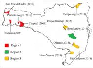
Map of the State of Santa Catarina, highlighting the regions and municipalities where samples were collected.
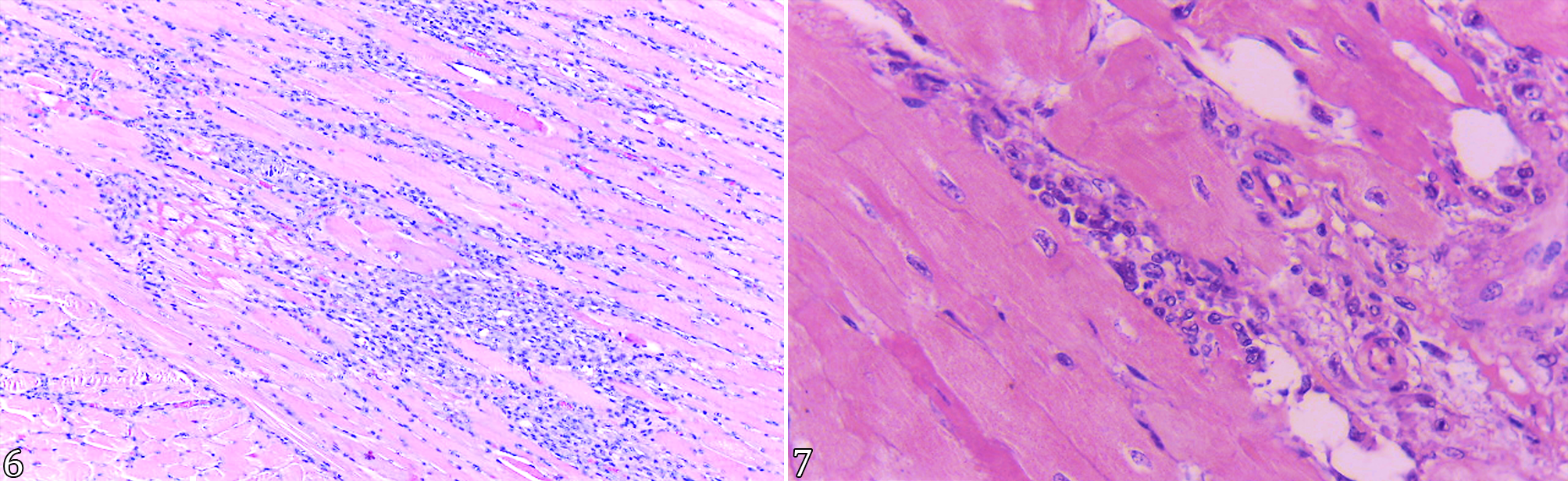
The animals affected by bluetongue, an acute form, exhibited clinical signs such as apathy (Fig. 2), hyperthermia ranging from 39.5 °C to 40.5 °C, hyperemia of the nasal and labial mucosa, accompanied by serous nasal discharge. Some had edema in the facial region, breathing difficulties, remnants of ruminal content in the nostrils, intense salivation, and purulent eye discharge. In advanced stages, there were ulcerations of the nostril, lips, and oral mucosa with crusting (Fig. 3), locomotor difficulties, black diarrhea, coughing, and regurgitation, followed by death. The course of the disease ranged from one to eight days, and those that did not die by the eighth day showed apparent recovery. According to the owners of the properties where the acute illness had occurred, some sheep were regurgitating food and progressively losing weight after 2-3 months. Of these sheep, necropsies were performed, one in Bom Retiro (animal acquired from Chapecó), one in Riqueza, and five in São Ludgero. The seven animals were euthanized within two to three months after the onset of clinical signs. Necropsies were performed on six sheep in the period of acute illness, of which two died naturally, three to five days after the onset of clinical signs, and four were euthanized. The main macroscopic lesions were multiple ulcers on the palate ranging from 1 to 5 mm in size; subcutaneous edema on the face, neck, and side of the thorax; hydrothorax and hydropericardium; pulmonary congestion and edema; petechial hemorrhages in the serosa of the esophagus near the esophageal hiatus; focal hemorrhages in the initial third of the pulmonary artery (Fig. 4); and multiple ulcers in the mucosa of the nasal cavity (turbinates). Ulcerations ranging in size from 1 to 2 cm were observed at the limits between the rumen (Fig. 5), reticulum, and omasum, in addition to splenomegaly. The histopathological examination revealed moderate to severe multifocal necrosis in the mucosa of the nasal cavity, oral cavity, rumen, reticulum, and omasum, sometimes accompanied by a large amount of cellular debris, bacterial colonies, and infiltration predominantly by neutrophils. Mild to moderate myofiber necrosis was observed in the esophagus (Fig. 6) and in the skeletal muscles, mainly the scapular region and the myocardium (Fig. 7), sometimes accompanied by light mononuclear infiltration. These lesions are not observed in sheep that survive several months after the acute clinical course. The clinical signs of chronic disease are limited to progressive weight loss, regurgitation, and, through histopathological evaluation. In the final third of the esophagus, smooth muscle fibers had been replaced by fibroblasts and adipocytes. There was extensive focal hemorrhaging in the pulmonary artery. Acute congestion and multifocal hemorrhages were found in the lungs and spleen. In the mediastinal and mesenteric lymph nodes, moderate edema, mainly in the medullary region, and multifocal hemorrhages, along with macrophage and neutrophil infiltration, were detected.
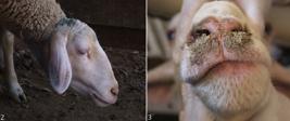
Sheep. (2) Apathetic sheep with a low head and drooping ears. (3) Crusty lesions in the nose and mouth region.

Sheep. (4) Heart: area of focal hemorrhaging at the base of the pulmonary artery. (5) Rumen: multiple ulcers ranging in diameter from 1 to 2 cm in the region between the rumen and the reticulum.

Sheep. (6) Esophagus: moderate, focally extensive lymphoplasmacytic infiltration, associated with individual necrosis of muscle fibers. HE, obj. 10x. (7) Heart: focally extensive inflammatory infiltration composed of lymphoplasmacytic cells, associated with necrosis of myofibers. HE, obj. 40x.
Of the 24 properties, 95.85% (23/24) had at least one animal that was seropositive for BTV. There was only one property located in the municipality of São Joaquim (Region 3) whose animals had all tested negative for BTV in the AGID test. Blood was collected from 388 sheep, of which 250 were positive and 138 were negative for BTV in the serologic test (Table 3). All the sheep on seven properties (29.16%) tested positive in the AGID test. Four of these properties were located in Region 1 (Riqueza 1 and 2, Cordilheira Alta 5, Lacerdópolis 7), and three were in Region 2 (São Ludgero 16, Braço do Norte 18, Campo Alegre 20).
Of the total number of sheep tested, 262 (67.52%) housed animals and five free-range (1.28%) animals were positive in the AGID test, while 96 (24.74%) housed sheep and 25 free-range sheep were negative in this same test. As for the sheep breeds that tested positive in the AGID test, there were 12 (3.09%) Criollo, 27 (6.95%) Lacaune, 36 (9.27%) Santa Inês, 86 (22.31%) of an undefined breed, and 106 (27.31%) Texel. There were two (0.51%) Santa Inês, 18 (4.63%) Texel, 19 (4.89%) Criollo, 29 (7.47%) Lacaune, and 86 (22) undefined breeds that tested negative in the AGID test. Of the 388 sheep assessed by the AGID test, 240 (61.85%) meat breeds and 27 (6.95%) dairy breeds tested positive, while 92 (23.71%) meat breeds and 29 (7.47%) dairy breeds tested negative.
Thirty-five of the 388 blood samples submitted to the AGID test presented clinical signs suggestive of bluetongue. It should be noted that all the sheep were randomly selected (Table 4).
Results of clinical diagnosis and agar gel immunodiffusion assay (AGID) for bluetongue virus (BTV) in sheep blood samples
From 2016 to 2019, we can observe an increase of up to one degree in the average annual temperatures of the municipalities of Água Doce in Region 1, Irineópolis and Campo Alegre in Region 2, and São Joaquim and Bom Retiro in Region 3. Despite this observation, it is not possible to correlate this increase in temperatures with the increase in diagnoses of BTV in sheep.
Discussion
Bluetongue is an important disease that affects sheep, and it can cause major direct and indirect economic losses through treatments, diagnoses, control measures, and trade restrictions. This disease has historically occurred between the 40th parallel north and the 35th parallel south (Taylor 1986, Purse et al. 2005, Samy & Peterson 2016). The region encompassing the state of Santa Catarina is located between the southern parallels 25°57’41” and 29°23’55”; therefore, this latitudinal margin favors the occurrence of BTV.
In the Mountain region, the disease was observed in 2019. The increase in average annual temperatures in recent years has probably contributed to the spread of the vector; on the other hand, there are contradictions as to the influence of average temperatures on the spread of the disease (Jacquot et al. 2017). Another plausible hypothesis for the onset of BTV in this region would be the introduction of asymptomatic positive animals in these places.
In Europe, coastal areas, mountain regions, and regions with altitudes above 300 meters (sites where vector replication is difficult) are considered to be barriers that limit the spread of Culicoides in certain locations (Jacquot et al. 2017). A similar situation occurs in the Mountain region of Santa Catarina, where the mosquito seems to have greater difficulty spreading in areas at higher altitudes. In this study, most of the animals that tested seropositive for BTV were located in regions with a lower altitude, such as the West and the Coast, which have an altitude below 300 meters. Some areas at higher altitudes in Santa Catarina probably have physical barriers due to topography, such as mountains that limit vector spread due to prevailing winds. James Spreull (1905) asserts that BTV does not normally circulate in high-altitude regions. Winds and their directions also play a crucial role in the dissemination of the vector, which can thus be dispersed passively over 300 kilometers (Purse et al. 2005, Jacquot et al. 2017). Only 16 of the 65 blood samples collected in the municipality of Água Doce (located in Region 1) for the AGID test were positive. This difference from the other properties in Region 1, which presented a greater number of positive sheep, may be attributed to the fact that this municipality is located 1,125 meters above sea level, with lower mean annual temperatures (16.7 °C). The other municipalities in this region (1) have higher mean annual temperatures (18.9 °C).
Similarly, only four sheep showed positive results in the samples collected in the municipalities of Lages and Correia Pinto in Region 3. In this region (3), samples collected in Bom Retiro resulted in 22 sheep testing positive and 15 testing negative. It is believed that the occurrence of the disease in this municipality was favored by its proximity to Region 2, which has a warmer climate, and the prolonged heat period recorded in 2019, which may have contributed to the mosquito’s expansion. It is important to note that the samples from Bom Retiro were collected in April 2019, a period that coincided with the clinical manifestation of the disease on the property. Samples collected from sheep in São Joaquim, a municipality with low temperatures, were negative. Low temperatures observed in previous years have probably hampered the proliferation of Culicoides in this region. In two outbreaks, one in the West and another in the High Valley of Itajaí, the sick sheep had been acquired from the Mountain region a few weeks earlier, a fact that can be explained by the nonproliferation of the mosquito vector of bluetongue in the region of origin. This study demonstrates that BTV is in circulation in all regions of the state of Santa Catarina. The largest number of positive results was observed in Regions 1 (Chapecó) and 2 (São Ludgero), which presented average annual temperatures of 19.9 °C and 20.4 °C, respectively, from 2018 to 2019.
Transmission of BTV depends on propitious temperatures and humidity that set off the following chain of events: The vector feeds off the blood of a host in the viremic period, ingests enough of the virus to exert the threshold for infection, survives the extrinsic incubation period of the virus, and feeds off a susceptible host (Purse et al. 2005). When a sheep is bitten by an infected mosquito and the virus is inoculated, the sheep can develop acute or persistent clinical signs, which characterize a chronic condition (Brown et al. 2007). In this study, BTV infection was observed in the acute form, with death within three to eight days after the onset of clinical signs. Episodes of regurgitation and weight loss two to three months after the acute clinical condition raised the suspicion of sequelae. In these cases, where clinical signs persist, the macroscopic change was limited to a dilation of the esophagus observed in two sheep in the municipality of Chapecó in 2009 (Property 4) and in five sheep in São Ludgero in 2019 (Property 16). Observed microscopic changes include multifocal fibrosis of the esophageal muscle and, sometimes, areas of proliferation of adipose cells. It is suggested that these changes are sequelae of an acute BTV infection, as in this condition, lesions in the esophageal muscles can make it difficult to swallow with the accumulation of food content, followed by weight loss and death.
Most infected sheep ruminants develop a subclinical infection, so they are not usually identified or removed from the herd and thus persist as a source of infection for vectors (Purse et al. 2005). Similarly, in this study, infections had not been identified previously and remained in the herds as a source of infection for Culicoides, which persisted on the properties. Sheep are more susceptible than other species (bovids, caprids, and camelids) to the development of the clinical disease; however, there may be an elevated number of subclinical seropositive animals in a sheep herd (Koumbati et al. 1999, Williamson et al. 2008, Lima et al. 2016). In the present study, the evaluated sheep tested positive in the AGID test but did not show clinical signs of the disease on Properties 9, 10, 11, 23, and 24. According to Takamatsu et al. (2003), there may be persistent infection in animals that had apparently recovered during the winter. When the proliferation of the vector occurs in the spring, it begins to feed off ruminants. The T cells that harbor the virus die, and the virus is ingested by the Culicoides, thereby initiating another transmission cycle.
To reduce costs, confirmation of the diagnosis by RT-PCR was performed by the property; therefore, samples were processed in a pool when blood was collected from more than one potentially infected sheep. RT-PCR is considered a fast, reliable, and robust diagnostic tool for detecting BTV. In this study, the amplified gene was VP1, the most conserved gene (Maan et al. 2016).
RT-PCR is considered to have high specificity, although other direct tests, such as inoculation into embryonated eggs in a cell culture, may also be used. Research into antibodies using ELISA and AGID can also be useful when there is a suspected circulation of BTV (Halder et al. 2016, Matos et al. 2016, Bumbarov et al. 2020).
To perform the diagnostic techniques described herein, blood was collected from sheep of various ages, but no younger than six months. In the young animals of Regions 1 and 2, the AGID technique generated a positive result for most animals. In the municipality of Bom Retiro (Region 3) at Property 22, blood was collected from 37 sheep, of which 10 were eight-month-old lambs that tested negative. These animals were kept sheltered in a shed. This observation corroborates James Spreull (1905) findings that shelter can inhibit the vector’s entry and prevent the virus from circulating.
In Regions 1 and 2, the sheep remained in paddocks during the day and were housed at night, and most tested positive by AGID. Nonetheless, there are disagreements regarding the housing of animals in close quarters, which would facilitate direct transmission. There are also reports that housing would make entry difficult for the vector (James Spreull 1905).
In Region 1, undefined sheep breeds accounted for the majority of positive sheep, although almost all were crossbreeds of wool sheep. The largest number of seropositive animals in Regions 2 and 3 were Texel and Lacaune, respectively. According to Taylor (1986), some meat and fine wool breeds are considered to be more sensitive to BTV as they develop more significant clinical signs.
It is worth noting that some strains of BTV can be transmitted horizontally, without the presence of vectors. There are still other types of BTV that are transmitted vertically (Batten et al. 2014). Density and distribution of cattle and sheep seem to play a vital role in the spread of BTV, and cattle are believed to be important reservoirs of infection, as Culicoides prefer them over sheep. The authors attribute this to the size of the cattle herd, in addition to emissions from semi-chemical substances, the absence of wool, and a longer detectable viremic period (Bonneau et al. 2002, Jacquot et al. 2017). It is important to note that the state of Santa Catarina is one of the states in Brazil with the highest agricultural technification; consequently, there is a higher density of cattle and sheep. As claimed by Jacquot et al. (2017), this approximation of animals would facilitate the faster spread of BTV.
Conclusions
Based on the data from this study, we can confirm the presence of bluetongue virus (BTV) in the state of Santa Catarina. In the three regions of the state, some sheep tested seropositive in the agar gel immunodiffusion (AGID) tests and positive in the reverse transcription polymerase chain reaction (RT-PCR) tests. In the Mountains of Santa Catarina, the spread of the virus may be related to the increase in temperature, which now meets the bionomic requirements for the vector of the virus.
BTV can cause sequelae in sheep, such as dysphagia, weight loss, and death, a few months after the manifestation of the acute disease. Control and prophylactic measures for BTV should be recommended among sheep farmers in the state of Santa Catarina. To ensure the effectiveness of these measures, the serotype circulating in the state should be confirmed. In the future, if possible, vaccination programs should be implemented in areas considered endemic.
Acknowledgments
This study was financed in part by the Brazilian fostering agency “Coordenação de Aperfeiçoamento de Pessoal de Nível Superior” (CAPES).
References
-
Antoniassi NAB, Pavarini SP, Ribeiro LAO, Silva MS, Flores EF, Driemeier D. Alterações clínicas e patológicas em ovinos infectados naturalmente pelo vírus da língua azul no Rio Grande do Sul. Pesq Vet Bras 2010; https://doi.org/10.1590/S0100-736X2010001200002
» https://doi.org/10.1590/S0100-736X2010001200002 -
Backx A, Heutink CG, Van Rooij EMA, Van Rijn PA. Clinical signs of bluetongue virus serotype 8 infection in sheep and goats. Vet Rec 2007; https://doi.org/10.1136/vr.161.17.591
» https://doi.org/10.1136/vr.161.17.591 -
Balaro MFA, Lima MS, Fava CD, Oliveira GR, Pituco EM, Brandão FZ. Outbreak of bluetongue virus serotype 4 in dairy sheep in Rio de Janeiro, Brazil. J Vet Diagn Invest 2014; https://doi.org/10.1177/1040638714538020
» https://doi.org/10.1177/1040638714538020 -
Baldini MHM, Rosa JCC, Matos ACD, Cubas ZS, Guedes MIMC, Moraes W, Oliveira MJ, Felippi DA, Lobato ZIP, Moraes AN. Multiple bluetongue virus serotypes causing death in Brazilian dwarf brocket deer (Mazama nana) in Brazil, 2015-2016. Vet Microbiol 2018; https://doi.org/10.1016/j.vetmic.2018.10.018
» https://doi.org/10.1016/j.vetmic.2018.10.018 -
Batten C, Darpel K, Henstock M, Fay P, Veronesi E, Gubbin S, Graves S, Frost L, Oura C. Evidence for transmission of bluetongue virus serotype 26 through direct contact. PloS One 2014; https://doi.org/10.1371/journal.pone.0096049
» https://doi.org/10.1371/journal.pone.0096049 -
Bianchi RM, Panziera W, Faccin TC, Almeida GL, Cargnelutti JF, Flores EF, Kommers GD, Fighera RA. Clinical, pathological and epidemiological aspects of outbreaks of bluetongue disease in sheep in the central region of Rio Grande do Sul. Pesq Vet Bras 2017; https://doi.org/10.1590/S0100-736X2017001200014
» https://doi.org/10.1590/S0100-736X2017001200014 -
Bonneau KR, DeMaula CD, Mullens BA, McLachlan NJ. Duration of viraemia infectious to Culicoides sonorensis in bluetongue vírus-infected cattle and sheep. Vet Microbiol 2002; https://doi.org/10.1016/S0378-1135(02)00106-2
» https://doi.org/10.1016/S0378-1135(02)00106-2 - Brown CC, Baker DC, Barker IK. Alimentary system. In: Maxie MG. Jubb, Kennedy, and Palmer’s Pathology of Domestic Animals. 5th ed. Vol. 1. Philadelphia: Saunders Elsevier; 2007.
-
Bumbarov V, Golender N, Jenckel M, Wernike K, Beer M, Khinich E, Zalesky O, Erster O. Characterization of bluetongue virus serotype 28. Transbound Emerg Dis 2020; https://doi.org/10.1111/tbed.13338
» https://doi.org/10.1111/tbed.13338 -
Clavijo A, Sepulveda L, Riva J, Pessoa-Silva M, Tailor-Ruthes A, Lopez JW. Isolation of bluetongue virus serotype 12 from an outbreak of the disease in South America. Vet Rec 2002; https://doi.org/10.1136/vr.151.10.301
» https://doi.org/10.1136/vr.151.10.301 -
D. Hutcheon MRCVS. The malarial catarrhal fever of sheep in South Africa. Vet J Ann Comp Pathol 1893; https://doi.org/10.1016/S2543-3377(17)49810-4
» https://doi.org/10.1016/S2543-3377(17)49810-4 -
Darpel KE, Batten CA, Veronesi E, Shaw AE, Anthony S, Bachanek-Bankowska K, Kgosana L, Bin-Tarif A, Carpenter S, Müller-Doblies UU, Takamatsu H-H, Mellor PS, Mertens PPC, Oura CAL. Clinical signs and pathology shown by British sheep and cattle infected with bluetongue virus serotype 8 derived from the 2006 outbreak in northern Europe. Vet Rec 2007; https://doi.org/10.1136/vr.161.8.253
» https://doi.org/10.1136/vr.161.8.253 -
Guimarães LLB, Rosa JCC, Matos ACD, Cruz RAS, Guedes MIMC, Dorella FA, Figueiredo HCP, Pavarini SP, Sonne L, Lobato ZIP, Driemeier D. Identification of bluetongue virus serotypes 1, 4, and 17 co-infections in sheep flocks during outbreaks in Brazil. Res Vet Sci 2017; https://doi.org/10.1016/j.rvsc.2017.09.001
» https://doi.org/10.1016/j.rvsc.2017.09.001 -
Halder A, Joardar SN, Isore DP, Samanta I, Parui P, Banerjee D, Lodh C. Seroepidemiology of bluetongue in South Bengal. Vet World 2016; https://doi.org/10.14202/vetworld.2016.1-5
» https://doi.org/10.14202/vetworld.2016.1-5 -
IBGE. Tabela 6720. Instituto Brasileiro de Geografia e Estatísticas, 2017. Accessed on Oct. 27, 2020. https://sidra.ibge.gov.br/tabela/6720#resultado
» https://sidra.ibge.gov.br/tabela/6720#resultado -
Jacquot M, Nomikou K, Palmarini M, Mertens P, Biek R. Bluetongue virus spread in Europe is a consequence of climatic, landscape and vertebrate host factors as revealed by phylogeographic inference. Proc R Soc B 2017; https://doi.org/10.1098/rspb.2017.0919
» https://doi.org/10.1098/rspb.2017.0919 -
James Spreull MRCVS. Malarial catarrhal fever (bluetongue) of sheep in South Africa. J Comp Pathol Ther 1905; https://doi.org/10.1016/S0368-1742(05)80073-6
» https://doi.org/10.1016/S0368-1742(05)80073-6 -
Jenckel M, Bréard E, Schulz C, Sailleau C, Viarouge C, Hoffmann B, Höper D, Beer M, Zientara S. Complete coding genome sequence of putative novel bluetongue virus serotype 27. Genome Announc 2015; https://doi.org/10.1128/genomea.00016-15
» https://doi.org/10.1128/genomea.00016-15 -
Kawanami AE, Oliveira JP, Arenales A, Crossley B, Woods LW, Duarte JMB, Werther K. Detection of bluetongue virus in Brazilian cervids in São Paulo state. Pesq Vet Bras 2018; https://doi.org/10.1590/1678-5150-PVB-4550
» https://doi.org/10.1590/1678-5150-PVB-4550 -
Koumbati M, Mangana O, Nomikou K, Mellor PS, Papadopoulos O. Duration of bluetongue viraemia and serological responses in experimentally infected European breeds of sheep and goats. Vet Microbiol 1999; https://doi.org/10.1016/S0378-1135(98)00255-7
» https://doi.org/10.1016/S0378-1135(98)00255-7 -
Lima PA, Utiumi KU, Nakagaki KYR, Biihrer DA, Albuquerque AS, Rezende FS, Matos ACD, Lobato ZIP, Driemeier D, Peconick AP, Varaschin MS, Raymundo DL. Diagnoses of ovine infection by the serotype-4 bluetongue virus on Minas Gerais, Brazil. Acta Scient Vet 2016; https://doi.org/10.22456/1679-9216.84210
» https://doi.org/10.22456/1679-9216.84210 -
Maan S, Maan NS, Belaganahalli MN, Potgieter AC, Kumar V, Batra K, Wright IM, Kirkland PD, Merten PPC. Development and evaluation of real time RT-PCR assays for detection and typing of bluetongue virus. PLoS One 2016; https://doi.org/10.1371/journal.pone.0163014
» https://doi.org/10.1371/journal.pone.0163014 -
Matos ACD, Rosa JCC, Nomikou K, Guimarães LLB, Costa ÉA, Guedes MIMC, Driemeier D, Lobato ZIP, Mertens PPC. Genome sequence of bluetongue virus serotype 17 isolated in Brazil in 2014. Genome Announc 2016; https://doi.org/10.1128/genomea.01161-16
» https://doi.org/10.1128/genomea.01161-16 -
Purse BV, Mellor PS, Rogers DJ, Samuel AR, Mertens PPC, Baylis M. Climate change and the recent emergence of bluetongue in Europe. Nat Rev Microbiol 2005; https://doi.org/10.1038/nrmicro1090
» https://doi.org/10.1038/nrmicro1090 - Quevedo LS, Zancan TD, Hugen GFGP, Morais RM, Ogliari D, Melchioretto E, Serzedello L, Gava A. Língua azul em ovinos no estado de Santa Catarina. Pesq Vet Bras 2019;39(Supl.)83-85. (Resumo).
- Riet-Correa F. Língua azul. In: Riet-Correa F, Schild AL, Lemos RAA, Borges JRJ. Doenças de Ruminantes e Equídeos. 3ª ed. Vol. 1. Santa Maria: Pallotti; 2007.
-
Samy AM, Peterson AT. Climate change influences on the global potential distribution of bluetongue virus. PLoS One 2016; https://doi.org/10.1371/journal.pone.0150489
» https://doi.org/10.1371/journal.pone.0150489 -
Takamatsu H, Mellor PS, Mertens PPC, Kirkham PA, Burroughs JN, Parkhouse RME. A possible overwintering mechanism for bluetongue virus in the absence of the insect vector. J Gen Virol 2003; https://doi.org/10.1099/vir.0.18705-0
» https://doi.org/10.1099/vir.0.18705-0 -
Taylor WP. The epidemiology of bluetongue. Rev Sci Off Int Epiz 1986; https://doi.org/10.20506/rst.5.2.256
» https://doi.org/10.20506/rst.5.2.256 - Thrusfield MV. Epidemiologia Veterinária. 2ª ed. São Paulo: Roca; 2004.
-
Williamson S, Woodger N, Darpel K. Differential diagnosis of bluetongue in cattle and sheep. In Practice 2008; https://doi.org/10.1136/inpract.30.5.242
» https://doi.org/10.1136/inpract.30.5.242 - Zachary JF. Mecanismos das infecções microbianas. In: Zachary JF, McGavin MD. Bases da Patologia em Veterinária. 5º ed. Rio de Janeiro: Elsevier; 2013.
-
Data Availability Statement
The authors confirm that the data supporting the findings of this study are available within the article. Derived data supporting the findings of this study are available from the corresponding author upon request.
The authors confirm that the data supporting the findings of this study are available within the article. Derived data supporting the findings of this study are available from the corresponding author upon request.