
 Retrospective study of canine non-Hodgkin lymphoma cases diagnosed from samples originating in northeastern Brazil (2012-2022)
ABSTRACT:
Lymphoma is a malignant hematopoietic neoplasm that affects canines. Its classification is based on anatomical, histomorphological, cytomorphological, and immunophenotypic characteristics. This study surveyed cases of canine non-Hodgkin lymphoma originating in Northeastern Brazil from 2012 to 2022, considering epidemiological, histomorphological, and immunophenotypic aspects. One hundred sixteen samples with a prior diagnosis of canine lymphoma were selected, re-evaluated, and classified according to the World Health Organization criteria. The cases originated from eight states in northeastern Brazil. The average age of the animals was 8.38 years, with 54.31% males and 40.52% females, and a prevalence of mixed-breed animals. Regarding the evaluated conditions, 33.62% corresponded to lymph nodes, 31.03% to the skin, and 25.86% to the spleen. Histomorphological and immunophenotypic results presented 50.86% of B-cell lymphomas, highlighting 45.76% diffuse large B-cell lymphoma, 32.20% splenic marginal zone lymphoma, and 49.13% T-cell lymphoma. Histomorphological and immunophenotypic findings presented 50.86% of B-cell lymphomas, highlighting 45.76% diffuse large B-cell lymphoma, 32.20% splenic marginal zone lymphoma, and 49.13% T-cell lymphoma. The cutaneous non-epitheliotropic lymphoma represented 43.85%, and the epitheliotropic was 17.54%. The epidemiological profile of canine non-Hodgkin lymphoma cases in northeastern Brazil is similar to that observed in other regions of Brazil and worldwide. Based on these findings, the authors hypothesize that regional environmental conditions could play a role in the development and progression of this neoplasm. Therefore, continuous monitoring and further regional studies are essential to elucidate its biological behavior.
INDEX TERMS:
Dog; immunohistochemistry; hematopoietic neoplasm; northeastern Brazil; World Health Organization
RESUMO:
O linfoma é uma neoplasia hematopoética maligna que acomete cães. Sua classificação baseia-se em características anatômicas, histomorfológicas, citomorfológicas e imunofenotípicas. Este estudo realizou um levantamento de casos de linfoma não-Hodgkin canino oriundos da região Nordeste do Brasil, no período de 2012 a 2022, considerando aspectos epidemiológicos, histomorfológicos e imunofenotípicos. Foram selecionadas 116 amostras com diagnóstico prévio de linfoma canino, as quais foram reavaliadas e classificadas de acordo com os critérios da Organização Mundial da Saúde (OMS). Os casos tiveram origem em oito estados da região Nordeste do Brasil. A idade média dos animais foi de 8,38 anos, sendo 54,31% machos e 40,52% fêmeas, com predomínio de animais sem raça definida. Com relação aos locais de acometimento, 33,62% dos casos corresponderam a linfonodos, 31,03% à pele e 25,86% ao baço. Os resultados histomorfológicos e imunofenotípicos indicaram 50,86% de linfomas de células B, destacando-se 45,76% de linfoma difuso de grandes células B e 32,20% de linfoma da zona marginal esplênica, além de 49,13% de linfomas de células T. Entre os linfomas cutâneos, o tipo não epiteliótropo representou 43,85% dos casos e o epiteliótropo, 17,54%. O perfil epidemiológico dos casos de linfoma não-Hodgkin canino no Nordeste do Brasil é semelhante ao observado em outras regiões do país e do mundo. Com base nesses achados, os autores levantam a hipótese de que condições ambientais regionais possam influenciar o desenvolvimento e a progressão dessa neoplasia. Portanto, o monitoramento contínuo e novos estudos regionais são essenciais para elucidar seu comportamento biológico.
TERMOS DE INDEXAÇÃO:
Cão; imunohistoquímica; neoplasia hematopoiética; Nordeste brasileiro; organização mundial da saúde
Introduction
Non-Hodgkin lymphoma (NHL) is a malignant hematopoietic neoplasm that affects dogs, accounting for approximately 8-10% of all neoplasms in this species and about 80% of hematopoietic neoplasms (Valli et al. 2016). Its classification is based on anatomical, histomorphological, cytomorphological, and immunophenotypic characteristics, demonstrating a biological behavior similar to human NHL (Zanini et al. 2013).
The Northeast region of Brazil lies between latitudes -2° and -18° and longitudes -34° and -46°, encompassing vast geographic and climatic diversity. Its strategic location makes it a key area for regional and environmental studies in the country, comprising approximately 30% (56.7 million) of the Brazilian human population. This region has revealed a hospital morbidity profile for NHL from 2017 to 2022 that included 19,648 hospitalizations and 1,747 human deaths. This scenario highlights a significant increase in NHL cases, underscoring the need for research on the disease and its preventive measures (Silva et al. 2022). Veterinary medicine presents another scenario, with a noticeable scarcity of studies on this type of neoplasm, particularly in the Northeast region of Brazil. Research conducted in various countries highlights immunophenotypic aspects with a similar profile for humans and animals, with B-cell lymphomas prevailing over T-cell lymphomas. The multicentric type prevailed in the anatomical classification (Van Rooyen et al. 2018, Jark et al. 2020, Pinello et al. 2023).
Given the limited research on canine NHL cases in Brazil, mainly in the Northeast region, this study surveyed cases in dogs originating in northeastern Brazil from 2012 to 2022, considering epidemiological, histomorphological, and immunophenotypic aspects.
Materials and Methods
Ethical approval. This study is exempt from an Animal Ethics License, as it involved the organ samples from paraffin-embedded biopsies donated by the Histopathology Laboratory teams of the investigated region, in accordance with the annex of CONCEA Normative Resolution No. 55 of October 5, 2022, which addresses the “Diretriz Brasileira para o Cuidado e a Utilização de Animais em Atividades de Ensino ou de Pesquisa Científica” (Brazilian Guidelines for the Care and Use of Animals in Teaching or Scientific Research Activities - DBCA).
Sample collection. The study sample consisted of 149 paraffin blocks containing canine tissues previously diagnosed with lymphoma by histopathological examination, submitted between 2012 and 2022. The samples originate from the collections of private and public institutions from eight states in Northeast Brazil that expressed interest in participating in the project. The study also gathered information regarding breed, sex, age, and affected tissue. The samples were re-evaluated and reclassified, and those without histomorphological and immunophenotypic features compatible with lymphoma were excluded. Thus, the study selected 116 samples.
Histopathological analysis. The 116 selected samples were sectioned using a semi-automatic rotary microtome to prepare slides and then stained with the hematoxylin and eosin (HE) technique according to the staining protocol of the Animal Diagnostic Laboratory (ADL) from the “Departamento de Morfologia e Fisiologia Animal” (Department of Animal Morphology and Physiology - DMFA) of the “Universidade Federal Rural de Pernambuco” (UFRPE). The slides were then mounted and evaluated under an Olympus BX41 light microscope, and classified according to the American College of Veterinary Pathologists (ACVP) review criteria adapted from the World Health Organization (WHO) guidelines for canine NHL (Valli et al. 2011).
Immunohistochemistry (IHC). The samples were sectioned to a thickness of 4 μm using a semi-automatic rotary microtome, just like the reactive canine lymph node tissue used as the positive control. The analysis followed the standard protocol of the immunohistochemistry laboratory at the Clinics Hospital (HC) of the UFPE. The used antibodies were anti-CD3 (Polyclonal, Catalog: 500-1254; AbboMax®; dilution 1:400) and anti-CD20 (Polyclonal, Catalog: PA5-16701, Invitrogen®; dilution 1:400).
The tissue sections remained overnight in a 75 °C oven. The next day, they underwent four washes in xylene for 5 minutes for deparaffinization, followed by rehydration with four washes in absolute alcohol for 5 minutes and one wash in distilled water for 7 minutes. Antigen retrieval was performed in a Pascal pressure cooker for 20 minutes in recovery EDTA buffer at pH 8.5 (EasyPath, São Paulo, BR). Subsequently, endogenous peroxidase was blocked for 20 minutes, followed by protein blocking for 20 minutes. The primary antibodies were then incubated for 1 hour (overnight) at room temperature, followed by 1x PBS wash, and the addition of the labeling amplifier (post-primary) for 30 minutes, polymer for 30 minutes, and DAB diluted according to the manufacturer’s instructions for 5 minutes. The slides were then counterstained with Harris hematoxylin, dehydrated, and mounted. The technique used the Novolink polymer detection system (Leica Biosystems Newcastle Ltd., Newcastle Upon Tyne, UK) according to the manufacturer’s instructions.
Results
The analyzed samples were from Northeast Brazil, represented by the states of Alagoas, Bahia, Ceará, Paraíba, Pernambuco, Piauí, Rio Grande do Norte, and Sergipe. Figure 1 shows the number of samples by state.
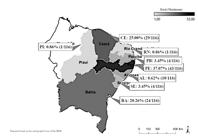
Canine lymphoma in Northeast Brazil. States in Northeast Brazil, and the number of samples sent for analysis in relative and absolute values.
The age of animals affected by the neoplasm ranged from two to 16 years, with an average of 8.38 years. Regarding sex, 54.31% (43/116) of the analyzed samples were from males, 40.52% (47/116) from females, and 5.17% (6/116) did not inform the animal’s sex.
Mixed-breed (28.45%; 33/116) and Poodles (10.34%; 12/116) were the most affected, and 8.62% (10/116) of the sample did not have breed information. Table 1 details the findings regarding animals’ breeds and sizes.
Regarding the affected tissues, lymph nodes were more prevalent in the sample, at 33.62% (39/116), followed by skin at 31.03% (36/116) and spleen at 25.86% (30/116). The intestine, at 6.9% (8/116) and the liver, at 2.59% (3/116), were the least observed.
According to the current WHO classification of canine lymphomas, histomorphological analyses showed a higher frequency of B-cell neoplasms, representing 50.86% (59/116) of the sample. Among these conditions, diffuse large B-cell lymphoma (DLBCL) prevailed at 45.76% (27/59) (Fig.2-5), followed by splenic marginal zone lymphoma (MZL) at 32.20% (19/59) (Fig. 6). T-cells appeared in 49.14% (57/116) of neoplasms, with a higher frequency of cutaneous non-epitheliotropic lymphomas at 43.85% (25/57) (Fig. 7), epitheliotropic lymphomas at 17.54% (10/57), and T-zone lymphoma at 10.52% (6/57) (Fig. 8). The systemic anaplastic large cell lymphoma, a peripheral lymphoma not otherwise specified (NOS), also stood out at 7.01% (4/57) (Fig. 9). Table 2 presents other histomorphological and immunophenotypic findings.
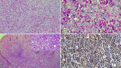
Canine lymphoma in Northeast Brazil. (2) B-cell lymphoma. Diffuse large B-cell lymphoma. Loss of nodal architecture with intense proliferation of neoplastic lymphoid cells. HE, obj. 40x. (3) Neoplastic cells of large immunoblastic type with a single central nucleolus (arrow). HE, obj. 200x. (4) Burkitt-like lymphoma. Diffuse proliferation of neoplastic cells, associated with macrophages containing apoptotic bodies (arrow), creating a starry-sky appearance. HE, obj. 100x. Inset: Higher magnification showing macrophages with apoptotic bodies (arrow). HE, obj. 600x. (5) B-cell lymphoma. IHC for CD20 showing staining of neoplastic cells. Anti-CD20, DAB chromogen, obj. 100x.

Canine lymphoma in Northeast Brazil. B- and T-cell lymphoma. (6) Marginal zone lymphoma. Spleen lymphoid follicles coalescing, with intense expansion of the marginal zone (arrow). HE, obj. 40x. Inset: IHC for CD20, showing staining of neoplastic cells. Anti-CD20, DAB chromogen, obj. 40x. (7) Systemic anaplastic T-cell lymphoma (NOS), spleen, exhibiting loss of architecture and diffuse, intense proliferation of neoplastic lymphoid cells. HE, obj. 40x. Upper inset: Higher magnification showing neoplastic cells with high pleomorphism and multiple nucleoli (arrow). HE, obj. 400x. Below inset: Immunohistochemistry for CD3, staining of neoplastic cells. Anti-CD3, DAB chromogen, obj. 200x. (8) T-zone lymphoma. IHC for CD3, lymphoid follicles compressed toward the periphery (arrow). Anti-CD3, DAB chromogen, obj. 40x. Inset: IHC for CD20, nodal follicle staining. Anti-CD20, DAB chromogen, obj. 100x. (9) Non-epitheliotropic cutaneous lymphoma. Proliferation of neoplastic lymphoid cells occupying the entire dermis, without invasion of the epidermal layer or skin adnexa (arrow). HE, obj. 200x. Inset: IHC for CD3, highlighting staining of neoplastic cells and noting the lack of invasion into skin adnexa (arrow). Anti-CD3, DAB chromogen, obj. 200x.
Discussion
This study found a high frequency of previously diagnosed cases of canine NHL in the Northeast region of Brazil, mainly in Pernambuco, Ceará, Bahia, and Alagoas, corresponding to 37.07%, 25%, 20.26%, and 8.62%, respectively. Jark et al. (2020) surveyed 203 canine multicentric lymphomas in Brazil, finding that 83.6% of cases occurred in the Southeast and only 1.2% in the Northeast region. Conversely, Cavalcanti & Lira (1968) diagnosed 57 cases of canine lymphoma in Recife (Pernambuco, BR) over six years (1960-1966), noting the limited research regarding this neoplasm in the region over the studied period.
The difference in the number of samples by state, as evidenced in this study, relates to the difficulties of partnership between research and pathology laboratories in the region. These difficulties impact the collected data and hinder the assessment of neoplasm rates in northeastern states. However, these factors do not compromise the validity of the research, as the samples were obtained from recognized laboratories that strictly follow the protocols for storage and processing of paraffin-embedded materials. An epidemiological database linking the findings among states and across species might strengthen communication and stimulate new research, emphasizing that dogs are sentinel animals for human diseases (Coleto et al. 2017, Pittaway et al. 2019, Holanda et al. 2022, INCA 2023).
The average age of the animals was 8.38 years, typical of patients affected by NHL, as highlighted in the literature (Vezzali et al. 2011, Coleto et al. 2017, Pittaway et al. 2019). Adult to elderly dogs are less immunocompetent, aiding the emergence of neoplastic diseases, including lymphoma, primarily due to exposure to environmental agents that favor neoplasm development (Holanda et al. 2022). The most affected age group in humans comprises adults and elderly individuals between 50 and 69 years, highlighting another similarity in the biological behavior of the neoplasm compared to the canine species (Melo et al. 2024).
Males were more affected than females, with 55.03% and 40.26% of cases, respectively. Veterinary and human literature reports that lymphomas occur more frequently in males (Neuwald et al. 2014, Coelho et al. 2019, Melo et al. 2024). The exact determining factor for canines is not explicit; however, in humans, elevated sex hormones, such as estrogen, may play a protective role in the development of certain lymphomas in women. Additionally, the immune response from cells, such as natural killer cells, slows the onset of the neoplasm more effectively in women than in men (Horesh & Horowitz 2014).
Small-breed animals were more frequent in the present study, diverging from the consulted literature, which reports that medium- and large-breed dogs are the most prone to developing neoplasia (Valli et al. 2013, Pinello et al. 2023). Mixed-breed dogs stood out, followed by Rottweilers, Pit Bulls, Cocker Spaniels, Poodles, and Shih Tzus.
The high frequency of cases in mixed-breed animals may reflect population behavior in developing countries or regions. These areas demonstrate a close relationship between socioeconomic aspects and animal care, revealing numerous neoplasm diagnoses in mixed-breed animals (Sanches et al. 2013, Cora et al. 2016, Coelho et al. 2019, Sanches et al. 2019, Pinello et al. 2023). This situation is changing in countries with greater economic power, such as those in the world’s northern regions (Pinello et al. 2023), particularly with the growing awareness of animal welfare and adoption in these countries, increasing the frequency of mixed-breed animals (Comazzi et al. 2018).
The risk factors for lymphoma development in certain breeds are multifactorial, with a strong genetic influence. Breeds such as Boxer, Boerboel, Rhodesian Ridgeback, English Bulldog, and Bullmastiff have an elevated risk, likely due to their common ancestry within the Molosser group, which shares ancestors and possibly genes related to lymphoma (Van Rooyen et al. 2018).
Besides the genetic predisposition of certain breeds to develop lymphoma, a synergistic combination with environmental factors may occur, such as exposure to chemicals and pollutants, increasing the likelihood of neoplasm occurrence (Craun et al. 2020). Luethcke et al. (2022) studied chronic environmental exposure as a risk factor for lymphoma development in 49 Golden Retriever dogs, highlighting the complexity of the relationship between such exposure and the risk of lymphoma in dogs. They suggested that additional factors, such as herbicides or nitrates in water, are more significant than atmospheric pollution, despite the lack of statistically significant associations.
The literature rarely mentions small breeds, such as Poodles and Shih Tzus, which were common in this study, as they are noted only occasionally (Kim et al. 2021). Another point to consider is population behavior regarding pet ownership, as the popularity of small dogs has increased with urban expansion and the prevalence of smaller homes in large cities. The samples submitted to this study were predominantly from state capitals that present a higher concentration of people per square meter, further favoring the acquisition of small breeds, such as Poodles and Shih Tzus (Mazon & Moura 2017, Gonzáles-Ramírez 2019).
The analyzed tissues showed a higher frequency of lymph node samples (33.62%), highlighting the solid hematopoietic organs affected by the neoplasm. Multicentric lymphomas are more frequent in various studies, even across Brazilian regions (Valli et al. 2013, Cora et al. 2016, Coelho et al. 2019, Jark et al. 2020, Pinello et al. 2023), similar to NHL in humans (Melo et al. 2024).
The skin was the second-most-affected organ by the neoplasm, accounting for 31.03% of cases. As the largest organ in the body and the most exposed to exogenous agents, the skin is particularly susceptible to the development of various neoplasms. Additionally, factors such as age, environmental influences, genetics (Kok et al. 2019, Craun et al. 2020, Luethcke et al. 2022), and microorganisms (Azuma et al. 2021) directly affect the development of cutaneous neoplasms.
Similar to human HIV patients, exposure to environmental agents and the influence of age and sex are risk factors for the disease (Cerroni et al. 2004). Thus, genetic aspects, such as mutations in TCR gene rearrangement and tumor suppressor genes p16 and p53, chromosomal abnormalities, hypermethylation of the CpG island, and tumor suppressor genes like CIDEA, MAL, and PCDH17, may characterize the onset of lymphoma. Conversely, these factors allow new research and therapeutic approaches to an early diagnosis (Montaner-Angoiti et al. 2023).
The spleen was the third most affected organ, which is hypothetically related to the numerous samples from this organ in this study. That may occur due to clinician-surgeon conduct in the region, as splenectomy is performed whenever any volume increase is detected, whether nodular or not, contributing to the high rates of splenic evaluations in various veterinary pathology laboratories. A study evaluated 57 cases of splenic tumors and the histopathological assessments found that 77.19% (44/57) were related to proliferative conditions, of which 27% were malignant and 73% were benign (Lee et al. 2018).
Other verified organs included the intestine, stomach, and liver. The low frequency of these anatomical sites in the Northeast region corroborates other local studies in Brazil and worldwide. These studies report a low frequency of alimentary lymphoma in canine species compared to other species, such as felids (Frank et al. 2007, Valli et al. 2016, Matsumoto et al. 2019). Similar studies have noted low rates of alimentary or gastrointestinal lymphoma in dogs (Moreno & Bracarense 2007, Coelho et al. 2019, Montaner-Angoiti et al. 2023). The samples evaluated in our study identified the compromised intestinal segments due to the significant morphological alteration caused by the lesion, and only three cases confirmed jejunum involvement. This intestinal segment is the most common in alimentary lymphoma development, followed by the stomach (Valls et al. 2022).
The liver was rarely affected by lymphoma when considered a primary neoplasm in this study. Usually, this organ is classified within a hepatosplenic lymphoid process, and several histomorphological lymphomas comprise this pattern. Detecting primary liver lymphomas is complicated, as it requires different immunohistochemical markers, such as γδ T lymphocytes, which are frequent in this neoplasm. Furthermore, few studies report the isolated involvement of this organ (Fry et al. 2003, Valli et al. 2016).
Considering the current WHO classification adapted for canine lymphomas (Valli et al. 2011), this study found a higher percentage of B-cell lymphomas, followed by T-cell lymphomas, findings similar to other studies (Neuwald et al. 2014, Mazaro et al. 2020, Pinello et al. 2023). Parameters have not been established to determine the reason for higher frequencies of B-lymphomas compared to T-lymphomas. A multifactorial process, primarily the genetic component, may influence B-lymphoma development, and the immune system may respond more effectively to neoplastic T-cells, resulting in fewer occurrences of this type (Modiano et al. 2005). This aspect is relevant for understanding the biological behavior of the neoplasm, highlighting the need for further studies at the molecular mutation level and risk factors.
The diffuse large B-cell lymphoma (DLBCL) stood out in the histomorphological classification, corroborating the consulted literature (Valli et al. 2011, Neuwald et al. 2014, Coelho et al. 2019, Jark et al. 2020, Houtana & Geine-Romanova 2022, Pinello et al. 2023). The high DLBCL frequency may be associated with various factors, such as a complex genetic interaction of mutations in growth regulatory genes, chromosomal alterations that contribute to malignancy, variations in the copy number of relevant genes, and epigenetic modifications such as DNA methylation, which may be crucial for DLBCL development and progression (Aresu et al. 2018). In humans, this lymphoma is frequently mentioned, with high hospitalization rates (Oliveira et al. 2023). Mudaliar et al. (2013) presented relevant points in the DLBCL’s global profile, with the nuclear-factor Kappa B factor (NF-kB) observed through immunohistochemistry for p65/p52 and NF-kB, indicating altered portions in at least 50% of cell nuclei and suggesting increased activation in this lymphoma.
Another verified aspect in DLBCL is the diagnosis of Burkitt lymphoma, which represents 45.76% (27/59) of the analyzed samples in this study. Human medicine has known this lymphoma since 1958, and its morphological characteristic is a diffuse pattern with a high apoptosis rate (Burkitt 1958). It specifically relates to the development of infection by the Epstein-Barr virus (EBV). However, its development in dogs has not been elucidated due to its multifactorial relationship with the low incidence of this lymphoma (Valli et al. 2016, Sueiro 2022). The present study acknowledges this aspect but cannot disregard the association with a virus similar to EBV; therefore, more in-depth studies are required in this field (Waugh et al. 2015).
Concerning B-cell lymphomas, the marginal zone lymphoma (MZL) was the second most diagnosed type. This lymphoma is more common in the spleen, and it is among the leading causes of splenectomy. It may also present peripheral blood infiltration in approximately 0.56% of patients, with 2.45% of these cases potentially involving invasion into the bone marrow (Cozzi et al. 2017). It is an indolent neoplasm according to biological behavior. A study evaluating nodular lesions that underwent splenectomy demonstrated that 17.12% of 111 diagnosed malignant neoplasms were lymphomas (Figueiredo et al. 2019).
Another identified B-cell lymphoma was follicular lymphoma, with a low occurrence compared to other types (Jark et al. 2020, Houtana & Geine-Romanova 2022). Additionally, it exhibits abnormal coding in transcription factors essential to regulating the cell cycle (Giannuzzi et al. 2020). Follicular lymphoma is the most common in humans. Freedman & Jacobsen (2019) state that behavioral aspects and molecular alterations, such as the absence of BCL-2 rearrangements in pediatric follicular lymphomas, promote the immunohistochemical expression of CD19, CD20, CD10, BCL-6, and BCL-2, favoring the determination of treatment with immunomodulators and representing a reference for designing future veterinary medicine research.
Cutaneous non-epithelioid B-cell lymphoma, also noted in this study, is a rare presentation in dogs (De Bosschere & Declercq 2008, Cortina et al. 2020, Antognoni et al. 2021) and in human medicine (Cerroni et al. 2004, Lima 2015). This type predominantly comprises T-cells (Valli et al. 2016). It is characterized by mature B-lymphoid cells in the dermis, promoting a monoclonal process of B receptor gene rearrangement (Cerroni et al. 2004).
Cutaneous lymphoma was more prevalent among T-cell lymphomas, with 25 cutaneous non-epithelioid and 10 epithelioid lymphomas, which differs from the consulted literature, even across different Brazilian regions, where few cases of these cases have been reported (Neuwald et al. 2014). Cutaneous epithelioid lymphoma, especially mycosis fungoides, is also the most common (Fatima et al. 2020) in humans (Lee et al. 2017). However, when analyzing cutaneous lymphomas according to their anatomopathological, epidemiological, and immunophenotypic aspects, Mazaro et al. (2023) found a higher prevalence of cutaneous non-epithelioid lymphoma, totaling 38 studied cases, corroborating our study, which also showed a higher predominance of this type. Furthermore, the anatomical region and presentation of this lymphoma were more common in the presence of nodules, especially in the trunk and limbs. In humans, the identified genetic alterations as a factor for TCRy gene rearrangement and CD30 overexpression may influence the emergence of this lymphoma (Lee et al. 2017, Fatima et al. 2020), which is relevant and applicable to veterinary evaluation. The authors suggest that in a broader analysis, cutaneous lymphomas constituted a statistically similar number to the nodal lymphomas. Different from what was expected in the literature consulted, where there is a great predominance of nodal lymphomas (Coleto et al. 2017, Houtana & Geine-Romanova 2022). This influence may relate to the high number of cytopathological examinations in lymph nodes, which often affect the veterinary oncologist’s decision not to perform the gold standard examination, such as histopathology and immunohistochemistry, for diagnosis. Additionally, a cutaneous anesthetic block in the surgical process, followed by nodulectomy, is easier than a lymphadenectomy that requires general anesthesia.
This study also identified intestinal lymphoma (another T-cell lymphoma subtype), corresponding to nine cases. This type is uncommon in canines compared to other species, such as felines (Valli et al. 2011). The challenge for veterinary pathologists is differentiating between intestinal lymphoma and inflammatory bowel disease, which always requires immunohistochemistry and polymerase chain reaction for antigen receptor rearrangements (PCR-PARR) (Carrasco et al. 2015, Couto et al. 2018, Matsumoto et al. 2019). It is also worth noting the relevance of correctly collecting material for diagnosis, as intestinal biopsies for lymphoma confirmation require evaluation of all layers (mucosa, submucosa, and muscular) to observe cellular behavior, which does not occur in conventional intestinal biopsies via endoscopy or colonoscopy (Caulfield et al. 2021).
The T-zone lymphoma represented 10.52% (6/57) of T lymphomas and 5.17% (6/116) of the total. It has a lower case rate than B-cell lymphomas (Valli et al. 2011), similar to reports in humans. It is a rare neoplasm of peripheral T-cell lymphoma, characterized as a treatment challenge (Sibon 2022). It also provides a broad field of study for this type of canine lymphoma.
This study highlights the occurrence of other T-cell lymphomas, such as peripheral T-cell lymphomas - not otherwise specified (PTCL-NOS). This type does not present a specific cytomorphological and anatomical classification and usually demonstrates diffuse proliferation of neoplastic cells. The present study classified them as anaplastic large T-cell lymphoma, T-lymphocytic lymphoma, and hepatosplenic T-cell lymphoma, all of which presented a low percentage in the evaluated samples. These lymphomas have a low occurrence even in humans, usually accounting for 15% of all diagnosed NHL (Savage et al. 2011). They present a variable prognosis, aggressiveness, and strong CD3 expression (Valli et al. 2011, Broccoli & Zinzani 2023).
Overall, lymphoma is a neoplasm with a significant public health interface. When considering united and collective health, the epidemiological profile of this disease in dogs reveals a unique aspect in the studied region. The authors suggest that the veterinary clinical profile, as evidenced by misdiagnoses, leads to underreporting at major veterinary centers in the analyzed location. An interconnected communication network through pathologic consortia among Brazilian northeastern states may improve communication regarding lymphoma and other neoplasms, helping understand their behavior and aiming for united healthcare.
Conclusions
The epidemiological and immunophenotypic profiles of canine non-Hodgkin lymphoma (NHL) cases in Northeast Brazil do not differ from those reported in the country and worldwide, with a higher incidence of B-cell lymphomas, followed by T-cell lymphomas. However, this study highlights a significant number of cutaneous lymphomas, which may indicate some interference from potential edaphoclimatic aspects in the studied region. Based on these findings, the authors hypothesize that regional environmental conditions could play a role in the development and progression of this neoplasm. Therefore, continuous monitoring and further regional studies are essential to elucidate its biological behavior.
Establishing a comprehensive information network on the occurrence of neoplasms, such as lymphomas, in the Northeast region of Brazil would be vital for monitoring development indices and biological behavior, similar to an existing system in human medicine through the National Cancer Institute (INCA). This network may be developed in partnership with regional and federal veterinary medicine councils and veterinary pathology associations, strengthening an entire chain of studies on animal neoplasms.
Acknowledgments
This study was partially financed by the “Coordenação de Aperfeiçoamento de Pessoal de Nível Superior” (CAPES), Brazil. We thank Karen Pena de Souza Cavalcanti for her valuable assistance in immunohistochemistry.
References
-
Antognoni MT, Misia AL, Brachelente C, Mechelli L, Paolini A, Miglio A. Non epitheliotropic B-cell lymphoma with plasmablastic differentiation vs. cutaneous plasmacytosis in a 12-years-old beagle: case presentation and clinical review. Vet Sci 2021; https://doi.org/10.3390/vetsci8120317
» https://doi.org/10.3390/vetsci8120317 -
Aresu L, Ferraresso S, Marconato L, Cascione L, Napoli S, Gaudio E, Kwee I, Tarantelli C, Testa A, Maniaci C, Ciulli A, Hillmann P, Bohnacker T, Wymann MP, Comazzi S, Milan M, Riondato F, Rovere GD, Giantin M, Giannuzzi D, Bertoni F. New molecular and therapeutic insights into canine diffuse large B-cell lymphoma elucidates the role of the dog as a model for human disease. J Eur Hematol Assoc 2018; https://doi.org/10.3324/haematol.2018.207027
» https://doi.org/10.3324/haematol.2018.207027 -
Azuma K, Ohmi A, Goto-Koshino Y, Tomiyasu H, Ohno K, Chambers JK, Uchida K, Namba H, Nagata M, Nagamine E, Nibe K, Irie M, Tsujimoto H. Outcomes and prognostic factors in canine epitheliotropic and nonepitheliotropic cutaneous T-cell lymphomas. Vet Comp Oncol 2021; https://doi.org/10.1111/vco.12752
» https://doi.org/10.1111/vco.12752 -
Broccoli A, Zinzani PL. Peripheral T-cell lymphomas. Hematol Oncol 2023; https://doi.org/10.1002/hon.3142
» https://doi.org/10.1002/hon.3142 -
Burkitt D. A sarcoma involving the jaws in African children. Brit J Surg 1958; https://doi.org/10.1002/bjs.18004619704
» https://doi.org/10.1002/bjs.18004619704 -
Carrasco V, Rodríguez-Bertos A, Rodríguez-Franco F, Wise AG, Maes R, Mullaney T, Kiupel M. Distinguishing intestinal lymphoma from inflammatory bowel disease in canine duodenal endoscopic biopsy samples. Vet Pathol 2015; https://doi.org/10.1177/0300985814559398
» https://doi.org/10.1177/0300985814559398 -
Caulfield S, Priestnall SL, Katharani A. Concordance of the histopathologic diagnosis of concurrent duodenal and ileal biopsy specimens in dogs. Animals 2021; https://doi.org/10.3390/ani11102938
» https://doi.org/10.3390/ani11102938 -
Cavalcanti MI, Lira V. Linfomas malignos observados em cães no Recife. Pesq Agropec Bras 1968; https://ainfo.cnptia.embrapa.br/digital/bitstream/item/192163/1/Linfomas-malignos-observados-em-caes.pdf
» https://ainfo.cnptia.embrapa.br/digital/bitstream/item/192163/1/Linfomas-malignos-observados-em-caes.pdf -
Cerroni L, Gatter K, Kerl H. An Illustrated Guide to Skin Lymphoma. 2004; https://doi.org/10.1002/9780470987445
» https://doi.org/10.1002/9780470987445 -
Coelho ACB, Echenique JVZ, Lemos BS, Estima-Silva P, Sallis ESV, Raffi MB, Schild AL. Canine lymphomas diagnosed in southern Brazil from 2000 to 2017: epidemiology and immunophenotype. Pesq Vet Bras 2019; https://doi.org/10.1590/1678-5150-PVB-6027
» https://doi.org/10.1590/1678-5150-PVB-6027 -
Coleto AF, Souza RR, Gundim LF, Moreira TA, Medeiros AA. Linfomas em cães: aspectos epidemiológicos. Rev Bras Ciênc Vet 2017; https://doi.org/10.4322/rbcv.2017.006
» https://doi.org/10.4322/rbcv.2017.006 -
Comazzi S, Marelli S, Cozzi M, Rizzi R, Finotello R, Henriques J, Pastor J, Ponce F, Rohrer-Bley C, Rütgen BC, Teske E. Breed-associated risks for developing canine lymphoma differ among countries: an European canine lymphoma network study. BMC Vet Res 2018; https://doi.org/10.1186/s12917-018-1557-2
» https://doi.org/10.1186/s12917-018-1557-2 -
Cora R, Gal AF, Tăbăran F, Taulescu M, Nagy A, Vidrighinescu R, Cătoi C. Epidemiological data concerning canine lymphoma over a ten-year period (2005-2014), in Cluj-Napoca, Romania. Bull UASVM Vet Med 2016; https://doi.org/10.15835/buasvmcn-vm:11673
» https://doi.org/10.15835/buasvmcn-vm:11673 -
Cortina B, Guenthner E, Sypniewski L. Subcutaneous B cell lymphoma in a dog from the West Indies. Hindawi 2020; https://doi.org/10.1155/2020/3695130
» https://doi.org/10.1155/2020/3695130 -
Couto KM, Moore PF, Zwingenberger AL, Willcox JL, Skorupski KA. Clinical characteristics and outcome in dogs with small cell T-cell intestinal lymphoma. Vet Comp Oncol 2018; https://doi.org/10.1111/vco.12384
» https://doi.org/10.1111/vco.12384 -
Cozzi M, Marconato L, Martini V, Aresu L, Riondato F, Rossi F, Stefanello D, Comazzi S. Canine nodal marginal zone lymphoma: Descriptive insight into the biological behaviour. Vet Comp Oncol 2017; https://doi.org/10.1111/vco.12374
» https://doi.org/10.1111/vco.12374 -
Craun K, Ekena J, Sacco J, Jiang T, Motsinger-Reif A, Trepanier LA. Genetic and environmental risk for lymphoma in boxer dogs. J Vet Intern Med 2020; https://doi.org/10.1111/jvim.15849
» https://doi.org/10.1111/jvim.15849 -
De Bosschere H, Declercq J. Cutaneous nonepitheliotropic B-cell lymphoma in a Golden retriever. Vlaams Diergeneeskundig Tijdschrift 2008; https://doi.org/10.21825/vdt.87224
» https://doi.org/10.21825/vdt.87224 -
Fatima S, Siddiqui S, Tariq MU, Ishtiaque H, Idrees R, Ahmed Z, Ahmed A. Mycosis fungoides: a clinicopathological study of 60 cases from a tertiary care center. Indian J Dermatol 2020; https://doi.org/10.4103/ijd.IJD_602_18
» https://doi.org/10.4103/ijd.IJD_602_18 -
Figueiredo RS, Muramoto C, Fontes TN, Meneses IDS, Cardoso PGS, Vieira Filho CHC, Estrela-Lima A, Peixoto TC. Lesions in 224 spleens of splenectomized dogs and evalution of alternative techniques for previous microscopic diagnosis. Pesq Vet Bras 2019; https://doi.org/10.1590/1678-5150-PVB-6266
» https://doi.org/10.1590/1678-5150-PVB-6266 -
Frank JD, Reimer SB, Kass PH, Kiupel M. Clinical outcomes of 30 cases (1997-2004) of canine gastrointestinal lymphoma. J Am Anim Hosp Assoc 2007; https://doi.org/10.5326/0430313
» https://doi.org/10.5326/0430313 -
Freedman A, Jacobsen E. Follicular lymphoma: 2020 update on diagnosis and management. Am J Hematol 2019; https://doi.org/10.1002/ajh.25696
» https://doi.org/10.1002/ajh.25696 -
Fry MM, Vernau W, Pesavento PA, Brömel C, Moore PF. Hepatosplenic lymphoma in a dog. Vet Pathol 2003; https://doi.org/10.1354/vp.40-5-556
» https://doi.org/10.1354/vp.40-5-556 -
Giannuzzi D, Giudice L, Marconato L, Ferraresso S, Giugno R, Bertoni F, Aresu L. Integrated analysis of transcriptome, methylome and copy number aberrations data of marginal zone lymphoma and follicular lymphoma in dog. Vet Comp Oncol 2020; https://doi.org/10.1111/vco.12588
» https://doi.org/10.1111/vco.12588 -
Gonzáles-Ramírez MT. Compatibility between humans and their dogs: benefits for both. Animals 2019; https://doi.org/10.3390/ani9090674
» https://doi.org/10.3390/ani9090674 -
Holanda LC, Silva BP, Pinheiro Júnior JW, Oliveira AAF. Linfoma canino: estudo caso-controle na região metropolitana do Recife-PE, Brasil. Res Soc Develop 2022; https://doi.org/10.33448/rsd-v11i3.26495
» https://doi.org/10.33448/rsd-v11i3.26495 -
Horesh N, Horowitz NA. Does gender matter in non-Hodgkin lymphoma? Differences in epidemiology, clinical behavior, and therapy. Rambam Maimonides Med J 2014; https://doi.org/10.5041/RMMJ.10172
» https://doi.org/10.5041/RMMJ.10172 -
Houtana IM-V, Geine-Romanova L. Retrospective analysis of 101 canine lymphoma cases diagnosed in surgical biopsies in Latvia (2011-2020). Rural Sustainability Res 2022; https://doi.org/10.2478/plua-2022-0011
» https://doi.org/10.2478/plua-2022-0011 -
INCA. Estimativa 2023: Incidência de Câncer no Brasil. Instituto Nacional de Câncer, Ministério da Saúde; 2023. Accessed on Jul 7, 2024. https://www.inca.gov.br/publicacoes/livros/estimativa-2023-incidencia-de-cancer-no-brasil
» https://www.inca.gov.br/publicacoes/livros/estimativa-2023-incidencia-de-cancer-no-brasil -
Jark PC, Fracacio CP, Anai LA, Silva MCL, Calazans SG, Senhorello ILS, Costa MT, Sequeira JL, Sueiro FAR. Histopathological and immunophenotypical characterization of canine multicentric lymphoma in Brazil: a study of 203 cases. Arq Bras Med Vet Zootec 2020; https://doi.org/10.1590/1678-4162-11484
» https://doi.org/10.1590/1678-4162-11484 -
Kim JM, Yi HJ, Song KH, Seo KW. Secondary lymphoma development after chemotherapy in three dogs. Vet Med Sci 2021; https://doi.org/10.1002/vms3.474
» https://doi.org/10.1002/vms3.474 -
Kok MK, Chambers JK, Tsuboi M, Nishimura R, Tsujimoto H, Uchida K, Nakayama H. Retrospective study of canine cutaneous tumors in Japan, 2008-2017. J Vet Med Sci 2019; https://doi.org/10.1292/jvms.19-0248
» https://doi.org/10.1292/jvms.19-0248 -
Lee M, Park J, Choi H, Lee H, Jeong SM. Presurgical assessment of splenic tumors in dogs: a retrospective study of 57 cases (2012-2017). J Vet Sci 2018; https://doi.org/10.4142/jvs.2018.19.6.827
» https://doi.org/10.4142/jvs.2018.19.6.827 -
Lima M. Cutaneous primary B-cell lymphomas: from diagnosis to treatment. An Bras Dermatol 2015; https://doi.org/10.1590/abd1806-4841.20153638
» https://doi.org/10.1590/abd1806-4841.20153638 -
Luethcke KR, Trepanier LA, Tindle AN, Labadie JD. Environmental exposures and lymphoma risk: a nested case-control study using the Golden Retriever Lifetime Study cohort. Canine Med Genet 2022; https://doi.org/10.1186/s40575-022-00122-9
» https://doi.org/10.1186/s40575-022-00122-9 -
Matsumoto I, Nakashima K, Goto-Koshino Y, Chambers JK, Tsujimoto H, Nakayama H, Uchida K. Immunohistochemical profiling of canine intestinal T-cell lymphomas. Vet Pathol 2019; https://doi.org/10.1177/0300985818800015
» https://doi.org/10.1177/0300985818800015 -
Mazaro RD, Lorenzetti DM, Leite SMG, Masuda EK, Da Costa LD, Vasconcelos RO, Dantas AFM, Lacerda LC, Da Silva TC, Motta MAA, Cogliati B, Fighera RA. Epidemiological, anatomopathological, and immunophenotypical aspects of cutaneous lymphomas in dogs. Pesq Vet Bras 2023; https://doi.org/10.1590/1678-5150-PVB-7124
» https://doi.org/10.1590/1678-5150-PVB-7124 -
Mazaro RD, Luz FS, Herbichi AP, Lorenzetti DM, Cogliati B, Fighera RA. Uncommon anatomical sites and anatomopathological presentations of lymphoma in dogs. Pesq Vet Bras 2020; https://doi.org/10.1590/1678-5150-PVB-6488
» https://doi.org/10.1590/1678-5150-PVB-6488 -
Mazon MS, Moura WG. Cachorros e humanos: mercado de rações pet em perspectiva sociológica. Civitas 2017; https://doi.org/10.15448/1984-7289.2017.1.25292
» https://doi.org/10.15448/1984-7289.2017.1.25292 -
Melo VCA, Rodovalho JMT, Pinto SAV, Rodrigues JX, Soares PHC, Silva GVM, Ramalho LTR, Fernando SMC, Prado CA. Epidemiological profile of non-Hodgkin lymphoma cases in Brazil. Res Soc Develop 2024; https://doi.org/10.33448/rsd-v13i4.45502
» https://doi.org/10.33448/rsd-v13i4.45502 -
Modiano JF, Breen M, Burnett RC, Parker HG, Inusah S, Thomas R, Avery PR, Lindblad-Toh K, Ostrander EA, Cutter GC, Avery AC. Distinct B-cell and T-cell lymphoproliferative disease prevalence among dog breeds indicates heritable risk. Cancer Res 2005; https://doi.org/10.1158/0008-5472.CAN-04-4613
» https://doi.org/10.1158/0008-5472.CAN-04-4613 -
Montaner-Angoiti E, Marín-García PJ, Llobat L. Epigenetic alterations in canine malignant lymphoma: future and clinical outcomes. Animals 2023; https://doi.org/10.3390/ani13030468
» https://doi.org/10.3390/ani13030468 -
Moreno K, Bracarense APFRL. Estudo retrospectivo de linfoma canino no período de 1990 - 2004 na região norte do Paraná. Braz J Vet Res Anim Sci 2007; https://doi.org/10.11606/issn.1678-4456.bjvras.2007.26589
» https://doi.org/10.11606/issn.1678-4456.bjvras.2007.26589 -
Mudaliar MAV, Haggart RD, Miele G, Sellar G, Tan KAL, Goodlad JR, Milne E, Vail DM, Kurzman I, Crowther D, Argyle DJ. Comparative gene expression profiling identifies common molecular signatures of NF-κB activation in canine and human diffuse large B cell lymphoma (DLBCL). Plos One 2013; https://doi.org/10.1371/journal.pone.0072591
» https://doi.org/10.1371/journal.pone.0072591 -
Neuwald EB, Teixeira LV, Conrado FO, Silva MOD, Hlavac NRC, González FHD. Epidemiological, clinical and immunohistochemical aspects of canine lymphoma in the region of Porto Alegre, Brazil. Pesq Vet Bras 2014; https://doi.org/10.1590/S0100-736X2014000400009
» https://doi.org/10.1590/S0100-736X2014000400009 -
Oliveira LAV, Siqueira BB, Silva EG, Carvalho EPM, Ramos GPFC, Ribeiro JVD, Lima MAC, Brito RG, Bezerra SGS, Fernandes VJ. Análise epidemiológica dos diagnósticos de linfoma não-Hodgkin difuso na região Nordeste do Brasil no período 2013-2022. Hematol Transf Cell Ther 2023; https://doi.org/10.1016/j.htct.2023.09.749
» https://doi.org/10.1016/j.htct.2023.09.749 -
Pinello K, Santos M, Dias-Pereira P, Niza-Ribeiro J, de-Matos A. A retrospective histopathological survey on canine lymphomas subtypes of Porto District. Open Vet J 2023; https://doi.org/10.5455/OVJ.2023.v13.i4.6
» https://doi.org/10.5455/OVJ.2023.v13.i4.6 -
Pittaway C, Schofield I, Dobson J, O’Neill DG, Brodbelt DC. Incidence and risk factors for the diagnosis of lymphoma in dogs in UK primary-care practice. J Small Anim Pract 2019; https://doi.org/10.1111/jsap.13054
» https://doi.org/10.1111/jsap.13054 - Sanches OC, Giuffrida R, Pereira LG, Tokawa PKA, Pereira VC. Avaliação epidemiológica em cães portadores de linfoma. Colloquium Agrariae 2013;9:70-84.
-
Savage KJ, Ferreri AJM, Zinzani PL, Pileri SA. Peripheral T-cell lymphoma - Not otherwise specified. Crit Rev Oncol/Hematol 2011; https://doi.org/10.1016/j.critrevonc.2010.07.007
» https://doi.org/10.1016/j.critrevonc.2010.07.007 -
Sibon D. Peripheral T-cell lymphomas: therapeutic approaches. Cancers 2022; https://doi.org/10.3390/cancers14092332
» https://doi.org/10.3390/cancers14092332 -
Silva RNC, Moura AKO, Filho MAF, Nogueira EBS, Rêgo MFND. Perfil de morbidade hospitalar por linfoma não-Hodgkin na região Nordeste, de 2017 a 2022. Hematol Transf Cell Ther 2022; https://doi.org/10.1016/j.htct.2022.09.1070
» https://doi.org/10.1016/j.htct.2022.09.1070 - Sueiro FAR. Histopatologia aplicada às neoplasias hematopoiéticas, p.101-118. In: Jark PC, Rodrigues LCS. Neoplasias Hematopoiéticas em Cães e Gatos. São Paulo: MedVet; 2022.
-
Valli VE, Bienzle D, Meuten DJ. Tumors of the hemolymphatic system, p.203-321. In: Meuten DJ. Tumors in Domestic Animals. 2016; https://doi.org/10.1002/9781119181200.ch7
» https://doi.org/10.1002/9781119181200.ch7 -
Valli VE, Kass PH, San Myint M, Scott F. Canine lymphomas: association of classification type, disease stage, tumor subtype, mitotic rate, and treatment with survival. Vet Pathol 2013; https://doi.org/10.1177/0300985813478210
» https://doi.org/10.1177/0300985813478210 -
Valli VE, San Myint M, Barthel A, Bienzle D, Caswell J, Colbatzky F, Durham A, Ehrhart EJ, Johnson Y, Jones C, Kiupel M, Labelle P, Lester S, Miller M, Moore P, Moroff S, Roccabianca P, Ramos-Vara J, Ross A, Scase T, Tvedten H, Vernau W. Classification of canine malignant lymphomas according to the World Health Organization criteria. Vet Pathol 2011; https://doi.org/10.1177/0300985810379428
» https://doi.org/10.1177/0300985810379428 -
Valls SM, Referral HS, Purzycka K. Canine alimentary lymphoma: clinical presentation and treatment options. Vet Ireland J 2022; https://www.veterinaryirelandjournal.com/images/2022/march2022/pdf/small_march_2022.pdf
» https://www.veterinaryirelandjournal.com/images/2022/march2022/pdf/small_march_2022.pdf -
Van Rooyen LJ, Hooijberg E, Reyers F. Breed prevalence of canine lymphoma in South Africa. J South African Vet Assoc 2018; https://doi.org/10.4102/jsava.v89i0.1530
» https://doi.org/10.4102/jsava.v89i0.1530 -
Vezzali E, Parodi AL, Marcato PS, Bettini G. Histopathologic classification of 171 cases of canine and feline non-Hodgkin lymphoma according to the WHO. Vet Comp Oncol 2011; https://doi.org/10.1111/j.1476-5829.2009.00201.x
» https://doi.org/10.1111/j.1476-5829.2009.00201.x -
Waugh EM, Gallagher A, McAulay KA, Henriques J, Alves M, Bell AJ, Morris JS, Jarrett RF. Gammaherpesviruses and canine lymphoma: no evidence for direct involvement in commonly occurring lymphomas. J Gen Virol 2015; https://doi.org/10.1099/vir.0.000106
» https://doi.org/10.1099/vir.0.000106 -
Zanini DA, Kimura KC, Nishiya AT, Ubukata R, Leandro RM, Brito CP, Trombetti M, Lagoa AC, Macedo TR, Rodrigues LCS, Rosendo JAS, Arndt HL, Dias RA, Dagli MLZ. Environmental risk factors related to the development of canine non-Hodgkin’s lymphoma. Ciência Rural 2013; https://doi.org/10.1590/S0103-84782013005000089
» https://doi.org/10.1590/S0103-84782013005000089


