The use of WhatsApp to identify previously undiagnosed and underreported ruminant and equine diseases in Mato Grosso do Sul
ABSTRACT:
This study explores the use of WhatsApp as a tool for identifying previously undiagnosed and underreported diseases in ruminants and horses in Mato Grosso do Sul, Brazil. The research, carried out from January 2022 to December 2023, involved a WhatsApp group of 302 public service and private sector veterinarians. The contacts were categorized as those that resulted in a necropsy by the veterinary diagnostic laboratory, the sending of a sample for histopathological analysis, or the non-sending of material. The results demonstrate the efficiency of using WhatsApp for diagnosing underreported diseases such as anaplasmosis, haemonchosis, rabies, blackleg disease, Mannheimia haemolytica pneumonia, bovine herpesvirus encephalitis, multiple mineral deficiency in cattle and copper poisoning in sheep, thus increasing diagnostic efficiency for veterinary laboratories. The tool also facilitated communication, resulting in an increase in conclusive diagnoses, the identification of previously undiagnosed diseases such as Chlamydia pecorum encephalomyelitis in buffalo, peripheral neuropathy in cattle, phosphorus deficiency in cattle, vitamin A deficiency, and perinatal pneumonia caused by Salmonella enterica subsp. arizonae in cattle. In addition, it was possible to identify underreported occurrences, such as cases of abortion and neonatal diarrhea and to implement the correction of diagnostic approaches in real-time via WhatsApp. The study concludes that WhatsApp improves communication between field veterinarians and diagnostic laboratories, providing valuable information on disease occurrence and diagnostic challenges.
INDEX TERMS:
Cattle; equine; diagnostic laboratories; underreported diseases; WhatsApp
RESUMO:
Este estudo explora o uso do WhatsApp como ferramenta para identificação de doenças até então não diagnosticadas e subnotificadas em ruminantes e equinos em Mato Grosso do Sul, Brasil. A pesquisa, realizada de janeiro de 2022 a dezembro de 2023, envolveu um grupo de WhatsApp composto por 302 veterinários do serviço público e da iniciativa privada. Os contatos foram categorizados naqueles que resultaram em necropsia pelo laboratório de diagnóstico veterinário, ao envio de amostra para análise histopatológica ou ao não envio de material. Os resultados demonstram a eficiência do uso do WhatsApp para o diagnóstico de doenças subnotificadas como a anaplasmose, hemoncose, raiva, carbúnculo sintomático, pneumonia por Mannheimia haemolytica, encefalite por herpesvírus bovino, deficiência mineral múltipla em bovinos e intoxicação por cobre em ovinos, ampliando assim a eficiência diagnóstica para os laboratórios veterinários. A ferramenta também facilitou a comunicação, resultando em aumento de diagnósticos conclusivos, identificação de doenças não diagnosticadas previamente como a encefalomielite por Chlamydia pecorum em búfalos, neuropatia periférica em bovinos, deficiência de fósforo em bovinos, deficiência de vitamina A e a pneumonia perinatal por Salmonella enterica subsp. arizonae em bovinos. Além disso, foi possível identificar ocorrências subnotificadas como casos de aborto e diarreia neonatal, e implementar a correção de abordagens diagnósticas em tempo real via WhatsApp. O estudo conclui que o WhatsApp melhora a comunicação entre veterinários de campo e laboratórios de diagnóstico, fornecendo informações valiosas sobre a ocorrência de doenças e desafios de diagnóstico.
TERMOS DE INDEXAÇÃO:
Bovinos; equinos; laboratórios de diagnóstico; doenças subnotificadas; WhatsApp
Introduction
Veterinary diagnostic laboratories are fundamental components of an integrated animal health system and are dedicated to studying diseases that cause losses or limit animal production. In addition to diagnosis, their work aims to determine the conditions in which diseases occur, enabling the adoption of efficient measures to control them (Lemos et al. 2023). Their actions are responsible for a significant reduction in economic losses caused by diseases (Schulz et al. 2018). Despite their importance, factors limiting the efficiency of veterinary diagnostic laboratories have been identified in various studies, such as the sending of inadequate materials for diagnosis and the lack of epidemiological and clinical information regarding the materials sent (Lucena et al. 2010, Rondelli et al. 2017, Pupin et al. 2019).
The use of the instant messaging application for smartphones, WhatsApp, as a tool to reduce these limitations has proven to be efficient, considerably reducing the number of inconclusive diagnoses (Lemos et al. 2023). However, using this tool revealed cases in which no material was sent despite contact between veterinarians working in the field and the veterinary diagnostic laboratory. This results in the underreporting of diseases when studies are carried out only based on samples sent to laboratories. In addition, diseases that had not been diagnosed in other years in the region were diagnosed after using this tool.
In this study, we evaluated the use of the WhatsApp application as a tool for diagnosing previously undiagnosed and underreported diseases in ruminants and horses in Mato Grosso do Sul.
Materials and Methods
Ethical approval. This study was not submitted to the Ethics Committee on the Use of Animals (CEUA) of the “Universidade Federal de Mato Grosso do Sul” (UFMS), because the cases studied were sick or dead cattle from laboratory routine and the legislation only provides for the use of live animals for experimentation.
The data were collected from a WhatsApp group made up of 302 veterinarians from the public and private sector, who were assessed by the team at the “Laboratório de Anatomia Patológica” (Pathology Laboratory) at the “Universidade Federal de Mato Grosso do Sul” (LAP-UFMS). The collection period was from January 2022 to December 2023, totaling 24 months.
The WhatsApp contacts were divided into three categories: (1) contact that resulted in the animal being sent for necropsy or where the necropsy was carried out on the property by the LAP-UFMS team, (2) contact that resulted in animal tissue samples being sent to the LAP-UFMS, collected by the group’s veterinarians, for histopathological analysis, and (3) contacts with no animal sent to necropsy or material for histopathological evaluation. In the first two situations, contacts were made after the cases had been notified, in other words, to obtain further information until the outbreak was closed.
In cases where samples were not sent to the LAP-UFMS, the methods used to obtain diagnostic suspicion or conclusive diagnosis were divided into three parts: 1 - collection of samples for other laboratory tests; 2 - images or video sent; 3 - positive response to the treatment instituted.
For analysis purposes, cases related to each other and on the same property were grouped together as a single contact, regardless of the number of necropsies performed.
Results
From January 2022 to December 2023, 63 contacts were obtained that resulted in necropsies carried out by the LAP-UFMS team, 62 samples were received (including biopsies and necropsy materials), and 86 contacts were made without sending material, all using WhatsApp to communicate and/or send.
The frequencies of occurrences diagnosed in ruminants and horses through WhatsApp contacts that resulted in necropsies by the LAP-UFMS are shown in Table 1.
The diagnoses made using the samples sent for histopathological evaluation by veterinarians outside the LAP-UFMS are listed with their respective frequencies in Table 2.
Diagnoses in ruminants and horses obtained through histopathological samples sent by veterinarians external to LAP from January 2022 to December 2023
Of the total of 125 cases analyzed during the study period, 63 of the necropsies carried out by the LAP-UFMS team and 62 of the samples sent by veterinarians outside the LAP for histopathology, 21 cases (16.8%) resulted in inconclusive diagnoses.
The diagnoses or suspected diagnoses of LAP obtained through contacts that did not result in animals being sent for necropsy or materials for histopathological examination are listed in Table 3, with the applicants’ complaints, the diagnostic methods used, and their frequency of occurrence.
In addition to the 29 cases in which no samples were sent for histopathology or necropsy but which were suspected or diagnosed using LAP-UFMS methods, in 57 cases, there was only clinical information about what was happening or the veterinarian’s suspicion (Table 4). Therefore, it was not possible to elucidate the cause of clinical signs and mortalities.
Diagnostic suspicions or clinical information from veterinarians in the group that did not result in suspicion or diagnosis by the LAP-UFMS due to lack or scarcity of information from January 2022 to December 2023
Discussion
WhatsApp has proven to be an efficient tool for identifying underreported diseases in production animals. The use of the tool to increase the efficiency of Veterinary Diagnostic Laboratories (VDLs) services has been demonstrated previously (Lemos et al. 2023). However, its use for identifying underreported diseases has not been evaluated. Its use for correcting diagnostic procedures has also not been evaluated.
Previously undiagnosed diseases. Among the diseases not previously diagnosed at LAP-UFMS are encephalomyelitis caused by Chlamydia pecorum in buffaloes, peripheral neuropathy in cattle, phosphorus deficiency in cattle, and vitamin A deficiency and perinatal pneumonia caused by Salmonella enterica subsp. arizonae in cattle. WhatsApp was an essential tool for the diagnosis of the first two as the diagnosis was not made by the first necropsies, and monitoring the continuity of the outbreaks through contact between producers and veterinarians responsible for these cases made it possible to correct the diagnostic approach. Chlamydia pecorum encephalomyelitis has not previously been described in ruminants in Brazil, and cases in buffaloes are rare worldwide (Astarita et al. 2007, Rigamonti et al. 2022). Peripheral neuropathy is also not reported in cattle, although a similar disease has been described in horses in other countries (Furuoka et al. 1994, 1999, Hanche-Olsen et al. 2008, 2017). Clinically, this disease is characterized by digital extensor dysfunction, mainly affecting the pelvic limbs, with the animal remaining alert without presenting other clinical manifestations (Fig.1). In these cases, the only lesion observed was degeneration of the peripheral nerves, which corroborated the diagnosis.

Peripheral neuropathy in cattle. Complete flexion of the metatarsophalangeal joint of a bovine in a quadrupedal position in the early stages of the disease.
Regarding phosphorus deficiency, its occurrence in soils and pastures is described in several Brazilian states, including Mato Grosso do Sul (Tokarnia et al. 1998). Although it is considered the main mineral deficiency in Brazil, the description of outbreaks of diseases with clinical and pathological follow-up is scarce in ruminants (Malafaia et al. 2023) and has not been previously reported in Mato Grosso do Sul (Pupin et al. 2019). In these cases, epidemiological, clinical, and pathological characterization, as well as monitoring the response to treatment, are facilitated with the use of WhatsApp. In the case diagnosed, it was possible to observe the occurrence of spontaneous fractures in the animals and the fragility of multiple bones, in addition to inadequate mineral supplementation (Fig.2-3).

Clinical and epidemiological findings of phosphorus deficiency in cattle. (2) Dislocation of the right scapula and lack of support on the floor of the thoracic limb on the corresponding side in cattle with humeral fracture. (3) Broken troughs in a batch with an excessive number of cattle.
The diagnosis of vitamin A deficiency was also made possible by monitoring the outbreak’s evolution. Initially, the suspicion was polioencephalomalacia based on the blindness presented by the cattle. However, as there was no regression of this clinical sign with treatment or evolution to other neurological signs, the case was reported to the LAP-UFMS via the app and diagnosed for the first time in Brazil (Pupin et al. 2023) with no other descriptions found in the literature.
Another diagnosis obtained in the present study that has not been described in previous studies is perinatal pneumonia caused by Salmonella enterica subsp. arizonae, which refers to a case of perinatal death of a calf that had been born weak, through normal birth, and did not get up or suckle. On histopathology, the calf presented marked interstitial pneumonia with foamy macrophages and a predominance of neutrophils with intralesional agents. In complementary lung exams, there was growth in the bacterial culture of Salmonella enterica subsp. arizonae and positive immunohistochemical staining for the genus Salmonella. Cases of salmonellosis in calves described in MS or other regions presented mononuclear interstitial pneumonia, associated or not with intestinal lesions, in calves over 30 days of age, caused by Salmonella enterica subsp. enterica serotype Dublin (Guizelini et al. 2019, 2020). The histological lesion’s characteristics, the age of the affected calf, and the bacterial agent isolated in the culture differ from typical cases of salmonellosis in calves. Salmonella Dublin is considered a cause of abortion and stillbirth (Sánchez-Miguel et al. 2018, Mee. 2023), and according to the incubation period of the disease, this case suggests that it is an intrauterine infection.
Diseases with changed conditions of occurrence. Some diseases have already been diagnosed in Mato Grosso do Sul, but their conditions of occurrence have changed. With the increase in the area devoted to agriculture and the planting of trees for cellulose production, there has been a significant reduction in the area devoted to pasture (McManus et al. 2016), resulting in an intensification of cattle production with the increase in crop-livestock integration systems and feedlots. The main consequence of this change is the concentration of herds destined for calving in the Pantanal region and that of finishing herds in the plateau region. As a result, diseases that previously occurred mainly in extensive production systems began to be diagnosed more frequently in these other farming systems. This was evidenced in this study with anaplasmosis, cobalt deficiency, sodium deficiency, hypothermia, haemonchosis, and dictyocaulosis.
Outbreaks of anaplasmosis affecting a significant number of cattle were diagnosed in integrated crop-livestock systems, feedlots, and extensive production systems. In all cases, the cattle came from properties in the Pantanal and were introduced into properties on the plateau. This condition is considered a predisposing factor for the occurrence of the disease (Puentes & Riet-Correa 2023), but its occurrence in different production systems has not been recorded previously.
Cobalt deficiency has previously been described in the state in extensive systems (Silva et al. 2020). In the present study, however, the disease occurred in a crop-livestock integration system, and some differences between the two systems related to the disease deserve to be highlighted. In integration systems, the pasture is fertilized, which reduces the need for phosphorus supplementation. However, cobalt is not essential for plants and is therefore not used in fertilization. In addition, the large amount of forage produced means that a large number of cattle can be brought onto the pastures. Therefore, mineral supplementation must be carried out properly, paying particular attention to the number of troughs available to the animals.
Sodium deficiency has not been described in Mato Grosso do Sul, as shown by a 24-year survey of diagnoses made at LAP-UFMS (Pupin et al. 2019). However, after using WhatsApp in a more recent study, an outbreak was described in cattle raised extensively (Lemos et al. 2023). In the present study, the deficiency was diagnosed again in feedlot cattle in two outbreaks. At necropsy, this deficiency does not cause characteristic lesions (Barbosa et al. 2021), and the finding of piloconcretions in the rumen is the main evidence, making the inspection of this organ at necropsy and the interpretation of the finding associated with the clinical picture fundamental for the diagnosis. In this sense, real-time communication between the laboratory and the veterinarian working in the field can contribute to the diagnosis.
Haemonchosis and dictyocaulosis in cattle have also been observed in integrated crop-livestock systems, with the case of haemonchosis diagnosed as a necropsy finding when attending to cases of mortality due to anaplasmosis. Given the need to acquire numerous cattle from different origins, which are certainly subject to different health protocols, it is believed that many of these cattle are resistant to the active principles used on the destination property. This situation is common to both infections and was observed in this study. One particularity regarding dictyocaulosis is that in integrated crop-livestock systems, there is often a lack of trees that could act as shelter and offer thermal comfort to the animals. This lack of shelter during periods of low temperatures favors the occurrence of parasitic pneumonia, as exposure to cold is a risk factor for the disease. Regarding the other cases of haemonchosis, these occurred in extensive production systems and were not the result of necropsies but rather those of complementary tests on live animals. The haemonchosis outbreaks previously described in the state were diagnosed only in cases of mortalities (Pupin et al. 2019). The occurrence of haemonchosis may have increased since the deworming of cattle herds used to be carried out at the time of vaccination against foot-and-mouth disease in the state. With the removal of mandatory vaccination, this practice may be neglected.
Although previously described in Mato Grosso do Sul (Santos et al. 2012), hypothermia occurred in places where the disease did not historically occur. Some of the outbreaks in 2023 are related to cattle transported from the Pantanal region after weaning and introduced into crop-livestock integration systems. In this situation, low body weight and the absence of trees are predisposing factors for outbreaks (Lemos et al. 2023).
Monitoring of outbreaks and underreporting of diseases. In several previously diagnosed diseases, monitoring the evolution of outbreaks has shown that the real number of cases is higher than that recorded when the material is sent to the laboratory or when the first contact is made. This situation was observed mainly in outbreaks of anaplasmosis, rabies, blackleg disease, Mannheimia haemolytica pneumonia, and bovine herpesvirus (BoHV) encephalitis. In rabies outbreaks, in addition to underreporting, several cases were reported in feedlots, a situation previously not observed in the state (Ribas et al. 2013, Pupin et al. 2019). Underreporting of rabies has been described previously (Mello et al. 2019). When monitoring BoHV encephalitis outbreaks, in addition to the repetition of outbreaks on the same property, it was possible to observe affected animals aged less than six months and more than 48 months, which is considered rare in BoHV-5 infection (Salvador et al. 1998, Gomes et al. 2002, Rissi et al. 2007, Ribas et al. 2013).
Suggestive diagnoses. Among the diseases monitored by the LAP-UFMS team that did not have a conclusive etiological diagnosis are hypocalcemia in pregnant cows, Microcystis aeruginosa poisoning, and equine viral encephalomyelitis. The first situation was observed in a Nelore cow lying down with a normal response to visual stimuli, limb sensitivity, decreased mandibular and tongue tone, abdominal breathing with short movements, and congested ocular and vulvar mucous membranes. She was raised on Brachiaria brizantha pasture and was not recently calved. No significant macro or microscopic lesions were observed. However, biochemical examination showed low serum calcium levels. Hypocalcemia in cattle is a disease that typically occurs in peripartum cows, but sporadic cases can occur when the cow is in another physiological stage and consumes pasture with a high oxalate content (Constable et al. 2017). This case shows the importance of measuring serum calcium in cattle that present signs compatible with neuromuscular syndromes, mainly in Mato Grosso do Sul, where other diseases, such as botulism, can cause similar conditions without macroscopic or histological changes.
The suspected M. aeruginosa poisoning, meanwhile, occurred in a batch of 780 steers, in which 35 died in less than 24 hours after being introduced into a pasture where there was a dam. In the past, 60 cattle died in the same way on the property. Three cattle were necropsied, one by the LAP-UFMS team and two by external veterinarians who sent samples for analysis. In all cases, the liver lesions were similar and were characterized by necrosis and marked hemorrhage in the centrilobular regions. The necropsied animal, in addition to the liver injury, had acute tubular nephrosis. Epidemiological characteristics such as the access of all affected cattle to the same water source, deaths concentrated over a short period, and the histological lesions identified are described in cases of poisoning by M. aeruginosa. However, the definitive diagnosis of this poisoning is made by detecting the microcystin toxin in the liver (Cullen & Stalker 2016), which was not performed in the present case.
In relation to the suggestive diagnosis of alphavirus encephalomyelitis in horses, the necropsied animal had a negative result for rabies in direct immunofluorescence (IFD) and immunohistochemistry (IHC), presenting mononuclear and neutrophilic inflammation that was more pronounced in the gray matter of the cerebral cortex, thalamus, and cerebellum than in the spinal cord. Such histological lesions are compatible with those caused by alphaviruses, both in terms of inflammatory components and anatomical location (Luethy 2023). However, it was not possible to identify the etiological agent involved in the molecular tests.
Inconclusive diagnoses. Regarding the number of inconclusive diagnoses in necropsies carried out by the LAP-UFMS team (3/63), two were cases of perinatal death or the birth of a weak calf. This difficulty occurs in most VDLs worldwide, where the diagnosis rates are typically low and do not appear to have improved even with the development of new diagnostic techniques. Different causes can be involved in the occurrence of these losses, both infectious and non-infectious ones, and there is a need for a well-consolidated investigative triad involving the owner, the veterinarian, and the veterinary pathologist to improve such results (Mee 2020). This was, however, not possible since the cases received for necropsy or the field veterinarian did not characterize the loss in detail or sent the carcass in an advanced state of autolysis.
Regarding the total number of inconclusive diagnoses in this study, the number observed among necropsies carried out by the LAP-UFMS team or by veterinarians external to the LAP-UFMS (16.8%) is significantly lower than that observed in previous studies carried out in the same laboratory, either before the use of WhatsApp, in which 53.79% of necropsies resulted in inconclusive diagnoses (Pupin et al. 2019), or using the tool, in which 21.42% of necropsies resulted in inconclusive diagnoses (Lemos et al. 2023).
Diseases that would not have been diagnosed without using WhatsApp. Among the diseases previously diagnosed at LAP-UFMS, but which, in this study, could only be diagnosed due to the possibility of the laboratory instructing the field veterinarian in real time via application regarding the best diagnostic procedure to be followed, four diseases deserve to be highlighted. Three refer to diseases that cause lesions to specific organs or sites, such as dictyocaulosis, eimeriosis, and secondary nutritional hyperparathyroidism in horses. In the first two diseases, the lesions and parasites are located in the lung. In contrast, eimeriosis also includes the ileum, cecum, and colon, and failure to evaluate these organs during necropsy with the collection and sending of materials for histological examination makes diagnosis impossible (Silva et al. 2005, Martins et al. 2020). In the case of dictyocaulosis, the diagnosis was made by visualizing the parasite, specifically in the lung bronchi, through photos. In the case of secondary nutritional hyperparathyroidism (fibrous osteodystrophy/”equine swollen face”), there were no significant histological lesions in the organ pieces sent, which did not include bones, and therefore, the characteristic lesions of fibrous osteodystrophy (Riet-Correa 2023) could not be observed. Consequently, the diagnosis was only possible by sending images and videos with clinical signs and necropsy findings (Fig.4). The other disease in this group is babesiosis, whose diagnosis was only possible due to the observation of suggestive clinical and pathological findings through photos and with a positive response to the implemented treatment.

Secondary nutritional hyperparathyroidism in horses. Marked bilateral bulging of the face (fibrous osteodystrophy).
In the situations in which no necropsies were performed by the LAP team or materials were sent for histopathology, four situations were identified: (1) the photos sent were sufficient to make the diagnosis or suspicions; (2) the disease was diagnosed based on visual inspection during visits due to other causes; (3) therapeutic diagnosis - no necropsies were performed due to the positive response to treatment; and (4) it was impossible to collect material due to the lack of a veterinarian at the time of the deaths, the animals were found a long time after the deaths, or deaths in which the owner or veterinarian believes they already know the cause and do not require necropsy. It should be noted that these situations can be combined, for example, when there is a positive response to patient treatment, and the patient subsequently dies, so performing a necropsy is considered unnecessary.
Considering that the majority of disease surveys in Brazil are based on data from pathology laboratories, these diseases would not be recorded using necropsies alone (Lucena et al. 2010, Sprenger et al. 2015, Mello et al. 2017, Rondelli et al. 2017, Pupin et al. 2019, Molossi et al. 2021). It is important that LDVs develop systems capable of collecting and systematizing such information. In this group, the occurrences of anaplasmosis, haemonchosis, and multiple mineral deficiencies in cattle and copper poisoning in sheep stand out, in which the suspected diagnosis was obtained based on the images sent and the diagnosis confirmed by ancillary laboratory tests and response to treatments. Two cases of hepatogenous photosensitization were observed in adult cows over 24 months old, both diagnosed based on visual inspection of the animals during visits to the property to attend to other health problems. The property’s pasture was predominantly composed of Brachiaria brizantha, and no other possible causes of hepatogenic photosensitization were identified. Cases of hepatogenous photosensitization due to Brachiaria poisoning in cattle are frequent in Mato Grosso do Sul (Souza et al. 2010), but its occurrence is considered uncommon in adult cattle (Faccin et al. 2014). Observation in cases of other diseases shows that cases of hepatogenous photosensitization, especially when attributed to Brachiaria poisoning, are underreported. Dewlap edema, which develops seasonally in Mato Grosso do Sul, although it can affect a high number of cattle, does not cause productive losses or deaths. Therefore, this is also an underreported condition when we rely only on diagnoses from pathology laboratories. Another case is polioencephalomalacia, which, although it is a lesion and not a specific disease, is also an underreported problem because when its diagnosis is based on a positive response to treatment, it leads to no deaths and, consequently, no necropsies and notification.
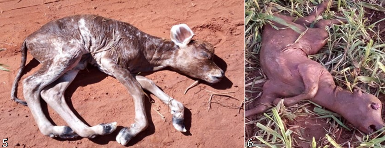
Under-reported occurrences. Among the complaints of field veterinarians who had sent images but did not send samples or animals for necropsy are abortions and neonatal diarrhea. Several causes can be associated in all these cases, and laboratory diagnosis is difficult. In the case of neonatal diarrhea, the number of deaths was lower than that of the animals that recovered, and establishing a precise diagnosis was not considered necessary by the owners. In this case, we also note the association between the behavior of ingesting soil (geophagy) by calves and the occurrence of diarrhea, which is not discussed in the literature. However, nutritional factors play an additional role in neonatal diarrhea, and inadequate nutrition can directly induce diarrhea or can further increase infectious diarrhea through osmosis or dysbiosis (Van Mol et al. 2022). Furthermore, this behavior is described in cases of mineral deficiencies such as sodium and iron deficiencies (Tokarnia et al. 2010). Another important point regarding this problem is the conclusion by the field veterinarian that the cause of neonatal diarrhea is Escherichia coli, based only on the culture test performed on the feces. Such a diagnosis is mistaken since there are non-pathogenic strains of this bacterium in the ruminant microbiota and because it can also grow in culture; in addition, virulence factors can be identified in both diarrheal and normal feces (Tutija et al. 2022). Regarding abortions, three incidents were reported, all with images sent but none with material sent (Fig.5-6). These data indicate a loss of animals and service time with the pregnant cow in these occurrences, showing economic losses. However, considering that abortion is a manifestation that can have several causes, including infectious agents, nutritional deficiencies, as well as genetic, toxic, and physical causes (Antoniassi et al. 2013, Mee 2023), the establishment of a diagnostic approach that allows addressing all these different causes is fundamental for an assertive etiological diagnosis and the reduction of these losses. In one of the cases, although it was not possible to make a diagnosis regarding the etiology involved in the abortion, malformations such as arthrogryposis and increased skull volume (Fig.5) were identified. In these cases, some suspicions can be raised regarding the cause. Congenital malformations can occur as a result of hereditary genetic factors and/or environmental factors, which can be of infectious origin when the female is infected by certain viruses in cattle, mainly the bovine viral diarrhea virus (BVDV), or even nutritional and toxic in nature (Schild 2023).

Cases of abortions in cattle without an etiological diagnosis. (5) Bovine fetus with congenital malformations (arthrogryposis and increased cranial volume). (6) Fetus in an advanced state of autolysis.
In contacts that did not result in diagnoses or suspicions by the LAP-UFMS due to the lack of information, apart from the notifier’s complaint, some points in the field veterinarian’s approach are noted. These include many incidents in which the sender’s suspicion or complaint was a neurological disease, including rabies. These data show the underreporting of suspected cases of this disease, which is relevant since there is an official herbivore rabies control program. However, despite the existence of this program, the underreporting of rabies has been described previously (Mello et al. 2019). Another situation is the number of cases with suspected urea poisoning, many of which had a clinical picture and epidemiology compatible with this poisoning, but none of which had material sent for histopathology. This situation has previously been observed in the LAP-UFMS area of activity (Gimelli et al. 2023).
In numerous contacts where no material was sent, the suspicion was not of a specific disease, and generic terms were used to refer to clinical signs or injuries such as nervous signs, plant poisoning, mortality, coughing, skin lesions, pneumonia, polioencephalomalacia, and tremors. This behavior shows that veterinarians often do not understand the importance of clinical and epidemiological characterization when diagnosing. In this category, the erroneous use of the term “clostridiosis” was also observed, referring to a single etiological agent. In contrast, more than one species of Clostridium is considered the causative agent of diseases in ruminants (Santos et al. 2019).
Diagnostic efficiency using WhatsApp. In addition to previously undiagnosed diseases and underreporting of diseases, this study shows an increase in the number of contacts via WhatsApp compared to a previous study that evaluated the same tool (Lemos et al. 2022). The previous study lasted 23 months, with 45 necropsies carried out by the LAP-UFMS team, 39 materials for histopathology, and 63 contacts without sending materials. In contrast, the present study lasted 24 months, with 63 necropsies carried out by the LAP, 62 materials for histopathological analysis, and 86 contacts without sending materials. This increase in the number of contacts was not proportional to the increase in the number of veterinarians who had joined the application group, which was greater than 100%. Furthermore, the number of contacts that did not result in diagnoses due to samples or information not being sent was higher than that of animals sent for necropsy or samples for histopathology individually, which demonstrates the need for improvements in communication via WhatsApp to increase the participation of veterinarians working in the diagnosis of diseases.
Conclusion
The use of WhatsApp facilitated communication and promoted different forms of contact between field veterinarians and the veterinary diagnostic laboratory, which increased the number of conclusive diagnoses and allowed the identification of diseases not previously diagnosed in Mato Grosso do Sul, diseases previously diagnosed but which occurred in different production systems, and underreported diseases. It also allows the continuous monitoring of outbreaks, which leads to underestimating the number of disease occurrences. Furthermore, it was possible to identify the use of mistaken diagnostic approaches and concepts by field veterinarians as well as the occurrence of health problems with challenging diagnoses, such as abortions, perinatal deaths, and deaths by neonatal diarrhea.
Acknowledgments
The author, R.A.A. Lemos, received a research fellowship from the “Conselho Nacional de Desenvolvimento Científico e Tecnológico” (CNPq). This study was carried out with the support of the “Coordenação de Aperfeiçoamento de Pessoa de Nível Superior” (CAPES), Brazil - Financing Code 001 and by the “Universidade Federal de Mato Grosso do Sul” (UFMS).
References
-
Antoniassi N.A.B., Juffo G.D., Santos A.S., Pescador C.A., Corbellini L.G. & Driemeier D. 2013. Causas de aborto bovino diagnosticadas no Setor de Patologia Veterinária da UFRGS de 2003 a 2011. Pesq. Vet. Bras. 33(2):155-160. <https://dx.doi.org/10.1590/S0100-736X2013000200004>
» https://doi.org/https://dx.doi.org/10.1590/S0100-736X2013000200004 - Astarita S., Marianelli C., Martucciello A., Capuano F., Iovane G. & Galiero G. 2007. Indagine sulla presenza di Chlamydophila spp. in feti di bufala mediterranea (Bubalus Bubalis) allevata in Italia. Large Anim. Rev. 13:159-160.
-
Barbosa J.D., Brito M.F., Oliveira C.M.C., Duarte M.D., Silveira N.S.S., Ferreira T.T.A., Silveira J.A.S., Bomjardim H.A., Barbosa C.C. & Malafaia P.A.M. 2021. Deficiências minerais em bovinos e bubalinos no Brasil: aspectos gerais, importância, diagnóstico, profilaxia e correção. Revta Bras. Buiatria, Enfermidades Metaból. 2(1):1-38. <https://dx.doi.org/10.4322/2763-955X.2021.010>
» https://doi.org/https://dx.doi.org/10.4322/2763-955X.2021.010 - Constable P.D., Hinchcliff K.W., Done S.H. & Grünberg W. 2017. Veterinary Medicine. 11th ed. Saunders Elsevier, St. Louis. 2278p.
-
Cullen J.M. & Stalker M.J. 2016. Liver and biliary system, p.258-352. In: Maxie M.G. (Ed.), Jubb, Kennedy and Palmer’s Pathology of Domestic Animals. Vol.2. 6th ed. Elsevier, St Louis. <https://dx.doi.org/10.1016/B978-0-7020-5318-4.00008-5>
» https://doi.org/https://dx.doi.org/10.1016/B978-0-7020-5318-4.00008-5 -
Faccin T.C., Riet-Correa F., Rodrigues F.S., Santos A.C., Melo G.K.A., Silva J.A., Ferreira R., Ítavo C.C.B.F. & Lemos R.A.A. 2014. Poisoning by Brachiaria brizantha in flocks of naïve and experienced sheep. Toxicon 82:1-8. <https://dx.doi.org/10.1016/j.toxicon.2014.02.008><PMid:24561120>
» https://doi.org/https://dx.doi.org/10.1016/j.toxicon.2014.02.008 -
Furuoka H., Hasegawa M., Kobayashi Y. & Matsui T. 1999. Peripheral nerve lesions in a case of equine motor neuron disease. J. Vet. Med. Sci. 61(5):557-560. <https://dx.doi.org/10.1292/jvms.61.557> <PMid:10379951>
» https://doi.org/https://dx.doi.org/10.1292/jvms.61.557 -
Furuoka H., Mizushima M., Miyazawa K. & Matsui T. 1994. Idiopathic peripheral neuropathy in a horse with knuckling. Acta Neuropathol. 88(4):389-393. <https://dx.doi.org/10.1007/BF00310385> <PMid:7839834>
» https://doi.org/https://dx.doi.org/10.1007/BF00310385 -
Gimelli A., Pupin R.C., Guizelini C.C., Gomes D.C., Franco G.L., Vedovatto M., Gaspar A.O. & Lemos R.A.A. 2023. Urea poisoning in cattle: A brief review and diagnostic approach. Pesq. Vet. Bras. 43:e07228. <https://dx.doi.org/10.1590/1678-5150-PVB-7228>
» https://doi.org/https://dx.doi.org/10.1590/1678-5150-PVB-7228 -
Gomes L.I., Rocha M.A., Costa E.A., Lobato Z.I.P., Mendes L.C.N., Borges A.S., Leite R.C. & Barbosa-Stancioli E.F. 2002. Detecção de herpesvírus bovino 5 (BoHV-5) em bovinos do Sudeste Brasileiro. Comun. Arq. Bras. Med. Vet. Zootec. 54(2):217-220. <https://dx.doi.org/10.1590/S0102-09352002000200014>
» https://doi.org/https://dx.doi.org/10.1590/S0102-09352002000200014 -
Guizelini C.C., Pupin R.C., Leal C.R.B., Ramos C.A.N., Pavarini S.P., Gomes D.C., Martins T.B. & Lemos R.A.A. 2019. Salmonellosis in calves without intestinal lesions. Pesq. Vet. Bras. 39(8):580-586. <https://dx.doi.org/10.1590/1678-5150-PVB-6328>
» https://doi.org/https://dx.doi.org/10.1590/1678-5150-PVB-6328 -
Guizelini C.C., Tutija J.F., Morais D.R., Bacha F.B., Ramos C.A.N., Leal C.R.B., Zaquetti M.E. & Lemos R.A.A. 2020. Outbreak investigation of septicemic salmonellosis in calves. J. Infect. Dev. Ctries. 14(1):104-108. <https://dx.doi.org/10.3855/jidc.12087> <PMid:32088691>
» https://doi.org/https://dx.doi.org/10.3855/jidc.12087 -
Hanche-Olsen S., Matiasek K., Molín J., Rosati M., Hahn C., Hultin Jäderlund K. & Gröndahl G. 2017. Acquired equine polyneuropathy of Nordic horses: A conspicuous inclusion body schwannopathy. Neuromuscular Disorders 27(10):931-941. <https://dx.doi.org/10.1016/j.nmd.2017.06.005> <PMid:28694072>
» https://doi.org/https://dx.doi.org/10.1016/j.nmd.2017.06.005 -
Hanche-Olsen S., Teige J., Skaar I. & Ihler C.F. 2008. Polyneuropathy associated with forage sources in Norwegian horses. J. Vet. Intern. Med. 22(1):178-184. <https://dx.doi.org/10.1111/j.1939-1676.2008.0023.x> <PMid:18289307>
» https://doi.org/https://dx.doi.org/10.1111/j.1939-1676.2008.0023.x - Lemos R.A.A., Guizelini C.C. & Araújo F.A. 2023. Hipotermia em Bovinos. Programa de Pós Graduação em Ciências Veterinárias, Faculdade de Medicina Veterinária e Zootecnia (FAMEZ), Universidade Federal de Mato Grosso do Sul (UFMS), Campo Grande, MS. 12p. (Nota técnica n°07/2023. Sanidade em animais de produção).
-
Lemos R.A.A., Guizelini C.C., Silva T.X., Souza L.L., Fonseca H.C.F., Bonato G.C., Gomes D.C. & Pupin R.C. 2022. Use of smartphone messaging applications to increase diagnostic efficiency in veterinary diagnostic laboratories. Pesq. Vet. Bras. 43:e07226. <https://dx.doi.org/10.1590/1678-5150-PVB-7226>
» https://doi.org/https://dx.doi.org/10.1590/1678-5150-PVB-7226 -
Lucena R.B., Pierezan F., Kommers G.D., Irigoyen L.F., Fighera R.A. & Barros C.S.L. 2010. Doenças de bovinos no Sul do Brasil: 6.706 casos. Pesq. Vet. Bras. 30(5):428-434. <https://dx.doi.org/10.1590/S0100-736X2010000500010>
» https://doi.org/https://dx.doi.org/10.1590/S0100-736X2010000500010 -
Luethy D. 2023. Eastern, Western, and Venezuelan equine encephalitis and West Nile viruses: Clinical and public health considerations. Vet. Clin. N. Am. Equine Pract. 39(1):99-113. <https://dx.doi.org/10.1016/j.cveq.2022.11.007> <PMid:36737290>
» https://doi.org/https://dx.doi.org/10.1016/j.cveq.2022.11.007 -
Malafaia P., Barbosa J.D., Brito M.F., Souza V.C. & Costa D.F.A. 2023. Phosphorus for cattle and buffaloes in Brazil: Clinical signs and diagnosis of its deficiency and relevance, and recommended strategies to alleviate issues observed under grazing conditions. Ruminants 3(1):55-75. <https://dx.doi.org/10.3390/ruminants3010006>
» https://doi.org/https://dx.doi.org/10.3390/ruminants3010006 -
Martins N.S., Motta S.P., Santos C.C., Moreira A.S., Farias N.A.R. & Ruas J.L. 2020. Eimeriose em bovinos e ovinos: uma inimiga invisível. Braz. J. Dev. 6(4):19421-19434. <https://dx.doi.org/10.34117/bjdv6n4-201>
» https://doi.org/https://dx.doi.org/10.34117/bjdv6n4-201 -
McManus C., Barcellos J.O.J., Formenton B.K., Hermuche P.M., Carvalho Jr. O.A., Guimarães R., Gianezini M., Dias E.A., Lampert V.N., Zago D. & Braccini Neto J. 2016. Dynamics of cattle production in Brazil. PLoS One 11(1):e0147138. <https://dx.doi.org/10.1371/journal.pone.0147138> <PMid:26814797>
» https://doi.org/https://dx.doi.org/10.1371/journal.pone.0147138 -
Mee J.F. 2020. Investigation of bovine abortion and stillbirth/perinatal mortality - similar diagnostic challenges, different approaches. Irish Vet. J. 73:20. <https://dx.doi.org/10.1186/s13620-020-00172-0> <PMid:32944225>
» https://doi.org/https://dx.doi.org/10.1186/s13620-020-00172-0 -
Mee J.F. 2023. Invited review: Bovine abortion-Incidence, risk factors and causes. Reprod. Domest. Anim. 58(Supl.2):23-33. <https://dx.doi.org/10.1111/rda.14366> <PMid:37186386>
» https://doi.org/https://dx.doi.org/10.1111/rda.14366 -
Mello A.K.M., Brumatti R.C., Neves D.A., Alcântara L.O.B., Araújo F.S., Gaspar A.O. & Lemos R.A.A. 2019. Bovine rabies: economic loss and its mitigation through antirabies vaccination. Pesq. Vet. Bras. 39(3):179-185. <https://dx.doi.org/10.1590/1678-5150-PVB-6201>
» https://doi.org/https://dx.doi.org/10.1590/1678-5150-PVB-6201 -
Mello L.S., Bianchi M.V., Bandinelli M.B., Sonne L., Driemeier D. & Pavarini S.P. 2017. Causas de morte em vacas leiteiras no Rio Grande do Sul. Pesq. Vet. Bras. 37(9):916-920. <https://dx.doi.org/10.1590/S0100-736X2017000900003>
» https://doi.org/https://dx.doi.org/10.1590/S0100-736X2017000900003 -
Molossi F.A., Cecco B.S., Pohl C.B., Borges R.B., Sonne L., Pavarini S.P. & Driemeier D. 2021. Causes of death in beef cattle in southern Brazil. J. Vet. Diagn. Investig. 33(4):677-683. <https://dx.doi.org/10.1177/10406387211007952> <PMid:33834923>
» https://doi.org/https://dx.doi.org/10.1177/10406387211007952 -
Puentes J.D. & Riet-Correa F. 2023. Epidemiological aspects of cattle tick fever in Brazil. Braz. J. Vet. Parasitol. 32(1):e014422. <https://dx.doi.org/10.1590/S1984-29612023007> <PMid:36722682>
» https://doi.org/https://dx.doi.org/10.1590/S1984-29612023007 -
Pupin R.C., Leal P.V., Paula J.P.L., Guizelini C.C., Möck T.B.M., Lemos R.A.A. & Gomes D.C. 2019. Cattle diseases in Mato Grosso do Sul, Brazil: a 24-year survey (1995-2018). Pesq. Vet. Bras. 39(9):686-695. <https://dx.doi.org/10.1590/1678-5150-PVB-6384>
» https://doi.org/https://dx.doi.org/10.1590/1678-5150-PVB-6384 -
Pupin R.C., Souza L.L., Silva T.X., Fonseca H.C.F., Silva E.A.S., Gomes D.C., Araújo M.A. & Lemos R.A.A. 2023. Vitamin A deficiency as a cause of blindness in feedlot calves. Pesq. Vet. Bras. 43:e07249. <https://dx.doi.org/10.1590/1678-5150-PVB-7249>
» https://doi.org/https://dx.doi.org/10.1590/1678-5150-PVB-7249 -
Ribas N.L.K.S., Carvalho R.I., Santos A.C., Valençoela R.A., Gouveia A.F., Castro M.B., Mori A.E. & Lemos R.A.A. 2013. Doenças do sistema nervoso de bovinos no Mato Grosso do Sul: 1082 casos. Pesq. Vet. Bras. 33(10):1183-1194. <https://dx.doi.org/10.1590/S0100-736X2013001000003>
» https://doi.org/https://dx.doi.org/10.1590/S0100-736X2013001000003 - Riet-Correa F. 2023. Doenças metabólicas: osteodistrofia fibrosa, p.423-426. In: Riet-Correa F., Schild A.L., Lemos R.A.A., Borges J.R.J, Mendonça F.S. & Machado M. (Eds), Doenças de Ruminantes e Equídeos. Vol.2. 4ª ed. MedVet, São Paulo.
-
Rigamonti S., Floriano A.M., Scaltriti E., Longbottom D., Livingstone M., Comandatore F., Pongolini S., Capucci L., Mandola M.L., Bazzucchi M., Prati P. & Vicari N. 2022. Comparative analysis of two genomes of Chlamydia pecorum isolates from an Alpine chamois and a water buffalo. BMC Genomics 23:645. <https://dx.doi.org/10.1186/s12864-022-08860-7> <PMid:36088280>
» https://doi.org/https://dx.doi.org/10.1186/s12864-022-08860-7 -
Rissi D.R., Rech R.R., Flores E.F., Kommers G.D. & Barros C.S.L. 2007. Meningoencefalite por herpesvírus bovino-5. Pesq. Vet. Bras. 27(7):251-260. <https://dx.doi.org/10.1590/S0100-736X2007000700001>
» https://doi.org/https://dx.doi.org/10.1590/S0100-736X2007000700001 -
Rondelli L.A.S., Silva G.S., Bezerra K.S., Rondelli A.L.H., Lima S.R., Furlan F.H., Pescador C.A. & Colodel E.M. 2017. Doenças de bovinos em Mato Grosso diagnosticadas no Laboratório de Patologia Veterinária da UFMT (2005- 2014). Pesq. Vet. Bras. 37(5):432-440. <https://dx.doi.org/10.1590/S0100-736X2017000500002>
» https://doi.org/https://dx.doi.org/10.1590/S0100-736X2017000500002 -
Salvador S.C., Lemos R.A.A., Riet-Correa F., Roehe P.M. & Osório A.L.A.R. 1998. Meningoencefalite em bovinos causada por herpesvírus bovino-5 no Mato Grosso do Sul e São Paulo. Pesq. Vet. Bras. 18(2):75-82. <https://dx.doi.org/10.1590/S0100-736X1998000200007>
» https://doi.org/https://dx.doi.org/10.1590/S0100-736X1998000200007 -
Sánchez-Miguel C., Crilly J., Grant J. & Mee J.F. 2018. Sensitivity, specificity and predictive probability values of serum agglutination test titers for the diagnosis of Salmonella Dublin culture-positive bovine abortion and stillbirth. Transbound. Emerg. Dis. 65(3):676-686. <https://dx.doi.org/10.1111/tbed.12784> <PMid:29230973>
» https://doi.org/https://dx.doi.org/10.1111/tbed.12784 -
Santos B.L., Ladeira S.R., Riet-Correa F., Soares M.P., Marcolongo-Pereira C., Sallis E.S.V., Raffi M.B. & Schild A.L. 2019. Clostridial diseases diagnosed in cattle from the South of Rio Grande do Sul, Brazil. A forty-year survey (1978-2018) and a brief review of the literature. Pesq. Vet. Bras. 39(7):435-446. <https://dx.doi.org/10.1590/1678-5150-PVB-6333>
» https://doi.org/https://dx.doi.org/10.1590/1678-5150-PVB-6333 -
Santos B.S., Pinto A.P., Aniz A.C.M., Almeida A.P.M.G., Franco G.L, Guimarães E.B. & Lemos R.A.A. 2012. Mortalidade de bovinos zebuínos por hipotermia em Mato Grosso do Sul. Pesq. Vet. Bras. 32(3):204-210. <https://dx.doi.org/10.1590/S0100-736X2012000300004>
» https://doi.org/https://dx.doi.org/10.1590/S0100-736X2012000300004 - Schild A.L. 2023. Defeitos congênitos, p.4-28. In: Riet-Correa F., Schild A.L., Lemos R.A.A., Borges J.R.J., Mendonça F.S. & Machado M. (Eds), Doenças de Ruminantes e Equídeos. Vol.2. 4ª ed. MedVet, São Paulo.
-
Schulz L.L., Hayes D.J., Holtkamp D.J. & Swenson D.A. 2018. Economic impact of university veterinary diagnostic laboratories: a case study. Prev. Vet. Med. 151:5-12. <https://dx.doi.org/10.1016/j.prevetmed.2017.12.018> <PMid:29496106>
» https://doi.org/https://dx.doi.org/10.1016/j.prevetmed.2017.12.018 -
Silva M.C., Barros R.R. & Graça D.L. 2005. Surto de dictiocaulose em bovinos no município de Santa Maria, RS, Brasil. Ciência Rural 35(3):629-632. <https://dx.doi.org/10.1590/S0103-84782005000300022>
» https://doi.org/https://dx.doi.org/10.1590/S0103-84782005000300022 -
Silva W.J., Guizelini C.C., Franco G.L., Veiga R.C.P., Arruda W.G., Nascimento V.A., Melo E.S.P. & Lemos R.A.A. 2020. Cobalt deficiency in cattle and its impact on production. Pesq. Vet. Bras. 40(11):837-841. <https://dx.doi.org/10.1590/1678-5150-PVB-6770>
» https://doi.org/https://dx.doi.org/10.1590/1678-5150-PVB-6770 -
Souza R.I.C., Riet-Correa F., Brum K.B., Fernandes C.E., Barbosa-Ferreira M. & Lemos R.A.A. 2010. Intoxicação por Brachiaria spp. em bovinos no Mato Grosso do Sul. Pesq. Vet. Bras. 30(12):1036-1042. <https://dx.doi.org/10.1590/S0100-736X2010001200006>
» https://doi.org/https://dx.doi.org/10.1590/S0100-736X2010001200006 -
Sprenger L.K., Risolia L.W., Gabardo L.B., Molento M.B., Silva A.W.C. & Sousa R.S. 2015. Doenças de ruminantes domésticos diagnosticadas no Laboratório de Patologia Veterinária da Universidade Federal do Paraná: 1075 casos. Arch. Vet. Sci. 20(4):45-53. <https://dx.doi.org/10.5380/AVS.V20I4.38525>
» https://doi.org/https://dx.doi.org/10.5380/AVS.V20I4.38525 - Tokarnia C.H., Döbereiner J. & Peixoto P.V. 1998. Deficiências minerais em animais de fazenda, principalmente bovinos criados em regime de campo, p.11-22. In: Gonzalez F.H.D., Opina H. & Barcellos J.O.J. (Eds), Nutrição Mineral em Ruminantes. 2ª ed. UFRGS, Porto Alegre.
- Tokarnia C.H., Peixoto P.V., Barbosa J.D., Brito M.F. & Döbereiner J. 2010. Deficiências Minerais em Animais de Produção. Helianthus, Rio de Janeiro. 200p.
-
Tutija J.F., Ramos C.A., Lemos R.A.A., Santos A.A.L., Reckziegel G.H., Freitas M.G. & Leal C.RB. 2022. Molecular and phenotypic characterization of Escherichia coli from calves in an important meat-producing region in Brazil. J. Infect. Dev. Ctries. 16(6):1030-1036. <https://dx.doi.org/10.3855/jidc.13377> <PMid:35797298>
» https://doi.org/https://dx.doi.org/10.3855/jidc.13377 -
Van Mol W., Clinquart J., Pas M.L., Bokma J. & Pardon B. 2022. Pathogen-oriented approaches for neonatal calf diarrhea. Vlaams Diergeneeskundig Tijdschrift 91(4):167-181. <https://dx.doi.org/10.21825/vdt.85202>
» https://doi.org/https://dx.doi.org/10.21825/vdt.85202