Infectious diseases in wild and exotic birds in Paraíba State, Northeast Brazil
ABSTRACT:
Wild and exotic birds play a pivotal role in the zoonotic transmission of various infectious diseases. As reservoirs for a diverse array of infectious agents, their interaction with domestic animals can facilitate the spread of diseases, emphasizing their importance in certain disease cycles. This study describes the occurrence of infectious diseases diagnosed in Paraíba from 2013 to 2018. Out of 189 wild birds necropsied, 50 (26.45%) had infectious diseases. Bacterial infections were the most prevalent, accounting for 38 (76%) cases, followed by fungi, with 10 (20%) affected birds, and lastly, protozoa with two (4%) cases. The most affected species included the blue-fronted amazon parrot (Amazona aestiva), red-shouldered macaw (Diopsittaca nobilis), Harris’s hawk (Parabuteo unicinctus), and saffron finch (Sicalis flaveola), respectively. The predominance of bacterial diseases was often associated with secondary infections resulting from debilitating conditions such as trauma or bronchoaspiration. Chronic aspergillosis emerged as the primary fungal disease, observed in various tissues and often linked to management failures and stress. Other fungal infections were predominantly suggestive of Candida sp. The presence of protozoa was limited to Sarcocystis sp. infections in two birds with no clinical signs, but they were responsible for the death of the birds. This survey reinforces the significance of bacterial, fungal and protozoan pathogens in wild birds and highlights the need for accurate diagnostic descriptions in these species. Accordingly, the birds in this survey are considered susceptible to the infections detailed herein.
INDEX TERMS:
Bacteria; fungi; protozoa; aspergillosis; Sarcocystis sp.; immunohistochemistry
RESUMO:
As aves silvestres e exóticas são responsáveis pelo ciclo zoonótico de diversas doenças infecciosas. As aves silvestres por sua vez, tem o papel de reservatório dos mais variados agentes infecciosos na natureza e, quando em contato com animais domésticos ou humanos, podem disseminar a enfermidade, se tornando fundamental no ciclo de algumas doenças. No presente estudo descrevem-se os casos de doenças infecciosas diagnosticadas na Paraíba de 2013 a 2018. De um total de 189 amostras de tecido de necropsia coletadas em aves silvestres e exóticas, 50 (26,45%) corresponderam a casos de doenças infecciosas. Das doenças infecciosas, as de etiologia bacteriana foram as mais prevalentes, representando 38 (76%) casos, seguidas dos fungos com 10 (20%) aves afetadas e por último dos protozoários com dois (4%) casos. As espécies mais afetadas foram papagaio-verdadeiro (Amazona aestiva), maracanã-nobre (Diopsittaca nobilis), gavião-asa-de-telha (Parabuteo unicinctus) e canário-da-terra-verdadeiro (Sicalis flaveola), respectivamente. A ocorrência maior de doenças bacterianas decorre de os casos apresentarem-se como uma doença secundária a um quadro debilitante, como, por exemplo, traumas e aspiração de conteúdo para o trato respiratório. Por vezes não foi possível uma classificação de gênero e espécie dos agentes patogênicos envolvidos. A aspergilose crônica destacou-se como a principal doença micótica e foi observada em diversos tecidos, representando uma condição primária ou secundária e, na maioria das vezes, associada a falhas de manejo e estresse. Os demais fungos foram representados por leveduras sugestivas de Candida sp. A presença de protozoários foi limitada à infecção por Sarcocystis sp. em tecidos de duas aves, sem manifestação clínica, porém foram responsáveis pela morte das aves. Este levantamento reforça a importância dos patógenos bacterianos, fúngicos e protozoários causadores de doenças em aves selvagens, bem como a descrição de diagnóstico nessas espécies. Nesse sentido, as aves desse levantamento podem ser consideradas susceptíveis às infecções descritas no presente trabalho.
TERMOS DE INDEXAÇÃO:
Bactérias; fungos; protozoários; aspergilose; Sarcocystis sp.; imuno-histoquímica
Introduction
The popularity of keeping wild and exotic birds as pets has grown due to their ability to mimic sounds and interact socially with both adults and children (Grespan & Raso 2014). Veterinary clinics must adapt to this trend, becoming essential services for pet bird owners, commercial breeders, conservationists, and birds targeted for scientific purposes (Sanches & Godoy 2014).
Infections in birds are extensively studied to understand the agents involved in tissue invasion, their reactions, clinical signs, and their ability to spread to other birds, mammals, and humans. A study by Seyedmousavi et al. (2015) involving animals and humans concluded that birds play a crucial role in zoonoses, demonstrating resistance profiles to various antimicrobials and having direct and indirect impacts on public health.
Pathogens such as bacteria, fungi, viruses, and protozoa can cause a range of diseases in wild and exotic birds. Preventing the occurrence and spread of these diseases is essential, as there is no treatment for many of them. Birds can act as reservoirs for these diseases, but in some cases, they may develop the disease and show clinical signs (Gargiulo et al. 2018).
Bacterial diseases are significant because of the high morbidity and often high mortality among wild birds, whether in captivity or not. Wild birds are considered reservoirs of several diseases, including zoonotic diseases, making them the focus of numerous studies aimed at identifying the infectious agents capable of persisting in these animals and spreading directly or indirectly, causing harm to other birds and to humans (Gargiulo et al. 2018).
Fungal diseases are recurrent in both domestic and wild animals as they are present in various environments and can exploit several factors leading to weakened immunity in animals. They can manifest as primary diseases, but are often diagnosed as secondary to immunosuppressive conditions. Notable examples include aspergillosis, cryptococcosis, candidiasis, and mycotoxicosis, which can cause significant lesions and spread easily in environments with inadequate management (Seyedmousavi et al. 2015, Konicek et al. 2016).
Among the parasites, Sarcocystis sp. is an intracellular protozoan that can multiply asexually in endothelial cells in the striated muscle, resulting in eosinophilic myositis (Gjerde et al. 2018). Morphological diagnosis is possible through the identification of protozoa in tissues (El-Morsey et al. 2013, Prakas et al. 2018).
The aim of this study was to identify the main infectious diseases in wild and exotic birds in the State of Paraíba, Northeast Brazil, and to describe the macroscopic and histopathological aspects of their different presentations.
Materials and Methods
Ethical approval. This study was waived by the Institutional Committee on Animal Use (CEUA) because it involved the use of cadavers. This is stated in section “6.1.10.” of CONCEA Resolution No. 30 of February 2, 2016.
The records of necropsies and biopsies performed on wild and exotic birds at the “Laboratório de Patologia Veterinária” (Veterinary Pathology Laboratory - LPV) of the “Universidade Federal da Paraíba” (UFPB) from January 2013 to December 2018 were reviewed. Some of these birds were seized from animal trafficking or illegal breeding sites and sent to the Rehabilitation Center or Zoo located in João Pessoa, Paraíba. Others were free-living birds rescued by environmental police or common folk due to illness or death and subsequently directed to these two institutions, or, lastly, to the “Hospital Veterinário Universitário” in Areia, Paraíba. Upon confirmation of death, the birds were refrigerated or frozen and subsequently sent to the LPV-UFPB for necropsy.
Based on screening of available records, cases of infectious diseases were classified as bacterial, fungal, or protozoal etiology. In addition to macroscopic and histopathological evaluations, results of other complementary exams, such as microbiological isolation, hematoxylin and eosin (HE) staining, histochemical stains with periodic acid-Schiff stain (PAS) and Grocott’s methenamine silver stain (GMS) technique, or immunohistochemistry (IHC), as in the study by Galiza et al. (2014), were considered. Information on the diagnostic method, the birds’ age and sex, and the epidemiological aspects was compiled.
For the microbiological examination, samples of Sporophila nigricollis (skin, liver, kidneys, lung, and spleen) were collected and sent to the laboratory for confirmation of the fungus’ species. There, they were manipulated in the fume hood, sterilized and flamed, and macerated. The maceration liquid was inoculated onto plates of Sabouraud dextrose agar, Löwenstein-Jensen, and Löffler’s growth mediums. Three samples of Amazona aestiva and two samples of Diopsittaca nobilis were sent for bacterial culture and identification. Microbiological examinations were standardized according to Moxley (2016). The fragments of tissues (skin, liver, kidneys, lung and spleen) were separated and sent for microbiological isolation and examination, where they were manipulated in a sterile environment and macerated. The maceration liquid was inoculated into the medium Salmonella-Shigella (SS) agar and Sorbitol-MacConkey agar. After 24 hours, catalase, oxidase, and Gram stain tests were performed to differentiate the bacteria.
For aspergillosis diagnosis, paraffin-embedded specimens were subjected to immunohistochemical examination, with 4 µm-thick sections. After deparaffinization in xylene, hydration in decreasing concentrations of ethanol, and washing in deionized water, the histological sections underwent antigen retrieval in 10 mM citrate solution (pH 6.0) in a pressurized chamber (Pascal®, Dako). Endogenous peroxidase was blocked with ready-to-use hydrogen peroxide (Peroxide Block®, Cell Marque, 925B-09) and non-specific protein block (Dako® Protein Block Serum-Free, X0909).
The primary antibody used was anti-Zygomycetes and anti-Aspergillus (Spring, E 4520) at a 1:50 dilution, incubated for 18 hours at 4 °C. Subsequently, the slides were washed in a TRIS pH 7.4 buffer solution and then incubated with a polymer detection system (Histofine®, Nichirei, 414154F) according to the manufacturer’s recommendations. Immuno-staining was obtained using DAB (3,3’-diaminobenzidine tetrachloride, Dako®). The sections were counterstained with Mayer’s hematoxylin and dehydrated in increasing concentrations of alcohol and xylene. Finally, the slides were mounted with resin and a coverslip. Positive and negative controls were added to the reaction to ensure the fidelity of the results.
Results
A total of 189 birds were necropsied, comprising 118 wild birds and 71 exotic birds. Infectious diseases were identified in 50 (26.45%) of the cases, of which 48 were wild birds, and two were exotic birds. Bacterial diseases were the most prevalent, accounting for 38 (76%) cases, followed by fungal infections in 10 (20%) cases, and protozoal infections in two (4%) birds. Twelve species of affected birds were recorded, as described in Table 1.
Distribution of necropsies in wild and exotic birds diagnosed with infectious disease at the LPV-UFPB, Paraíba, Brazil, according to the species of the bird
Bacterial diseases
Bacterial diseases represented over half of the cases. Escherichia coli infection was the most common bacterial disease, with a total of five affected birds, all diagnosed with extraintestinal colibacillosis. It was not possible to isolate bacteria from the lesions of the other birds due to the advanced state of autolysis of their tissues. The birds diagnosed with extraintestinal colibacillosis were three blue-fronted amazon parrots (Amazona aestiva) and two red-shouldered macaws (Diopsittaca nobilis), presented in the form of liver granulomas caused by the bacteria, with lesions ranging from moderate to severe. In these cases, pure cultures of E. coli were isolated from the lesions.
In the cases of extraintestinal colibacillosis, there was massive infection with similar clinical and pathological presentations. The birds exhibited extreme cachexia, emaciation, multifocal areas of apteria, and dull feathers. Necropsy revealed multifocal, whitish hepatic and splenic coligranulomas, while histopathology showed central necrosis and an inflammatory infiltrate composed of histiocytes, macrophages and lymphocytes surrounding bacterial colonies near granulomatous infiltrate. Additionally, a moderate lymphohistiocytic inflammatory infiltrate was noted in the kidneys and air sacs.
Fungal diseases
Fungal infections were identified in 10 birds, with nine cases of aspergillosis and one of candidiasis. All infected species exhibited involvement of the lungs and air sacs, ranging from moderate to severe inflammation, with involvement of other systems, demonstrating the fungal infection’s potential to spread. Fungal hyphae were found in the lungs, heart, liver, spleen, kidneys and skin of infected birds.
Aspergillosis was the most commonly diagnosed fungal disease. In one case, there was a concomitant infection of aspergillosis and zygomycosis. In another case, suspected pulmonary candidiasis was confirmed in a collared forest falcon (Micrastur semitorquatus), with multifocal interstitial involvement. In both situations, diagnosis was achieved through histopathological and histochemical exams, with isolation, culture and identification of one aspergillosis case and IHC analysis of all the others.
Aspergillus sp. was present in the following species: blue-fronted amazon parrots (Amazona aestiva), Harris´s hawk (Parabuteo unicinctus), yellow-bellied seedeater (Sporophila nigricollis), smooth-billed ani (Crotophaga ani) and magnificent frigatebird (Fregata magnificens). In confirmed cases of Aspergillus sp. infection, the affected organs included the lungs, heart, liver, spleen, kidneys and skin. Granulomatous lesions with septated hyphae were observed in all these species of birds, with the presence of septated hyphae better evidenced after histochemical stainings of PAS and GMS.
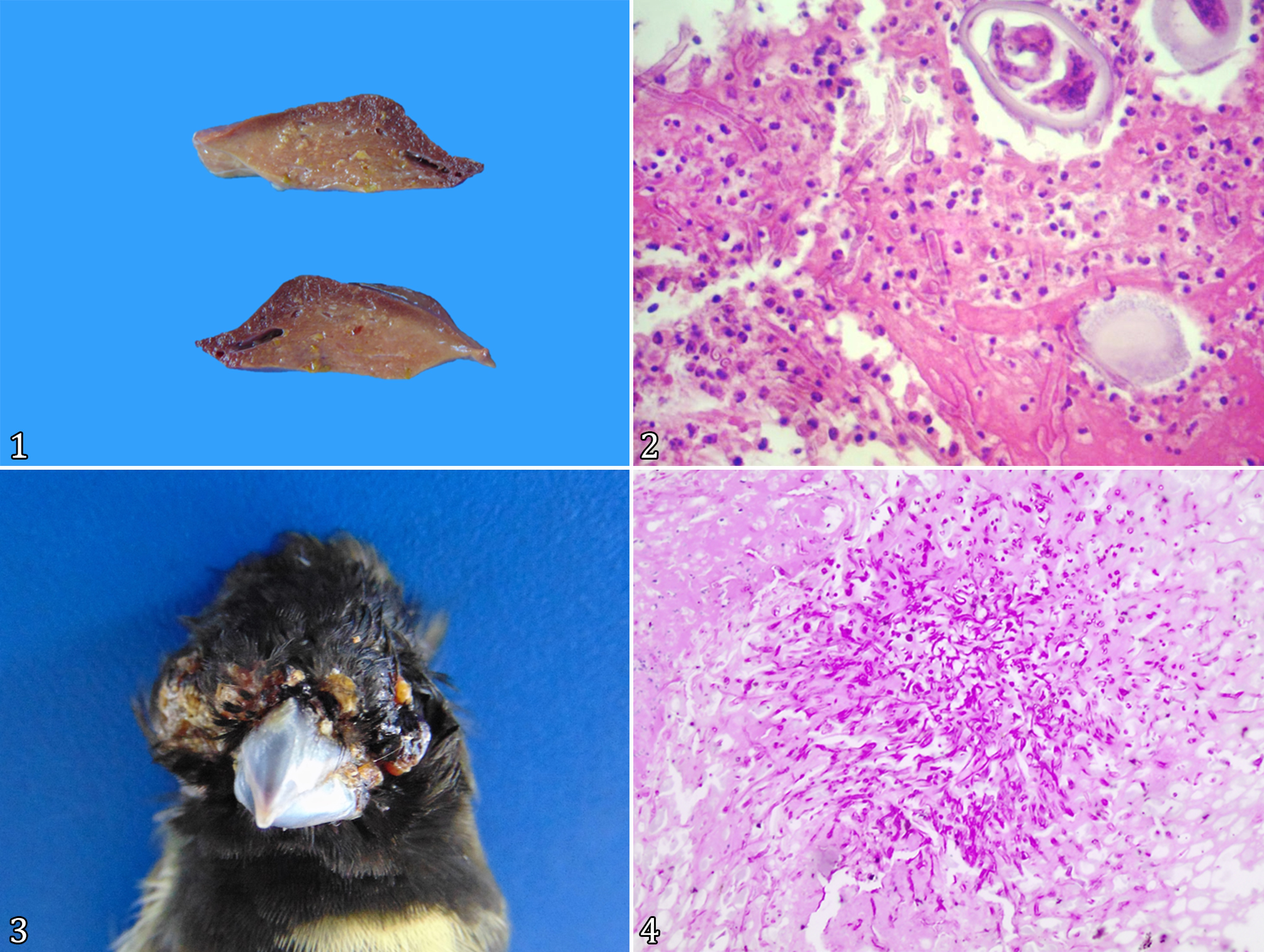
The three infected parrots exhibited significant pulmonary impairment, similar to that observed in this study with bacterial infections. Diagnosis was confirmed by macroscopic (Fig. 1) and microscopic lesions, histochemical staining, and immunohistochemical analysis. In one case, the liver’s parenchyma showed multifocal to coalescent necrosis and a moderate lymphoplasmacytic inflammatory infiltrate, with negative images of hyphae. Additionally, rounded to oval eggs with a striated capsule and multifocal non-protruding bipolar operculum, compatible with Capillaria sp. (Fig. 2), were found. Major circulatory lesions were observed in all lungs of affected parrots, including marked multifocal congestion and focally extensive hemorrhage with marked edema, ranging from moderate to severe. These lesions were associated with granulomas under microscopy. Subsequently, IHC strongly marked for Aspergillus spp.
Infectious diseases in wild and exotic birds in Paraíba. (1) Amazona aestiva (blue-fronted amazon). Macroscopically pale liver diagnosed with generalized aspergillosis. (2) Amazona aestiva (blue-fronted amazon). Fungal hepatitis associated with secondary parasitism, evidenced by the presence of negative images of septate hyphae intermingling with mononuclear inflammation and parasitic structures consistent with capillariid eggs, compatible with Capillaria sp. HE, obj. 100x. (3) Skin nodule dorsal to the beak of Sporophila nigricollis measuring 1.3 cm, with a crusty and blackish appearance, featuring multifocal areas varying from white to gray. (4) Histochemical staining revealing acute-angled septate hyphae marked in magenta in a cutaneous nodule of S. nigricollis, with a morphology compatible with Aspergillus sp. PAS, obj. 20x.
Both S. nigricollis and C. ani exhibited similar fungal lesions, despite being different species. Crotophaga ani developed a cutaneous granuloma on the pelvic limbs, which stained positive for Aspergillus sp. in IHC analysis. Macroscopically, the lesion was a white skin elevation with 1 cm of diameter, circumscribed and well adhered to the limb. Microscopically, it was a rounded structure with moderate necrosis, surrounded by macrophages, lymphocytes, and plasma cells, with negative image structures of multifocal hyphae. In S. nigricollis, the lesion appeared as an isolated nodule on the cere, measuring 1.3 cm, with a white to gray crusted appearance (Fig. 3) and multifocal white and yellow necrotic areas. Microscopically, it was characterized by focally extensive necrotic tissue, a mixed inflammatory infiltrate composed of lymphocytes, plasma cells, macrophages, and a multifocal-to-severe coalescing infiltrate of heterophils, with filamentous hyphal structures distributed multifocally and well aggregated in the peripheral necrotic part of the lesion. PAS histochemical staining highlighted acute-angled septate hyphae in magenta in a cutaneous nodule of S. nigricollis, with a morphology similar to Aspergillus sp. (Fig. 4).
In histochemistry, PAS and Grocott staining enabled visualization of hyphae with a morphology similar to Aspergillus sp. Isolation and IHC confirmed the fungal histopathological findings in blue-fronted amazon parrots, Harris’s hawks, and one magnificent frigatebird. In the latter, infection and concomitant spread of two species of fungi were observed in the esophageal lesion, marked in IHC with anti-Zygomycetes and anti-Aspergillus antibodies.
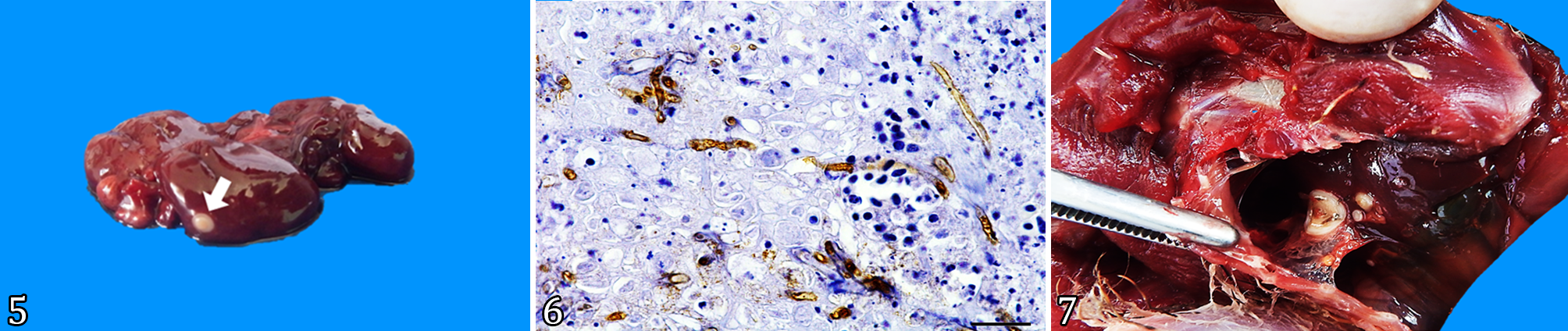
Similar to parrots, all Harri’s hawks (P. unicinctus) exhibited pulmonary involvement, but the gastrointestinal form was particularly prominent in this species, with several macroscopic and microscopic chronic lesions. In the hawks, marked involvement of the gastrointestinal tract was noted, with multiple nodules distributed in the organ serosa, as well as in the kidneys (Fig. 5), which were confirmed as aspergillosis in IHC (Fig. 6). Significant circulatory and nodular lesions were observed throughout the coelomic cavity of affected hawks, ranging from moderate to severe (Fig. 7). Following microscopic evaluation, these lesions were submitted to IHC, which strongly marked for Aspergillus spp. The diagnostic methods varied in each case, as specified in Table 2.

Infectious diseases in wild and exotic birds in Paraíba. (5) Right kidney of Harri’s hawks (Parabuteo unicinctus) with a white nodule, corresponding to a granuloma at histological evaluation (arrow) and marked congestion of the renal parenchyma. (6) Immunohistochemistry of the kidney from a Harri’s Hawk (P. unicinctus) displaying filamentous, septate, brown hyphae at diconic bifurcation in the necrotic areas, positively marked with anti-Aspergillus antibody. IHC, obj. 100x. (7) Coelomic cavity of P. unicinctus exhibiting multifocal cream-colored nodular areas with a central halo attached to air sacs and liver, later diagnosed as aspergillosis.
Methods used to diagnose infectious diseases in wild and exotic birds at the Veterinary Hospital of UFPB from 2013 to 2018
Diseases caused by protozoa
The Sarcocystis sp. was diagnosed as the cause of lesions in two birds: a rufous-bellied thrush (Turdus rufiventris) and a red-shouldered macaw (D. nobilis), with disseminated development in the striated skeletal muscles of the chest, the muscular layer of the ventricle, and the myocardium, respectively.
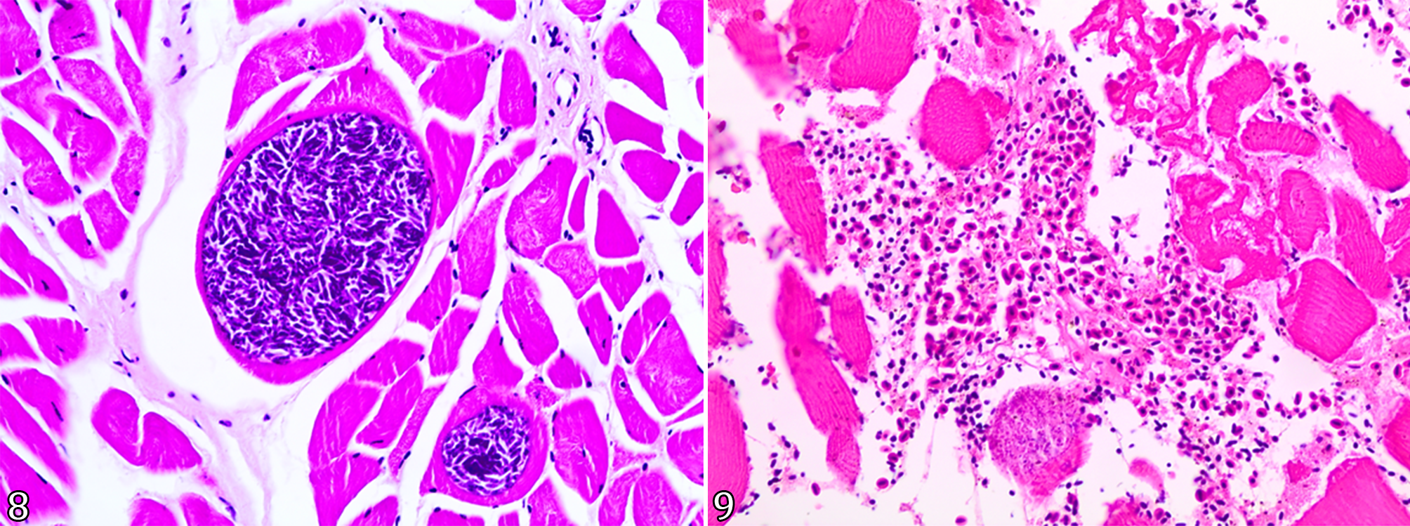
Macroscopic evaluation of the T. rufiventris revealed multiple whitish spots on its pectoral muscle’s surface, ranging from 0.1 to 0.4 cm, which extended into the muscle tissue. Histologically, Sarcocystis sp. cysts, with a delicate wall containing several bradyzoites, were distributed in a multifocal and disseminated manner in the skeletal muscle, proventriculus and ventricle. The inflammatory processes were discrete, essentially composed of lymphocytes and other granulocytes. This confirmed the disseminated form of sarcocystosis in the muscles. In the pectoral muscle, there was distension of the muscle fibers and a marked inflammatory infiltrate, predominantly consisting of lymphocytes and granulocytes (Fig. 8 and 9). In the myocardium, a discrete inflammatory process composed of lymphocytes and cysts was observed.
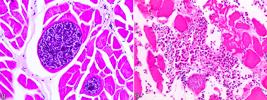
Infectious diseases in wild and exotic birds in Paraíba. (8) Microscopic view of cysts of Sarcocystis sp. in the muscle layers of the proventriculus, intracytoplasmatic within the tissue in a Turdus rufiventris. HE, obj. 40x. (9) Turdus rufiventris (rufous-bellied thrush). Necrosis of skeletal muscle fibers associated with an inflammatory infiltrate composed of eosinophils, and to a lesser extent, lymphocytes, plasma cells, and macrophages, with bradyzoites from Sarcocystis sp. in the pectoral skeletal muscle. HE, obj. 40x.
In the macroscopic evaluation of the D. nobilis, multifocal pallor not limited to the myocardium was observed, without other relevant changes. Microscopically, there was distension of the myocardium fibers with multifocal necrosis and a discrete inflammatory infiltrate, primarily composed of granulocytes and a few macrophages. Additionally, inflammatory and necrotic processes were observed, with intact cysts containing bradyzoites of Sarcocystis sp.
Discussion
The results of this study demonstrate that wild and exotic birds in the State of Paraíba, Northeast Brazil, can be affected by various infectious diseases, especially those originating from captures related to animal trafficking or rescue operations. Among these illnesses, bacterial diseases stand out. Bacterial infections, particularly intestinal and extraintestinal infections by Escherichia coli, are prevalent in wild birds (Konicek et al. 2016, Borges et al. 2017). In this sense, the trafficking of birds poses a risk of spreading E. coli, as these birds are reservoirs for the bacteria. Infected birds can intermittently shed bacteria through their feces for months or years, potentially transmitting this pathogen to the environment and posing a health risk to humans and birds (Borges et al. 2017). Identifying histopathological lesions and isolating the bacteria are essential steps in definitively diagnosing these bacterial infections.
Colibacillosis was the most recurrent bacterial disease in wild and exotic birds, as noted in studies on diseases (Konicek et al. 2016, Cigler et al. 2020). It is crucial to emphasize that E. coli is present in a commensal form in the gastrointestinal tract of these birds, and the pathogenic forms must be investigated using molecular methods. A study with birds in northeastern Brazil suggested that captive birds from screening centers and wildlife can be reservoirs of antibiotic-resistant E. coli, acting as disseminators of this bacterium in the environment (Silva et al. 2021). This finding is consistent with other studies that investigated the resistance genes of E. coli and concluded that birds are hosts and disseminators of this bacterium (Ong et al. 2020).
Extraintestinal infections caused by the bacterium E. coli can affect the renal, reproductive and nervous systems (Dórea et al. 1984), especially in young animals and reach the circulatory system by spreading to the other organs and may be responsible for the clinical picture of septicemia that can lead to the bird’s death (Borges et al. 2017). In the present study, parrots were diagnosed with liver granulomas caused by the bacteria, highlighting that the infection can evolve to an irreversible condition.
In confirmed cases of Aspergillus sp., the affected organs included the lungs, heart, liver, spleen, kidneys, and skin. These organs were severely affected by fungal hyphae infiltration, circulatory and inflammatory lesions, similar to reports in other wild bird species (Latge 1999). Septate hyphae can be identified by their morphological characteristics, which are crucial for differentiating filamentous fungi. Histochemical stains of PAS and GMS aid in this differentiation, as noted in previous studies that used special staining as a diagnostic tool in ostriches (Struthio camelus) and other animal species (Paixão et al. 2004, Desoubeaux & Cray 2018). In the parrot, which was affected by aspergillosis and Capillaria sp., no adult worms were observed in the adjacent liver tissue. This finding is consistent with a study on capillariasis in other animals (Soares et al. 2011). In the respiratory system, lesions of both parrots and hawks, similar lung lesions and hyphae are seen in domestic mammals and different wild species, including birds (Spanamberg et al. 2012, Xavier & Madrid 2014, Seyedmousavi et al. 2015). This may be attributed to the susceptibility of birds and other wild and exotic species to lower their immune response in the face of stress and an unhealthy environment.
Fungal infections were observed in a disseminated form or in multiple organs in most cases. Notably, in one of the cases, which involved a magnificent frigatebird (Fregata magnificens), two fungi caused lesions in its tissues, similar to cases reported in other birds such as chickens (Gallus gallus domesticus), ducks (Cairina moschata), ostriches (S. camelus), pigeons (Columba livia), penguins (Spheniscus), and canaries (Serinus canaria). In these reports, other agents were concomitant to the fungal infection, making it challenging to determine the primary agent responsible for the initial lesion in the affected organs (Severo et al. 2010, Xavier & Madrid 2014). The free-ranging magnificent frigatebird was found on the ground and cachectic; a traumatic lesion was identified in the proventriculus, caused by a fishing hook found within the lesion. This puncturing object created an entry point for infectious agents. The importance of studying the presence of Aspergillus spp. in aquatic birds and their potential role in One Health is essential, as these species can harbor zoonotic pathogens (Melo et al. 2020).
In weakened birds, infection by Sarcocystis sp. should be differentiated, as the literature indicates that at least 25 species of these parasites can encyst in striated muscles of these birds (Prakas et al. 2018). Histopathological findings of notable lesions in various organs are consistent with those observed in other species of wild and domestic birds (Mayr et al. 2016, Gondim et al. 2017), including an inflammatory infiltrate in organs affected by the protozoan. In the present study, this is evidenced by the presence of histiocytes, eosinophils and lymphocytes, present in the inflammation of the birds’ tissues. Given that several species of Sarcocystis sp. can be pathogenic or not for wild birds, as some might be present with clinically significant lesions. In contrast, others may only harbor the pathogen (Lacerda et al. 2023, Moura et al. 2023).
The rufous-bellied thrush (Turdus rufiventris) had disseminated muscular infection of the ventricle and proventriculus’ muscles, pectoral skeletal muscle and myocardium. At the same time, the red-shouldered macaw (Diopsittaca nobilis) was affected only in the skeletal muscle. Consequently, the two species affected by Sarcocystis sp. in the present study should be considered as intermediate hosts (IH) of the agent. Previous studies (Gjerde et al. 2018) indicate that species considered IH exhibit asexual protozoan reproduction in striated muscle and vascular cells, while definitive hosts carry the parasitic agent only in the intestinal mucosa. It is crucial to note that the protozoan can be considered eventually fatal when it occurs systemically (Prakas et al. 2018), and some of these Sarcocystis spp. can be more pathogenic (Moura et al. 2023).
Conclusions
This study demonstrates that the birds of the species Amazona aestiva, Diopsittaca nobilis, Parabuteo unicinctus, Sicalis flaveola, Cyanoloxia brissonii, Nymphicus hollandicus, Crotophaga ani, Cyanocorax cyanopogon, Fregata magnificens, Turdus rufiventris, whether free-living, zoo-housed, or bred in captivity, are susceptible to various infectious agents.
In the present study, bacterial diseases were the most prevalent, followed by fungal infections and protozoal infections. Escherichia coli was notably associated with granuloma lesions in organs such as the liver and kidneys. Aspergillosis was the predominant fungal infection, with one case involving aspergillosis and zygomycosis. Histopathological findings were characterized by fungal granulomas and prominent filamentous hyphae. Additionally, Sarcocystis spp. were identified as the causative agent of protozoal infections, with parasitic cysts observed in the muscle tissues of affected birds.
This survey highlights the need to investigate infectious pathogens in wild and exotic birds. Further research is needed to understand the prevalence and manifestation of these diseases across different avian species, which will contribute to the development of effective diagnostic and management strategies.
Acknowledgments
We thank the members of the “Laboratório de Patologia Veterinária” of the “Universidade Federal da Paraíba” (UFPB). The “Conselho Nacional de Desenvolvimento Científico e Tecnológico” (CNPq) for the aid and financing, and “Coordenação de Aperfeiçoamento de Pessoal de Nível Superior” (CAPES) for the granting of a scholarship throughout the research.
References
-
Borges CA, Beraldo LG, Maluta RP, Cardozo MV, Barboza KB, Guastalli EAL, Kariyawasam S, DebRoy C, Ávila FA. Multidrug-resistant pathogenic Escherichia coli isolated from wild birds in a veterinary hospital. Avian Pathol 2017; https://doi.org/10.1080/03079457.2016.1209298
» https://doi.org/10.1080/03079457.2016.1209298 -
Cigler P, Kvapil P, Kastelic M, Gombač M, Švara T, Vobr J, Račnik J, Bartova E. Retrospective study of causes of animal mortality in Ljubljana Zoo 2005-2015. J Zoo Wildl Med 2020; https://doi.org/10.1638/2019-0206
» https://doi.org/10.1638/2019-0206 -
Desoubeaux G, Cray C. Animal models of aspergillosis. Comp Med 2018; https://pmc.ncbi.nlm.nih.gov/articles/PMC5897967/pdf/cm2018000109.pdf
» https://pmc.ncbi.nlm.nih.gov/articles/PMC5897967/pdf/cm2018000109.pdf -
Dórea EMA, Santos JA, Santangelo C. Aspergilose cerebral em pintos. Pesq Agro Bras 1984; https://apct.sede.embrapa.br/pab/article/view/15790/9844
» https://apct.sede.embrapa.br/pab/article/view/15790/9844 -
El-Morsey A, El-Seify M, Desouky A-RY, Abdel-Aziz MM, Sakai H, Yanai T. Morphologic identification of a new Sarcocystis sp. in the common moorhen (Gallinula chloropus) (Aves: Gruiformes: Rallidae) from Brolos Lake, Egypt. Parasitol Res 2013; https://doi.org/10.1007/s00436-013-3667-x
» https://doi.org/10.1007/s00436-013-3667-x -
Galiza GJN, Tochetto C, Rosa FB, Panziera W, Silva TM, Caprioli RA, Kommers GD. Utilização de três métodos imuno-histoquímicos na detecção de aspergilose e zigomicose em animais. Pesq Vet Bras 2014; https://doi.org/10.1590/S0100-736X2014000700005
» https://doi.org/10.1590/S0100-736X2014000700005 -
Gargiulo A, Fioretti A, Russo TP, Varriale L, Rampa L, Paone S, De Luca Bossa LM, Raia P, Dipineto L. Occurrence of enteropathogenic bacteria in birds of prey in Italy. Lett Appl Microbiol 2018; https://doi.org/10.1111/lam.12836
» https://doi.org/10.1111/lam.12836 -
Gjerde B, Vikøren T, Hamnes IS. Molecular identification of Sarcocystis halieti n. sp., Sarcocystis lari and Sarcocystis truncata in the intestine of a white-tailed sea eagle (Haliaeetus albicilla) in Norway. Int J Parasitol Parasites Wildl 2018; https://doi.org/10.1016/j.ijppaw.2017.12.001
» https://doi.org/10.1016/j.ijppaw.2017.12.001 -
Gondim LSQ, Jesus RF, Ribeiro-Andrade M, Silva JCR, Siqueira DB, Marvulo MFV, Aléssio FM, Mauffrey J-F, Julião FS, Savani ESMM, Soares RM, Gondim LFP. Sarcocystis neurona and Neospora caninum in Brazilian opossums (Didelphis spp.): Molecular investigation and in vitro isolation of Sarcocystis spp. Parasitol Vet 2017; https://doi.org/10.1016/j.vetpar.2017.07.002
» https://doi.org/10.1016/j.vetpar.2017.07.002 - Grespan A, Raso TF. Psittaciformes (araras, papagaios, periquitos, calopsitas e cacatuas). In: Cubas ZS, Silva JCR, Catão-Dias JL. Tratado de Animais Selvagens: medicina veterinária. Vol. 1. 2ª ed. São Paulo: Roca; 2014.
-
Konicek C, Vodrážka P, Barták P, Knotek Z, Hess C, Račka K, Troxler S. Detection of zoonotic pathogens in wild birds in the cross-border region Austria - Czech Republic. J Wildl Dis 2016; https://doi.org/10.7589/2016-02-038
» https://doi.org/10.7589/2016-02-038 -
Lacerda MSC, Santos WHM, Lopes MC, Fonseca CS, Carvalho MPN, Martins NRS, Ecco R. A Survey of diseases in different species of wild, captive, and illegally traded birds in Brazil. Animals 2023; https://doi.org/10.3390/ani14010025
» https://doi.org/10.3390/ani14010025 -
Latge J-P. Aspergillus fumigatus and aspergillosis. Clin Microbiol Rev 1999; https://doi.org/10.1128/cmr.12.2.310
» https://doi.org/10.1128/cmr.12.2.310 -
Mayr SL, Maier K, Müller J, Enderlein D, Gruber AD, Lierz M. Accipiter hawks (Accipitridae) confirmed as definitive hosts of Sarcocystis turdusi, Sarcocystis cornixi and Sarcocystis sp. ex Phalacrocorax carbo Parasitol Res 2016; https://doi.org/10.1007/s00436-016-5059-5
» https://doi.org/10.1007/s00436-016-5059-5 -
Melo AM, Silva-Filho RP, Poester VR, von Groll A, Fernandes CG, Stevens DA, Sabino R, Xavier MO. Aspergillosis in free-ranging aquatic birds. Medical Mycol Case Rep 2020; https://doi.org/10.1016/j.mmcr.2020.04.005
» https://doi.org/10.1016/j.mmcr.2020.04.005 -
Moura GHF, Nascimento GS, Oliveira PRF, Santana VS, Shimabukuro A, Valença YM, Calabuig C, Melo RPB, Mota RA. Occurrence of Apicomplexa protozoa in wild birds in the Northeast region of Brazil. Rev Bras Parasitol 2023; https://doi.org/10.1590/S1984-29612023020
» https://doi.org/10.1590/S1984-29612023020 - Moxley RA. Enterobacteriaceae: Escherichia, p.56-74. In: McVey DS, Kennedy M, Chengappa MM. Microbiologia Veterinária. 3ª ed. Rio de Janeiro: Guanabara Koogan; 2016.
-
Ong KH, Khor WC, Quek JY, Low ZX, Arivalan S, Humaidi M, Chua C, Seow KLG, Guo S, Tay MYF, Schlundt J, Ng LC, Aung KT. Occurrence and antimicrobial resistance traits of Escherichia coli from wild birds and rodents in Singapore. Int J Environ Res Public Health 2020; https://doi.org/10.3390/ijerph17155606
» https://doi.org/10.3390/ijerph17155606 -
Paixão TA, Nascimento EF, Parra PNS, Santos RL. Aspergilose em avestruz (Struthio camelus) no Brasil. Ciência Rural 2004; https://doi.org/10.1590/S0103-84782004000200037
» https://doi.org/10.1590/S0103-84782004000200037 -
Prakas P, Butkauskas D, Švažas S, Juozaitytė-Ngugu E, Stanevičius V. Morphologic and genetic identification of Sarcocystis fulicae n. sp. (Apicomplexa: Sarcocystidae) from the Eurasian coot (Fulica atra). J Wildl Dis 2018; https://doi.org/10.7589/2017-11-279
» https://doi.org/10.7589/2017-11-279 - Sanches TC, Godoy SN. Passeriformes (canário, sabiá, pássarro-preto e trinca-ferro). In: Cubas ZS, Silva JCR, Catão-Dias JL. Tratado de Animais Selvagens: medicina veterinária. Vol. 1. 2ª ed. São Paulo: Roca; 2014.
-
Severo CB, Guazzelli LS, Severo LC. Chapter 7 - Zygomycosis. J Bras Pneumol 2010; https://doi.org/10.1590/s1806-37132010000100018
» https://doi.org/10.1590/s1806-37132010000100018 -
Seyedmousavi S, Guillot J, Arné P, Hoog GS, Mouton JW, Melchers WJG, Verweij PE. Aspergillus and aspergilloses in wild and domestic animals: a global health concern with parallels to human disease. Med Mycol 2015; https://doi.org/10.1093/mmy/myv067
» https://doi.org/10.1093/mmy/myv067 -
Silva GGC, Campana EH, Vasconcelos PC, Silva NMV, Santos Filho L, Leite EL, Givisiez PEN, Gebreyes WA, Oliveira CJB. Occurrence of KPC-producing Escherichia coli in Psittaciformes rescued from trafficking in Paraíba, Brazil. Int J Environ Res Public Health 2021; https://doi.org/10.3390/ijerph18010095
» https://doi.org/10.3390/ijerph18010095 -
Soares MCP, Nunes HM, Silveira FAA, Alves MM, Souza AJS. Capillaria hepatica (Bancroft, 1893) (Nematoda) entre populações indígenas e mamíferos silvestres no noroeste do Estado do Mato Grosso, Brasil, 2000. Rev Pan-Amaz Saúde 2011; https://doi.org/10.5123/S2176-62232011000300005
» https://doi.org/10.5123/S2176-62232011000300005 -
Spanamberg A, Casagrande RA, Ferreiro L, Rolim VM, Souza SO, Gonçalves ICM, Oliveira LGS, Wouters F, Wouters ATB, Fontana CS, Driemeier D. Aspergilose em trinca-ferros (Saltator similis) competidores de canto. Acta Scient Vet 2012; https://www.ufrgs.br/actavet/40-4/PUB%201089.pdf
» https://www.ufrgs.br/actavet/40-4/PUB%201089.pdf - Xavier MO, Madrid IM. Doenças fúngicas em aves, p.1527-1536. In: Cubas ZS, Silva JCR, Catão-Dias JL. Tratado de Animais Selvagens: medicina veterinária. Vol. 1. 2ª ed. São Paulo: Roca; 2014.
-
Data availability statement
The data used in this study are available and can be accessed upon request to the corresponding author.
Edited by
-
Editor-in-Chief Fabiano José Ferreira de Sant’Ana.
The data used in this study are available and can be accessed upon request to the corresponding author.