Anatomopathological findings and causes of death in wild canids rescued in São Paulo State (2021-2023): implications for wildlife health and conservation
ABSTRACT:
Wild canids in Brazil are increasingly affected by habitat fragmentation and emerging infectious diseases, some of which are zoonotic. This study aimed to investigate the pathological findings and causes of death of wild canids admitted to the Mata Ciliar Association (Jundiaí/SP) from 2021 to 2023, including 28 crab-eating foxes (Cerdocyon thous), six maned wolves (Chrysocyon brachyurus), and one hoary fox (Lycalopex vetulus). Tissue samples collected during necropsies were subjected to histopathological and molecular examination. The causes of death were road mortality (11/35, 31.4%), euthanasia (8/35, 22.9%), respiratory disorders (7/35, 20.0%), cachexia/malnutrition (4/35, 11.4%), sepsis (2/35, 5.7%), gastrointestinal disease (2/35, 5.7%), and hypovolemic shock associated with severe flea infestation (1/35, 2.8%). Notable pathological findings included granulomatous pneumonia associated with Angiostrongylus spp. (5/28, 17.85%) in crab-eating foxes, canine distemper virus infection (5/35, 14.3%), sarcoptic mange (2/35, 5.7%), renal parasitism by Dioctophyme renale in maned wolves (3/6, 50%),and an oral squamous cell carcinoma in the hoary fox. These findings highlight the importance of post mortem examinations for wildlife disease surveillance and emphasize the role of health monitoring in biodiversity conservation and the prevention of zoonotic risks.
INDEX TERMS:
Pathology; infectious diseases; wildlife disease surveillance; conservation medicine
RESUMO:
Canídeos silvestres no Brasil vêm sendo cada vez mais impactados pela fragmentação de hábitat e por doenças infecciosas emergentes, algumas de importância zoonótica. Este estudo teve como objetivo investigar os achados anatomopatológicos e as causas de morte de canídeos silvestres admitidos na Associação Mata Ciliar (Jundiaí/SP) de 2021 a 2023, incluindo 28 cachorros-do-mato (Cerdocyon thous), seis lobos-guará (Chrysocyon brachyurus) e uma raposinha-do-campo (Lycalopex vetulus). Amostras de tecidos coletadas durante as necropsias foram submetidas a exames histopatológicos e moleculares. As causas de morte foram atropelamentos (11/35; 31,4%), eutanásia (8/35; 22,9%), distúrbios respiratórios (7/35; 20,0%), caquexia/desnutrição (4/35; 11,4%), sepse (2/35; 5,7%), doenças gastrointestinais (2/35; 5,7%) e choque hipovolêmico associado à severa infestação por pulgas (1/35; 2,8%). Entre os achados anatomopatológicos mais relevantes destacam-se pneumonia granulomatosa associada a Angiostrongylus spp. (5/28; 17,85%) em cachorros-do-mato, infecção pelo vírus da cinomose canina (5/35; 14,3%), sarna sarcóptica (2/35; 5,7%), parasitismo renal por Dioctophyme renale em lobos-guará (3/6; 50%), e carcinoma de células escamosas oral na raposinha-do-campo. Esses resultados reforçam a importância dos exames post mortem para a vigilância de doenças na fauna silvestre e destacam o papel do monitoramento da saúde animal na conservação da biodiversidade e na prevenção de riscos zoonóticos.
TERMOS DE INDEXAÇÃO:
Patologia; doenças infecciosas; vigilância de doenças da vida selvagem; medicina da conservação
Introduction
Habitat loss and fragmentation driven by agricultural expansion, livestock farming, and urbanization are major drivers of global biodiversity decline (Haddad et al. 2015, Piano et al. 2020). Agricultural intensification profoundly alters ecosystems, often prioritizing food production over habitat integrity and wildlife health. These impacts are particularly evident in biodiversity hotspots such as Brazil, where rapid land-use change poses severe threats to native fauna (Schmid & Schöb 2023).
In this context, wild canids are particularly vulnerable. In the Cerrado, one of Brazil’s most threatened biomes, species such as the maned wolf (Chrysocyon brachyurus) are increasingly affected by pasture expansion, intensive agriculture, and urban encroachment (Myers et al. 2000, Queirolo et al. 2011). These pressures intensify human-wildlife conflict, increasing road mortality and retaliatory killings, especially among generalist species that frequently use anthropogenic landscapes (Caceres 2011, Grilo et al. 2018).
Beyond direct anthropogenic threats, wild canids are also exposed to a wide range of infectious and parasitic diseases, many of which are zoonotic. Rabies, leishmaniasis, canine distemper, and emerging infections such as angiostrongyliasis have been documented in Brazilian canids, with some species acting as important reservoirs (Araújo et al. 2014, Caprioli et al. 2018, Slaviero et al. 2019). Despite this growing body of research, regionally representative post mortem studies remain scarce, limiting our understanding of disease burden, mortality patterns, and their implications for conservation.
Post mortem examinations are essential for identifying pathological processes, causes of death, and health threats relevant to wildlife conservation and One Health approaches (Cooper 2002). To address these gaps, this study aimed to investigate the pathological findings and causes of death of wild canids admitted to the “Associação Mata Ciliar” (Mata Ciliar Association, Jundiaí, São Paulo, Brazil) from 2021 to 2023, contributing to wildlife health surveillance and conservation planning.
Materials and Methods
Ethical approval. All procedures followed the guidelines of the Committee on Ethics in the Use of Animals (CEUA) of the “Faculdade de Medicina Veterinária e Zootecnia” (Faculty of Veterinary Medicine and Animal Science - FMVZ) of the “Universidade de São Paulo” (USP), which approved the use of biological samples for teaching and research purposes (protocol 9185230222). This study was also authorized by the “Sistema de Autorização e Informação em Biodiversidade” (Biodiversity Authorization and Information System - Sisbio) of the “Instituto Brasileiro do Meio Ambiente e dos Recursos Naturais Renováveis” (Brazilian Institute of Environment and Renewable Natural Resources - IBAMA) (protocol 81825-A).
Animals and post mortem examination. The study included 35 wild canids: 28 crab-eating foxes (Cerdocyon thous), six maned wolves (Chrysocyon brachyurus), and one hoary fox (Lycalopex vetulus). Most individuals were rescued by the Environmental Police and admitted alive, though in a debilitated clinical condition, to the Wild Animal Rehabilitation Center of the “Associação Mata Ciliar” (Jundiaí, São Paulo).
Upon admission, all animals underwent veterinary evaluation, including clinical examination and documentation of rescue circumstances and geographic origin. According to clinical progression and prognosis, the canids either received medical treatment or were humanely euthanized. After death, all animals were subjected to a complete post mortem examination. Necropsies were performed following standard procedures, and tissue samples were collected for histopathology and complementary diagnostic investigations.
Diagnosis of canine distemper virus (CDV). Real-time reverse transcription polymerase chain reaction (RT-qPCR) was performed on blood samples from four crab-eating foxes and one maned wolf. Viral RNA was extracted from 200 µL of each sample using a magnetic-bead-based automated system, following the manufacturer’s instructions, and eluted into RNase-free buffer. A one-step RT-qPCR assay was then performed in a 25 µL reaction containing a commercial master mix with reverse transcriptase and primers/probe targeting a conserved region of the CDV genome. Cycling conditions consisted of reverse transcription at 50 °C for 10 min, initial denaturation at 95 °C for 5 min, followed by 45 cycles of 95 °C for 15 s and 60 °C for 30 s, with fluorescence acquisition at each extension step. No-template controls, extraction negative controls, and positive controls were included in all runs. Samples with a cycle threshold (Ct) < 40 were considered positive.
Tumor characterization. During necropsy, tissue samples (approximately 1 cm³) were collected from suspected neoplastic lesions, fixed in 10% phosphate-buffered formalin (pH 7.4), and processed using routine histological techniques. Sections (3 µm thick) were stained with hematoxylin and eosin (HE) for histopathological evaluation. For tumor characterization, immunohistochemistry (IHC) was performed with the following monoclonal antibodies: AE1/AE3 (1:300, Dako, Pan-cytokeratin Clone, pH 5.6), Ki67 (1:300, Dako, Mib-1 Clone, pH 8.9), p63 (1:400, Dako, DAK-p63 Clone, pH 8.9), CK14 (1:100, BioSb, LL002 Clone, pH 5.6), and Melanoma Cocktail (1:400, CellMarque, Pool HMB45 + MART-1 + Tyrosinase Clone, pH 8.9). Antigen retrieval was performed using heat-induced epitope retrieval in a steamer for 20-30 minutes. The membranes were incubated with primary antibodies at 4 °C for 18 hours in the Advance System. Staining was performed with DAB, followed by counterstaining with Harris hematoxylin.
Results
Epidemiology
During the study period, necropsies were performed on 35 wild canids: 28 crab-eating foxes, six maned wolves, and one hoary fox. Table 1 summarizes the epidemiological characteristics of these canids, including age, sex, time in captivity, and city of origin.
Epidemiological data of wild canids (Cerdocyon thous, Chrysocyon brachyurus, and Lycalopex vetulus) subjected to necropsy from June 2021 to June 2023
Causes of death
Considering all canids evaluated (n = 35), road traffic collisions were the leading cause of death, accounting for 11 cases (31.4%). Euthanasia due to poor clinical prognosis was the second most frequent outcome (8/35; 22.9%). Among euthanized individuals, canine distemper virus infection was diagnosed in three cases. In contrast, the remaining cases were associated with severe trauma from previous road collisions, cachexia, neoplasia, severe respiratory disease, or neurological signs of unknown cause. Respiratory disease was the third most frequent cause of death, with pneumonia responsible for seven cases (7/35; 20.0%), including interstitial (3/7), granulomatous (2/7), and suppurative bronchopneumonia (2/7). Cachexia or severe malnutrition accounted for four deaths (4/35; 11.4%), including two individuals affected by sarcoptic mange. Septicemia was the primary cause of death in two canids (2/35; 5.7%). Gastrointestinal disease was responsible for two deaths (2/35; 5.7%): one due to parasitic hemorrhagic enteritis associated with necrotizing neutrophilic hepatitis, and one due to necrotizing and hemorrhagic lymphoplasmacytic enteritis progressing to the mesentery. One individual (1/35; 2.9%) died from hypovolemic shock associated with severe flea infestation.
Among crab-eating foxes (n = 28), road mortality was the leading cause of death (10/28; 35.7%). Euthanasia accounted for seven cases (7/28; 25%), three of which were associated with confirmed canine distemper virus infection. Pneumonia was responsible for six deaths (6/28; 21.4%), followed by cachexia or malnutrition (3/28; 10.7%), septicemia (1/28; 3.6%), and gastrointestinal involvement with hepatitis and enteritis (1/28; 3.6%).
The six maned wolves exhibited heterogeneous causes of death, including roadkill (1/6), septicemia (1/6), severe flea infestation (1/6), cachexia/malnutrition associated with sarcoptic mange (1/6), severe respiratory failure due to suppurative bronchopneumonia associated with distemper (1/6), and gastrointestinal disease characterized by necrotizing hemorrhagic enteritis (1/6). The single hoary fox was euthanized due to a poor prognosis associated with a highly metastatic oral squamous cell carcinoma.
Pathological findings
The main pathological findings observed in the maned wolves, crab-eating foxes, and the hoary fox evaluated in this study are summarized in Table 2 and described below by pathological category.
Endoparasites
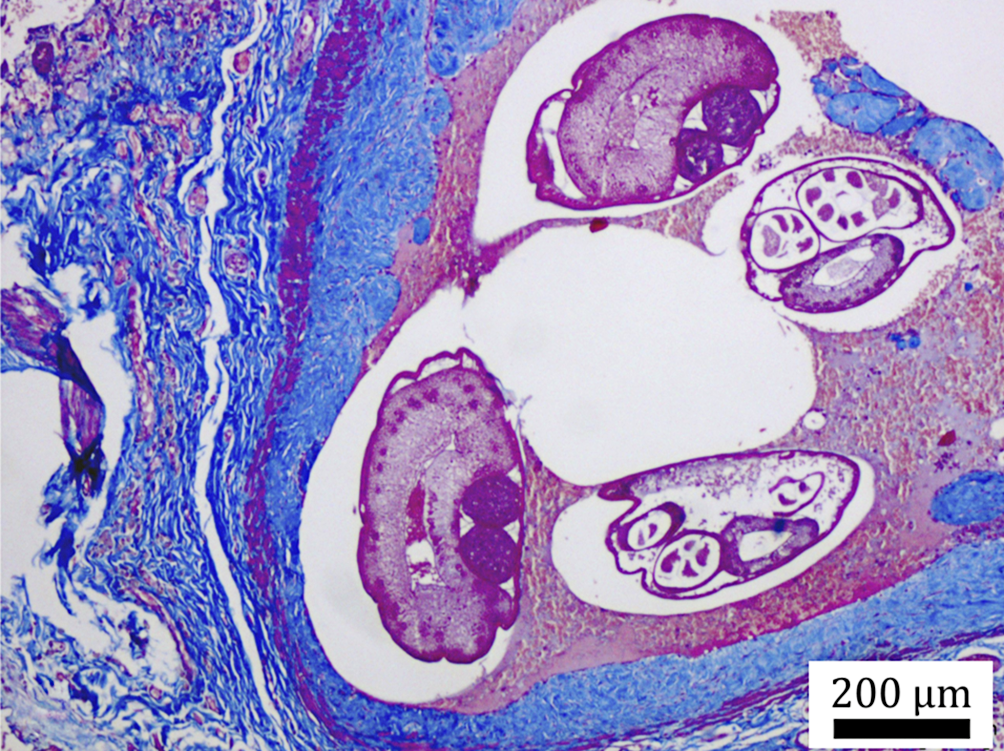
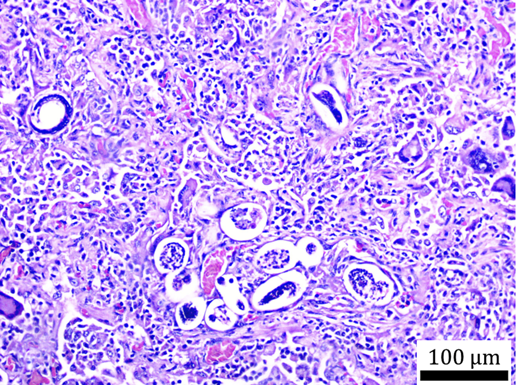
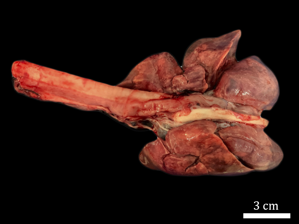
Eight of the 28 crab-eating foxes (28.6%) harbored pulmonary nematodes. Based on the tissue sections evaluated, five cases presented adult worms in the pulmonary arteries, whereas three showed only eggs and larvae in the alveolar and bronchiolar lumens. These parasites were morphologically compatible with Angiostrongylus spp.; they had a thin eosinophilic cuticle, a pseudocoelom with gastrointestinal and reproductive tracts, and coelomic musculature. In arteries containing nematodes, the vessel wall was thickened due to fibrous connective tissue proliferation, as evidenced by Masson’s trichrome staining (Fig. 1). Granulomatous pneumonia was observed in five of the eight infected crab-eating foxes (62.5%), characterized by lymphocytic, macrophagic, plasmacytic, and multinucleated giant cell infiltration (Fig. 2). Grossly, the lungs of these canids were not collapsed but were distended and showed multifocal dark red areas with a shiny surface (Fig. 3). One crab-eating fox (1/28; 3.6%) harbored an adult trematode morphologically compatible with Platynosomum or Athesmia in the bile duct, causing ductal distension without an associated ductular reaction. Only mild lymphoplasmacytic hepatitis and focal fibrosis were present. In the gastrointestinal tract of crab-eating foxes, enteritis was identified in 23/25 individuals examined histologically (92%), mostly lymphoplasmacytic. Nematode eggs and larvae were observed in seven of these 23 cases (7/23; 30.4%). Colitis was observed in 8/15 crab-eating foxes examined histologically (53.3%). Muscle cysts compatible with Sarcocystis spp. were detected in 9/24 crab-eating foxes examined histologically (37.5%). However, there was no inflammatory response associated with the cysts. Additionally, Dioctophyme renale nematodes were macroscopically observed in the right kidney of three maned wolves (3/6; 50%). In these individuals, the right kidney was reduced to its capsule, and the contralateral kidney was hypertrophic as a compensatory change.
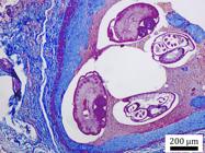
Granulomatous pneumonia caused by nematodes compatible with Angiostrongylus spp. in a crab-eating fox (Cerdocyon thous). Pulmonary artery with marked wall thickening due to fibrous connective tissue proliferation; adult nematodes are visible in the lumen. Masson’s trichrome, obj. 4x, bar = 200 µm.
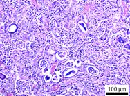
Granulomatous pneumonia caused by nematodes compatible with Angiostrongylus spp. in a crab-eating fox (Cerdocyon thous). Larvated eggs and larvae of nematodes in alveolar and interstitial spaces, associated with dense lymphohistiocytic infiltrate and multinucleated giant cells. HE, obj. 10x, bar = 100 µm.
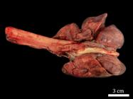
Granulomatous pneumonia caused by nematodes compatible with Angiostrongylus spp. in a crab-eating fox (Cerdocyon thous). Noncollapsed lung lobes showing multifocal dark red areas of consolidation. Bar = 3 cm.
Ectoparasites
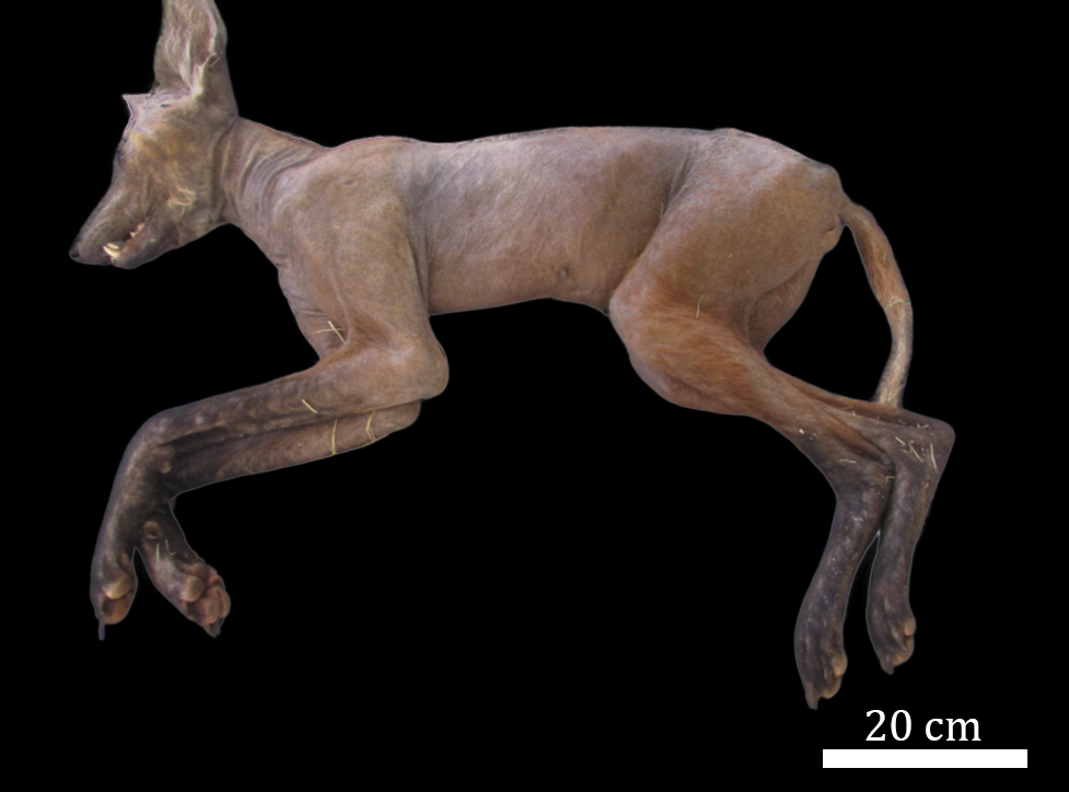
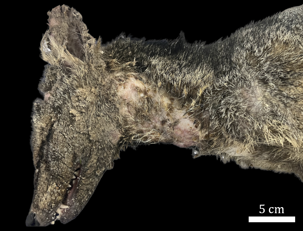
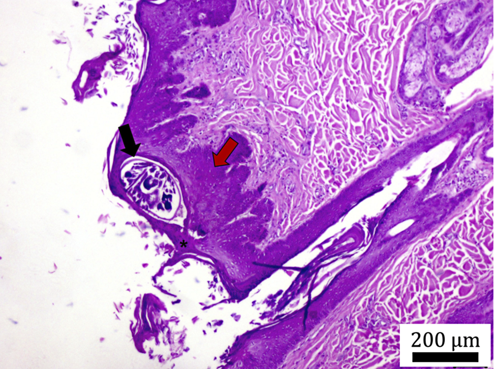
Mites (Sarcoptes scabiei) were identified in two canids - one maned wolf and one crab-eating fox - causing sarcoptic mange in both. Another maned wolf (rescued in Mogi-Mirim) showed lesions highly suggestive of mange (alopecia on the limbs and ventral body); however, it had been frozen prior to necropsy, preventing visualization of mites on histopathology. In the confirmed case, the maned wolf (rescued in Araçatuba) exhibited generalized alopecia affecting the face, neck, muzzle, ears, ventral trunk, tail, and dorsum, with marked hyperkeratosis and crusting (Fig. 4). The affected crab-eating fox showed alopecia, crusts, and hyperkeratosis on the elbows, knees, ischial region, and face, especially the ears, with wrinkled skin in affected areas (Fig. 5). These canids exhibited poor body condition, with evident signs of weakness and dehydration. Histopathology in both individuals revealed hyperkeratosis, acanthosis, epidermal hyperplasia, ulcerated areas with intralesional adult mites (Fig. 6), and neutrophilic dermal inflammation.
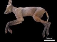
Sarcoptic mange in wild canids. Maned wolf (Chrysocyon brachyurus) from Araçatuba (São Paulo state) with severe alopecia due to sarcoptic mange. Bar = 20 cm.
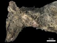
Sarcoptic mange in wild canids. Crab-eating fox (Cerdocyon thous) with severe hyperkeratosis, alopecia, and crust formation caused by sarcoptic mange. Bar = 5 cm.
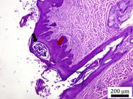
Histopathological lesions of sarcoptic mange in a maned wolf (Chrysocyon brachyurus). Adult mite of Sarcoptes scabiei within an epidermal tunnel (black arrow); marked parakeratotic hyperkeratosis with retained nuclei in the stratum corneum (*) and acanthosis of the spinous layer (red arrow). HE, obj. 4x, bar = 200 μm.
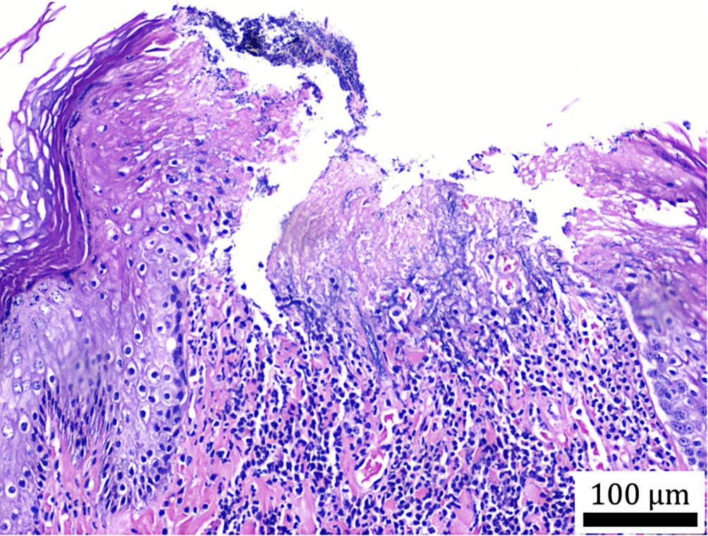
Lice (Trichodectes canis) were found in 35.7% of crab-eating foxes (10/28). Among these, 30% (3/10) showed clinical pediculosis, with histopathological lesions including parakeratotic hyperkeratosis, acanthosis, ballooning degeneration, neutrophilic dermatitis, and secondary bacterial infection (Fig. 7).
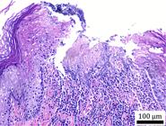
Histopathological lesions caused by lice in a crab-eating fox (Cerdocyon thous). Ulcerative dermatitis with dense neutrophilic inflammation, bacterial colonies, epithelial barrier rupture, ballooning degeneration of keratinocytes, marked hyperkeratosis, and acanthosis. Obj. 10x, bar = 100 µm.
Bacterial infections
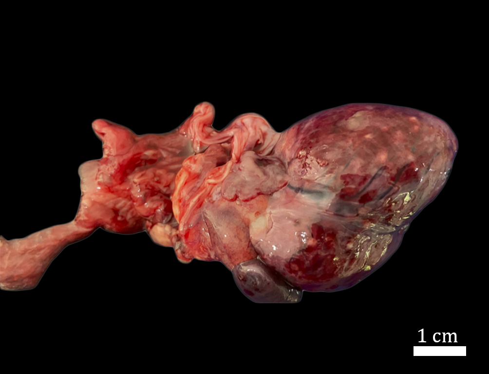
One crab-eating fox died from sepsis. Macroscopically, the epicardium exhibited multifocal to coalescing whitish-yellow dots corresponding to areas of necrosis and inflammation (Fig. 8), and the pancreas was markedly enlarged. Histopathology revealed severe suppurative myocarditis with predominantly neutrophilic inflammation, extensive cardiomyocyte necrosis, and bacterial colonies composed mainly of Gram-positive cocci with fewer Gram-negative cocci (Fig. 9). The pancreas showed severe interstitial necrotizing pancreatitis with mixed (lymphocytic, plasmacytic, and neutrophilic) but predominantly neutrophilic inflammation. Additional lesions included lymphoplasmacytic meningitis, neutrophilic lymphadenitis, neutrophilic myositis, and mixed (neutrophilic and lymphoplasmacytic) nephritis and hepatitis. Bacterial colonies were identified in most affected tissues, except in the meninges.
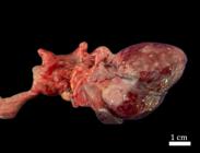
Sepsis in a crab-eating fox (Cerdocyon thous). Epicardium with multifocal whitish-yellow rounded foci of necrosis and inflammation. Bar = 1 cm.
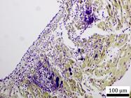
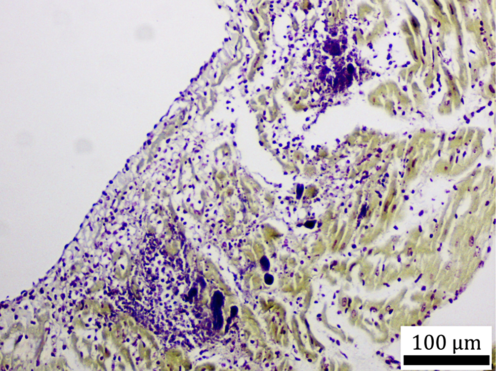
Sepsis in a crab-eating fox (Cerdocyon thous). Severe suppurative myocarditis with abundant Gram-positive bacterial colonies. Gram stain, obj. 4x, bar = 100 µm.
Among the eight crab-eating foxes with pulmonary nematodes, three (37.5%) had secondary bacterial infections in the lungs associated with neutrophilic inflammation. The maned wolf diagnosed with CDV also exhibited bacterial colonies associated with severe suppurative bronchopneumonia. Secondary bacterial infections were also observed in canids infested with lice and mites, consistent with the severe skin lesions described. Eight crab-eating foxes had gastritis. In two cases of gastritis, ulcers were observed in association with inflammation, and Gram-negative bacteria were identified.
Diagnosis of canine distemper virus
Canine distemper virus infection was assessed by real-time PCR in five crab-eating foxes and one maned wolf, with a confirmed diagnosis in four crab-eating foxes and one maned wolf (14.3%; 5/35). Testing was only performed on individuals presenting clinical signs compatible with distemper; therefore, other canids in this study were not screened for the disease. The infected maned wolf presented predominantly respiratory clinical signs (dyspnea and nasal and ocular discharge), along with diarrhea and head tremors. All affected crab-eating foxes arrived at the “Associação Mata Ciliar” with advanced neurological signs, including head-pressing, myoclonus, and circling.
Among these foxes, two showed marked lymphohistioplasmacytic granulomatous pneumonia associated with nematode parasitism, one had predominantly mononuclear interstitial pneumonia, and one had mixed broncho-interstitial pneumonia with neutrophils and lymphocytes. Scattered eosinophilic intracytoplasmic inclusion bodies were observed in the bronchial and bronchiolar epithelium.
In the central nervous system, all distemper-positive individuals showed cerebellar white-matter demyelination ranging from mild to moderate, while encephalitis with mild perivascular cuffing was observed in four of the five cases.
Tumor characterization
A single neoplasm was identified: an extensive oral squamous cell carcinoma in the hoary fox. Grossly, the mass was verrucous, multinodular, and friable, affecting the right upper and lower lips and extending to the ipsilateral maxillary and mandibular gingiva, with invasion of the hard palate. Histopathology revealed atypical squamous proliferation with inflammatory features, consistent with squamous cell carcinoma, and confirmed by positive immunohistochemistry for AE1/AE3, p63, and CK14. The proliferation marker Ki67 was expressed in approximately 80% of neoplastic cells.
Metastatic lesions were observed in the lungs and kidneys. The pulmonary lobes were distended and turgid, with firm yellowish multifocal to coalescing nodules throughout the parenchyma. Histopathology revealed severe suppurative bronchopneumonia associated with metastatic squamous cells of oral origin. In the kidneys, there was acute tubular nephritis, with neoplastic cells forming nests within the parenchyma.
Trauma
Twelve canids (34.2%; 12/35) were victims of road traffic collisions, including 11 crab-eating foxes (39% of this species) and one maned wolf (16.6% of this species). Among the crab-eating foxes, fractures were identified in the pelvis (2/11; 18%), mandible (2/11; 18%), tibia (2/11; 18%), fibula (1/11; 9%), and skull (2/11; 18%). All crab-eating foxes exhibited good body condition at the time of the accident. In contrast, the maned wolf was underweight and presented alopecic areas suggestive of sarcoptic mange, although no mites were detected histologically. Other common traumatic lesions observed in road-killed individuals included hematomas, hemothorax, hemoperitoneum and pulmonary hemorrhage.
Other conditions
Follicular hyperplasia of the thyroid gland was identified in 14 of the 16 canids in which the thyroid was examined (87.5%). This alteration was characterized by irregular thyroid follicles with reduced luminal diameter and epithelial cell proliferation forming papillary projections into the follicular lumen. The colloid was generally weakly eosinophilic, with peripheral resorption vacuoles located near the apical surface of the cells.
Alopecia affected two crab-eating foxes (2/28; 7%), involving multiple body regions in one individual and presenting as generalized in the other. No mites were detected in either skin scrapings or histopathological sections. Histopathological examination of the skin revealed follicular dysplasia and atrophy, along with melanin aggregates within hair follicles and perifollicular melanosis.
Discussion
The pulmonary nematodes identified in the crab-eating foxes were morphologically compatible with the genus Angiostrongylus. In Brazil, infection with Angiostrongylus vasorum has previously been reported not only in this species (Ferreira-Júnior et al. 2017) but also in the hoary fox (Lima et al. 1994). Adult worms typically reside in the right atrium and ventricle and the pulmonary artery, whereas larvae and eggs are found in the lungs. Mollusks act as intermediate hosts (Spratt 2015).
The histopathological lesions we observed in parasitized canids - granulomatous pneumonia with eggs and larvae surrounded by lymphocytes, plasma cells, macrophages, and giant cells, together with adult nematodes in pulmonary arteries - were consistent with previous descriptions (Kistler et al. 2014, Santoro et al. 2015, Caprioli et al. 2018). Depending on the severity, these lesions can compromise respiratory function (Kistler et al. 2014, Santoro et al. 2015, Caprioli et al. 2018). One of the most severe cases of verminotic pneumonia in this study occurred in a crab-eating fox co-infected with the distemper virus, which contributed to the severity of the lung lesions.
We detected pulmonary parasitic infections in 28.6% of the crab-eating foxes examined, which is lower than the 57.7% reported for this species in southeastern Brazil (Caprioli et al. 2018). This discrepancy may reflect methodological differences, as we identified nematodes solely by histopathology, whereas Caprioli et al. (2018) also used PCR, which can detect subclinical infections not visible in tissue sections.
The public health-relevant species Angiostrongylus cantonensis has been reported in crab-eating foxes in southeastern Brazil, including larvae in a focal area of hemorrhage and malacia in the brain (Caprioli et al. 2018). In our series, no larvae were detected in the central nervous system. Wildlife cases can precede human infections, as illustrated in Florida, where detection of A. cantonensis in a primate at Miami Zoo in 2003 was followed by human cases, especially in children (Chance et al. 2024). In Brazil, a recent report identified A. cantonensis larvae in the leptomeninges and neuropil of a Peruvian spider monkey (Ateles chamek) at Sorocaba Zoo, highlighting local transmission risk via infected mollusks (Fernandes et al. 2025). Human cases remain rarely reported in Brazil, likely due to underdiagnosis and limited surveillance (Morassutti et al. 2014).
Half of the maned wolves examined were parasitized by the nematode Dioctophyme renale, which typically infects the right kidney of mammals and has been reported in canids and mustelids worldwide, including the Middle East, Asia, the Americas, and Europe (Eiras et al. 2021). Although widely distributed, human infections remain rare (Chauhan et al. 2016).
Severe skin lesions caused by the mite Sarcoptes scabiei were documented in both a maned wolf and a crab-eating fox. Affected canids showed poor body condition, dehydration, weakness, and debility associated with extensive dermatitis. Histological features were consistent with previous reports in maned wolves and crab-eating foxes (Teodoro et al. 2018, Fiori et al. 2023). They included marked hyperkeratosis, acanthosis, epidermal hyperplasia, and ulceration with intralesional mites.
Sarcoptic mange is highly contagious in domestic and wild mammals worldwide (Pence & Ueckermann 2002) and occasionally affects humans (Larsson 1978). In Brazil, it has been increasingly reported in maned wolves, especially in anthropized areas (Fiori et al. 2023, 2025). The disease is concentrated in the southeastern region of Brazil, mainly in the State of São Paulo, and appears to be an emerging disease in maned wolves in Brazil. Camera-trap studies show overlap of maned wolves with domestic dogs and cattle, which may facilitate mite transmission (Fiori et al. 2023). This ectoparasitosis has also been described in invasive wild boar (Sus scrofa) (Haas et al. 2015). Our detection of sarcoptic mange in both a maned wolf and a crab-eating fox reinforces its emerging threat and underscores the need for surveillance and control strategies at the wildlife-domestic interface.
One maned wolf died from a severe flea infestation (pulicosis) during a hot, rainy period. These climatic conditions favor flea development (Perez et al. 2023). Because no ectoparasites were found on admission, we believe that heavy infestation likely occurred in captivity. Fleas such as Ctenocephalides felis can carry bacteria from the genera Rickettsia and Bartonella, which were previously detected in free-ranging crab-eating foxes in southern Brazil (Schott et al. 2019). Thus, we recommend that preventive ectoparasite control in captive facilities be performed periodically to ensure the health of canids.
Canine distemper is a major threat to wild canids (Giacinti et al. 2022). The course of the disease varies according to the host’s age and immune status, environmental conditions, and the virulence of the viral strain. In susceptible species, the respiratory, gastrointestinal, nervous, and integumentary systems can be affected (Deem et al. 2000). All four crab-eating foxes with confirmed CDV in this study presented severe neurological signs at admission and either died or were euthanized. Our histopathological examination revealed demyelination of the cerebellar white matter, the hallmark lesion in crab-eating foxes and Pampas foxes (Slaviero et al. 2019). This process typically begins about three weeks post-infection and is associated with profound immunosuppression (Vandevelde & Zurbriggen 2005).
Although the crab-eating fox is listed as Least Concern by the IUCN, its adaptability to degraded and human-dominated landscapes, omnivorous diet, and frequent contact with domestic dogs can facilitate CDV transmission to threatened species (Echeverry-Bonilla et al. 2022). Canine distemper is a very common disease in suburban and farm dogs in Brazil due to the lack of vaccination, which increases spillover risk (Cleaveland et al. 2000, Megid et al. 2009). Vaccinating wildlife is impractical, but targeted vaccination of domestic dogs at urban-wildland interfaces is an effective preventive strategy (Silva et al. 2023). The virus circulates widely among carnivores and has caused fatalities even in free-ranging American black bears (Ursus americanus) in the USA (Cottrell et al. 2013). Uncontrolled populations of stray dogs and cats - often unneutered and unvaccinated - exacerbate this scenario, posing both public health risks (e.g., rabies) and biodiversity threats, including predation, competition with native predators, environmental contamination, and disease spread (Doherty et al. 2016, 2017, Hughes & Macdonald 2013).
Thyroid follicular cell hyperplasia was frequent among the canids examined. Sonne et al. (2009) described morphological alterations, including C-cell hyperplasia and follicular cysts, in the thyroid glands of Arctic foxes (Vulpes lagopus) exposed to environmental pollutants, although without clinical signs of goiter. Studies in rats have also shown that polybrominated and polychlorinated biphenyls can induce thyroid hyperplasia and goiter (Kasza et al. 1978). Our findings highlight the importance of histopathological thyroid screening across Brazilian canid populations and its potential use as a sentinel marker of environmental contamination.
Alopecia was observed in two crab-eating foxes. Histopathological examination revealed follicular dysplasia and atrophy, along with melanin aggregates within hair follicles. The causes of alopecia in animals may involve inflammatory, infectious, endocrine, nutritional, immune-mediated, or traumatic factors (Mauldin & Peters-Kennedy 2016). When these causes are absent, the condition is poorly understood and has been more thoroughly characterized in domestic dogs than in wild species (Deering et al. 2023). Although the cause of alopecia in the crab-eating foxes examined in this study could not be determined, a possible association with follicular dysplasia involving melanocytes cannot be excluded. In domestic dogs, melanocytic abnormalities have been described and are occasionally associated with alopecia (Muller & Kirk 2012).
One hoary fox (Lycalopex vetulus) presenting a poorly differentiated oral squamous cell carcinoma affecting the gingiva and right lip and extending beyond the bony palate, previously described by our group as a separate case report (Zanoti et al. 2024), also showed pulmonary and renal metastases. High Ki-67 expression indicated aggressive biological behavior.
In Brazil, roadkill is a major cause of mortality among wild mammals and affects multiple species, including those that are threatened (Navas-Suárez et al. 2022). In this study, roadkill was one of the main causes of death, resulting in the deaths of ten crab-eating foxes and one maned wolf. In southeastern Brazil, the crab-eating fox (Cerdocyon thous) is among the most frequently road-killed species, likely due to its foraging behavior along road edges, scavenging habits, and tendency to freeze in front of vehicle headlights - a predator-avoidance response that may increase the risk of being hit (Caceres 2011, Ferreira et al. 2014). Most road-killed canids in our study were in good body condition, and fractures resulting from trauma involved the pelvis, tibia, mandible, and skull. These findings are consistent with those reported by Navas-Suárez et al. (2022), who analyzed traumatic injuries in 430 road-killed neotropical mammals in Brazil and found that the crab-eating fox was one of the most affected species. Similar to our results, that study reported skull, pelvis, and mandible fractures as the most frequent injuries, and most canids also had good body condition at the time of the accident. In São Paulo State, roadkill contributes significantly to biodiversity loss and mortality of wild canids. Therefore, road planning and monitoring initiatives are essential for conservation, as well as anatomopathological studies of road-killed wildlife, which can serve as tools for disease surveillance and health assessment of free-ranging populations.
Conclusion
This study provides valuable insights into the overall health status and diseases affecting wild canids in São Paulo State. Post mortem examinations proved essential for identifying the main health threats to these canids. Expanding such surveillance to other Brazilian biomes is critical for effective disease control and wildlife conservation. Additionally, our findings highlight the importance of the One Health approach by identifying zoonotic diseases such as sarcoptic mange and dioctophymosis. Monitoring the health of wild fauna plays a key role in preventing diseases that can impact both humans and domestic animals.
Acknowledgments
This study was supported by the Mata Ciliar Association (Jundiaí-SP).
References
-
Araújo JL, Dantas AFM, Galiza GJN, Pedroso PMO, Silva MLCR, Pimentel LA, Riet-Correa F. Aspectos histopatológicos e imuno-histoquímicos da raiva em raposas Cerdocyon thous Acta Scient Vet 2014; https://www.ufrgs.br/actavet/42-suple-1/CR_67.pdf
» https://www.ufrgs.br/actavet/42-suple-1/CR_67.pdf -
Caceres NC. Biological characteristics influence mammal road kill in an Atlantic Forest-Cerrado interface in south-western Brazil. Italian J Zoology 2011; https://doi.org/10.1080/11250003.2011.566226
» https://doi.org/10.1080/11250003.2011.566226 -
Caprioli RA, Andrade CP, Argenta FF, Ehlers LP, Soares JF, Pavarini SP, Driemeier D, Sonne L. Angiostrongylosis in Cerdocyon thous (crab-eating fox) and Lycalopex gymnocercus (Pampas fox) in southern Brazil. Parasitology 2018; https://doi.org/10.1017/S0031182018001865
» https://doi.org/10.1017/S0031182018001865 -
Chance MD, Noel AD, Thompson AB, Marrero N, Bula-Rudas F, Horvat CM, Green J, Armstrong JE, Levent F, Dudas RA, Shaffren S, Samide A, Martinez K, Stockdale K, Chancey RJ. Angiostrongylus cantonensis meningoencephalitis in three pediatric patients in Florida, USA. J Pediatric Infect Dis Soc 2024; https://doi.org/10.1093/jpids/piae113
» https://doi.org/10.1093/jpids/piae113 -
Chauhan S, Kaval S, Tewari S. Dioctophymiasis: a rare case report. J Clin Diagn Res 2016; https://doi.org/10.7860/JCDR/2016/17394.7305
» https://doi.org/10.7860/JCDR/2016/17394.7305 -
Cleaveland S, Appel MGJ, Chalmers WSK, Chillingworth C, Kaare M, Dye C. Serological and demographic evidence for domestic dogs as a source of canine distemper virus infection for Serengeti wildlife. Vet Microbiol 2000; https://doi.org/10.1016/S0378-1135(99)00207-2
» https://doi.org/10.1016/S0378-1135(99)00207-2 -
Cooper JE. Diagnostic pathology of selected diseases in wildlife. Rev Sci Tech 2002; https://doi.org/10.20506/rst.21.1.1320
» https://doi.org/10.20506/rst.21.1.1320 -
Cottrell WO, Keel MK, Brooks JW, Mead DG, Phillips JE. First report of clinical disease associated with canine distemper virus infection in a wild black bear (Ursus americana). J Wildl Dis 2013; https://doi.org/10.7589/2013-02-027
» https://doi.org/10.7589/2013-02-027 -
Deem SL, Spelman LH, Yates RA, Montali RJ. Canine distemper in terrestrial carnivores: a review. J Zoo Wildl Med 2000; https://doi.org/10.1638/1042-7260(2000)031[0441:CDITCA]2.0.CO;2
» https://doi.org/10.1638/1042-7260(2000)031[0441:CDITCA]2.0.CO;2 -
Deering KM, Clarke LL, Lennix SV, Layne E, Long L, Businga N. Follicular dysplasia and hair loss in white-tailed deer (Odocoileus virginianus). Vet Pathol 2023; https://doi.org/10.1177/03009858231185106
» https://doi.org/10.1177/03009858231185106 -
Doherty TS, Dickman CR, Glen AS, Newsome TM, Nimmo DG, Ritchie EG, Vanak AT, Wirsing AJ. The global impacts of domestic dogs on threatened vertebrates. Biol Conserv 2017; https://doi.org/10.1016/j.biocon.2017.04.007
» https://doi.org/10.1016/j.biocon.2017.04.007 -
Doherty TS, Glen AS, Nimmo DG, Ritchie EG, Dickman CR. Invasive predators and global biodiversity loss. Proc Natl Acad Sci USA 2016; https://doi.org/10.1073/pnas.1602480113
» https://doi.org/10.1073/pnas.1602480113 -
Echeverry-Bonilla DF, Buriticá-Gaviria EF, Orjuela-Acosta D, Chinchilla-Cardenas DJ, Ruiz-Saenz J. The first report and phylogenetic analysis of canine distemper virus in Cerdocyon thous from Colombia. Viruses 2022; https://doi.org/10.3390/v14091947
» https://doi.org/10.3390/v14091947 -
Eiras J, Zhu X-Q, Yurlova N, Pedrassani D, Yoshikawa M, Nawa Y. Dioctophyme renale (Goeze, 1782) (Nematoda, Dioctophymidae) parasitic in mammals other than humans: a comprehensive review. Parasitology 2021; https://doi.org/10.1016/j.parint.2020.102269
» https://doi.org/10.1016/j.parint.2020.102269 -
Fernandes NCCA, Ferreira-Machado E, Garcia JM, Navas-Suarez PE, Simões SRJS, Carvalho ACSR, Matsumoto PSS, Costa ALM, Martins MC, Teixeira RHF, Gonzaga CRR, Medeiros MA, Leandro SFS, Matos FN, Melo LCV, Baccin AO, Motta DJG, Gava R, Luca LR, Buti TEM, Ennser JRP, Etlinger-Colonelli D, Guerra JM. A tale of a snail: Fatal meningoencephalitis caused by Angiostrongylus cantonensis in a spider monkey (Ateles chamek) and detection among snails, São Paulo, Brazil. Vet Parasitol Reg Stud Reports 2025; https://doi.org/10.1016/j.vprsr.2025.101262
» https://doi.org/10.1016/j.vprsr.2025.101262 -
Ferreira CMM, Ribas ACA, Casella J, Mendes SL. Spatial variation of mammal road kills in a restinga area in Espírito Santo State, southeast Brazil. Neotrop Biol Conserv 2014; https://doi.org/10.4013/nbc.2014.93.02
» https://doi.org/10.4013/nbc.2014.93.02 -
Ferreira-Júnior JA, Blume GR, Sousa SKH, Carvalho CM, Gardiner C. Anatomo-pathological aspects of parasitism by nematodes of the superfamily Metastrongyloidea in wild crab-eating fox (Cerdocyon thous) in Midwestern Brazil. Ciência Rural 2017; https://doi.org/10.1590/0103-8478cr20160547
» https://doi.org/10.1590/0103-8478cr20160547 -
Fiori F, Paula RC, Boulhosa RLP, Dias RA. Clinical evaluation of sarcoptic mange (Sarcoptes scabiei) in maned wolves (Chrysocyon brachyurus). Vet Res Commun 2025; https://doi.org/10.1007/s11259-025-10774-0
» https://doi.org/10.1007/s11259-025-10774-0 -
Fiori F, Paula RC, Navas-Suárez PE, Boulhosa RLP, Dias RA. The sarcoptic mange in Maned Wolf (Chrysocyon brachyurus): mapping an emerging disease in the largest South American Canid. Pathogens 2023; https://doi.org/10.3390/pathogens12060830
» https://doi.org/10.3390/pathogens12060830 -
Giacinti JA, Pearl DL, Ojkic D, Campbell GD, Jardine CM. Genetic characterization of canine distemper virus from wild and domestic animal submissions to diagnostic facilities in Canada. Prev Vet Med 2022; https://doi.org/10.1016/j.prevetmed.2021.105535
» https://doi.org/10.1016/j.prevetmed.2021.105535 -
Grilo C, Coimbra MR, Cerqueira RC, Barbosa P, Dornas RAP, Gonçalves LO, Teixeira FZ, Coelho IP, Schmidt BR, Pacheco DLK, Schuck G, Esperando IB, Anza JA, Beduschi J, Oliveira NR, Pinheiro PF, Bager A, Secco H, Guerreiro M, Carvalho CF, Veloso AC, Custódio AEI, Marçal Jr O, Ciocheti G, Assis J, Ribeiro MC, Francisco BSS, Cherem JJ, Trigo TC, Jardim MMA, Franceschi IC, Espinosa C, Tirelli FP, Rocha VJ, Sekiama ML, Barbosa GP, Rossi HR, Moreira TC, Cervini M, Rosa CA, Silva LG, Ferreira CMM, César A, Casella J, Mendes SL, Zina J, Bastos DFO, Souza RAT, Hartmann PA, Deffaci ACG, Mulinari J, Luzzi SC, Rezzadori T, Kolcenti C, Reis TX, Fonseca VSC, Giorgi CF, Migliorini RP, Kasper CB, Bueno C, Sobanski M, Pereira APFG, Andrade FAG, Fernandes MEB, Corrêa LLC, Nepomuceno A, Banhos A, Hannibal W, Fonseca R, Costa LA, Medici EP, Croce A, Werther K, Oliveira JP, Ribeiro JM, Santi M, Kawanami AE, Perles L, Couto C, Figueiró DS, Eizirik E, Correia Jr AA, Corrêa FM, Queirolo D, Quagliatto AL, Saranholi BH, Galetti Jr PM, Rodriguez-Castro KG, Braz VS, França FGR, Buss G, Rezini JA, Lion MB, Cheida CC, Lacerda ACR, Freitas CH, Venâncio F, Adania CH, Batisteli AF, Hegel CGZ, Mantovani JA, Rodrigues FHG, Bagatini T, Curi NHA, Emmert L, Erdmann RH, Costa RRGF, Martinelli A, Santos CVF, Kindel A. Brazil road-kill: A data set of wildlife terrestrial vertebrate road-kills. Ecology 2018; https://doi.org/10.1002/ecy.2464
» https://doi.org/10.1002/ecy.2464 -
Haas C, Origgi FC, Akdesir E, Batista Linhares M, Giovannini S, Mavrot F, Casaubon J, Ryser-Degiorgis M-P. First detection of sarcoptic mange in free-ranging wild boar (Sus scrofa) in Switzerland. Schweiz. Arch Tierheilkd 2015; https://doi.org/10.17236/sat00020
» https://doi.org/10.17236/sat00020 - Haddad NM, Brudvig LA, Clobert J, Davies KF, Gonzalez A, Holt RD, Lovejoy TE, Sexton JO, Austin MP, Collins CD, Cook WM, Damschen EI, Ewers RM, Foster BL, Jenkins CN, King AJ, Laurance WF, Levey DJ, Margules CR, Melbourne BA, Nicholls AO, Orrock JL, Song D-X, Townshend JR. Habitat fragmentation and its lasting impact on Earth’s ecosystems. Sci Adv 2015; https://doi.org/10.1126/sciadv.1500052
-
Hughes J, Macdonald DW. A review of the interactions between free-roaming domestic dogs and wildlife. Biol Conserv 2013; https://doi.org/10.1016/j.biocon.2012.07.005
» https://doi.org/10.1016/j.biocon.2012.07.005 - Kasza L, Collins WT, Capen CC, Garthoff LH, Friedman L. Comparative toxicity of polychlorinated biphenyl and polybrominated biphenylin the rat thyroid gland: Light and electron microscopic alterations after subacute dietary exposure. J Environ Pathol Toxicol 1978;1(5):587-599. PMid:214505.
- Kistler WM, Brown JD, Allison AB, Nemeth NM, Yabsley MJ. First report of Angiostrongylus vasorum and Hepatozoon from a red fox (Vulpes vulpes) from West Virginia, USA. Vet Parasitol 2014; https://doi.org/10.1016/j.vetpar.2013.12.007
-
Larsson MHMA. Evidências epidemiológicas da ocorrência de escabiose, em humanos, causada pelo Sarcoptes scabiei (DeGeer, 1778) var. canis (Bourguignon, 1853). Rev Saúde Pública 1978; https://doi.org/10.1590/S0034-89101978000300007
» https://doi.org/10.1590/S0034-89101978000300007 -
Lima WS, Guimaraes MP, Lemos IS. Occurrence of Angiostrongylus vasorum in the lungs of the Brazilian fox Dusicyon vetulus J Helminthol 1994; https://doi.org/10.1017/S0022149X00013547
» https://doi.org/10.1017/S0022149X00013547 -
Mauldin EA, Peters-Kennedy J. Integumentary system. In: Maxie GM. Jubb, Kennedy and Palmer’s Pathology of Domestic Animals. 2016; https://doi.org/10.1016/B978-0-7020-5317-7.00006-0
» https://doi.org/10.1016/B978-0-7020-5317-7.00006-0 -
Megid J, Souza VAF, Teixeira CR, Cortez A, Amorin RL, Heinemman MB, Cagnini DQ, Richtzenhain LJ. Canine distemper virus in a crab-eating fox (Cerdocyon thous) in Brazil: case report and phylogenetic analyses. J Wildl Dis 2009; https://doi.org/10.7589/0090-3558-45.2.527
» https://doi.org/10.7589/0090-3558-45.2.527 -
Morassutti AL, Thiengo SC, Fernandez M, Sawanyawisuth K, Graeff-Teixeira C. Eosinophilic meningitis caused by Angiostrongylus cantonensis: An emergent disease in Brazil. Mem Inst Oswaldo Cruz 2014; https://doi.org/10.1590/0074-0276140023
» https://doi.org/10.1590/0074-0276140 - Muller GH, Kirk RW. Congenital and hereditary defects. In: Muller WH, Griffin CE, Campbell KL. Muller & Kirk’s Small Animal Dermatology. 7th ed. St Louis: Elsevier; 2012.
-
Myers N, Mittermeier RA, Mittermeier CG, Fonseca GAB, Kent J. Biodiversity hotspots for conservation priorities. Nature 2000; https://doi.org/10.1038/35002501
» https://doi.org/10.1038/35002501 -
Navas-Suárez PE, Diaz-Delgado J, Caiaffa MG, Silva MC, Yogui DR, Alves MH, Cereda JF, Silva MP, Cremer MJ, Ascensão F, Lorigados CAB, Medici EP, Desbiez ALJ, Catão-Dias JL. Characterization of traumatic injuries due to motor vehicle collisions in neotropical wild mammals. J Comp Pathol 2022; https://doi.org/10.1016/j.jcpa.2022.06.003
» https://doi.org/10.1016/j.jcpa.2022.06.003 -
Pence DB, Ueckermann E. Sarcoptic mange in wildlife. Sci Tech Rev 2002; https://doi.org/10.20506/rst.21.2.1335
» https://doi.org/10.20506/rst.21.2.1335 -
Perez R, Pérez-Cutillas P, Gonzálvez M, Muñoz-Hernández C, Arcenillas-Hernández I, Ybáñez RR, Escribano F, Martínez-Carrasco C. Predictive factors for flea occurrence in red foxes (Vulpes vulpes) from semi-arid Mediterranean environments. Med Vet Entomol 2023; https://doi.org/10.1111/mve.12611
» https://doi.org/10.1111/mve.12611 -
Piano E, Souffreau C, Merckx T, Baardsen LF, Backeljau T, Bonte D, Brans KI, Cours M, Dahirel M, Debortoli N, Decaestecker E, De Wolf K, Engelen JMT, Fontaneto D, Gianuca AT, Govaert L, Hanashiro FTT, Higuti J, Lens L, Martens K, Matheve H, Matthysen E, Pinseel E, Sablon R, Schön I, Stoks R, Van Doninck K, Van Dyck H, Vanormelingen P, Van Wichelen J, Vyverman W, De Meester L, Hendrickx F. Urbanization drives cross-taxon declines in abundance and diversity at multiple spatial scales. Glob Change Biol 2020; https://doi.org/10.1111/gcb.14934
» https://doi.org/10.1111/gcb.14934 -
Queirolo D, Moreira JR, Soler L, Emmons LH, Rodrigues FHG, Pautasso AA, Cartes JL, Salvatori V. Historical and current range of the Near Threatened maned wolf Chrysocyon brachyurus in South America. Oryx 2011; https://doi.org/10.1017/S0030605310000372
» https://doi.org/10.1017/S0030605310000372 -
Santoro M, D’Alessio N, Di Prisco F, Neola B, Restucci B, Pagano TB, Veneziano V. Angiostrongylus vasorum infection in red foxes (Vulpes vulpes) in southern Italy. Acta Parasitol 2015; https://doi.org/10.1515/ap-2015-0050
» https://doi.org/10.1515/ap-2015-0050 -
Schmid B, Schöb C. Ecological intensification of agriculture through biodiversity management: introduction. J Plant Ecol 2023; https://doi.org/10.1093/jpe/rtad018
» https://doi.org/10.1093/jpe/rtad018 -
Schott D, Souza UA, Dall’Agnol B, Webster A, Doyle R, Peters F, Favarini M, Mazim F, Rosa OA, Jardim MMA, Trigo TC, Reck J. Detection of Rickettsia spp. and Bartonella spp. in Ctenocephalides felis fleas from free-ranging crab-eating foxes (Cerdocyon thous). Med Vet Entomol 2019; https://doi.org/10.1111/mve.12371
» https://doi.org/10.1111/mve.12371 -
Silva ML, Caiaffa MG, Costa ALM, Teixeira RHF, Ervedosa TB, Machado EF, Suárez PEN, Réssio RA, Borges CC, Jesus IP, Carvalho J, Figueiredo KB, Carvalho ACSR, Brandão PE, Azevedo-Fernandes NCC, Guerra JM. Canine distemper virus and canine adenovirus type 1 co-infection in a free-living hoary fox (Lycalopex vetulus) from Brazil. Braz J Microbiol 2023; https://doi.org/10.1007/s42770-023-00921-7
» https://doi.org/10.1007/s42770-023-00921-7 -
Slaviero M, Ehlers LP, De Lorenzo C, Zafalon-Silva B, Driemeier D, Pavarini SP, Alievi MM, Sonne L. Anatomopathological and immunohistochemical apects of distemper virus in crab-eating-foxes and pampa-foxes. Acta Scient Vet 2019; https://doi.org/10.22456/1679-9216.90120
» https://doi.org/10.22456/1679-9216.90120 -
Sonne C, Wolkers H, Leifsson PS, Iburg T, Jenssen BM, Fuglei E, Ahlstrøm Ø, Dietz R, Kirkegaard M, Muir DCG, Jørgensen EH. Chronic dietary exposure to environmental organochlorine contaminants induces thyroid gland lesions in Arctic foxes (Vulpes lagopus). Environ Res 2009; https://doi.org/10.1016/j.envres.2009.04.008
» https://doi.org/10.1016/j.envres.2009.04.008 -
Spratt DM. Species of Angiostrongylus (Nematoda: Metastrongyloidea) in wildlife: a review. Int J Parasitol Parasites Wildl 2015; https://doi.org/10.1016/j.ijppaw.2015.02.006
» https://doi.org/10.1016/j.ijppaw.2015.02.006 -
Teodoro TGW, Lima PA, Stehling PC, Oliveira Junior IM, Varaschin MS, Wouters F, Wouters ATB. Sarcoptic mange (Sarcoptes scabiei) in wild canids (Cerdocyon thous). Pesq Vet Bras 2018; https://doi.org/10.1590/1678-5150-PVB-5700
» https://doi.org/10.1590/1678-5150-PVB-5700 -
Vandevelde M, Zurbriggen A. Demyelination in canine distemper virus infection: a review. Acta Neuropathol 2005; https://doi.org/10.1007/s00401-004-0958-4
» https://doi.org/10.1007/s00401-004-0958-4 -
Zanoti IC, Gouveia BA, Ramos FR, Klein M, Momo C. Oral squamous cell carcinoma with lung and kidney metastases in a hoary fox (Lycalopex vetulus). Braz J Vet Pathol 2024; https://doi.org/10.24070/bjvp.1983-0246.v17i2p99-103
» https://doi.org/10.24070/bjvp.1983-0246.v17i2p99-103