
 Mechanical, Optical, and Biofilm Accumulation Properties of Heat-Cured, CAD-CAM, and 3D-Printed Denture Base Resins After Simulated Brushing
ABSTRACT
Objective: To evaluate the effects of simulated brushing on Candida albicans biofilm formation, surface roughness, hardness, mass loss, and color stability of denture base resins fabricated using different materials.
Material and Methods: The study included three groups of resins: heat-cured PMMA (Vipicril Plus), CAD-CAM milled PMMA (Ivotion Base), and 3D-printed resin (Cosmos Denture), with 10 samples per group. Surface roughness, Vickers hardness, mass loss, color change (∆E00), and Candida albicans biofilm formation were measured before and after brushing. Statistical analyses were conducted using ANOVA and Tukey's post-hoc tests at a significance level of 5%.
Results: Heat-cured resin exhibited lower hardness values compared to milled resin (p < 0.05), while the hardness of the 3D-printed resin showed more significant variability (p > 0.05). The 3D-printed resin had the lowest surface roughness values before and after brushing (p < 0.05). In contrast, heat-cured resin demonstrated minimal color change and mass loss (p < 0.05). Candida albicans biofilm formation was lower on 3D-printed resin before brushing, although no significant difference was noted post-brushing.
Conclusion: Simulated brushing significantly altered the optical and mechanical properties of all resins. Heat-cured resin showed superior color stability and resistance to mass loss, while 3D-printed resin demonstrated the lowest surface roughness but higher biofilm formation after brushing.
Keywords:
Candida albicans; Computer-Aided Design; Dental Prosthesis; Dental Materials.
Introduction
Complete edentulism poses a significant global health challenge, especially among elderly populations. Traditionally, denture bases made from polymethyl methacrylate (PMMA) have been the gold standard for complete dentures due to their favorable aesthetic properties, affordability, and ease of manipulation [1]. Resins used in complete dentures that exhibit lower Candida albicans biofilm adhesion offer advantages in the prevention of denture stomatitis, a common condition among denture wearers [2]. Likewise, materials with higher mechanical resistance and optical stability are preferable to ensure the long-term esthetic and functional durability of the prostheses [3]. Therefore, understanding the differences among materials processed by different techniques (heat polymerization, 3D printing, and CAD-CAM milling) allows for a more informed selection by the clinician, taking into account not only clinical performance but also the patient’s profile and specific needs [4].
However, conventional heat-cured PMMA has several inherent limitations, including dimensional instability, fracture susceptibility, color degradation, and surface wear [5]. These issues can compromise both the longevity and functionality of the prosthesis. Recent advancements in digital technologies, such as computer-aided design and manufacturing (CAD-CAM) and additive manufacturing (3D printing), have created alternative methods for producing denture bases [6]. These techniques promise enhanced material homogeneity, reduced porosity, and improved mechanical properties. Subtractive methods, like milling, yield pre-cured PMMA bases that exhibit greater hardness and lower microbial adhesion [7].
In contrast, 3D-printed resins provide greater design flexibility and material efficiency, although they may face surface properties and durability challenges [8]. Maintaining hygiene is crucial for the longevity of dentures and overall oral health [9]. Brushing with dentifrices is the most common cleaning method among denture users [10]. However, the abrasiveness of these cleaning agents, combined with the mechanical forces involved in brushing, can damage the surface quality, alter mechanical properties, and increase microbial colonization, particularly by Candida albicans [11,12]. Such changes can reduce the functionality of the prosthesis and heighten the risk of infections related to the prosthesis [13,14]. With the increasing adoption of digital manufacturing technologies in dentistry, it is vital to understand how these methods affect the wear resistance, color stability, and microbial colonization of denture base materials under simulated brushing conditions [15,16]. This study evaluates the optical and mechanical changes in heat-cured, milled, and 3D-printed denture base resins after simulated brushing and their susceptibility to Candida albicans colonization [17,18]. These findings guide material selection and care protocols to enhance clinical outcomes [19,20].
Surface roughness plays a crucial role in adhesion and the performance of prosthetics. Increased roughness can promote microbial retention, particularly for Candida albicans, an opportunistic pathogen that flourishes in the oral environment [21-23]. This pathogen is closely linked to denture stomatitis, a common condition among denture wearers [24,25]. CAD-CAM-milled denture bases typically exhibit smoother surfaces, reducing microbial adhesion, whereas 3D-printe d bases are more susceptible to biofilm formation due to their inherent surface characteristics [26-28].Color stability is another important property that affects the aesthetics of denture materials [29,30]. Factors influencing color changes include surface roughness, water absorption, intrinsic pigment degradation, and exposure to staining agents [31,32]. CAD-CAM-milled resins demonstrate superior color stability compared to heat-cured and 3D-printed resins, which tend to show significant discoloration over time [33,34]. Simulated brushing studies have revealed differences in mechanical properties such as hardness and roughness across various fabrication methods [35,36]. CAD-CAM-milled PMMA resins consistently outperform heat-cured and 3D-printed resins regarding wear resistance and dimensional stability [37,38]. However, the clinical implications of these laboratory findings, particularly regarding patient satisfaction, have not yet been fully explored [39]. Understanding how different fabrication methods affect the optical, mechanical, and microbiological properties of denture base materials is essential for optimizing clinical outcomes [40]. The present study contributes to this growing body of knowledge by evaluating how simulated brushing affects the performance of heat-cured, milled, and 3D-printed denture resins, focusing on their mechanical properties, color stability, and susceptibility to microbial colonization. The null hypothesis is that there is no significant difference in biofilm formation, mechanical properties, or optical properties among dentures fabricated using conventional heat-curing, 3D printing, or CAD/CAM milling techniques after simulated brushing.
Material and Methods
Materials and Procedures
This study evaluated the effects of simulated brushing on the mechanical and optical properties of three types of denture base materials. Group HCR was composed of heat-cured resin (Vipicril Plus, VIPI Indústria, Comércio, Exportação e Importação de Produtos Odontológicos Ltda, Pirassununga, SP, Brazil), group CAD-CAM was composed of milled PMMA (Ivotion Base Pink V Denture Disc, Ivoclar Vivadent Ltda, Barueri, SP, Brazil), and group PRINTED was composed of 3D-printed denture base resin (Cosmos Denture, Yller Biomaterials S/A, Pelotas, RS, Brazil). Each group consisted of 10 specimens, resulting in 30 samples. All specimens were fabricated per the manufacturer's instructions and standardized to 10 mm × 10 mm × 3 mm.
Specimen Preparation
Specimens were prepared using consistent protocols to minimize variability. For surface finishing, specimens were manually polished with constant irrigation using silicon carbide abrasive papers (400, 600, and 1200 grit; Norton Saint-Gobain, Guarulhos, SP, Brazil). The final polishing involved felt discs impregnated with alumina solutions of 1 μm, 0.3 μm, and 0.05 μm (Buehler Ltd, Lake Bluff, USA), applied for one minute each using a polishing machine (APL-4 Universal Polisher, Arotec S/A Indústria e Comércio, Cotia, SP, Brazil) (Figure 1).

Surface roughness (Ra) was measured before and after simulated brushing using a surface roughness tester (SJ-201; Mitutoyo Sul Americana Ltda, Jundiaí, SP, Brazil). Measurements were conducted in six directions-three along each axis-using a diamond-tipped stylus with a 5-μm radius and a 90° angle. The stylus moved linearly over a length of 2 mm at a speed of 1 mm/second, applying a force of 6 mN, with a cutoff value of 0.25 mm.
Vickers Hardness was measured before brushing using a microhardness tester (ISH-TDV1000A; Insize do Brasil Ltda, Boituva, SP, Brazil). A 25-g load was applied for 10 seconds at three random locations on each specimen, ensuring at least 2 mm spacing between indents. Mean values were calculated for each sample.
Color stability was assessed using a spectrophotometer (CM2600d; Konica Minolta Sensing Americas, Inc., Ramsey, USA) and the CIEDE2000 color difference formula. The device was calibrated before each test as per the manufacturer's recommendations. Measurements were conducted before and after brushing to calculate the total color change (∆E00) using the formula: ∆E00 = [(∆L′ / KL, SL)2 + (∆C′ / KC, SC)2 + (∆H′ / KH, SH)2 + RT × (∆C′ / KC, SC) × (∆H′ / KH, SH)]1/2. Here, ∆L', ∆C', and ∆H' represent the mathematical differences in luminosity, chroma, and hue between two moments. RT denotes the rotation function, while SL, SC, and SH are the weighing functions for luminosity, chroma, and hue components. KL, KC, and KH are the parametric factors adjusted for the evaluated conditions. Color was assessed before and after simulated brushing.
Simulated brushing was conducted using a toothbrushing simulator (Odeme Equipamentos Médicos e Odontológicos Ltda., Luzerna, SC, Brazil) equipped with soft nylon-bristled brushes (Sorriso Standard Macia; Colgate-Palmolive Co., São Paulo, SP, Brazil) and a toothpaste-water solution (1:1 ratio by weight; Colgate Máxima Proteção Anticáries). Each specimen underwent 10,000 brushing cycles at 4.5 cycles per second under a load of 200 g. Specimen weight was recorded before and after brushing using an analytical balance (Marte Científica & Instrumentação Industrial Ltda., Santa Rita do Sapucaí, MG< Brazil) to determine mass loss. A subset of specimens (n = 6 per group) was sterilized using ultraviolet light and inoculated with Candida albicans (ATCC 102310) suspensions. After incubation at 37°C for 48 hours, colony-forming units (CFUs) were quantified using Sabouraud Dextrose Agar plates. The specimens were divided into brushed and unbrushed groups to assess the impact of simulated brushing on microbial adhesion.
Data Analysis
Data normality was verified using the Shapiro-Wilk test. One-way ANOVA followed by Tukey's post hoc test was used to compare groups. Paired t-tests were performed for within-group comparisons of preand post-brushing measurements. A significance level of 5% (p < 0.05) was adopted for all analyses. Sample size was determined using a power analysis, assuming a standard deviation of the difference of 0.5, indicating that a sample of 10 would have >90% power to detect differences in the primary outcome measure, surface roughness, among the three groups.
Results
The Vickers hardness test indicated that the the HCR group exhibited significantly lower hardness values than CAD-CAM group (p < 0.05). In contrast, the PRINTED group demonstrated the greatest variation in hardness, with no statistically significant difference to the CAD-CAM and HCR groups (p > 0.05) (Figure 2).

Data on the alterations in surface roughness caused by simulated toothbrushing are presented in Figure 3, and simulated toothbrushing reduced surface roughness for all three materials (p < 0.05). The PRINTED group presented the lowest surface roughness among the three tested materials at baseline and after brushing (p < 0.05). At the same time, no significant differences were noted for the surface roughness between the CAD-CAM and HCR groups.

The color stability data are presented in Table 1 and demonstrate significant differences (ANOVA, p=0.01) for total color change after brushing among the HCR (∆E00 = 6.2 ± 0.8), the CAD-CAM (∆E00 = 7.6 ± 1.6), and PRINTED (∆E00 = 8.0 ± 1.3) groups. The HCR exhibited significantly less color variations than the CAD-CAM (p = 0.03) and PRINTED groups (p = 0.006), while no significant differences were noted between the latter.
Color variation and mass variation values of the different groups before and after brushing.
The mass data are presented in Table 1 and show that HCR group demonstrated significantly smaller mass loss (3.2 ± 1.2 mg) after brushing than the CAD-CAM (p=0.0004) and 3D PRINTED groups (p = 0.0005). Meanwhile, the CAD-CAM group (9.5 ± 1.0 mg) exhibited significantly more mass loss than the PRINTED (6.2 ± 0.6 mg) group (p = 0.0003).
Biofilm accumulation in brushed and unbrushed samples is shown in Figure 4. In unbrushed samples, biofilm accumulation was lower in the PRINTED (171.1 ± 74.9) group than in the HCR (221.12 ± 88.2) and CAD=CAM (278.58 ± 116.9) groups; however, these differences did not reach statistical significance (ANOVA, p = 0.13). In unbrushed samples, biofilm accumulation was smaller, albeit not statistically significantly (ANOVA, p = 0.24), in HCR (225.1 ± 89.6) than in PRINTED (293.4 ± 123.2) and CAD=CAM (332.2 ± 132.5) groups. Paired intra-group comparisons revealed that brushing did not significantly increase biofilm accumulation in HCR and CAD=CAM groups; however, significant (p = 0.015) biofilm accumulation occurred in the PRINTED group (171.1 ± 74.9 versus 293.4 ± 123.2).

Discussion
The findings of this study highlight significant differences in the Candida albicans biofilm formation, mechanical, and optical properties of denture base resins made using heat-cured, CAD-CAM milled, and 3D printing techniques, particularly under simulated brushing conditions, rejecting the null hypothesis [4,6,7,16,25,28]. These results provide valuable insights into the clinical implications of material selection and hygiene protocols for complete denture bases [4,23].
The data showed that the HCR group exhibited the lowest hardness, mass loss, and color variation among the test groups; however, biofilm accumulation after brushing was similar to the other groups. The PRINTED group exhibited smoother surfaces, the most significant mass changes, and increased biofilm accumulation after brushing. The CAD-CAM group showed higher hardness and mass loss than the HCR group, and higher roughness and mass loss than the PRINTED group. Among the groups, only the PRINTED groups exhibited significantly increased in biofilm accumulation after brushing.
The lower hardness values observed for the thermally cured resin (HCR group) are consistent with previous research, indicating that conventional PMMA is more susceptible to surface wear and deformation under mechanical stress [17,18,27]. This susceptibility can be attributed to the polymerization process, which typically results in a higher residual monomer content and increased porosity compared to CAD-CAM milled and 3D printed resins [16,25,28]. The milled resin is suitable for high-wear clinical applications, such as those involving patients with bruxism or increased chewing forces [9,11,12,20]. Interestingly, the hardness variability in 3D printing was comparable to that of milled resin, suggesting potential improvements in 3D printing technologies that warrant further exploration [17,18,27].
Surface roughness significantly impacts microbial adhesion, patient comfort, and the longevity of the prosthesis [20,21,26]. The low initial roughness values of the 3D printed resin, which further decreased after brushing, are consistent with findings that emphasize the smoothness achievable with advanced resin formulations [7,13,14,17]. The increased roughness observed in post-brushing heat-cured resins highlights the importance of professional polishing during maintenance visits to mitigate microbial adhesion and surface deterioration [17,20]. Color stability is a critical factor for denture bases [6,15]. Thermally cured resins demonstrated superior color stability compared to 3D printed and milled resins, corroborating previous studies that attribute this performance to differences in material composition and polymerization processes [20,22,24]. The higher ∆E00 values of the 3D printed resin indicate that color change is influenced more by intrinsic factors, such as the resin matrix's composition and the pigment's stability, rather than merely surface texture [6,15,22,24]. The significant mass loss in the milled and 3D printed resins raises concerns about their durability under prolonged mechanical stress, highlighting the issue of abrasive wear [3,4,13,17]. The lower mass loss in heat-cured resin corresponds with its higher resistance to dimensional changes, making it a cost-effective choice for long-term use in low-stress environments [18,19]. These findings emphasize the need for tailored brushing protocols based on material properties to minimize wear [21,26,30]. Despite its lower initial roughness, the increased colonization of 3D-printed resin post-brushing suggests that factors beyond surface texture influence microbial adhesion [38-40]. Changes in hydrophobicity or material composition during wear may play a role [10-12]. This finding underscores the importance of combining mechanical and chemical cleaning methods for 3D-printed dentures to reduce the risk of prosthesis-related infections [4,23,33-35].
These results emphasize the need for material-specific hygiene protocols to optimize clinical performance [19,21]. Thermally polymerized resins offer better color stability and resistance to mass loss but may require frequent polishing to address increased roughness [17,20,21,26,28]. With their superior hardness, milled resins are ideal for patients who need durable prostheses despite higher production costs [9,11,12,20]. 3D-printed resin shows potential for initial aesthetics and microbial resistance, but its susceptibility to post-brushing
This study has some limitations that should be acknowledged. A single brand was used for each material, which may limit the generalizability of the findings. Future studies should evaluate a broader range of brands and incorporate extended brushing cycles, thermocycling, and different cleaning protocols to better simulate clinical conditions. In addition, further investigation into the effects of polishing and the role of resin composition on microbial adhesion and wear resistance is essential for improving 3D-printed denture materials.
Conclusion
Heat-cured resin showed minimal mass loss following brushing; however, this resin exhibited the lowest hardness values, indicating greater susceptibility to abrasion. The 3D-printed resin had the smoothest surface roughness; however, the colonization of Candida albicans significantly increased after the brushing cycle. Material properties should be carefully considered in order to establish evidence-based clinical material selection and hygiene protocols for complete denture bases.
-
Financial SupportNone.
Acknowledgments
The authors thank Professors Luise Gomes da Motta, Larissa Maria Assad Cavalcante, and Wayne José Batista Cordeiro for their valuable contributions.
Data Availability
The data used to support the findings of this study can be made available upon request to the corresponding author.
References
-
[1] Alfouzan AF, Alotiabi HM, Labban N, Al-Otaibi HN, Al Taweel SM, AlShehri HA. Color stability of 3D-printed denture resins: Effect of aging, mechanical brushing and immersion in staining medium. J Adv Prosthodont 2021; 13(3):160-171. https://doi.org/10.4047/jap.2021.13.3.160
» https://doi.org/10.4047/jap.2021.13.3.160 -
[2] Perea-Lowery L, Gibreel M, Vallittu PK, Lassila LV. 3D-printed vs. heat-polymerizing and autopolymerizing denture base acrylic resins. Materials 2021; 14(19):5781. https://doi.org/10.3390/ma14195781
» https://doi.org/10.3390/ma14195781 -
[3] Campos DES, Ferreira Muniz ÍA, da Costa TKVL, Lima RBW, Neppelenbroek KH, Batista AUD. Effect of simulated brushing with dentifrices on surface roughness and the mass loss of acrylic resin: A systematic review and meta-analysis of in vitro studies. J Prosthet Dent 2025; 133(5):1209-1220. https://doi.org/10.1016/j.prosdent.2023.06.027
» https://doi.org/10.1016/j.prosdent.2023.06.027 -
[4] Badaró MM, Salles MM, de Arruda CNF, Oliveira VC, de Souza RF, Paranhos HFO, et al. In vitro analysis of surface roughness of acrylic resin exposed to the combined hygiene method of brushing and immersion in ricinus communis and sodium hypochlorite. J Prosthodont 2017; 26(6):516-521. https://doi.org/10.1111/jopr.12436
» https://doi.org/10.1111/jopr.12436 -
[5] Moslehifard E, Ghaffari T, Abolghasemi H, Maleki Dizaj S. Comparison of conventional pressure-packed and injection molding processing methods for an acrylic resin denture based on microhardness, surface roughness, and water sorption. Int J Dent 2022; 2022:7069507. https://doi.org/10.1155/2022/7069507
» https://doi.org/10.1155/2022/7069507 -
[6] Dayan C, Guven MC, Gencel B, Bural C. A comparison of the color stability of conventional and CAD/CAM polymethyl methacrylate denture base materials. Acta Stomatol Croat 2019; 53(2):158-167. https://doi.org/10.15644/asc53/2/8
» https://doi.org/10.15644/asc53/2/8 -
[7] Ayaz EA, Aladağ SÜ. Effect of cigarette smoke and denture cleansers on the surface properties and color stability of CAD-CAM and conventional denture base resins. Dent Mater J 2023; 42(2):167-176. https://doi.org/10.4012/dmj.2022-117
» https://doi.org/10.4012/dmj.2022-117 -
[8] Anadioti E, Musharbash L, Blatz MB, Papavasiliou G, Kamposiora P. 3D printed complete removable dental prostheses: a narrative review. BMC Oral Health 2020; 20(1):343. https://doi.org/10.1186/s12903-020-01328-8
» https://doi.org/10.1186/s12903-020-01328-8 -
[9] Charoenphol K, Peampring C. Fit accuracy of complete denture base fabricated by CAD/CAM milling and 3D-printing methods. Eur J Dent 2023; 17(3):889-894. https://doi.org/10.1055/s-0042-1757211
» https://doi.org/10.1055/s-0042-1757211 -
[10] Emera RMK, Shady M, Alnajih MA. Comparison of retention and denture base adaptation between conventional and 3D-printed complete dentures. J Dent Res Dent Clin Dent Prospects 2022; 16(3):179-185. https://doi.org/10.34172/joddd.2022.030
» https://doi.org/10.34172/joddd.2022.030 -
[11] Srinivasan M, Kamnoedboon P, McKenna G, Angst L, Schimmel M, Özcan M, et al. CAD-CAM removable complete dentures: A systematic review and meta-analysis of trueness of fit, biocompatibility, mechanical properties, surface characteristics, color stability, time-cost analysis, clinical and patient-reported outcomes. J Dent 2021; 113:103777. https://doi.org/10.1016/j.jdent.2021.103777
» https://doi.org/10.1016/j.jdent.2021.103777 -
[12] Srinivasan M, Kalberer N, Kamnoedboon P, Mekki M, Durual S, Özcan M, et al. CAD-CAM complete denture resins: An evaluation of biocompatibility, mechanical properties, and surface characteristics. J Dent 2021; 114:103785. https://doi.org/10.1016/j.jdent.2021.103785
» https://doi.org/10.1016/j.jdent.2021.103785 -
[13] Çakmak G, Molinero-Mourelle P, De Paula MS, Akay C, Cuellar AR, Donmez MB, et al. Surface roughness and color stability of 3D-printed denture base materials after simulated brushing and thermocycling. Materials 2022; 15(18):6441. https://doi.org/10.3390/ma15186441
» https://doi.org/10.3390/ma15186441 -
[14] Helal MA, Fadl-Alah A, Baraka YM, Gad MM, Emam AM. In-vitro comparative evaluation for the surface properties and impact strength of CAD/CAM milled, 3D printed, and polyamide denture base resins. J Int Soc Prev Community Dent 2022; 12(1):126-131. https://doi.org/10.4103/jispcd.JISPCD_293_21
» https://doi.org/10.4103/jispcd.JISPCD_293_21 -
[15] Mugri MH, Jain S, Sayed ME, Halawi AHA, Hamzi SAI, Aljohani RAS, et al. Effects of smokeless tobacco on color stability and surface roughness of 3D-printed, CAD/CAM-milled, and conventional denture base materials: An in vitro study. Biomedicines 2023; 11:491. https://doi.org/10.3390/biomedicines11020491
» https://doi.org/10.3390/biomedicines11020491 -
[16] Al-Qarni FD, Gad MM. Printing accuracy and flexural properties of different 3D-printed denture base resins. Materials 2022; 15(7):2410. https://doi.org/10.3390/ma15072410
» https://doi.org/10.3390/ma15072410 -
[17] Ozyilmaz OY, Akin C. Effect of cleansers on denture base resins structural properties. J Appl Biomater Funct Mater 2019; 17(1):2280800019827797. https://doi.org/10.1177/2280800019827797
» https://doi.org/10.1177/2280800019827797 -
[18] Chang YH, Lee CY, Hsu MS, DU JK, Chen KK, Wu JH. Effect of toothbrush/dentifrice abrasion on weight variation, surface roughness, surface morphology and hardness of conventional and CAD/CAM denture base materials. Dent Mater J 2021; 40(1):220-227. https://doi.org/10.4012/dmj.2019-226
» https://doi.org/10.4012/dmj.2019-226 -
[19] Nam NE, Shin SH, Lim JH, Shim JS, Kim JE. Effects of artificial tooth brushing and hydrothermal aging on the mechanical properties and color stability of dental 3D rinted and CAD/CAM materials. Materials 2021; 14(20):6207. https://doi.org/10.3390/ma14206207
» https://doi.org/10.3390/ma14206207 -
[20] Zeidan AAE, Abd Elrahim RA, Abd El Hakim AF, Harby NM, Helal MA. Evaluation of surface properties and elastic modulus of CAD-CAM milled, 3D printed, and compression moulded denture base resins: An in vitro study. J Int Soc Prev Community Dent 2022; 12(6):630-637. https://doi.org/10.4103/jispcd.JISPCD_158_22
» https://doi.org/10.4103/jispcd.JISPCD_158_22 -
[21] Çakmak G, Donmez MB, Akay C, Atalay S, Silva de Paula M, Schimmel M, et al. Effect of simulated brushing and disinfection on the surface roughness and color stability of CAD-CAM denture base materials. J Mech Behav Biomed Mater 2022; 134:105390. https://doi.org/10.1016/j.jmbbm.2022.105390
» https://doi.org/10.1016/j.jmbbm.2022.105390 -
[22] Takhtdar M, Azizimoghadam N, Kalantari MH, Mohaghegh M. Effect of denture cleansers on color stability and surface roughness of denture bases fabricated from three different techniques: Conventional heat-polymerizing, CAD/CAM additive, and CAD/CAM subtractive manufacturing. Clin Exp Dent Res 2023; 9(5):840-850. https://doi.org/10.1002/cre2.763
» https://doi.org/10.1002/cre2.763 -
[23] Gad MM, Fouda SM. Current perspectives and the future of Candida albicans-associated denture stomatitis treatment. Dent Med Probl 2020; 57(1):95-102. https://doi.org/10.17219/dmp/112861
» https://doi.org/10.17219/dmp/112861 -
[24] Al Taweel SM, Fouzan AA, Al-Otaibi HN, Labban N, AlShehri HA. Thermal-cycling, simulated brushing, and beverages induced color changes and roughness of CAD/CAM poly (methyl methacrylate) denture resins. Mater Res Express 2021; 8(12):125401. https://doi.org/10.1088/2053-1591/ac406e
» https://doi.org/10.1088/2053-1591/ac406e -
[25] Shinawi LA. Effect of denture cleaning on abrasion resistance and surface topography of polymerized CAD CAM acrylic resin denture base. Electron Physician 2017; 9(5):4281-4288. https://doi.org/10.19082/4281
» https://doi.org/10.19082/4281 -
[26] Žilinskas J, Junevičius J, Česaitis K, Junevičiūtė G. The effect of cleaning substances on the surface of denture base material. Med Sci Monit 2013; 19:1142-1145. https://doi.org/10.12659/MSM.889568
» https://doi.org/10.12659/MSM.889568 -
[27] Alzaid M, AlToraibily F, Al-Qarni FD, Al-Thobity AM, Akhtar S, Ali S, et al. The effect of salivary pH on the flexural strength and surface properties of CAD/CAM denture base materials. Eur J Dent 2023; 17(1):234-241. https://doi.org/10.1055/s-0042-1749160
» https://doi.org/10.1055/s-0042-1749160 -
[28] Alammari MR. The influence of polishing techniques on pre-polymerized CAD\CAM acrylic resin denture bases. Electron Physician 2017; 9(10):5452-5458. https://doi.org/10.19082/5452
» https://doi.org/10.19082/5452 -
[29] Ghazal ARA, Idris G, Hajeer MY, Alawer K, Cannon RD. Efficacy of removing Candida albicans from orthodontic acrylic bases: An in vitro study. BMC Oral Health 2019; 19(1):71. https://doi.org/10.1186/s12903-019-0765-x
» https://doi.org/10.1186/s12903-019-0765-x -
[30] Nunes TSBS, Silva MDDD, Coelho SRG, Viotto HEDC, Pero AC. Effectiveness of disinfectant solutions associated or not with brushing on the biofilm control of a 3D printed-denture base resin. J Appl Oral Sci 2023; 31:e20230104. https://doi.org/10.1590/1678-7757-2023-0104
» https://doi.org/10.1590/1678-7757-2023-0104 -
[31] Osman RB, Khoder G, Fayed B, Kedia RA, Elkareimi Y, Alharbi N. Influence of fabrication technique on adhesion and biofilm formation of Candida albicans to conventional, milled, and 3D-printed denture base resin materials: A comparative in vitro study. Polymers 2023; 15(8):1836. https://doi.org/10.3390/polym15081836
» https://doi.org/10.3390/polym15081836 -
[32] Meirowitz A, Rahmanov A, Shlomo E, Zelikman H, Dolev E, Sterer N. Effect of denture base fabrication technique on Candida albicans adhesion in vitro. Materials 2021; 14(1):221. https://doi.org/10.3390/ma14010221
» https://doi.org/10.3390/ma14010221 -
[33] Alfouzan AF, Tuwaym M, Aldaghri EN, Alojaymi T, Alotiabi HM, Taweel SMA, et al. Efficacy of denture cleansers on microbial adherence and surface topography of conventional and CAD/CAM-processed denture base resins. Polymers 2023; 15(2):460. https://doi.org/10.3390/polym15020460
» https://doi.org/10.3390/polym15020460 -
[34] Silva MDDD, Nunes TSBS, Viotto HEDC, Coelho SRG, Souza RF, Pero AC. Microbial adhesion and biofilm formation by Candida albicans on 3D-printed denture base resins. PLoS One 2023; 18(10):e0292430. https://doi.org/10.1371/journal.pone.0292430
» https://doi.org/10.1371/journal.pone.0292430 -
[35] Fiore AD, Meneghello R, Brun P, Rosso S, Gattazzo A, Stellini E, et al. Comparison of the flexural and surface properties of milled, 3D-printed, and heat polymerized PMMA resins for denture bases: An in vitro study. J Prosthodont Res 2022; 66(3):502-508. https://doi.org/10.2186/jpr.JPR_D_21_00116
» https://doi.org/10.2186/jpr.JPR_D_21_00116 -
[36] Larijani M, Zareshahrabadi Z, Alhavaz A, Hajipour R, Ranjbaran A, Giti R, et al. Evaluation of Candida albicans biofilm formation on conventional and computer-aided-design/computer-aided manufacturing (CAD/CAM) denture base materials. Curr Med Mycol 2022; 8(3):23-29. https://doi.org/10.18502/cmm.8.3.11208
» https://doi.org/10.18502/cmm.8.3.11208 -
[37] Chladek G, Nowak M, Pakieła W, Mertas A. Effect of Candida albicans suspension on the mechanical properties of denture base acrylic resin. Materials 2022; 15(11):3841. https://doi.org/10.3390/ma15113841
» https://doi.org/10.3390/ma15113841 -
[38] Manikandan S, Vinesh E, Selvi DT, Kannan RK, Jayakumar A, Dinakaran J. Prevalence of Candida among denture wearers and nondenture wearers. J Pharm Bioallied Sci 2022; 14(Suppl 1):S702-S705. https://doi.org/10.4103/jpbs.jpbs_781_21
» https://doi.org/10.4103/jpbs.jpbs_781_21 -
[39] Al-Fouzan AF, Al-Mejrad LA, Albarrag AM. Adherence of Candida to complete denture surfaces in vitro: A comparison of conventional and CAD/CAM complete dentures. J Adv Prosthodont 2017; 9(5):402-408. https://doi.org/10.4047/jap.2017.9.5.402
» https://doi.org/10.4047/jap.2017.9.5.402 -
[40] Sultana N, Ahmed S, Nandini VV, Lathief J, Boruah S. An in vitro comparison of microbial adhesion on three different denture base materials and its relation to surface roughness. Cureus 2023; 15(4):e37085. https://doi.org/10.7759/cureus.37085
» https://doi.org/10.7759/cureus.37085
Edited by
-
Academic Editor: Alessandro Leite Cavalcanti


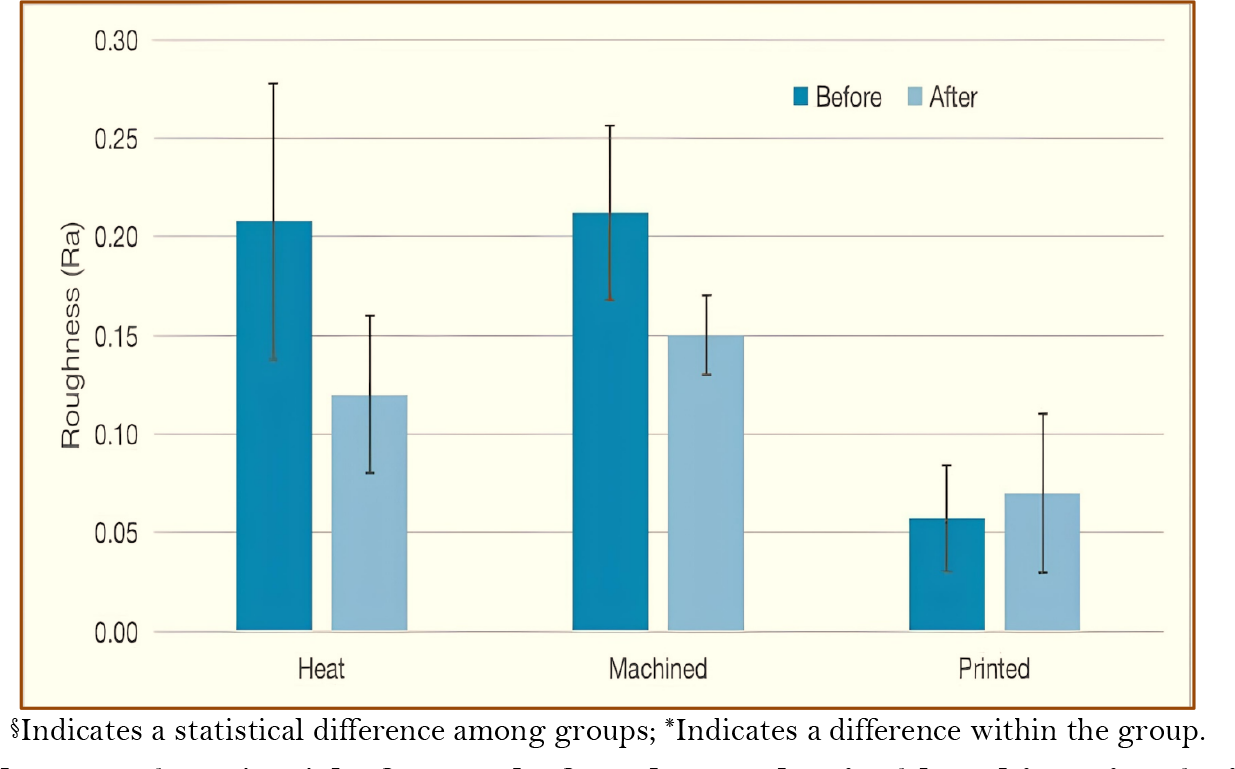 §Indicates a statistical difference among groups; *Indicates a difference within the group.
