
 Effect of Different Professional Prophylaxis Systems in Induced White Spot Lesions: An in Vitro Study
ABSTRACT
Objective: To evaluate the effect of different prophylaxis systems on the surface of sound enamel and in induced white spot lesions using confocal microscopy analysis.
Material and Methods: 40 specimens were obtained from sound premolars, and the buccal surface was divided into two parts: sound enamel and with an induced white spot lesion. The specimens were randomly divided into 4 groups (n=10/group): Group I: ClinproTM Prophy Paste (Perlite Particles); Group II: Extra-thin Pumice Stone; Group III: ClinproTM Prophy Powder (Glycine Amino Acid Powder); and Group IV: Sodium Bicarbonate Jet Powder. The specimens were analyzed by a confocal laser scanning microscope for surface roughness, preand post-prophylaxis. Data were analyzed by T-test and ANOVA (α= 0,05%).
Results: Confocal microscopy analysis showed that ClinproTM Prophy Paste and ClinproTM Prophy Powder did not cause an increase in the surface roughness (p > 0.05). On the other hand, the Sodium Bicarbonate Jet Powder caused a significant increase in the surface roughness only in the induced white spot lesion (p = 0.015).
Conclusion: Professional prophylaxis with Sodium Bicarbonate Jet Powder should be avoided in case of white spot lesions, and the ClinproTM Prophy Paste and ClinproTM Prophy Powder seem to be a good option for professional dental prophylaxis, especially in these cases.
Keywords:
Dental Prophylaxis; Sodium Bicarbonate; Glycine.
Introduction
The oral cavity is a fertile environment for bacterial proliferation since this environment provides a variety of substrates, such as teeth, tongue, cheeks, and gums, which are great habitats for several microbial communities [1]. In general, bacteria tend to organize themselves into biofilms as a way of increasing their resistance and ensuring greater survival. Oral biofilm consists of a polymicrobial aggregate that adheres to and colonizes the enamel and root surfaces of teeth and dental implants. This biofilm can initiate oral dysbiosis and promote the development of diseases such as caries and periodontal disease, which can even affect systemic health [2].
Dental caries is a prevalent disease worldwide and a public health problem for both populations and governments. According to the World Health Organization (WHO) [3], more than a third of the world's population lives with untreated tooth decay. In orthodontic patients, the prevalence is even higher due to the presence of appliance structural elements that serve as retention sites for biofilm accumulation [4]. In turn, periodontal disease is the leading cause of tooth loss, with approximately 1 billion cases worldwide [3].
The most commonly used instrument for biofilm removal is the toothbrush, often used with dentifrices [5]. However, the task may not be one of the easiest, since the effectiveness of brushing depends on several factors, such as brushing frequency [6], time [7], dexterity [8], and individual education and motivation [9]. Therefore, professional prophylaxis is necessary [10].
However, the abrasive effect caused by professional dental prophylaxis systems, which are routinely used in clinical practice, either with a Pumice Stone or Sodium Bicarbonate Jet Powder, can promote micro-damage to sound or demineralized enamel [11-13] and also cause an increase in roughness and a decrease in enamel gloss [14].
Meanwhile, in addition to traditional prophylaxis systems, products with low abrasive capacity have been launched on the Market, including materials containing Glycine or Perlite Particles [15]. According to the manufacturer’s instructions, ClinproTM Prophy Powder, a bicarbonate Powder with water-soluble Amino Acid Glycine, and ClinproTM Prophy Paste, a prophylactic paste with Perlite Particles, have minimal or low abrasiveness, maintaining their effectiveness in removing biofilm [16]. So far, these materials with low abrasiveness have been evaluated in the specific literature regarding their effect on cementum [17,18], soft tissues of periodontal and peri-implant pockets [15,19], biofilm removal [20], periodontal disease [21], composite surface [13] and compatibility with sound enamel [22].
On the other hand, white spot lesions represent the first stage of tooth decay, and they are characterized by a significant mineral loss on the subsurface of the enamel, which leads to greater susceptibility to cavitation [23]. Demineralization is a process that causes enamel mineral loss by dissolving hydroxyapatite in an acidic environment, thereby creating porosities in the enamel [24]. This decrease in the mineral’s quantity also leads to a lower resistance of the enamel to wear, since it becomes more vulnerable and influenced by environmental conditions [24-26]. Although Barnes et al. [27] have observed that the air polishing powders, including Glycine, are compatible for use on sound enamel, there are no studies, to date, evaluating the effect of professional dental prophylaxis with Glycine or Perlite Particles on demineralized enamel.
Thus, the present study aimed to evaluate the effect of professional dental prophylaxis with the water-soluble Amino Acid Glycine and Perlite Particles, compared with Sodium Bicarbonate Jet Powder, on sound enamel and white-spot-induced lesions in permanent human teeth. The null hypothesis was that sound and demineralized enamel would not differ from baseline in all intervention groups.
Material and Methods
Ethical Clearance
The Research Ethics Committee of Ribeirão Preto School of Dentistry (Opinion number #3.292.594) approved this research protocol. All experimental procedures were performed by a calibrated examiner (Kappa > 0.8).
Sample Size
The sample size calculation was based on the results of a previous study [28], resulting in the requirement of 9 specimens per group (Two Tails, effect size 1.37; α prob 0.05; Power 0.95). However, due to the possibility of losses, 10 specimens were used per group.
The sample consisted of 40 sound premolars (first and second, upper and lower - obtained from the Human Teeth Biobank of Ribeirão Preto School of Dentistry, Ribeirão Preto, SP, Brazil), which were washed in running water, analyzed by a stereoscopic magnifier glass, with a 10x magnification (Carl Zeiss Spectroscopy GmbH, Jena, Germany) and tactile examination with a dental explorer instrument. Teeth with cracks, fractures, caries lesions, or structural anomalies were excluded from the sample.
Sample Preparation
The root portion of each tooth was removed with a diamond disk 2mm beneath the cementoenamel junction. The vestibular surface of the enamel was divided into two areas of 6mm x 4mm, with one area being isolated with adhesive tape (Silver taper red, Adelbras Indústria e Comércio de Adesivos Ltda, São Paulo, SP, Brazil). Then, the specimens were dipped twice in pink wax #7 (Polidental Indústria e Comércio Ltda, Cotia, SP, Brazil) to cover the remaining parts. Afterward, the adhesive tape was removed with the aid of a scalpel blade and the exposed area was submitted to artificial induction of an initial carious lesion (white spot lesion) by immersion in a calcium nitrate demineralizing solution containing 1.28mM Calcium, 0.74mM Phosphate, 0.03µg Fluorine/mL, and incubated at 37ºC for 43 hours (pH 4.5) [29,30].
The teeth were fixed in PVC tubes (2cm high x 2.5 cm in diameter) filled with chemically activated acrylic resin (JET - Artigos Odontológicos Clássico Ltda, Campo Limpo Paulista, SP, Brazil), with the buccal enamel surface facing upwards, for analysis by confocal microscopy.
After induction of the white spot, each specimen presented two specific areas: surface A (control): sound enamel (SE); and, surface B: enamel with white spot lesion (WS). The pre-treatment analysis of surface roughness, expressed as Ra (µm), was performed using laser confocal microscopy (LEXT OLS4000®, Olympus Corp., Tokyo, Japan), with images captured at 5x and magnified to 10x.
After the baseline measurements, the specimens were stored for 24 hours, at 37ºC and 100% relative humidity, and randomly assigned into four groups (n=10/group), according to a professional prophylaxis system applied: Group I (Perlite Particles) prophylaxis with ClinproTM Prophy Paste (3M do Brasil, Sumaré, SP, Brazil); Group II Prophylaxis with Pumice Stone Extra Thin (SSWhite Duflex, São Cristovão, RJ, Brazil); Group III (Glycine Amino Acid) Prophylaxis with ClinproTM Prophy Powder (3M do Brasil, Sumaré, SP, Brazil); and Group IV - Sodium Bicarbonate Jet Powder prophylaxis (Schuster Equipamentos, Santa Maria, RS, Brazil), following the manufacturer's instructions. After treatment, all specimens were stored at 100% relative humidity for 24 hours and then reevaluated by confocal laser microscopy.
The values of surface roughness analysis demonstrated, according to the Shapiro-Wilk normality test, a normal distribution in part of the data. Thus, these data were normalized by a logarithmic transformation in 10 bases, and ANOVA and T-test were performed. The level of significance adopted was 5% for all analyses.
Results
Compared to sound enamel, an increase in roughness surface was observed in the induced white spot lesion groups (p<0.05), demonstrating the effectiveness of the white spot lesion induction protocol. According to the confocal laser microscopy data (Table 1), an increase in surface roughness was observed only on the side of the induced white spot lesion in the Sodium Bicarbonate Jet Powder group (p=0.015) (Figure 1).

Comparison of surface roughness (Sa - µm) before and after different professional dental prophylaxis.
Figure 2 shows representative confocal microscopy images of surface roughness in the group with a statistically significant difference.

Representative confocal laser microscopy images before and after sodium bicarbonate jet powder prophylaxis.
Discussion
Through laser confocal microscopy, a tool to obtain high-resolution images, 3D reconstructions, and optical sections through 3D samples [31,32], it was found, in the present study, that the prophylactic paste ClinproTM Prophy Paste, with Perlite Particles, did not increase the surface roughness, which agrees with the study of Wang [33], that reported that the Perlite Particles were added to promote greater polish and gloss in the enamel.
According to our results, the ClinproTM Prophy Powder, with Glycine, also did not show an increase in the surface roughness of sound enamel, nor in those with induced white spots, indicating that it can be used without causing damage, as well as the results shown by Cobb et al. [17], and the manufacturer's recommendation.
Bühler et al. [34] concluded, in a study with human molars, that air polishing powders with glycine caused significantly less alterations on root roughness compared to powders containing Sodium Bicarbonate, in the sound enamel. In this study, these results were also observed in the induced white spot lesion groups that underwent professional dental prophylaxis with Glycine Powder.
Dental prophylaxis performed with Sodium Bicarbonate Jet Powder, which has high abrasive power, in the present study, showed significant damage (p>0.05) only in the induced white spot lesion group (Group IV), as evidenced by a significant increase in surface roughness. These data corroborate the results of Honório et al. [11] and Poormoradi et al. [12], which confirm that professional prophylaxis can promote micro-damage to sound or demineralized enamel. Guma et al. [35] observed that after 150 seconds of air polishing, there was a non-significant increase in surface roughness in healthy enamel, both with sodium bicarbonate and with erythritol; however, in cases of white spot lesions, this increase was significant. Ratzska et al. [36] concluded that both air polishing powders applied, sodium bicarbonate and erythritol, can cause changes to the surface of the enamel; however, sodium bicarbonate was significantly more abrasive than erythritol.
The main limitation of this in vitro study is the difficulty in reliably reproducing in vivo conditions. New studies must be conducted to assess the repetition of this procedure at 3-month intervals in patients at higher risk of dental caries and periodontal disease [37] to determine whether the damage observed in this study could result in a substantial loss of enamel surface.
Thus, although professional prophylaxis should be indicated for mechanical control of biofilm, in patients with more frequent indications for prophylaxis or who present more pronounced dental wear, according to our results, ClinproTM Prophy Paste and ClinproTM Prophy Powder could be the best options. This way, the null hypothesis was not accepted. However, additional studies, mainly clinical, are needed to provide stronger support for the results obtained in the present ex vivo study.
Conclusion
Professional prophylaxis with Sodium Bicarbonate Jet Powder should be avoided in cases of white spot lesions, as it has been shown to significantly increase surface roughness. On the other hand, ClinproTM Prophy Paste and ClinproTM Prophy Powder seem to be a good option for professional dental prophylaxis, especially in these cases, since they did not increase surface roughness.
Financial Support
Data Availability
The data used to support the findings of this study can be made available upon request to the corresponding author.
References
-
[1] Mark Welch JL, Ramírez-Puebla ST, Borisy GG. Oral microbiome geography: Micron-Scale habitat and niche. Cell Host Microbe 2020; 28(2):160-168. https://doi.org/10.1016/j.chom.2020.07.009
» https://doi.org/10.1016/j.chom.2020.07.009 -
[2] Mosaico G, Pinna M, Grassi R, Orrù G, Scribante A, Maiorani C, et al. Oral health and caries prevention: How tongue hygiene helps maintain balance of microbiota and overall health in pediatric patients. Children 2024; 11(7):816. https://doi.org/10.3390/children11070816
» https://doi.org/10.3390/children11070816 - [3] World Health Organization. Global oral health status report: towards universal health coverage for oral health by 2030. Geneva: World Health Organization; 2022.
-
[4] Khachatryan G, Markaryan M, Vardanyan I, Manrikyan M, Manrikyan G. Morphological characteristics and prevention of tooth enamel demineralization during orthodontic treatment with non-removable appliances. Int J Environ Res Public Health 2022; 20(1):540. https://doi.org/10.3390/ijerph20010540
» https://doi.org/10.3390/ijerph20010540 -
[5] Davies RM, Davies GM, Ellwood RP. Prevention. Part 4: Toothbrushing: what advice should be given to patients? Br Dent J 2003; 195(3):135-141. https://doi.org/10.1038/sj.bdj.4810396
» https://doi.org/10.1038/sj.bdj.4810396 -
[6] Kumar S, Tadakamadla J, Johnson NW. Effect of toothbrushing frequency on incidence and increment of dental caries: A systematic review and meta-Analysis. J Dent Res 2016; 95(11):1230-1236. https://doi.org/10.1177/0022034516655315
» https://doi.org/10.1177/0022034516655315 -
[7] Pelka AK, Nagler T, Hopp I, Petschelt A, Pelka MA. Professional brushing study comparing the effectiveness of sonic brush heads with manual toothbrushes: A single blinded, randomized clinical trial. Clin Oral Investig 2011; 15(4):451-460. https://doi.org/10.1007/s00784-010-0411-0
» https://doi.org/10.1007/s00784-010-0411-0 -
[8] Shin NR, Choi JS. Manual dexterity and dental biofilm accumulation in independent older adults without hand disabilities: A cross-sectional study. Photodiagnosis Photodyn Ther 2019; 25:74-83. https://doi.org/10.1016/j.pdpdt.2018.11.007
» https://doi.org/10.1016/j.pdpdt.2018.11.007 -
[9] Favero R, Fabiane M, Zuccon A, Conte D, Ludovichetti FS. Maintaining hygiene in orthodontic miniscrews: Patient management and protocols - A literature review. Dent J 2024; 12(7):227. https://doi.org/10.3390/dj12070227
» https://doi.org/10.3390/dj12070227 -
[10] Finucane D. Rationale for restoration of carious primary teeth: A review. Eur Arch Paediatr Dent 2012; 13(6):281-292. https://doi.org/10.1007/BF03320828
» https://doi.org/10.1007/BF03320828 -
[11] Honório HM, Rios D, Abdo RC, Machado MA. Effect of diferente prophylaxis methods on sound and demineralized enamel. J Appl Oral Sci 2006; 14(2):117-123. https://doi.org/10.1590/s1678-77572006000200010
» https://doi.org/10.1590/s1678-77572006000200010 -
[12] Poormoradi B, Tamasoki S, Shahbazi A, Hooshyarfard A, Vahdatinia F, Behgozin F, et al. The comparison of two professional prophylaxis systems in plaque removal and debonding of orthodontic brackets. J Indian Soc Periodontol 2018; 22(5):414-418. https://doi.org/10.4103/jisp.jisp_387_17
» https://doi.org/10.4103/jisp.jisp_387_17 -
[13] Valian A, Ansari ZJ, Rezaie MM, Askian R. Composite surface roughness and color change following airflow usage. BMC Oral Health 2021; 21(1):398. https://doi.org/10.1186/s12903-021-01745-3
» https://doi.org/10.1186/s12903-021-01745-3 - [14] Covey DA, Barnes C, Watanabe H, Johnson WW. Effects of paste-free prophylaxis polishing cup and various prophylaxis polishing pastes on tooth enamel and restorative materials. Gen Dent 2011; 59(6):466-473.
-
[15] Flemmig TF, Arushanov D, Daubert D, Rothen M, Mueller G, Leroux BG. Randomized controlled trial assessing efficacy and safety of glycine powder air polishing in moderateto-deep periodontal pockets. J Periodontol 2012; 83:444-452. https://doi.org/10.1902/jop.2011.110367
» https://doi.org/10.1902/jop.2011.110367 -
[16] 3M. 3M Oral Care Division. 2016. Available from: https://www.3m.com/propypowder [Accessed on September 02, 2024].
» https://www.3m.com/propypowder - [17] Cobb CM, Daubert DM, Davis K, Deming J, Flemmig TF, Pattison A, et al. Consensus conference findings on supragingival and subgingival air polishing. Compend Contin Educ Dent 2017; 38(2):e1-e4.
-
[18] Tamilselvi S, Nagate RR, Al-Ahmari MMM, Kokila G, Tikare S, Chaturvedi S. Comparison of the effect of sodium bicarbonate and glycine air polishing systems on tooth surface roughness: An atomic force microscopic analysis. Technol Health Care 2021; 29(3):489-498. https://doi.org/10.3233/THC-202210
» https://doi.org/10.3233/THC-202210 -
[19] Mussano F, Rovasio S, Schierano G, Baldi I, Carossa S. The effect of glycine-powder airflow and hand instrumentation on peri-implant soft tissues: A split-mouth pilot study. Int J Prosthodont 2013; 26(1):42-44. https://doi.org/10.11607/ijp.3063
» https://doi.org/10.11607/ijp.3063 -
[20] Park BY, Kim M, Park J, Jeong JH, Noh H. Research on dental plaque removal methods for efficient oral prophylaxis: With a focus on air polishing and rubber cup polishing. Int J Dent Hyg 2020; 19(3):255-261. https://doi.org/10.1111/idh.12481
» https://doi.org/10.1111/idh.12481 -
[21] Caygur A, Albaba MR, Berberoglu A, Yilmaz HG. Efficacy of glycine powder air-polishing combined with scaling and root planning in the treatment of periodontitis and halitosis: A randomized clinical study. J Int Med Res 2017; 45(3):1168-1174. https://doi.org/10.1177/0300060517705540
» https://doi.org/10.1177/0300060517705540 -
[22] Castanho GM, Arana-Chavez VE, Fava M. Roughness of human enamel surface submitted to different prophylaxis methods. J Clin Pediatr Dent 2008; 32(4):299-303. https://doi.org/10.17796/jcpd.32.4.5p372ph43w216p41
» https://doi.org/10.17796/jcpd.32.4.5p372ph43w216p41 -
[23] Lopes PC, Carvalho T, Gomes ATPC, Veiga N, Blanco L, Correia MJ, et al. White spot lesions: Diagnosis and treatment - A systematic review. BMC Oral Health 2024; 24(1):58. https://doi.org/10.1186/s12903-023-03720-6
» https://doi.org/10.1186/s12903-023-03720-6 -
[24] Featherstone J. The continuum of dental caries - Evidence for a dynamic disease process. J Dent Res 2004; 83:C39-C42. https://doi.org/10.1177/154405910408301s08
» https://doi.org/10.1177/154405910408301s08 - [25] Fejerskov O, Kidd E. Dental Caries - The disease and its clinical management. 2nd ed. Hoboken, New Jersey: Wiley-Blackwell; 2008.
-
[26] Belli R, Rahiotis C, Schubert EW, Baratieri LN, Petschelt A, Lohbauer U. Wear and morphology of infiltrated white spot lesions. J Dent 2011; 39(5):376-385. https://doi.org/10.1016/j.jdent.2011.02.009
» https://doi.org/10.1016/j.jdent.2011.02.009 - [27] Barnes CM, Covey D, Watanabe H, Simetich B, Schulte JR, Chen H. An in vitro comparison of the effects of various air polishing powders on enamel and selected esthetic restorative materials. J Clin Dent 2014; 25(4):76-87.
- [28] Chicalé-Ferreira AB, Palma-Dibb RG, Faraoni JJ, Gatón-Hernández P, Silva LAD, Silva RAD, et al. Effect of manual and electrical brushing on the enamel of sound primary teeth and teeth with induced white spot lesions. Am J Dent 2020; 33(1):25-28.
-
[29] Queiroz CS, Hara AT, Paes Leme AF, Cury JA. pH-cycling models to evaluate the effect of low fluoride dentifrice on enamel deand remineralization. Braz Dent J 2008; 19(1):21-27. https://doi.org/10.1590/s0103-64402008000100004
» https://doi.org/10.1590/s0103-64402008000100004 -
[30] Bergamo AZN, Miguel LP, Delgado RZR, Palma-Dibb RG, Faraoni JJ, Gatón-Hernández P, et al. Efficacy of ClinproTMXT Varnish surrounding brackets on the enamel surface of white spot lesion. Braz Dent J 2023; 34(4):143-149. https://doi.org/10.1590/0103-6440202305405
» https://doi.org/10.1590/0103-6440202305405 -
[31] Austin RS, Giusca CL, Macaulay G, Moazzez R, Bartlett DW. Confocal laser scanning microscopy and area-scale analysis used to quantify enamel surface textural changes from citric acid demineralization and salivary remineralization in vitro. Dent Mater 2016; 32(2):278-284. https://doi.org/10.1016/j.dental.2015.11.016
» https://doi.org/10.1016/j.dental.2015.11.016 -
[32] Wilson SM, Lien W, Lee DP, Dunn WJ. Confocal microscope analysis of depth of etch between self-limiting and traditional etchant systems. Angle Orthod 2017; 87(5):766-773. https://doi.org/10.2319/120816-880.1
» https://doi.org/10.2319/120816-880.1 - [33] Wang B. Cleaning, abrasion, and polishing effect of novel perlite toothpaste abrasive. J Clin Dent 2013; 24(3):88-93.
-
[34] Bühler J, Schmidli F, Weiger R, Walter C. Analysis of the effects of air polishing powders containing sodium bicarbonate and glycine on human teeth. Clin Oral Investig 2015; 19(4):877-885. https://doi.org/10.1007/s00784-014-1317-z
» https://doi.org/10.1007/s00784-014-1317-z -
[35] Guma E, Kiliaridis S, Scherrer SS, Antonarakis GS. An in vitro evaluation of the effects of air-polishing powders on sound and demineralised enamel. Materials 2023; 16(13):4811. https://doi.org/10.3390/ma16134811
» https://doi.org/10.3390/ma16134811 -
[36] Ratzka P, Zaslansky P, Jost-Brinkmann PG. Scanning electron microscopy evaluation of enamel surfaces using different air-polishing powders in the orthodontic setting: An in vitro study. J Orofac Orthop 2024; 85(6):404-413. https://doi.org/10.1007/s00056-023-00466-2
» https://doi.org/10.1007/s00056-023-00466-2 - [37] American Academy of Pediatric Dentistry. Caries-risk assessment and aanagement for infants, children, and adolescents. The reference manual of pediatric dentistry. Chicago, III.: American Academy of Pediatric Dentistry; 2024:306-312.

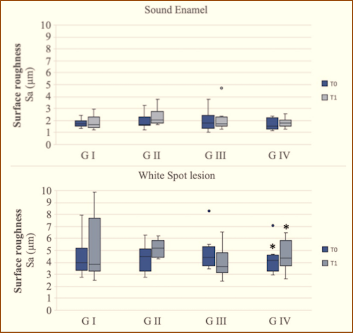 Group I: ClinproTM Prophy Paste (Perlite Particles); Group II: Extra fine pumice; Group III: ClinproTM Prophy Powder (Amino Acid Glycine); and Group IV: Sodium Bicarbonate Jet Powder; T1: After treatment; *Statistical significance.
 Capture images before treatment. A: Group IV: Sodium Bicarbonate Jet Powder prophylaxis; Capture images after treatment. B: Group IV: Sodium Bicarbonate Jet Powder prophylaxis.