Structural Complexity of the Bone Trabecular in Children Exposed to Different Sunlight Conditions: A Cross-Sectional Study
ABSTRACT
Objective: To assess trabecular bone structural complexity in children with different sunlight exposure levels using panoramic radiographs (PR). Fractal dimension (FD) and pixel intensity (PI) were evaluated.
Material and Methods: Panoramic radiographs of 120 children aged 6 to 9 years were analyzed, divided equally into groups with more (n=60) and less (n=60) sunlight exposure. Three regions of interest (ROI) on each radiograph were analyzed using ImageJ software. FD was calculated by the box-counting method, and PI values were measured. Data were compared between exposure groups and genders using Kruskal-Wallis and t-tests (p<0.05).
Results: Children with less sunlight exposure showed significantly higher FD values (3.60 ± 0.29) than those with more exposure (3.31 ± 0.29) (p<0.001), notably in ROI2 and ROI3. Overall, PI values did not differ between exposure groups (p=0.735) or genders (p=0.553).
Conclusion: Lower sunlight exposure in children was associated with increased trabecular bone structural complexity, indicated by higher fractal dimension values. Routine panoramic radiographs analyzed using fractal dimension and pixel-intensity measurements may help dentists detect early trabecular bone changes in children.
Keywords:
Fractals; Radiography; Panoramic; Child; Bone and Bones.
Introduction
Over the years, scientific evidence has shown positive associations between regular exposure to sunlight and health benefits [1], while low exposure to sunlight can be a triggering factor in bone growth damage in children. As the bones of the human skeleton undergo growth, modeling, and remodeling - and these events occur mainly during childhood and adolescence [2,3] - regulating sun exposure at this stage of life can contribute to maintaining vitamin D3 (“sunshine vitamin”), which indirectly acts on the mineralization process, helping to prevent possible bone alterations [4].
Bone quality is closely related to trabecular microarchitecture [5], and changes in the bone matrix alter bone density and texture, which can be detected by imaging [6]. The most common methods for evaluating bone trabeculation include computed tomography (CT), bone densitometry, and dual-energy X-ray absorptiometry (DXA) [7]. However, compared to routine two-dimensional dental exams, these techniques emit higher radiation doses, raising significant concerns, especially in children and adolescents, where the biological effects of ionizing radiation are more pronounced [8,9].
Given that panoramic radiography is the most widely used two-dimensional imaging technique in Dentistry and is often sufficient to support the diagnosis of bone alterations [10,11], it represents a safer and more accessible alternative for the initial assessment of pediatric patients [12]. Therefore, assessing trabecular bone characteristics through structural complexity analysis using panoramic radiographs is very useful [6,13], as it can assist in the detection of systemic and metabolic conditions [6].
Such assessments can be performed using the calculation of the fractal dimension (FD) [13], which provides a numerical descriptor of structural complexity derived from fractal analysis (FA) [14]. The FD quantifies how complex a trabecular pattern is, identifying self-similar structures at different scales [15]. It has been widely applied to detect bone abnormalities and to quantify the severity of skeletal disorders [15]. Pixel intensity (PI), in turn, measures the average grayscale value within a region of interest and reflects the radiographic density of the bone [13]. This parameter is commonly used in medical and dental imaging to indirectly estimate bone mass. Both FD and PI offer complementary information: while FD captures the complexity and architecture of the trabecular network, PI reflects variations in bone radiodensity, which is often related to mineral content. Therefore, analyzing these parameters together enhances the assessment of both qualitative (structural) and quantitative (density) aspects of bone health.
Despite the relevance of these metrics, few studies have jointly evaluated FD and PI, and to the best of our knowledge, none have investigated their relationship with environmental factors, such as sunlight exposure in children. Moreover, previous research [6,7] has primarily focused on adult populations or on pathological conditions such as osteoporosis, without considering pediatric populations under varying sunlight exposure conditions. Since children’s bone metabolism is highly dynamic and susceptible to environmental influences, including sunlight, which affects vitamin D synthesis and, consequently, bone remodeling, there is a critical need to investigate whether differences in sunlight exposure are reflected in trabecular bone structure and density, as visualized on panoramic radiographs.
Therefore, this study aims to answer the following research question: Does the trabecular bone structural complexity and radiographic density, as measured by FD and PI, differ in children exposed to varying levels of sunlight? We hypothesize that reduced sunlight exposure is associated with increased trabecular complexity (higher FD) and potential changes in bone radiodensity (PI), reflecting adaptive or compensatory mechanisms in bone metabolism. The objective is to compare FD and PI values in children living in regions with higher versus lower sunlight exposure, using panoramic radiographs as a low-radiation, accessible diagnostic tool.
Material and Methods
Study Design and Ethical Aspects
This is a cross-sectional study that received prior approval from the local Ethics Committee under protocol #6.180.196. The study adhered to the guidelines for observational studies outlined in the STROBE (Strengthening the Reporting of Observational Studies in Epidemiology) checklist [16].
Sample and Eligibility Criteria
The sample consisted of digital panoramic radiographs of children aged 6 to 9 years old, from two cities with different solar exposure conditions: one located in the coastal region of Rio de Janeiro, RJ, Brazil (high solar exposure) and the other in the Zona da Mata Mineira region, MG, Brazil (low solar exposure). These solar radiation data were obtained from automatic meteorological stations of the National Institute of Meteorology (INMET), available on the institution's website [17]. The aim was to determine whether different levels of sunlight exposure influence trabecular bone structure, as measured by FD and PI values.
All radiographs were obtained using the same device (Orthophos 3D, Dentsply Sirona, Erlangen, Germany) with identical acquisition parameters (64 kVp and 10 mA). Images were included if they presented optimal sharpness, appropriate contrast, and minimal distortion. Radiographs with motion artefacts, positioning errors, intraosseous retained teeth in ectopic positions, fractures, or osteolytic lesions in the mandible were excluded.
It is important to note that sunlight exposure was classified solely based on the city of residence. Individual sunlight exposure habits, including time spent outdoors, sunscreen use, and clothing, were not measured.
Examiner Training and Calibration
A radiologist with expertise in dental radiology trained two examiners using video tutorials demonstrating the procedures for ROI selection and FD calculation using ImageJ® software [18]. Afterwards, in-person clarification was provided.
Twenty panoramic radiographs were used for calibration, and examiners independently measured FD and PI within the same predefined ROIs. Inter-examiner agreement was excellent, with intraclass correlation coefficients (ICC) ranging from 0.84 to 0.98 (ICC=0.984), indicating high reproducibility [19]. These calibration images were not included in the final sample. Inter-examiner agreement was verified during calibration, and the main study was only initiated after this verification (kappa=1). The main analysis of the study was conducted by a single examiner, previously trained and calibrated, as in a previous study [13].
Pilot Study and Sample Calculation
Due to the lack of prior studies for direct reference, a pilot study was conducted using 60 radiographs (30 from each exposure group) to obtain preliminary FD and PI values. ROIs were placed in three anatomically consistent locations: midway between the neck of the mandibular condyle and the mandibular angle, adjacent to the outer cortex of the ramus (ROI1); centered in the mandibular angle (ROI2); near the mandibular base, between the mesial and distal roots of the first permanent molar (ROI3).
Based on pilot data (FD: G1 = 3.36 ± 0.31; G2 = 3.55 ± 0.29; PI: G1 = 244.9 ± 33.1; G2 = 267 ± 54.3), sample size was calculated using BioEstat (version 5.3), assuming 80% power, a 5% significance level, and a two-tailed test. The minimum required was 39 radiographs per group for PI analysis and 60 for FD analysis. The larger number (n = 60 per group) was adopted for consistency across analyses.
Image Analysis
Following calibration, a single examiner performed all measurements in a blinded and randomized manner (i.e., unaware of the radiograph's origin regarding the sunlight exposure group). A 21-inch high-resolution LCD monitor under soft ambient lighting was used. A maximum of five images per day were analyzed to minimize visual fatigue [20].
Three ROIs, each measuring 30 × 30 pixels, were selected based on anatomical landmarks rather than purely random placement, and were always positioned on the right side of the mandible to ensure consistency. The selection was based on established protocols from the literature [21]. While the location of ROIs followed predefined anatomical guidelines, the selection within the defined area was determined manually by the examiner, introducing an element of subjectivity. This approach is consistent with prior studies but is recognized as a methodological limitation.
Fractal Dimension (FD) Analysis
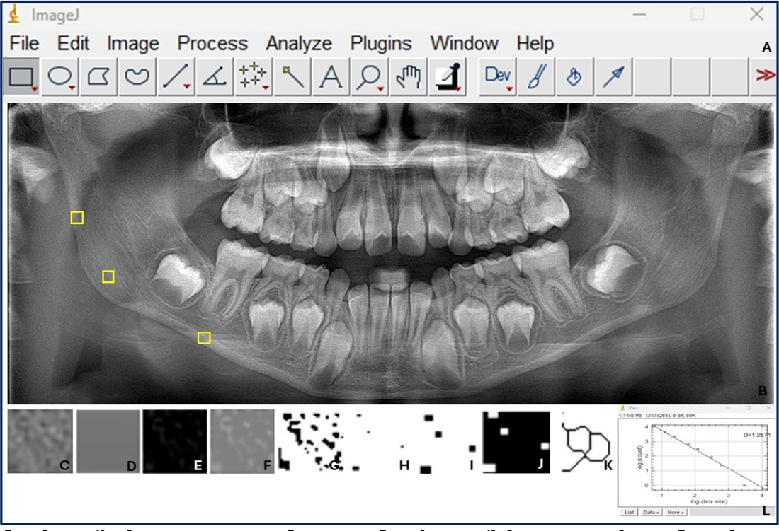
The structural complexity of the bone trabeculae was obtained by calculating the FD in the 3 selected ROIs, for each group of radiographs (G1; n=60 and G2; n=60). A Gaussian filter was applied to the original image (sigma = 35 pixels, kernel size = 33x33), enabling the correction of large variations in density (low-pass filtering) and removing discrepancies between pixels of similar intensity. Then, the resulting image, highly blurred, was subtracted from another duplicate of the original ROI, and a third image was generated, in which gray values (128) were added to each pixel, thus preventing some structures from standing out compared to others. Next, the third image was binarized and subsequently segmented into components that visually approximate the trabeculae. Then, noise reduction was performed, and the image was transformed into a single-pixel line, highlighting the texture and patterns that were subjected to FD analysis [13].
Superimposing the trabecular image onto the original bone image demonstrates that the skeletal structure studied corresponds to the trabeculae in the original image. The FD was calculated using the box-counting method, with square boxes of widths previously established in the literature: 2, 3, 4, 6, 8, 12, 16, 32, and 64 pixels [21] (Figure 1).

Digital analysis of the structural complexity of bone trabeculae by processing regions of interest (ROI) located in the mandible, using the box-counting method to calculate the fractal dimension (FD). (A) ImageJ™ interface; (B) Panoramic radiograph illustrating the selection of ROI; (C) duplication; (D) application of the Gaussian filter; (E) subtraction; (F) addition of 128 gray units; (G) binarization; (H) erosion; (I) dilation; (J) inversion; (K) skeletonization; (L) box counting procedure.
Statistical Analysis
The results were analyzed using the Statistical Package for the Social Sciences (SPSS for Windows, version 21.0; SPSS Inc., Chicago, IL, USA). Descriptive and inferential analyses were performed. The Shapiro-Wilk test was used to verify the normality of the data, for both the total PI and FD (sum of ROI1, ROI2, and ROI3) and the individual means. To verify differences in total and individual image values between children less and more exposed to the sun, as well as between genders, the Kruskal-Wallis and Student's t-tests were used. A significance level of 5% was considered (p<0.05).
Pixel Intensity Analysis
The PI values were also calculated for each of the established ROIs, using the program's own “Histogram” tool. When calculating the PI, average, minimum, and maximum values were obtained, in addition to the specific histogram for each region, highlighting the threshold (interval between grayscale values) selected (Figure 2).

Steps in the process of obtaining pixel intensity (PI). (A) ImageJ™ interface showing the “histogram” analysis tool; (B) panoramic radiograph illustrating the selection of a ROI; (C) histogram generated from the selected ROI, showing the mean and standard deviation values, as well as the minimum and maximum pixel intensity values.
Results
Sample Description
The images included in the study belonged to children with an average age of 7.39 ± 1.11 years. Of the sample (n=120), 62 x-rays were from girls (51.7%), while 58 were from boys (48.3%). Furthermore, the following total DF and PI values were observed: 3.45 ± 0.32 and 258.30 ± 58.38, respectively.
Comparison of Fractal Dimension Between Groups, Considering Sun Exposure and Sex
When analyzing the sum of the average FD values of the three ROIs, there was a significant difference between the children most exposed (G1=3.31±0.29) and those least exposed to the sun (G2=3.60±0.29) (p=0.000). When analyzing each ROI separately, a significant difference was found concerning the sum of the mean values of ROI2 (p=0.000) and ROI3 (p=0.000) regarding sun exposure, as shown in Table 1.
Total fractal dimension (FD) values of regions of interest (ROI), presented in panoramic radiographs of children most exposed (G1) and least exposed (G2) to sunlight.
Regarding gender, considering the total sample, no significant difference was observed between FD values between girls (3.48±0.28) and boys (3.43±0.36) (p=0.607), and adjusting for sun exposure, there was also no difference between girls (n=32; 3.33±0.25) and more exposed boys (n=28; 3.28±0.34) (p=0.491); as well as girls (n=30; 3.63±0.24) and less exposed boys (n=30; 3.77±0.33) (p=0.408).
Comparison of Pixel Intensity Between Groups, Considering Sun Exposure and Sex
There was no difference between the total PI values between children (G1=261.26±69.54) and those with less exposure to sunlight (G2=255.33±44.95) (p=0.735). However, when analyzing each ROI separately, there was a significant difference in ROI2 between the most exposed (G1=78.30±65.20) and the least exposed (G2=62.76±20.48) (p=0.013), as shown in Table 2.
Total pixel intensity (PI) values of regions of interest (ROI), analyzed in panoramic radiographs of children, most exposed (G1) and least exposed (G2) to sunlight.
As with FD, the total PI values (p=0.553) of the total sample were not influenced by gender, with no difference being observed between girls (257.88±70.12) and boys (258.74±43.05). When sun exposure was considered, there was also no difference between girls (n=32; 269.82±88.83) and more exposed boys (n=28; 251.48±36.33) (p=0.291) or girls (n=30; 245.15±39.76) and less exposed boys (n=30; 265.51±48.12) (p=0.079).
Discussion
This study compared the structural complexity of bone trabeculae in children residing in areas with different levels of sunlight exposure using FD (fractal dimension) and PI (pixel intensity) values derived from digital panoramic radiographs. While the use of FD and PI to assess trabecular bone has been established primarily in adult populations, cadavers, and animals [13,22], this study is among the first to apply these methods to a pediatric population in the context of environmental factors, such as sunlight exposure. This approach is particularly relevant since sunlight stimulates vitamin D3 synthesis, a micronutrient crucial for bone metabolism during growth.
A review of the literature revealed that no studies have assessed trabecular bone complexity in children based on sunlight exposure using panoramic radiography. Most existing studies have employed more advanced imaging techniques, such as computed tomography (CT), dual-energy X-ray absorptiometry (DXA), or bone densitometry, and have typically focused on skeletal regions, such as the vertebrae or the forearm [23,24]. Although one study employing panoramic radiography was identified, it assessed different parameters in adult patients, including fractal dimension (FD), panoramic mandibular index (PMI), mandibular cortical index (MCI), and mandibular cortical thickness (MCT). However, that study did not specifically focus on fractal dimension or pixel intensity (PI) analyses [6].
Panoramic radiography offers several advantages: it is widely available, cost-effective, easy to perform, and emits significantly lower radiation compared to three-dimensional imaging methods, an essential consideration for pediatric populations [25]. These factors, combined with the absence of prior studies evaluating FD in association with PI in children, justified the choice of these analyses in the present study. The methodological strengths include non-invasiveness, accessibility, low radiation exposure, and the use of rapid, reproducible methods implemented with open-source software. In contrast, the use of PI complements FD, as combining these two values allows differentiation regarding whether denser bone does or does not exhibit greater complexity in its trabecular structure.
A significant finding was that children with less sunlight exposure exhibited higher FD values. While higher FD is generally interpreted as indicative of greater trabecular complexity [26], this does not always translate into better bone quality. Previous studies have shown that regions affected by pathological bone remodeling, such as osteosarcoma, bone healing after surgery, and sites of implant osseointegration, often exhibit elevated FD values despite compromised bone integrity [27,28]. Therefore, increased FD in this context may reflect disorganized or altered trabecular patterns resulting from suboptimal bone metabolism, potentially related to lower vitamin D levels. This explains why the relationship between FD and bone health is not strictly linear and why a higher FD is not always beneficial. Calculating PI values complements the clinical interpretation, as regions with lower PI values (lower bone density) and higher PI values (higher bone density) may exhibit similarly low FD values. This indicates that, despite differing densities, these regions may be structurally comparable in terms of complexity.
Similarly, our PI findings, which are often used as a proxy for bone density, showed no overall difference between groups but revealed localized reductions in the mandibular angle (ROI2) in less sun-exposed children. This supports the interpretation that while trabecular complexity may increase (reflected in FD), bone mineral density does not necessarily follow the same trend, indicating a decoupling between architectural complexity and mineral content in response to environmental stressors like reduced sunlight.
A recent study evaluated potential changes in the jawbone in children and adolescents with Rheumatic Heart Disease [29]. However, no studies were found that used this test to assess children without previous disease, to detect bone changes. The focus on a pediatric population during a critical period of bone development addresses a significant gap in the literature related to bone health in children and its public health implications. Furthermore, adopting standardized regions of interest (ROI) selection and rigorous examiner calibration procedures enhances the methodological reliability and reproducibility of the findings.
Despite its contributions, this study has several limitations that should be acknowledged. Sunlight exposure was determined solely by participants' city of residence, without accounting for individual habits such as time spent outdoors, sunscreen use, or clothing, which could influence actual exposure. Additionally, no biochemical markers, such as serum vitamin D or calcium levels, were measured, limiting the ability to directly correlate radiographic findings with bone metabolism. FD and PI analyses were performed by a single examiner; although examiner calibration demonstrated high reliability, inter-observer reproducibility during the primary analysis was not assessed. The inherent technical limitations of panoramic radiographs, including image distortion, magnification, and overlapping anatomical structures, may also affect measurement accuracy. Furthermore, although ROI placement was guided by standardized anatomical landmarks, a degree of subjectivity remains, which could impact the consistency and reproducibility of the results.
Future research should incorporate direct measurement of biochemical markers, such as serum vitamin D, calcium, and other indicators of bone metabolism, to strengthen correlations between imaging findings and biological status. Longitudinal studies are recommended to monitor changes in trabecular bone over time, considering variables such as seasonal solar variation, growth stages, and dietary factors. Larger multicenter studies that consider behavioral factors that influence sun exposure, including outdoor activity levels and clothing habits, are also needed to refine exposure assessment. Additionally, the tools used in the present study can be combined with other methodologies, as they help to measure objective data in a simple, fast, and cost-effective way.
This pioneering study highlights that FD and PI analysis applied to routine panoramic radiographs can detect variations in trabecular bone related to differences in sunlight exposure in children. While the findings suggest that lower sunlight exposure may lead to increased trabecular complexity (higher FD) and localized reductions in density (lower PI), these patterns should be interpreted with caution, given limitations in individual exposure assessment and the absence of biochemical data. Nonetheless, FD and PI represent promising, non-invasive, and accessible tools for preliminary bone health assessment in pediatric dental practice, opening new avenues for research at the intersection of dental imaging and systemic bone health.
Conclusion
Children with lower sun exposure had higher fractal dimension values, indicating changes in trabecular bone complexity, while no differences were observed in total pixel intensity values or between sexes. These findings suggest a potential association between reduced sun exposure and changes in bone microarchitecture. However, clinical interpretation should be made with caution, as fractal dimension reflects structural complexity rather than directly indicating bone strength. Analysis of fractal dimension and pixel intensity using panoramic radiographs may serve as complementary tools for preliminary bone assessment in pediatric populations.
-
Financial Support
None.
Data Availability
The data used to support the findings of this study can be made available upon request to the corresponding author.
References
-
[1] Rhee HVD, Vries E, Coomans C, Velde PVD, Coebergh J. Sunlight: For better or for worse? A review of positive and negative effects of sun exposure. Cancer Res Front 2016; 2:156-183. https://doi.org/10.17980/2016.156
» https://doi.org/10.17980/2016.156 -
[2] Weller RB. Sunlight: Time for a rethink? J Invest Dermatol 2024; 144(8):1724-1732. https://doi.org/10.1016/j.jid.2023.12.027
» https://doi.org/10.1016/j.jid.2023.12.027 -
[3] Taylor SN. Vitamin D in toddlers, preschool children, and adolescents. Ann Nutr Metab 2020; 76:30-41. https://doi.org/10.1159/000505635
» https://doi.org/10.1159/000505635 -
[4] Holick MF. Vitamin D and bone health: What vitamin D can and cannot do. Adv Food Nutr Res 2024; 109:43-66. https://doi.org/10.1016/bs.afnr.2024.04.002
» https://doi.org/10.1016/bs.afnr.2024.04.002 -
[5] Vadiati Saberi B, Khosravifard N, Nooshmand K, Dalili Kajan Z, Ghaffari ME. Fractal analysis of the trabecular bone pattern in the presence/absence of metal artifact-producing objects: Comparison of cone-beam computed tomography with panoramic and periapical radiography. Dentomaxillofac Radiol 2021; 50(6):20200559. https://doi.org/10.1259/dmfr.20200559
» https://doi.org/10.1259/dmfr.20200559 -
[6] Zihni Korkmaz M, Yemenoğlu H, Günaçar DN, Ustaoğlu G, Ateş Yildirim E. The effects of vitamin D deficiency on mandibular bone structure: A retrospective radiological study. Oral Radiol 2023; 39(1):67-74. https://doi.org/10.1007/s11282-022-00602-5
» https://doi.org/10.1007/s11282-022-00602-5 -
[7] Burt LA, Billington EO, Rose MS, Raymond DA, Hanley DA, Boyd SK. Effect of high-dose vitamin D supplementation on volumetric bone density and bone strength: A randomized clinical trial. JAM 2019; 322(8):736-745. https://doi.org/10.1001/jama.2019.11889 Erratum in: JAMA 2019; 322(19):1925. https://doi.org/10.1001/jama.2019.17910
» https://doi.org/10.1001/jama.2019.11889» https://doi.org/10.1001/jama.2019.17910 -
[8] Tomà P, Bartoloni A, Salerno S, Granata C, Cannatà V, Magistrelli A, et al. Protecting sensitive patient groups from imaging using ionizing radiation: Effects during pregnancy, in fetal life and childhood. Radiol Med 2019; 124(8):736-744. https://doi.org/10.1007/s11547-019-01034-8
» https://doi.org/10.1007/s11547-019-01034-8 -
[9] Abalo KD, Rage E, Leuraud K, Richardson DB, Le Pointe HD, Laurier D, et al. Early life ionizing radiation exposure and cancer risks: Systematic review and meta-analysis. Pediatr Radiol 2021; 51(1):45-56. https://doi.org/10.1007/s00247-020-04803-0 Erratum in: Pediatr Radiol 2021; 51(1):157-158. https://doi.org/10.1007/s00247-020-04883-y
» https://doi.org/10.1007/s00247-020-04803-0» https://doi.org/10.1007/s00247-020-04883-y -
[10] International Commission on Radiological Protection. The 2007 Recommendations of the International Commission on Radiological Protection. ICRP publication 103. Ann ICRP 2007; 37(2-4):1-332. https://doi.org/10.1016/j.icrp.2007.10.003
» https://doi.org/10.1016/j.icrp.2007.10.003 -
[11] Sams CM, Dietsche EW, Swenson DW, DuPont GJ, Ayyala RS. Pediatric panoramic radiography: Techniques, artifacts, and interpretation. Radiographics 2021; 41(2):595-608. https://doi.org/10.1148/rg.2021200112
» https://doi.org/10.1148/rg.2021200112 -
[12] Mupparapu M, Akintoye SO. Application of panoramic radiography in the detection of osteopenia and osteoporosis-current state of the art. Curr Osteoporos Rep 2023; 21(4):354-359. https://doi.org/10.1007/s11914-023-00807-5
» https://doi.org/10.1007/s11914-023-00807-5 -
[13] Arrepia BF, Rocha TG, Medeiros AS, Ferreira MD, Fonseca-Gonçalves A, Visconti MA. The mandibular bone structure in children by fractal dimension and its correlation with pixel intensity values: A pilot study. Oral Radiol 2023; 39(4):771-778. https://doi.org/10.1007/s11282-023-00693-8
» https://doi.org/10.1007/s11282-023-00693-8 -
[14] Kato CN, Barra SG, Tavares NP, Amaral TM, Brasileiro CB, Mesquita RA, et al. Use of fractal analysis in dental images: A systematic review. Dentomaxillofac Radiol 2020; 49(2):20180457. https://doi.org/10.1259/dmfr.20180457
» https://doi.org/10.1259/dmfr.20180457 -
[15] Apolinário AC, Sindeaux R, de Souza Figueiredo PT, Guimarães AT, Acevedo AC, Castro LC, et al. Dental panoramic indices and fractal dimension measurements in osteogenesis imperfecta children under pamidronate treatment. Dentomaxillofac Radiol 2016; 45(4):20150400. https://doi.org/10.1259/dmfr.20150400
» https://doi.org/10.1259/dmfr.20150400 -
[16] Cuschieri S. The STROBE guidelines. Saudi J Anaesth 2019; 13:S31-S34. https://doi.org/10.4103/sja.SJA_543_18
» https://doi.org/10.4103/sja.SJA_543_18 -
[17] Instituto Nacional de Meteorologia. Available from: https://portal.inmet.gov.br/ [Accessed on May 26, 2025]. [In Portuguese].
» https://portal.inmet.gov.br/ -
[18] Schneider CA, Rasband WS, Eliceiri KW. NIH Image to ImageJ: 25 years of image analysis. Nat Methods 2012; 9(7):671-675. https://doi.org/10.1038/nmeth.2089
» https://doi.org/10.1038/nmeth.2089 - [19] Landis JR, Koch GG. The measurement of observer agreement for categorical data. Biometrics 1977; 33(1):159-174.
-
[20] Rosado LPL, Barbosa IS, Junqueira RB, Martins APVB, Verner FS. Morphometric analysis of the mandibular fossa in dentate and edentulous patients: A cone beam computed tomography study. J Prosthet Dent 2021; 125(5):758.e1-758.e7. https://doi.org/10.1016/j.prosdent.2021.01.014
» https://doi.org/10.1016/j.prosdent.2021.01.014 -
[21] White SC, Rudolph DJ. Alterations of the trabecular pattern of the jaws in patients with osteoporosis. Oral Surg Oral Med Oral Pathol Oral Radiol Endod 1999; 88(5):628-635. https://doi.org/10.1016/s1079-2104(99)70097-1
» https://doi.org/10.1016/s1079-2104(99)70097-1 -
[22] Soğur E, Baksı BG, Gröndahl HG, Sen BH. Pixel intensity and fractal dimension of periapical lesions visually indiscernible in radiographs. J Endod 2013; 39(1):16-19. https://doi.org/10.1016/j.joen.2012.10.016
» https://doi.org/10.1016/j.joen.2012.10.016 -
[23] Cheung TF, Cheuk KY, Yu FW, Hung VW, Ho CS, Zhu TY, et al. Prevalence of vitamin D insufficiency among adolescents and its correlation with bone parameters using high-resolution peripheral quantitative computed tomography. Osteoporos Int 2016; 27(8):2477-2488. https://doi.org/10.1007/s00198-016-3552-4
» https://doi.org/10.1007/s00198-016-3552-4 -
[24] Alghadir AH, Gabr SA, Rizk AA. Physical fitness, adiposity, and diets as surrogate measures of bone health in schoolchildren: A biochemical and cross-sectional survey analysis. J Clin Densitom 2018; 21(3):406-419. https://doi.org/10.1016/j.jocd.2017.12.006
» https://doi.org/10.1016/j.jocd.2017.12.006 -
[25] Choi YJ. Dual-energy x-ray absorptiometry: Beyond bone mineral density determination. Endocrinol Metab 2016; 31(1):25-30. https://doi.org/10.3803/EnM.2016.31.1.25
» https://doi.org/10.3803/EnM.2016.31.1.25 -
[26] Hichijo N, Tanaka E, Kawai N, van Ruijven LJ, Langenbach GE. Effects of decreased occlusal loading during growth on the mandibular bone characteristics. PLoS One 2015; 10(6):e0129290. https://doi.org/10.1371/journal.pone.0129290
» https://doi.org/10.1371/journal.pone.0129290 -
[27] Jung JH, Huh KH, Yong TH, Kang JH, Kim JE, Yi WJ, et al. Differentiation of osteosarcoma from osteomyelitis using microarchitectural analysis on panoramic radiographs. Sci Rep 2022; 12(1):12339. https://doi.org/10.1038/s41598-022-16504-9
» https://doi.org/10.1038/s41598-022-16504-9 -
[28] Kış HC, Güleryüz Gürbulak A. Evaluation of the peri-implant bone trabecular microstructure changes in short implants with fractal analysis. Int J Implant Dent 2020; 6(1):13. https://doi.org/10.1186/s40729-020-00209-7
» https://doi.org/10.1186/s40729-020-00209-7 -
[29] Temur KT, Magat G, Cosgunarslan A, Ozcan S. Evaluation of jaw bone change in children and adolescents with rheumatic heart disease by fractal analysis. Niger J Clin Pract 2024, 27(2):260-267. https://doi.org/10.4103/njcp.njcp_346_23
» https://doi.org/10.4103/njcp.njcp_346_23
Edited by
-
Academic Editor:
Alessandro Leite Cavalcanti