
 Osseointegration of Surface Coated and Surface Uncoated Titanium Dental Implants: A Systematic Review and Meta-Analysis
ABSTRACT
Objective: To compare the osseointegration of surface-coated and uncoated titanium dental implants.
Material and Methods: PubMed, Cochrane Library, SCOPUS, Web of Science, Google Scholar, and trial registries with no language or date of publication restrictions were searched on electronic databases. The risk of bias was assessed using the Revised Cochrane Risk of Bias Tool for Randomized Trials (RoB-2) and the Risk of Bias in Non-randomized Studies - of Interventions (ROBINS - I) tool. Data were pooled for bone-implant contact percentage (BIC%) of the included studies and forest plots were produced accordingly. A funnel plot was generated to detect the publication bias and GRADEpro was used to assess the certainty of evidence.
Results: Four studies with full reports satisfying the eligibility criteria were included. Three studies had a moderate risk and one study had a high risk. The BIC% at 6 months after implant placement was high in surface-coated titanium dental implants with a mean difference of 16.66% (p=0.0005) and a 95% confidence interval (CI) of 7.28% to 26.03% and heterogeneity of I2 of 0% compared to uncoated titanium dental implants.
Conclusion: With moderate methodological risk of bias, certainty of evidence, and low heterogeneity, we suggest that surface-coated titanium dental implants can be used to increase osseointegration and implant survival rate.
Keywords:
Titanium; Dental Implants; Osseointegration; Bone-Implant Interface.
Introduction
The original work of Branemark and his coworkers on dental implants has been a reliable treatment modality for restoring edentulous jaws [1,2]. Titanium, for its good biocompatibility, osseointegration, mechanical, anti-bacterial, and corrosion resistance properties, is the commonly used dental implant material [3]. However, poor bone quality is the reason for high rates of early implant failure [4,5]. Surface modifications of titanium implants are the only solution to this clinical problem. These modifications will improve osseointegration [6], reduce the rate of early implant failure, and ensure increased survival of implants [7].
Rough, acid-etched, sandblasted, and bioactive material-coated implant surfaces have enhanced osseointegration compared to smooth implant surfaces, due to their increased wettability with bone [8-10]. These surface modifications are achieved either by additive or subtractive methods. In the additive process, materials are added to the superficial surface or integrated into the surface, called a surface coating or a surface impregnation [11,12]. In the impregnation method, the chemical agent, such as calcium phosphate crystals, will be fully integrated into the titanium core. In the surface coating, either the materials or the agents of varying thickness will be added to the surface of the core material [11,12]. On the other hand, the subtractive technique involves the removal of a layer from the core material or deform the superficial layer [11,12]. The common additive techniques include plasma-sprayed hydroxyapatite (HA) coating, titanium plasma spraying (TPS), alumina coating, and biomimetic calcium phosphate (CaP) coating. Similarly, large-grit sanding, ceramic particle blasting, acid etch, and anodization are common subtractive techniques [11,12].
Physical and chemical treatments of titanium alloys are performed to improve aesthetic performance, alter surface roughness, modify composition, and increase wettability and surface energy [13]. Further, ion deposition, laser treatment, and sputtering are also carried out to improve osseointegration [14-16]. With this background on the necessity of surface-modified titanium dental implants, the present systematic review aims to compare the osseointegration of surface-coated and uncoated titanium dental implants.
Material and Methods
Protocol and Eligibility Criteria
This systematic review was registered in PROSPERO with registered number CRD420245883176 and followed the Preferred Reporting Items for Systematic Review and Meta-analysis (PRISMA) 2020 guidelines in reporting this systematic review and meta-analysis [17]. All prospective human trials, such as randomized controlled trials, clinical trials, non-randomized trials, and split-mouth trials, were included. Partially or completely edentulous patients who need implant placement between the ages of 30 and 70 years were considered. Surface-coated titanium dental implants were compared with surface-uncoated titanium dental implants as interventions, using the PICO criteria. Bone-implant contact percentage (BIC%) and bone area density percentage (BA%) were the outcomes measured for this systematic review.
Sources and Search Strategy
We searched PubMed, Cochrane Library, SCOPUS, Web of Science, Google Scholar, and trial registries, with no language or publication date restrictions. The search terms and MeSH terms used for this search, following the PICO criteria, are given in Table 1. A hand search in all implantology and periodontology journals was made with the help of a librarian.
Study Selection
The RAYYAN software, an artificial intelligence tool for systematic review, was used to remove duplicate studies from different databases. Studies not suitable for this review were excluded after screening for title and abstract by the reviewers (KKP and AKS reviewers) independently. The full text of the remaining studies was subjected to eligibility criteria and eliminated after discussion with reviewers. Any disagreement in study selection between the reviewers was solved by the third reviewer.
Data Extraction
The following data were extracted and recorded for each included trial by the 1st and 2nd reviewers individually on a pilot-tested data extraction sheet: 1. Author, year of publication, country where the trial was carried out, type of study (randomized or non-randomized); 2. Participants demographic details and criteria for inclusion; 3. Intervention (surface-coated and surface-uncoated) type and relevant details; and 4. Outcome details, follow-up details. Any conflicts between the reviewers in data extraction were resolved by the third reviewer through discussion. Corresponding authors were reached out to for clarification or missing details. Data lacking the necessary additional information were omitted.
Assessment of Risk of Bias
The risk of bias was assessed by the reviewers independently. The reviewers were not blinded to the authors of the included studies. The Revised Cochrane Risk of Bias Tool for Randomized Trials (RoB-2) with five domains [18] was used to analyze the randomized studies. The RoB-2 tool was used to assess the included studies across the following five domains: 1. Bias arising from the randomization process; 2. Bias due to deviations from intended interventions; 3. Bias resulting from missing outcome data; 4. Bias in outcome measurement; and 5. Bias in the selection of reported results
Each of these domains comprised two to three subdomains, assessed through signaling questions. The responses to these questions guided the overall risk of bias judgment for each study as follows:
-
Low risk of bias: when all subdomains were rated as “low risk”.
-
Some concerns: when at least one subdomain was rated as “some concerns”.
-
High risk of bias: when one or more subdomains were rated as “high risk,” or when more than two domains were rated as “some concerns”.
Risk of Bias in Non-randomized Studies - of Interventions (ROBINS -I) tool with seven domains was used to analyze non-randomized studies [19]. Each of the seven domains contained multiple signaling questions that required responses, which collectively informed the overall risk of bias judgment. The seven domains assessed were: 1. Bias due to confounding; 2. Bias in the selection of participants into the study; 3. Bias in the classification of interventions; 4. Bias due to deviations from intended interventions; 5. Bias resulting from missing data; 6. Bias in the measurement of outcomes; and 7. Bias in the selection of reported results
The response options for the signaling questions were: Yes, probably yes, No, probably no, and No information. Certain signaling questions were contingent on the responses to earlier questions, being answered only if the preceding response was Yes/Probably yes or No/Probably no.
Based on these responses, the overall risk of bias for each study was categorized as follows:
-
Low risk of bias: studies comparable to a well-conducted randomized trial.
-
Moderate risk of bias: studies methodologically sound for a non-randomized trial but not comparable to a high-quality randomized trial.
-
Serious risk of bias: studies with notable concerns in one or more domains.
-
Critical risk of bias: studies with substantial methodological flaws, rendering them uninformative regarding the effects of the intervention.
-
No information: Studies lacking sufficient data to permit a risk of bias judgment.
Data Synthesis
The mean bone-implant contact percentages were pooled and summarised as mean differences to represent the effect size of the interventions. The weighted mean difference using the inverse variance of DerSimonian and Laird (DL) with a random effect model was used. An I2 test for heterogeneity was used to assess the discrepancies across the studies. An I2 > 40% was considered as high heterogeneity. A funnel plot was used to assess publication bias across the included studies.
Quality of Evidence Assessment
The grading of Recommendations Assessment, Development, and Evaluation (GRADE) approach was used to assess the quality of evidence from the meta-analysis. GRADEpro GDT software was used with results as very low, low, moderate, or high quality of evidence [20].
Results
Study Search and Study Selection
In the initial search across PubMed, Cochrane Library, SCOPUS, Web of Science, Google Scholar, hand searches, and trial registries, we identified 434 studies. About 29 studies were screened for titles and abstracts, resulting in the removal of 405 duplicates. A total of 16 studies, not suitable for the systematic review, were excluded. Of the remaining 13 studies, full reports of three studies were not retrieved after contacting the authors. So, 10 studies with full reports were assessed for eligibility criteria (Figure 1). About five reviews and one study comparing two different titanium implant systems were excluded, as given in Table 2 [21-26]. Thus, full reports of 4 studies were included in the qualitative synthesis [27-30], and 2 studies were included in the quantitative synthesis [28,29].

Characteristics of the Included Studies
The characteristics of the four included studies were summarized in Table 3. About 107 patients with a mean age of 53.75 years were evaluated for osseointegration using surface-coated and uncoated titanium dental implants. About 59 females and 48 males had participated in the trials. Three studies used a non-randomized [27,29,30] and one study used a randomized controlled study design [28]. Two studies [27,30] included partially edentulous patients, one study [29] included completely edentulous patients, and one study included both partially and completely edentulous patients [28]. Three studies placed implants in the posterior regions [27-29], and one study placed them in the anterior region of the maxilla and mandible [30]. One study added a 10µm oxide layer to the screw-type turned titanium dental implant [27], one other study added nano-sized calcium phosphate particles on the dual-acid etched surface [28], another study impregnated bioceramic molecules on dual acid-etched titanium dental implants [29], and the other study coated hydroxyapatite, and bioactive glass on the titanium dental implants [30]. All studies assessed osseointegration regarding bone-implant contact percentage [27-29], except one study [30], which assessed mesial and distal interface radiodensity.
Risk of Bias Assessment
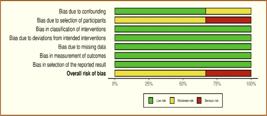
The risk of bias for one study [28] assessed using the Revised Cochrane Risk of Bias Tool for Randomized Trials (RoB-2) is presented in Figure 2. The results reported some concerns arising from the randomization process. Similarly, the risk of bias assessed using the Risk of Bias in Non-randomized Studies - of Interventions (ROBINS - I) tool is presented in Figures 3 and 4. Two non-randomized studies [27,29] showed some concerns, and one study [30] showed serious risk of bias in the selection of participants. The summary of ROBINS - I assessment showed a 35% moderate risk of bias due to confounding, 65% moderate, and 35% serious risk of bias due to the selection of participants.


Meta-Analysis
Two studies that assessed the mean bone-implant contact percentage (BIC%) histomorphometrically at 6 months after implant placement were included for meta-analysis [28,29]. The results of the forest plot favored surface-coated titanium dental implants with a significantly high mean difference in BIC% of 16.66% (p=0.0005), 95% confidence interval (CI) of 7.28% to 26.03%, with heterogeneity of I2 =0% between them, as presented in Figure 5.

Forest plot comparing BIC% among surface-coated and surface-uncoated titanium dental implants.
Publication Bias
The funnel plot of the comparison of BIC% between surface-coated and uncoated titanium surfaces revealed a strongly suspected publication bias with a high standard error in the sample selection from the actual population, as shown in Figure 6.

Funnel plot comparing BIC% among surface-coated and surface-uncoated titanium dental implants.
Certainty of Evidence
There was a moderate level of certainty of evidence with a moderate risk of bias and publication bias in the meta-analysis of BIC% of surface-coated and uncoated titanium dental implants is given in Table 4.
Discussion
The four studies [27-30] with a moderate level of risk of bias in this review reported a high bone-implant contact percentage (BIC%), bone area percentage (BA%), and marginal interface radiodensity in histomorphometry with surface-coated titanium dental implants compared to uncoated titanium dental implants.
One study that compared a 10µm oxide layer added to conventional titanium dental implants showed significantly more bone-implant contact (BIC%) and bone inside the threaded area in oxidized implants [27]. The oxidized implants in this study were subjected to normal clinical situations such as chewing, risk of infection, and load from the denture [27]. On exposure to normal clinical situations, oxidized implants exhibited stronger bone reaction, which may be attributed to the thicker oxide layer, and changes in the size and distribution of pores, which is lacking in turned titanium dental implants [27]. The change in the amorphous oxide crystallinity to anatase and rutile forms creates a stronger bone reaction in increased oxide thickness [31,32].
The mean BIC% and interfacial strengths are high in discrete crystalline deposition of nano-sized calcium phosphate particles in dual acid-etched titanium dental implants [28]. It was found that bone was adapted closely to the implant threads, satisfying the principle of contact osteogenesis [33]. Also, there was a stereographic structure of the bone-implant interface throughout the implant. Whereas the dual acid-etched implants without any coating showed osteogenesis only in the central areas of the implant threads [28].
Shibli et al. demonstrated increased BIC% and Oi index in bioceramic molecular impregnation compared to dual acid-etched dental implants [29]. The CaPO4 molecule was integrated into the titanium oxide surface in the bioceramic dental implants. It has been proposed that CaP-coated surfaces increase the platelets, providing a stimulus to accelerate early bone healing [34]. A large portion of the bioceramic implant threads was covered with a thin, newly formed bone showing contact osteogenesis [35,36]. Also, it has been found that physicochemical interactions between molecules and cells in peri-implant areas influence protein adsorption, proliferation, and bone tissue deposition [37].
Another research showed the least marginal bone loss, high marginal interface density, and less interfacial gaps in Hydroxyapatite (HAp) and bioactive glass powders (BAG) added to titanium dental implants [30]. HAp and BAG powders were applied by air microplasma spraying technique, which enables moderately high bonding strength with minimal structural change in composition and crystallinity [30]. Increased blood supply around the implants with firm clot adherence on the HAp and BAG-coated implant surface suggests increased wettability compared to machined implants [38]. Also, a preclinical study showed an improved osseointegration on the addition of rhBMP-7 to nano HA-coated implants [39]. However, implants coated with liquid platelet-rich fibrin in a split-mouth trial showed no significantly improved osseointegration compared to the controls [40]. A narrative review that assessed the osseointegration of anti-sclerotin coated dental implants found accelerated bone remodeling and new bone formation [41].
This systematic review is the implication of four trials with a moderate level of risk of bias in methodological quality and certainty of evidence. The data analysis with no heterogeneity (I2 = 0%) suggests that surface-coated titanium dental implants exhibit high BIC%. However, the meager sample size in the included studies suggests a strong suspicion of publication bias with high standard error. The differences in the protocols across the studies make the comparison a complicated one.
To the best of our knowledge, this is the first systematic review that aims to compare the osseointegration of surface-coated and uncoated titanium dental implants. We made an extensive search in electronic databases, hand searches, and trial registries. Also, we have followed the PRISMA 2020 guidelines for transparent reporting of systematic review and meta-analysis. The limitation is that only a few trials with meager sample sizes have been included in this review. Further trials with a larger sample size, considering the confounders and following the CONSORT guidelines, should be directed to strengthen the evidence.
Conclusion
With moderate methodological risk of bias, certainty of evidence, and strong publication bias, we suggest that surface-coated titanium dental implants with two-fold increased osseointegration than uncoated dental implants can be used to increase their survival rate. Hope that our results will guide researchers to carry out more randomized controlled trials with rigorous protocols to assess the osseointegration of additive and subtractive titanium surface coatings.
-
Financial Support
None.
Data Availability
The data used to support the findings of this study can be made available upon request to the corresponding author.
References
- [1] van Steenberghe D, Quirynen M, Naert I. Survival and success rates with oral endosseous implants. In: Lang NP, Karring T, Lindhe J, eds. Proceedings of the 3rd European workshop on periodontology. Berlin, Germany: Quintessence Books; 1999:242-254.
- [2] Naert I, Alsaadi G, Quirynen M. Prosthetic aspects and patient satisfaction with two-implant-retained mandibular overdentures: A 10-year randomized clinical study. Int J Prosthodont 2004; 17(4):401-410.
-
[3] Pandey C, Rokaya D, Bhattarai BP. Contemporary concepts in osseointegration of dental implants: A review. BioMed Res Int 2022; 2022:6170452. https://doi.org/10.1155/2022/6170452
» https://doi.org/10.1155/2022/6170452 -
[4] Kochar SP, Reche A, Paul P. The etiology and management of dental implant failure: A review. Cureus 2022; 14(10):e30455. https://doi.org/10.7759/cureus.30455
» https://doi.org/10.7759/cureus.30455 -
[5] Gehrke SA, Cortellari GC, de Oliveira Fernandes GV, Scarano A, Martins RG, Cançado RM, et al. Randomized clinical trial comparing insertion torque and implant stability of two different implant macrogeometries in the initial periods of osseointegration. Medicina 2023; 59(1):168. https://doi.org/10.3390/medicina59010168
» https://doi.org/10.3390/medicina59010168 -
[6] Remísio MJDS, Borges T, Castro F, Gehrke SA, Fernandes JCH, Fernandes GVO. Histologic osseointegration level comparing titanium and zirconia dental implants: Meta-analysis of preclinical studies. Int J Oral Maxillofac Implants 2023; 38(4):667-680. https://doi.org/10.11607/jomi.10142
» https://doi.org/10.11607/jomi.10142 -
[7] Kokubo T, Pattanayak DK, Yamaguchi S, Takadama H, Matsushita T, Kawai T, et al. Positively charged bioactive Ti metal prepared by simple chemical and heat treatments. J R Soc Interface 2010; 7 Suppl 5(Suppl 5):S503-513. https://doi.org/10.1098/rsif.2010.0129.focus
» https://doi.org/10.1098/rsif.2010.0129.focus -
[8] Gittens RA, Scheideler L, Rupp F, Hyzy SL, Geis-Gerstorfer J, Schwartz Z, et al. A review on the wettability of dental implant surfaces II: Biological and clinical aspects. Acta Biomater 2014; 10(7):2907-2918. https://doi.org/10.1016/j.actbio.2014.03.032
» https://doi.org/10.1016/j.actbio.2014.03.032 -
[9] Albertini M, Fernandez-Yague M, Lázaro P, Herrero-Climent M, Rios-Santos JV, Bullon P, et al. Advances in surfaces and osseointegration in implantology. Biomimetic surfaces. Med Oral Patol Oral Cir Bucal 2015; 20(3):e316-325. https://doi.org/10.4317/medoral.20353
» https://doi.org/10.4317/medoral.20353 -
[10] Eliaz N, Shmueli S, Shur I, Benayahu D, Aronov D, Rosenman G. The effect of surface treatment on the surface texture and contact angle of electrochemically deposited hydroxyapatite coating and on its interaction with bone-forming cells. Acta Biomater 2009; 5(8):3178-3191. https://doi.org/10.1016/j.actbio.2009.04.005
» https://doi.org/10.1016/j.actbio.2009.04.005 -
[11] Le Guéhennec L, Soueidan A, Layrolle P, Amouriq Y. Surface treatments of titanium dental implants for rapid osseointegration. Dent Mater 2007; 23(7):844-854. https://doi.org/10.1016/j.dental.2006.06.025
» https://doi.org/10.1016/j.dental.2006.06.025 -
[12] Jemat A, Ghazali MJ, Razali M, Otsuka Y. Surface modifications and their effects on titanium dental implants. Biomed Res Int 2015; 2015:791725. https://doi.org/10.1155/2015/791725
» https://doi.org/10.1155/2015/791725 -
[13] Liu X, Chu PK, Ding C. Surface modification of titanium, titanium alloys, and related materials for biomedical applications. Mater Sci Eng R Rep 2004; 47(3-4):49-121. https://doi.org/10.1016/j.mser.2004.11.001
» https://doi.org/10.1016/j.mser.2004.11.001 -
[14] Darimont GL, Cloots R, Heinen E, Seidel L, Legrand R. In vivo behaviour of hydroxyapatite coatings on titanium implants: A quantitative study in the rabbit. Biomaterials 2002; 23(12):2569-2575. https://doi.org/10.1016/s0142-9612(01)00392-1
» https://doi.org/10.1016/s0142-9612(01)00392-1 -
[15] Ochsenbein A, Chai F, Winter S, Traisnel M, Breme J, Hildebrand HF. Osteoblast responses to different oxide coatings produced by the sol-gel process on titanium substrates. Acta Biomater 2008; 4(5):1506-1517. https://doi.org/10.1016/j.actbio.2008.03.012
» https://doi.org/10.1016/j.actbio.2008.03.012 -
[16] Braceras I, Alava JI, Oñate JI, Brizuela M, Garcia-Luis A, Garagorri N, et al. Improved osseointegration in ion implantation-treated dental implants. Surf Coat Tech 2002; 158-159:28-32. https://doi.org/10.1016/S0257-8972(02)00203-7
» https://doi.org/10.1016/S0257-8972(02)00203-7 -
[17] Page MJ, McKenzie JE, Bossuyt PM, Boutron I, Hoffmann TC, Mulrow CD, et al. The PRISMA 2020 statement: An updated guideline for reporting systematic reviews. BMJ 2021; 372:n71. https://doi.org/10.1136/bmj.n71
» https://doi.org/10.1136/bmj.n71 -
[18] Sterne JAC, Savović J, Page MJ, Elbers RG, Blencowe NS, Boutron I, et al. Rob 2: A revised tool for assessing risk of bias in randomised trials. Br Med J 2019; 366:l4898. https://doi.org/10.1136/bmj.l4898
» https://doi.org/10.1136/bmj.l4898 -
[19] Sterne JA, Hernán MA, Reeves BC, Savović J, Berkman ND, Viswanathan M et al. ROBINS-I: A tool for assessing risk of bias in non-randomised studies of interventions. BMJ 2016; 355:i4919. https://doi.org/10.1136/bmj.i4919
» https://doi.org/10.1136/bmj.i4919 -
[20] GRADEpro. GRADEpro GDT: GRADEpro Guideline Development Tool. McMaster University and Evidence Prime; 2024. Available from: https://www.gradepro.org/ [Accessed on December 12, 2024].
» https://www.gradepro.org/ -
[21] Jacobs R, Pittayapat P, van Steenberghe D, De Mars G, Gijbels F, Van Der Donck A, et al. A split-mouth comparative study up to 16 years of two screw-shaped titanium implant systems. J Clin Periodontol 2010; 37(12):1119-1127. https://doi.org/10.1111/j.1600-051X.2010.01626.x
» https://doi.org/10.1111/j.1600-051X.2010.01626.x -
[22] Barfeie A, Wilson J, Rees J. Implant surface characteristics and their effect on osseointegration. Br Dent J 2015; 218(5):E9. https://doi.org/10.1038/sj.bdj.2015.171
» https://doi.org/10.1038/sj.bdj.2015.171 -
[23] Sotova C, Yanushevich O, Kriheli N, Grigoriev S, Evdokimov V, Kramar O, et al. Dental implants: Modern materials and methods of their surface modification. Materials 2023; 16(23):7383. https://doi.org/10.3390/ma16237383
» https://doi.org/10.3390/ma16237383 -
[24] Sun XD, Liu TT, Wang QQ, Zhang J, Cao MS. Surface modification and functionalities for titanium dental implants. ACS Biomater Sci Eng 2023; 9(8):4442-4461. https://doi.org/10.1021/acsbiomaterials.3c00183
» https://doi.org/10.1021/acsbiomaterials.3c00183 -
[25] Aneksomboonpol P, Mahardawi B, Nan PN, Laoharungpisit P, Kumchai T, Wongsirichat N, et al. Surface structure characteristics of dental implants and their potential changes following installation: A literature review. J Korean Assoc Oral Maxillofac Surg 2023; 49(3):114-124. https://doi.org/10.5125/jkaoms.2023.49.3.114
» https://doi.org/10.5125/jkaoms.2023.49.3.114 -
[26] Kligman S, Ren Z, Chung CH, Perillo MA, Chang YC, Koo H, et al. The impact of dental implant surface modifications on osseointegration and biofilm formation. J Clin Med 2021; 10(8):1641. https://doi.org/10.3390/jcm10081641
» https://doi.org/10.3390/jcm10081641 - [27] Ivanoff CJ, Widmark G, Johansson C, Wennerberg A. Histologic evaluation of bone response to oxidized and turned titanium micro-implants in human jawbone. Int J Oral Maxillofac Implants 2003; 18(3):341-348.
-
[28] Orsini G, Piattelli M, Scarano A, Petrone G, Kenealy J, Piattelli A, et al. Randomized, controlled histologic and histomorphometric evaluation of implants with nanometer-scale calcium phosphate added to the dual acid-etched surface in the human posterior maxilla. J Periodontol 2007; 78(2):209-218. https://doi.org/10.1902/jop.2007.060297
» https://doi.org/10.1902/jop.2007.060297 -
[29] Shibli JA, Grassi S, Piattelli A, Pecora GE, Ferrari DS, Onuma T, et al. Histomorphometric evaluation of bioceramic molecular impregnated and dual acid-etched implant surfaces in the human posterior maxilla. Clin Implant Dent Relat Res 2010; 12(4):281-288. https://doi.org/10.1111/j.1708-8208.2009.00174.x
» https://doi.org/10.1111/j.1708-8208.2009.00174.x -
[30] Mistry S, Roy R, Kundu B, Datta S, Kumar M, Chanda A, et al. Clinical outcome of hydroxyapatite coated, bioactive glass coated, and machined Ti6Al4V threaded dental implant in human jaws: A short-term comparative study. Implant Dent 2016; 25(2):252-260. https://doi.org/10.1097/ID.0000000000000376
» https://doi.org/10.1097/ID.0000000000000376 - [31] Hall J, Lausmaa J. Properties of a new porous oxide surface on titanium implants. Appl Osseointegration Res 2000; 1:5-8.
-
[32] Sul Y-T, Johansson CB, Petronis S, Krozer A, Jeong Y, Wennerberg A, et al. Characteristics of the surface oxides on turned and electrochemically oxidized pure titanium implants up to dielectric breakdown: The oxide thickness, micropore configurations, surface roughness, crystal structure and chemical composition. Biomaterials 2002; 23(2):491-501. https://doi.org/10.1016/s0142-9612(01)00131-4
» https://doi.org/10.1016/s0142-9612(01)00131-4 - [33] Davies JE. Understanding peri-implant endosseous healing. J Dent Educ 2003; 67(8):932-949.
-
[34] Shibli JA, Naddeo V, Cotrim KC, Kalil EC, de Avila ED, Faot F, et al. Osteoporosis’ effects on dental implants osseointegration and survival rate: A systematic review of clinical studies. Quintessence Int 2025; 56(3):206-216. https://doi.org/10.3290/j.qi.b5927487
» https://doi.org/10.3290/j.qi.b5927487 -
[35] Kikuchi L, Park JY, Victor C, Davies JE. Platelet interactions with calcium-phosphate-coated surfaces. Biomaterials 2005; 26(26):5285-5295. https://doi.org/10.1016/j.biomaterials.2005.01.009
» https://doi.org/10.1016/j.biomaterials.2005.01.009 -
[36] Davies J. Bone bonding at natural and biomaterial surfaces. Biomaterials 2007; 28(34):5058-5067. https://doi.org/10.1016/j.biomaterials.2007.07.049
» https://doi.org/10.1016/j.biomaterials.2007.07.049 -
[37] Schneider GB, Perinpanayagam H, Clegg M, Zaharias R, Seabold D, Keller J, et al. Implant surface roughness affect osteoblast gene expression. J Dent Res 2003; 82(5):372-376. https://doi.org/10.1177/154405910308200509
» https://doi.org/10.1177/154405910308200509 -
[38] Bodhak S, Bose S, Bandyopadhyay A. Role of surface charge and wettability on early-stage mineralization and bone cell-materials interactions of polarized hydroxyapatite. Acta Biomater 2009; 5(6):2178-2188. https://doi.org/10.1016/j.actbio.2009.02.023
» https://doi.org/10.1016/j.actbio.2009.02.023 -
[39] Bonato RS, Fernandes GVdO, Calasans-Maia MD, Mello A, Rossi AM, Carreira ACO, et al. The influence of rhBMP-7 associated with nanometric hydroxyapatite coatings titanium implant on the osseointegration: A pre-clinical study. Polymers 2022; 14(19):4030. https://doi.org/10.3390/polym14194030
» https://doi.org/10.3390/polym14194030 -
[40] de Oliveira Fernandes GV, Malta Santos NB, Correia de Sousa MF, Fernandes JCH. Liquid platelet-rich fibrin coating implant surface to enhance osseointegration: A double-blinded, randomized split-mouth trial with 1-year follow-up. Int J Oral Maxillofac Implants 2022; 37(1):159-170. https://doi.org/10.11607/jomi.9107
» https://doi.org/10.11607/jomi.9107 -
[41] Couto BAdA, Fernandes JCH, Saavedra-Silva M, Roca H, Castilho RM, Fernandes GVdO. Antisclerostin effect on osseointegration and bone remodeling. J Clin Med 2023; 12(4):1294. https://doi.org/10.3390/jcm12041294
» https://doi.org/10.3390/jcm12041294
Edited by
-
Academic Editor:
Alidianne Fábia Cabral Cavalcanti


