Correlation of Salivary Proline-Rich Proteins (PRPs), Salivary pH, and Caries Index in Coastal and Plateau Areas of Jember Regency, Indonesia
ABSTRACT
Objective: To evaluate the correlation between salivary Proline Rich Proteins (PRPs) levels, salivary pH, and the caries index in children aged 10-12 years from coastal and plateau areas of Jember, East Java, Indonesia.
Material and Methods: A cross-sectional design was used, involving 136 students from the district of Puger and Jelbuk, Jember regency. Caries index was assessed using DMF-T examinations, salivary pH measurements, and PRP quantification by ELISA; data were analyzed using the Spearman correlation test.
Results: The average DMF-T index in the coastal area was 1.8, with an average salivary pH of 8.25 and PRP level of 8.076 ng/ml. The plateau area had an average DMF-T index of 1.7, a salivary pH of 6.51, and a PRP level of 0.338 µg/ml. In both regions, a significant inverse correlation was found between salivary pH and DMF-T (p < 0.05). No significant correlation was observed between PRP levels and DMF-T or between salivary pH and PRP levels.
Conclusion: The higher caries index is due to low PRP levels and a low salivary pH. Conversely, the lower the caries index, the higher the levels of PRPs and salivary pH.
Keywords:
Salivary Proline-Rich Protein; Dental Caries; Saliva.
ν Introduction
Untreated dental caries can affect quality of life, especially in children [1]. The World Health Organization (WHO) reports that 28.8%-46.9% of Indonesian children are affected by dental caries, which attacks all age groups [2]. Generally, children who are susceptible to dental caries are aged 6-12 years, as the transition from primary to permanent teeth occurs. There are variations in primary and permanent teeth in the oral cavity that mark the mixed dentition period in children [3,4]. This mixed dentition period, combined with children's preference for sweet foods and inadequate prevention efforts, increases the risk of caries [5,6].
Jember Regency, located in southern East Java, has a population of 2.5 million spread across 31 districts, including six coastal districts [7]. One of these is Puger, which borders the Indian Ocean to the south. In 2022, Puger recorded 174 cases of dental caries reported by the Puger Health Center, the highest among Jember's coastal areas [8,9]. While Jelbuk, a plateau area in Jember Regency, is among the regions with high caries rates, ranking third in the number of caries cases in 2020. Factors such as limited calcium intake, lower levels of education, and early marriage practices affect parenting approaches and, consequently, children's oral health [10,11].
Caries is caused by Streptococcus mutans, a bacterium that metabolizes sugar to produce acid, leading to enamel demineralization [12]. Environmental factors such as oral hygiene behaviors, water mineral content, ethnicity, and socioeconomic status also play a role in caries susceptibility [13]. The condition can cause pain, difficulty chewing, bad breath, and eating problems. Host factors, including saliva, significantly influence caries development, as saliva contains electrolytes and proteins that protect enamel and regulate oral microbiota [14,15]. The DMF-T index is used to assess caries levels based on the number of decayed, missing, or filled teeth [16].
Salivary proteins, particularly Proline-Rich Proteins (PRPs), play a critical role. Proline-rich protein (PRP), a key component of saliva, forms a protective enamel pellicle that inhibits calcium phosphate crystal formation [17]. PRP is classified into acidic, basic, and glycosylated types, each with a specific role in preventing demineralization and protecting teeth from bacteria. Acidic PRP (aPRP) binds strongly to enamel. At the same time, basic PRP (bPRP) attaches to bacterial cell walls, protecting enamel and neutralizing acids, while gPRP binds to and agglutinates bacteria such as Fusobacterium nucleatum, facilitating their removal during swallowing [18]. However, studies on the correlation between PRP levels and caries have yielded mixed results, underscoring the need for further research [15,19].
Saliva pH also plays a critical role in caries development. A normal saliva pH is approximately 7, but when it drops to ≤5.5, the risk of caries increases due to the proliferation of cariogenic bacteria [20]. Research shows a link between lower saliva pH and higher caries indices. The relationship between PRP levels, salivary pH, and caries indices, such as DMF-T (for permanent teeth), is under-researched, particularly in children [21]. Given these findings, additional research is needed to examine the relationships among salivary PRP levels, salivary pH, and the DMF-T index in children aged 10 to 12, particularly in coastal and plateau areas of Jember regency, Indonesia.
ν Material and Methods
Study Design and Ethical Clearance
This study employs an observational-analytic, cross-sectional design to examine the relationships among salivary Proline-Rich Protein (PRP) levels, salivary pH, and the DMF-T index in children aged 10-12 years. The research was conducted in October 2024 in the coastal area (Puger district) and the plateau area (Jelbuk district) for clinical examinations and saliva sampling, and at the Center for Development of Advanced Science and Technology, Jember University (CDAST UNEJ), for laboratory analyses. Ethical approval of this study was obtained from the Research Ethics Commission of the Faculty of Dentistry, Jember University (No.2736/UN25.8/KEPK/DL/2024).
Subjects Preparation
The study involved a population of children aged 10 to 12 years from coastal and plateau areas, with 74 subjects from the coastal region (Puger) and 62 from the plateau region (Jelbuk) in Jember Regency, Indonesia. These subjects were selected through purposive sampling based on specific inclusion and exclusion criteria. The study population and minimum sample size were calculated using Slovin's formula, with a 10% margin of error. Inclusion criteria required subjects to reside in coastal or plateau areas, brush their teeth daily, and provide consent to participate, while exclusion criteria excluded those who had received fluoride treatment within the past six months. The study subjects who met the inclusion criteria provided informed consent, which their parents also approved.
Saliva Collection
The preparation for saliva sample collection involved sterilizing all necessary tools and materials. Subjects were instructed to brush their teeth one hour before sample collection and refrain from eating or drinking during that hour. Clean, labeled 1.5 ml Eppendorf tubes and medicine pots were prepared, each labeled with the subject's name, age, and gender. The saliva samples collected were unstimulated, obtained by the spitting method, in which subjects kept their lips closed and allowed saliva to accumulate naturally. The collection process was conducted between 9 am and 11 am. Subjects were asked to sit upright, lean slightly forward with their heads down, and hold the medicine pot in their right hand. They were then instructed to close their mouths and let saliva collect at the bottom of the oral cavity before spitting it into the pot. Once 2 ml of saliva was collected, 1 ml was transferred to a labeled Eppendorf tube for storage, and the remaining 1 ml was used immediately to measure pH with a digital pH meter. The saliva samples stored in Eppendorf tubes (Onemed, Jakarta, Indonesia) were kept at 4°C in an ice box with ice gel and later transported to CDAST, University of Jember, where they were stored at -80°C for further analysis.
Measurement of Salivary pH
The 1 ml saliva sample in the medicine pot was tested for pH using a digital pocket pH meter (Onemed, Jakarta, Indonesia). The meter was calibrated, cleaned, and prepared before use. The probe was immersed in the saliva sample, ensuring complete contact, and the pH reading was recorded once stabilized. Afterward, the electrode was cleaned with distilled water and dried for subsequent use.
Measurement of Salivary PRP Levels
All reagents were brought to room temperature (18-25°C). The wash buffer was prepared by diluting 20 ml of concentrate with 1 liter of distilled water. A standard working solution of 80 ng/ml was prepared, followed by serial dilutions (40, 20, 10, 5, 2.5, and 0 ng/ml) in six Eppendorf tubes, each containing 120 µl standard diluent. Saliva samples and the ELISA kit biotinylated Human PRB1 antibody (Elabscience Bionovation Inc., Houston, TX, USA) were allowed to reach room temperature. Samples were vortexed, centrifuged at 3000 rpm for 20 minutes at 4°C, and 50 µl of each diluted sample, standard, and blank was added to designated wells. After sealing, 40 µl of each sample and 10 µl of biotinylated antibody were added, incubated at 37°C for 1 hour, and washed three times with wash buffer. Next, 50 µl of HRP conjugate was added, incubated for 30 minutes, and washed five times. Substrate solutions A and B (50 µl each) were added, incubated for 10 minutes at 37°C, and protected from light. Finally, 50 µl of stop solution was added, and optical density (OD) at 450 nm was measured using an ELISA reader (Elabscience Bionovation Inc., Houston, TX, USA). The results were recorded for analysis.
Measurement of Caries Index
The DMF-T index was used to assess the dental caries experience in a population, specifically focusing on the number of teeth that have decayed, been filled, or are missing due to caries. This index, recommended by the World Health Organization (WHO), is used to measure the prevalence of caries [22]. Permanent teeth were examined using a disposable dental examination kit (Amazon JMU Dental Inc., California, USA). The components evaluated included D (decayed) for teeth with caries, M (missing) for teeth that were lost, and F (filled) for teeth that had been restored. After the examination, scores were calculated and the results recorded in a data table for further analysis.
Statistical Analysis
Data analysis was performed using SPSS software (version 29.0, SPSS Inc., Chicago, IL, USA), and descriptive statistics were applied to examine data distribution and normality. The Kolmogorov-Smirnov test was used to assess the normality of quantitative variables. For normally distributed data, the mean and standard deviation were reported. Spearman's correlation coefficient was used to evaluate the relationships among salivary PRP levels, pH, and the DMF-T index, achieving a study power of over 90% at a significance level of <0.05, ensuring robust, reliable statistical results to support the study's findings.
ν Results
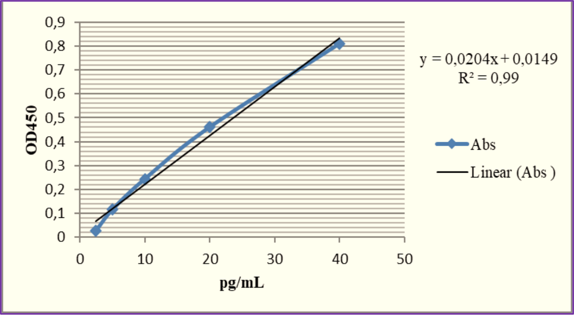
The ELISA standard curve shows a clear positive correlation between concentration (ng/ml) and optical density (OD450). The linear regression equation y = 0.0204x + 0.0149y = 0.0204x + 0.0149 reflects the correlation, with a high R2 value of 0.99 indicating a strong linear fit (Figure 1). This demonstrates the high accuracy of the ELISA test for determining analyte concentrations within the tested range, ensuring reliable PRP data.

Table 1 presents results by age in the coastal area. It can be observed that, in this study, the number of subjects aged 10 years was higher than those aged 11 and 12 years, with the latter representing the highest percentage at 41.9%. The highest average DMF-T value among the three age categories was also observed in 10-year-old subjects, at 2.5, although it still falls within the low category range of 1.2-2.6. Subjects aged 11 and 12 years had the same average DMF-T value of 1.4, which also falls under the low category. Meanwhile, the salivary pH values of subjects aged 10 to 12 years tended to decrease, although not significantly. The normal range for salivary pH is between 6 and 7.4, while the critical salivary pH is ≤5.5. The average salivary pH was 8.28 for 10-year-olds, 8.23 for 11-year-olds, and 8.22 for 12-year-olds. The highest average salivary PRP level was observed in 11-year-olds at 8.403 ng/mL, followed by 10-year-olds at 7.969 ng/mL and 12-year-olds at 7.805 ng/mL.
Distribution of participants according to age and DMF-T Score, Salivary pH, and Salivary PRP in the Coastal area.
Table 2 presents the distribution of study subjects by age along with the average DMF-T index, salivary pH, and PRP levels in the plateau area. Among the 62 participants, 21 (34%) were 10 years old, 20 (32%) were 11 years old, and 21 (34%) were 12 years old. The average DMF-T index increased with age: 1.1 for 10-year-olds, 1.7 for 11-year-olds, and 2.2 for 12-year-olds. Corresponding salivary pH values were 6.68, 6.47, and 6.39, while PRP levels were 3,381 ng/ml, 3,713 ng/ml, and 3,051 ng/ml, respectively. Overall, the average DMF-T was 1.7, the salivary pH was 6.51, and the PRP was 3,376 ng/ml.
Distribution of participants according to age and DMF-T score, salivary pH, and salivary PRP in the Plateau area.
Table 3 presents results by gender in the coastal area, where the number of male and female participants was equal (50%). The average DMF-T value was slightly higher in male participants (1.9) compared to females, although the difference was minimal, and both remained in the low category. Regarding salivary pH, male participants had a slightly lower average (8.2) compared to females. Similarly, the average salivary PRP levels were lower in males (7.128 ng/ml) than in females (8.935 ng/ml).
Distribution of participants according to gender and DMF-T score, salivary pH, and salivary PRP in the Coastal area.
Table 4 presents results by gender in the plateau area: 35 male students (56%) and 27 female students (44%). Male participants have an average DMF-T score of 1.7, an average saliva pH of 6.68, and an average saliva PRP level of 3,147 ng/ml. Meanwhile, female participants have an average DMF-T score of 1.6, a saliva pH of 6.52, and a saliva PRP level of 3.674 ng/ml.
Distribution of participants according to gender and DMF-T score, salivary pH, and salivary PRP in the Plateau area.
Figure 2 shows a comparison of DMF-T, pH, and PRP between the coastal and plateau areas.

Because the data were not normally distributed, the nonparametric Spearman correlation test was used to assess relationships among the DMF-T index, pH, and PRP. The results show a value of 0.427 (p > 0.05) for the correlation between PRP and the DMF-T index and 0.217 (p > 0.05) for the correlation between PRP and pH, indicating no significant relationship. In contrast, the correlation between pH and the DMF-T index is 0.000 (p < 0.05), indicating a significant relationship. The correlation coefficient is -0.535, indicating a strong relationship. The negative correlation coefficient between pH and the DMF-T index suggests an inverse relationship between the two variables.
The results of the nonparametric Spearman correlation test show a value of 0.976 (p > 0.05) for the correlation between PRP and the DMF-T index and 0.816 (p > 0.05) for the correlation between PRP and pH, indicating no significant relationship. In contrast, the correlation between pH and the DMF-T index is 0.000 (p < 0.05), indicating a significant relationship. The correlation coefficient is -0.647, indicating a strong relationship. The negative correlation coefficient between pH and the DMF-T index suggests an inverse relationship between the two variables.
ν Discussion
In unstimulated saliva, PRP concentrations range from 270 to 1.335 ng/mL, indicating a normal average level [22]. PRP levels are influenced by dietary habits, with research showing that salivary composition varies across species with different eating patterns. Additionally, genes encoding salivary proteins differ between species. Studies report that PRP presence in saliva depends on tannin intake [23]. Specifically, bPRP affects the astringent taste caused by polyphenols (tannins). A tannin-rich diet induces PRP synthesis and production of other salivary proteins [24]. Foods high in tannins, such as tea, coffee, chocolate, and certain fruits, can increase PRP secretion. bPRP, in particular, is effective at binding tannins to form insoluble complexes, thereby preventing their interaction with other biological molecules. As such, a tannin-rich diet can stimulate increased PRP secretion [25,26].
The findings also showed higher salivary PRP levels in females compared to males in both areas (Tables 3 and 4). This aligns with Melis et al. [27], who reported that unstimulated salivary PRP levels were lower in males with normal weight than in females. The difference may stem from gender-related variations in salivary gland activity. Gender also impacts PRP secretion, with females generally having higher levels than males, likely due to elevated estrogen levels. Estrogen is known to enhance salivary gland activity and protein production across tissues.
Additionally, salivary gland contributions and the distinction between stimulated and unstimulated saliva influence PRP levels. The parotid gland is the primary source of stimulated saliva, secreting approximately 70% of PRPs [28]. Other factors that may affect PRP secretion include differences in laboratory sample processing, such as variations in the ELISA kit used, equipment conditions, and suboptimal handling or preparation procedures [29].
Normal salivary pH ranges from 6 to 7.4, while the critical pH for demineralization is ≤5.5. At a lower pH (4.5-5.5), acidogenic bacteria such as Streptococcus mutans and Lactobacillus are key contributors to caries [30]. Saliva maintains oral pH near neutral through two mechanisms: first, by flushing out metabolizable carbohydrates, and second, by neutralizing acids from food, drinks, and microbial activity. Low salivary pH promotes aciduric bacterial growth, favoring cariogenic bacteria and creating a cycle that further lowers pH. This shift destabilizes oral microbial balance, enhancing the risk of caries. Other risk factors include high blood glucose levels, reduced salivary flow rate, low buffering capacity, and poor dietary control [31].
Subjects in coastal areas have higher pH levels, suggesting a more protective environment against caries, as acidogenic bacteria are more active in acidic saliva. However, consistently high pH may increase the risk of tartar formation [32]. Factors contributing to elevated pH include diet, salivary stimulation, flow rate, timing, oral microorganisms, and buffering capacity [33]. However, research indicates that age and gender do not significantly affect salivary pH, but salivary pH may vary with hormonal fluctuations [34,35].
The average DMF-T scores for ages 10, 11, and 12 are 1.1, 1.7, and 2.2, respectively, indicating an increase in caries prevalence with age in children at the plateau area. This rise occurs as children transition from primary to permanent teeth. Erupting teeth are more prone to caries due to difficulty in cleaning until the teeth reach occlusal contact [36]. No significant differences in DMF-T were observed by age or gender, consistent with the findings of Hartami et al. [37]. However, Hu et al. [38] reported higher DMF-T with age due to more prolonged exposure to caries risk factors.
By gender, males had an average DMF-T of 1.9, while females averaged 1.8 in the coastal area. In the plateau area, the average DMF-T score was 1.7 for males and 1.6 for females. However, caries prevalence is generally higher in females due to earlier puberty, which accelerates tooth eruption by 1-6 months compared to males, making females more prone to caries at the same chronological age. Additionally, females' preference for cariogenic foods contributes to this trend [39]. In this study, the slightly higher DMF-T score in males may be due to the greater number of male participants. Factors such as genetics and oral hygiene habits also play a role. External factors such as environmental factors, behavior, and dental health knowledge also contribute to caries risk [13,14].
Based on Tables 5 and 6, the correlation between PRP and the DMF-T index is not significant (p > 0.05) in either coastal or plateau areas. In other words, PRP is not significantly associated with the DMF-T index. Various types of salivary PRPs serve specific functions in the oral cavity. aPRP helps form the enamel pellicle, which protects teeth by regulating calcium phosphate deposition and maintaining calcium homeostasis. bPRP protects enamel by binding to Streptococcus mutans, neutralizing acids, and preventing microbial adhesion. Meanwhile, gPRP has dual functions: facilitating bacterial adhesion to enamel and agglutinating bacteria in saliva for elimination through swallowing. However, PRPs can also support adhesion of certain bacteria, such as Actinomyces viscosus and S. gordonii, and promote Candida albicans colonization, highlighting their influence on oral microbiota balance [18,20]. Several previous studies have shown that high salivary PRP levels were found in subjects with low DMF-T values [15,18,40]. However, other studies also reported that subjects with high PRP levels had higher DMF-T values [41].
Spearman correlation test of DMF-T Index, Saliva pH, and Salivary PRP in children aged 10-12 years in the Coastal area.
Spearman correlation test of DMF-T Index, Saliva pH, and Salivary PRP in children aged 10-12 years at Plateau area.
This study used unstimulated saliva samples, with two-thirds of the secretions originating from the submandibular glands. In stimulated saliva, the parotid glands account for up to 70% of total saliva secretion. aPRP, encoded by the PRH1 and PRH2 genes, is secreted only by the submandibular glands, while bPRP and gPRP, encoded by the PRB1-PRB4 genes, are secreted by the parotid glands [42]. The insignificant findings may be due to distinct protective mechanisms and secretion patterns among PRP types. The study measured overall PRP levels rather than individual PRP types, making it unclear which specific PRPs are directly associated with caries. Additionally, the non-significant results may be attributed to the limited sample size, measurement inaccuracies, participants' diets and oral hygiene, or the multifactorial nature of caries itself.
Correlation between PRP and salivary pH was not statistically significant (p > 0.05) in both the coastal and plateau areas. According to the literature, PRPs play a protective role in maintaining oral environment balance by binding calcium ions and inhibiting bacterial adhesion to tooth surfaces. This reduction in bacterial adhesion decreases acid production from carbohydrate metabolism, preventing salivary pH from dropping to acidic levels [43]. While this function helps stabilize salivary pH, the statistical relationship between PRPs and pH was not significant in this study. The balance of PRPs and salivary pH can also be influenced by diet and nutritional sources [44]. As a coastal area, Puger's seafood-rich diet may contribute to better dental health due to its high mineral content. In plateau areas like Jelbuk, calcium-rich sources such as fish are difficult to find and relatively expensive, making it challenging for the local population to meet their calcium needs [10].
The insignificant correlation results are likely due to a combination of factors, including sample size, potential measurement inaccuracies, and the distinct mechanisms by which PRP and salivary pH function. PRPs primarily contribute to oral health by forming a protective pellicle on the enamel, binding calcium ions, and interacting with specific bacteria. At the same time, salivary pH is more directly involved in buffering acids and maintaining a neutral environment. These differing roles and interactions with the oral environment may contribute to the lack of a direct statistical correlation observed in this study.
A statistically significant correlation (p < 0.05) between salivary pH and the DMF-T index in both coastal and plateau areas indicates a strong negative relationship, meaning that as salivary pH decreases, the DMF-T index tends to increase. This aligns with previous studies, which also found a negative correlation between salivary pH and DMF-T, showing that lower salivary pH is associated with higher caries incidence [31]. Salivary pH can predict caries status and reflect saliva's buffering capacity. Individuals with a resting salivary pH around 7.0 have lower caries activity compared to those with a pH of 5.5, who exhibit higher caries rates. When pH is between 5.5 and 7.0, caries incidence is moderate. Lower salivary pH prolongs exposure of tooth structures to acidic conditions, increasing the risk of demineralization and caries. Dental caries also exacerbates carbohydrate retention, prolonging acid production and further lowering salivary pH. This cycle promotes bacterial adhesion and reduces the effectiveness of saliva's natural cleansing action [45]. The study used unstimulated saliva samples, which may not fully reflect PRP secretion from the parotid and submandibular glands, as stimulated saliva contains higher levels of PRP.
The limitations of the study were that it tested only PRP levels, not overall protein levels, so it cannot compare overall protein levels with PRP levels in saliva samples. The PRPs used were whole in unstimulated saliva, so they cannot determine the nature of the most dominant PRP subclass or play a direct role in caries risk.
ν Conclusion
There is no significant correlation between salivary PRP levels, salivary pH, and DMF-T index in children aged 10-12 years in both areas, but there is a significant correlation between salivary pH and DMF-T index in children aged 10-12 years in both regions.
-
Financial Support
None.
Acknowledgements
We gratefully thank Melok Aris Wahyukundari, DDS, Sp. Perio and Neira Najatus Shakinah, DDS, Sp. Perio for advice and contributions to the investigation.
Data Availability
The data used to support the findings of this study can be made available upon request to the corresponding author.
ν References
-
[1] Pakkhesal M, Riyahi E, Naghavi A, Amdjadi P, Behnampour N. Impact of dental caries on oral health related quality of life among preschool children: Perceptions of parents. BMC Oral Health 2021; 21(1):68. https://doi.org/10.1186/s12903-021-01396-4
» https://doi.org/10.1186/s12903-021-01396-4 -
[2] World Health Organization. Global oral health status report: towards universal health coverage for oral health by 2030. Regional summary of the South-East Asia Region. Geneva: World Health Organization; 2023. Available from: https://www.who.int/publications/i/item/9789240070844 [Accessed on December 1, 2024].
» https://www.who.int/publications/i/item/9789240070844 -
[3] Choudhary A, Bhat M, Choudhary H, Joshi V, Singh WS, Soni RK. Prevalence of dental caries with salivary assessment in six to twelve years old school-going children in Shahpura Tehsil, Jaipur. Cureus 2022; 14(8):e27802. https://doi.org/10.7759/cureus.27802
» https://doi.org/10.7759/cureus.27802 -
[4] Utami SP, Mahata IBE, Ningrum V, Amran R, Wiyanti R. Relationship between tooth brushing behavior and dental caries in children at 27 Sungai Sapih Public Elementary School Kuranji, Padang City. E-GiGi 2023; 12(2):162-167. https://doi.org/10.35790/eg.v12i2.48841
» https://doi.org/10.35790/eg.v12i2.48841 -
[5] Utami SP, Mahata IBE, Wiyanti R. Dental caries and tooth brushing practices among students elementary school. Dentika: Dental Journal 2023; 26(2):115-121. https://doi.org/10.32734/dentika.v26i2.12470
» https://doi.org/10.32734/dentika.v26i2.12470 -
[6] Decker RT, Loveren CV. Sugars and dental caries. Am J Clin Nutr 2003; 78(4):881S-892S. https://doi.org/10.1093/ajcn/78.4.881S
» https://doi.org/10.1093/ajcn/78.4.881S -
[7] Sukandar, Dewi CSU, Harsindhi CJ, Handayani M, Maulana AW, Supriyadi, et al. Profil desa pesisir provinsi Jawa Timur. Volume 2. Bidang Kelautan, Pesisir, dan Pengawasan Dinas Perikanan dan Kelautan Provinsi Jawa Timur. 2016. Available from: https://bpp.fpik.ub.ac.id/wp-content/uploads/2017/02/PROFIL-DESA-PESISIR-SELATAN-JAWA-TIMUR-Vol-2.pdf [Accessed on March 5, 2024]. [In Indonesian].
» https://bpp.fpik.ub.ac.id/wp-content/uploads/2017/02/PROFIL-DESA-PESISIR-SELATAN-JAWA-TIMUR-Vol-2.pdf -
[8] Wikipedia. Jember Regency. 2024. Available from: https://en.wikipedia.org/wiki/Jember_Regency#cite_ref-Badan_Pusat_Statistik_2024_2-3 [Accessed on February 3, 2025].
» https://en.wikipedia.org/wiki/Jember_Regency#cite_ref-Badan_Pusat_Statistik_2024_2-3 -
[9] Komarudin K, Mashud M, Andriati R, Apriasari ML. The social resilience of fishermen to oral diseases during the Covid-19 pandemic in Puger Jember East Java. Odonto: Dental Journal 2022; 9(2):279-289. https://doi.org/10.30659/odj.9.2.279-289
» https://doi.org/10.30659/odj.9.2.279-289 -
[10] Barid I, Indahyani DE, Utami SE. Analysis of jaw arch size in children aged 10-13 years and 14-16 years in the highlands region (Study in Jelbuk District, Jember Regency). e-GiGi 2022; 11(1):56-63. https://doi.org/10.35790/eg.v11i1.44391
» https://doi.org/10.35790/eg.v11i1.44391 - [11] Kiswaluyo, Sulistiyani, Sutjiati R, Juslily M. Relationship between dental and oral health behavior with caries in the covid-19 pandemic in the community of Jelbuk sub-district, Jember Regency. Int J Sci Dev Res 2022; 7(6):301-304.
- [12] Rathee M, Sapra A. Dental Caries. 2023. In: StatPearls. Treasure Island (FL): StatPearls Publishing; 2025.
-
[13] de Abreu MHNG, Cruz AJS, Borges-Oliveira AC, Martins RC, Mattos FF. Perspectives on social and environmental determinants of oral health. Int J Environ Res Public Health 2021; 18(24):13429. https://doi.org/10.3390/ijerph182413429
» https://doi.org/10.3390/ijerph182413429 -
[14] Laputková G, Schwartzová V, Bánovčin J, Alexovič M, Sabo J. Salivary protein roles in oral health and as predictors of caries risk. Open Life Sci 2018; 13:174-200. https://doi.org/10.1515/biol-2018-0023
» https://doi.org/10.1515/biol-2018-0023 -
[15] Alamoudi A, Alamoudi R, Gazzaz Y, Alqahtani AM. Role of salivary biomarkers in diagnosis and detection of dental caries: A systematic review. Diagnostics 2022; 12(12):1-18. https://doi.org/10.3390/diagnostics12123080
» https://doi.org/10.3390/diagnostics12123080 - [16] Moradi G, Mohamadi Bolbanabad A, Moinafshar A, Adabi H, Sharafi M, Zareie B. Evaluation of oral health status based on the Decayed, Missing and Filled Teeth (DMFT) Index. Iran J Public Health 2019; 48(11):2050-2057.
-
[17] Komatsu T, Kobayashi K, Morimoto Y, Helmerhorst E, Oppenheim F, Lee MC. Direct evaluation of the antioxidant properties of salivary proline-rich proteins. J Clin Biochem Nutr 2020; 67(2):131-136. https://doi.org/10.3164/jcbn.19-75
» https://doi.org/10.3164/jcbn.19-75 -
[18] Lynge Pedersen AM, Belstrøm D. The role of natural salivary defences in maintaining a healthy oral microbiota. J Dent 2019; 80(Suppl 1):S3-S12. https://doi.org/10.1016/j.jdent.2018.08.010
» https://doi.org/10.1016/j.jdent.2018.08.010 -
[19] Ahmad P, Hussain A, Carrasco-Labra A, Siqueira WL. Salivary proteins as dental caries biomarkers: A systematic review. Caries Res 2022; 56(4):385-398. https://doi.org/10.1159/000526942
» https://doi.org/10.1159/000526942 -
[20] Mazurkiewicz D, Pustułka M, Ambrozik-Haba J, Bienkiewicz M. Dietary habits and oral hygiene as determinants of the incidence and intensity of dental caries-A Pilot Study. Nutrients 2023; 15(22):4833. https://doi.org/10.3390/nu1522483322
» https://doi.org/10.3390/nu1522483322 -
[21] Antonelli R, Massei V, Ferrari E, Gallo M, Pertinhez TA, Vescovi P, et al. Salivary diagnosis of dental caries: A Systematic Review. Curr Issues Mol Biol 2024; 46(5):4234-4250. https://doi.org/10.3390/cimb46050258
» https://doi.org/10.3390/cimb46050258 - [22] Sabermahani A, Jafari Sirizi M, Shaker Z, Hassanzadeh R, Malekmohammadi T, Hasheminejad N. Dental health status and associated factors in the deprived population covered by rural dental insurance program in the province of Kerman/Iran. J Dent Indones 2024;31(2):101-109.
-
[23] Levine M. Susceptibility to dental caries and the salivary proline-rich proteins. Int J Dent 2011; 953412. https://doi.org/10.1155/2011/953412
» https://doi.org/10.1155/2011/953412 -
[24] Wang M, Brignot H, Septier C, Martin C, Canon F, Feron G. Astringency sensitivity to tannic acid: Effect of ageing and salivary proline-rich protein levels. Food Chem 2024; 8:100192. https://doi.org/10.1016/j.fochms.2023.100192
» https://doi.org/10.1016/j.fochms.2023.100192 -
[25] Simões C, Caeiro I, Carreira L, Silva FCE, Lamy E. How different snacks produce a distinct effect in salivary protein composition. Molecules 2021; 26(9):2403. https://doi.org/10.3390/molecules26092403
» https://doi.org/10.3390/molecules26092403 -
[26] Rinaldi A, Moio L. Salivary protein-tannin interaction: The binding behind astringency. In: Cosme F, Nunes FM, Filipe-Ribeiro L. Chemistry and Biochemistry of Winemaking, Wine Stabilization and Aging. London: IntechOpen; 2021. https://doi.org/10.5772/intechopen.93611
» https://doi.org/10.5772/intechopen.93611 -
[27] Melis M, Mastinu M, Pintus S, Cabras T, Crnjar R, Barbarossa IT. Differences in salivary proteins as a function of PROP taster status and gender in normal weight and obese subjects. Molecules 2021; 26(8):2244. https://doi.org/10.3390/molecules26082244
» https://doi.org/10.3390/molecules26082244 -
[28] Patriarca EJ, Cermola F, D’Aniello C, Fico A, Guardiola O, De Cesare D, et al. The multifaceted roles of proline in cell behavior. Front Cell Dev Biol 2021; 9:728576. https://doi.org/10.3389/fcell.2021.728576
» https://doi.org/10.3389/fcell.2021.728576 -
[29] Almhöjd U, Fisic A, Cevik-Aras H, Tuomi L, Finizia C, Almståhl A. Explorative study of stimulated saliva proteome in head and neck cancer patients preand post-treatment. Heliyon 2024; 10(20):e39033. https://doi.org/10.1016/j.heliyon.2024.e39033
» https://doi.org/10.1016/j.heliyon.2024.e39033 -
[30] Song JG, Baral KC, Kim GL, Park JW, Seo SH, Kim DH, et al. Quantitative analysis of therapeutic proteins in biological fluids: Recent advancement in analytical techniques. Drug Deliv 2023; 30(1):2183816. https://doi.org/10.1080/10717544.2023.2183816
» https://doi.org/10.1080/10717544.2023.2183816 -
[31] Hans R, Thomas S, Garla B, Dagli RJ, Hans MK. Effect of various sugary beverages on salivary ph, flow rate, and oral clearance rate amongst adults. Scientifica 2016; 2016:5027283. https://doi.org/10.1155/2016/5027283
» https://doi.org/10.1155/2016/5027283 -
[32] Seethalakshmi C, Reddy RC, Asifa N, Prabhu S. Correlation of salivary pH, incidence of dental caries and periodontal status in diabetes mellitus patients: A Cross-sectional Study. J Clin Diagn Res 2016; 10(3):ZC12-14. https://doi.org/10.7860/JCDR/2016/16310.7351
» https://doi.org/10.7860/JCDR/2016/16310.7351 -
[33] Fons-Badal C, Fons-Font A, Labaig-Rueda C, Fernanda Solá-Ruiz M, Selva-Otaolaurruchi E, Agustín-Panadero R. Analysis of predisposing factors for rapid dental calculus formation. J Clin Med 2020; 9(3):858. https://doi.org/10.3390/jcm9030858
» https://doi.org/10.3390/jcm9030858 -
[34] Annapoorna HB, Datta D. Effects of the salivary flow rate, ph, viscosity and buffering capacity on dental caries experience in government school children in Bangalore City. Int J Community Med Public Heal 2020; 7(12):4865-4868. https://doi.org/10.18203/2394-6040.ijcmph20205154
» https://doi.org/10.18203/2394-6040.ijcmph20205154 -
[35] Ferrer MD, Pérez S, Lopez AL, Sanz JL, Melo M, Llena C, et al. Evaluation of clinical, biochemical and microbiological markers related to dental caries. Int J Environ Res Public Health 2021; 18(11):6049. https://doi.org/10.3390/ijerph18116049
» https://doi.org/10.3390/ijerph18116049 -
[36] lves LS, Zenkner JE, Wagner MB, Damé-Teixeira N, Susin C, Maltz M. Eruption stage of permanent molars and occlusal caries activity/arrest. J Dent Res 2014; 93(7 Suppl):114S-119S. https://doi.org/10.1177/0022034514537646
» https://doi.org/10.1177/0022034514537646 -
[37] Hartami E, Rachmawati YL, Puspitasari A, Herlambang AN. Differences in caries severity based on sociodemographic factors in pediatric patients at Universitas Brawijaya Hospital. Dentika: Dental Journal 2024; 27(1):1-5. https://doi.org/10.32734/dentika.v27i1.15615
» https://doi.org/10.32734/dentika.v27i1.15615 -
[38] Hu J, Jiang W, Lin X, Zhu H, Zhou N, Chen Y, et al. Dental caries status and caries risk factors in students ages 12-14 years in Zhejiang, China. Med Sci Monit 2018; 1(24):3670-3678. https://doi.org/10.12659/MSM.907325
» https://doi.org/10.12659/MSM.907325 -
[39] Lukacs JR, Largaespada LL. Explaining sex differences in dental caries prevalence: Saliva, hormones, and "life-history" etiologies. Am J Hum Biol 2006; 18(4):540-555. https://doi.org/10.1002/ajhb.20530
» https://doi.org/10.1002/ajhb.20530 -
[40] Salehabadi N, Moallem Savasari A, Nahvi A. Salivary proteins, enzymes and immune factors associated with early childhood caries: A Narrative Review. J Pediatr Rev 2022; 1(10):305-314. https://doi.org/10.32598/jpr.10.4.870.3
» https://doi.org/10.32598/jpr.10.4.870.3 -
[41] Sadashivappa Pateel DG, Gunjal S, Dutta S. Association of salivary statherin, calcium, and proline-rich proteins: A potential predictive marker of dental caries. Contemp Clin Dent 2022; 13(1):84-89. https://doi.org/10.4103/ccd.ccd_859_20
» https://doi.org/10.4103/ccd.ccd_859_20 -
[42] Russell MW, Bobek LA, Brock JH, Hajishengallis G, Tenovuo J .Mucosal Immunology (Third Edition): Chapter 5 - Innate Humoral Defense Factors. Academic Press 2005. pages 73-93. ISBN 9780124915435. https://doi.org/10.1016/B978-012491543-5/50009-7
» https://doi.org/10.1016/B978-012491543-5/50009-7 -
[43] Santacroce L, Passarelli PC, Azzolino D, Bottalico L, Charitos IA, Cazzolla AP, et al. Oral microbiota in human health and disease: A perspective. Exp Biol Med 2023; 248(15):1288-1301. https://doi.org/10.1177/15353702231187645
» https://doi.org/10.1177/15353702231187645 -
[44] Gasmi Benahmed A, Gasmi A, Dadar M, Arshad M, Bjørklund G. The role of sugar-rich diet and salivary proteins in dental plaque formation and oral health. J Oral Biosci 2021; 63(2):134-141. https://doi.org/10.1016/j.job.2021.01.007
» https://doi.org/10.1016/j.job.2021.01.007 -
[45] Spatafora G, Li Y, He X, Cowan A, Tanner ACR. The evolving microbiome of dental caries. Microorganisms 2024; 12(1):121. https://doi.org/10.3390/microorganisms12010121
» https://doi.org/10.3390/microorganisms12010121
Edited by
-
Academic Editor:
Alessandro Leite Cavalcanti