
 Decision-Making regarding Interproximal Caries Stages Among Dental Students in a Public and Private Institution
ABSTRACT
Objective: To evaluate the treatment decision-making of dental students from two dental schools for different stages of interproximal carious lesions in permanent and deciduous teeth.
Material and Methods: This is a cross-sectional study, where a questionnaire with schematic diagrams and radiographs of deciduous and permanent teeth showing five stages of interproximal carious lesions (external third of enamel, internal third of enamel, enamel-dentin junction (EDJ), external third of dentin and internal third of dentin) were presented to dental students of one Public University and one Private University, to determine the need for invasive intervention.
Results: For diagrams, significant differences were found between schools in dichotomized responses (external or internal to EDJ) for deciduous and permanent teeth (p=0.019 and p=0.015, respectively), with a higher frequency of responses for early intervention (external to EDJ) in the private school. For radiographs, a higher response frequency for early intervention was observed in permanent teeth; meanwhile, for deciduous teeth, a late intervention (internal to the EDJ) was preferred, without significant differences between schools.
Conclusion: Students from both schools showed a high frequency of responses to early invasive intervention (external to EDJ) in interproximal caries lesions when diagrams and radiographs of permanent teeth were evaluated, with the private schools evaluating more frequently. Only in the deciduous teeth radiograph, the prevalence of late intervention (internal to the EDJ) was higher.
Keywords:
Academic Performance; Decision Making; Dental Caries; Diagnosis; Oral.
Introduction
Recent advances in understanding the process of dental caries have led to a paradigm shift in caries management. Preventive strategies and noninvasive therapies, such as fluorotherapy and remineralization, play an essential role in the caries disease approach and, depending on the cavitation stage, are preferable to invasive treatments [1].
Decision-making regarding invasive or noninvasive intervention in the interproximal region usually involves evaluating bite-wing radiographs. Pitts and Rimmer [2] have shown that when there is radiolucency in permanent teeth only on the outer half of the enamel, there is no cavitation (0%). However, when the radiographic image extends into the enamel's internal half, the dentin's external half, and the dentin's internal half, cavitation is present in 10.5%, 40.9%, and 100% of cases, respectively. In the primary teeth, the percentages of cavitation for the same scenario were 2%, 2.9%, 28.3%, and 95.5%, respectively.
Previous studies [3-11] evaluated decision-making regarding interproximal carious lesions at different progression stages in primary and/or permanent teeth, using schematic diagrams and bite-wing radiographs, conducted by professionals and dental students in various countries. In general, there are variations in the results regarding the moment/stage at which the decision to perform invasive treatment in interproximal carious lesions is made. A recent study showed that less severe lesions and clinical experience influence decision-making [12]. Despite the variability in decision-making reported in the literature, Pitts and collaborators developed a specific guide for practices in individualand tooth-level caries diagnosis and management [13].
In some situations, the information derived from schematic drawings, usually used in books and classes, does not correspond to the same information when radiographs are evaluated, even if they bring the same information. Thus, this study aimed to assess the decision-making treatment for different interproximal carious lesion stages in permanent and deciduous teeth, comparing the results with those shown in schematic diagrams and radiographs, among dental students from public and private dental schools.
Material and Methods
Study Design, Sample and Ethical Clearance
The sample for this cross-sectional study consisted of dental students enrolled at two higher education institutions in the city of Santa Maria a public (Federal University of Santa Maria) and a private (Franciscan University) institution. Before the study, the respective curriculum grid was evaluated together with the coordinator of each course to determine the disciplines encompassing the programmatic contents of cariology and radiology. Thus, all students who had already been approved by the disciplines comprising cariology and radiology were considered eligible. Those who agreed to participate and signed the free informed consent form were included. The data was collected between March and August 2019 from all eligible students in the two institutions. This research was approved by the Committee of Ethics in Research in Human Beings of the Franciscan University (Protocol n. 299.2011.2).
Questionnaires
A questionnaire was structured from a pilot study and then applied to the students. Demographic variables and the year of the course were collected. Two diagrams, one for deciduous teeth and one for permanent teeth, each with five schemes simulating five different stages of interproximal carious lesions (external third of enamel, internal third of enamel, enamel-dentin junction (EDJ), external third of dentin and internal third of dentin), and photographs of radiographs from deciduous and permanent teeth with interproximal caries lesions (deciduous = carious lesion on the distal face of the right lower first molar / permanent = carious lesion on the distal face of the right upper second premolar) with the same stages mentioned in the schematic diagrams were presented to the students (Figure 1A, 1B and 1C). For the radiographs, additional patient information was available. Deciduous tooth, 6-year-old child (dmft = 1) and permanent tooth, 15-year-old adolescent (dmft = 1). For all diagrams and radiographs of deciduous and permanent teeth, a question to check the decision-making was performed: From what stage of the interproximal carious lesion progression do you believe that immediate restorative treatment should be made?

For the questionnaire application, the students were in a large room and had the diagrams and questionnaires printed on high-quality paper, as they would for a test. All questions were administered simultaneously, all data were collected individually, and the test was administered by another professor unrelated to the study to prevent the researcher from influencing responses. The questions concerned the stage of the lesion and the decision about whether immediate restorative treatment was indicated.
Data Analysis
According to the institution, the therapeutic decision for each diagram or radiograph was described in absolute and relative frequencies. The carious lesion was dichotomized by progression (external to EDJ/internal to EDJ; the option "EDJ" was considered external for EDJ in the dichotomization) and described for both institutions. Associations were tested using the chi-square test and odds ratios (in this case, they resemble prevalence reasons) and were calculated considering the therapeutic decision according to the dichotomized intervention (external to the EDJ/internal to the EDJ) as the outcome and the type of institution (public/private) as an independent variable for both diagrams and radiographs. Data were analyzed using SPSS (IBM Corp., Armonk, NY, USA) version 22.0.
Results
Of the 442 eligible students, 186 (42.1%) were from the private school and 256 (57.9%) from the public school. The response rate was 356 students (80.5%), comprising 172 (67.2%) from public schools and 184 (98.9%) from private schools. For gender, 240 (67.4%) were female, and 116 (32.6%) were male; ages ranged from 18 to 46 years, with a mean of 22.28 (±3.07).
The frequencies of the responses for public and private schools about the stage of the proximal carious lesion in which the immediate restorative treatment should occur, according to the diagram and radiograph, are presented in Table 1, and the frequencies of the dichotomized responses (external to EDJ/internal to EDJ) are presented in Table 2. For the private school, the immediate restoration decision when the lesion was external to the EDJ was chosen by approximately 70%, 58%, 68%, and 39% in the permanent diagram, deciduous diagram, and radiograph of the deciduous, respectively. These percentages were 58%, 45%, 71%, and 38% for public schools, respectively.
Absolute and relative frequencies to decision making by immediate intervention according to the type of institution.
Decisions of immediate intervention externally to the EDJ of the private schools in reference to the public, considering the diagrams according to the type of institution.
Table 2 provides information on the likelihood of immediate restorative decision-making when the carious lesion is external to the EDJ, and the diagrams were analyzed. Table 3 shows the same information when the radiographs were evaluated. Regarding the public schools, students from private institutions have a 60% (OR 1.66, CI95% 1.1-2.5) greater chance and a 73% greater chance (OR 1.73, CI95% 1.1-2.6) of restoring external lesions to EDJ when analyzed in deciduous and permanent tooth diagrams, respectively. However, considering the radiographs of deciduous and permanent teeth, there are no significant differences in the pattern of responses between the two institutions.
Decisions of immediate intervention externally to the EDJ of the private schools in reference to the public, considering the radiographs according to the type of institution.
Table 4 shows the association between decision-making based on diagrams versus radiographs for deciduous and permanent teeth. Although the stages of carious lesions were the same in the diagrams and radiographs (Figure 1), significant differences were observed in the patterns of responses to dichotomous restorative decision-making across the two institutions.
Association between immediate intervention decision based on the diagram and radiograph according to the institution.
ν Discussion
The present study evaluated and compared decision-making treatments based on information from different stages of proximal carious lesions at two dental education institutions (private vs. public). In the association between schools (public and private) and therapeutic decision, a difference was observed in students' responses to deciduous and permanent dental diagrams, with the highest chance of intentional invasive intervention among private students, with ORs of 67% for the deciduous diagram and 73%for the permanent diagram. When these dental students evaluated the radiographs of deciduous and permanent teeth, there was no difference in the frequency of responses between schools. However, in permanent teeth radiographs, the academics of both schools mainly chose to restore external to EDJ.
The classification of an interproximal lesion depends on the extent of penetration into dental tissues. The present study divided penetration into an external third of enamel, an internal third of enamel, the enamel-dentin junction (EDJ), an external third of dentin, and an internal third of dentin [4,6,7]. In the questionnaire administered, the clinical profile of patients was described, including age and dmft, providing the necessary support for the restorative therapeutic decision. Radiographic information was exemplified by diagrams and photographs of radiographs for both deciduous and permanent teeth. Using radiographs to diagnose interproximal caries lesions is highly specific. However, the amount of demineralized material compromises its sensitivity, making it difficult to obtain a reliable radiographic record; therefore, it is always recommended to be used in conjunction with the clinical examination [6].
Bervian et al. [7] reported similar results when evaluating six private and three public schools, finding a 72% higher likelihood that students from private schools opted for restorative treatment in the permanent dentition with a caries lesion external to the EDJ than did students from public schools. The authors note that differences in dental philosophy between public and private schools may account for the results and that no previous literature comparing public and private schools was found, which makes it more challenging to establish the reason for this observation. This difference in teaching philosophies can also contribute to the diversity of responses in the present study. However, in the private school in our study, cariology is taught alongside Pediatric Dentistry. Its concepts are not revisited in theoretical classes later, leading students to have a strong tendency to make a less precocious restorative decision in deciduous teeth than in permanent ones, probably because they relate preventive concepts only to deciduous teeth. In public schools, cariology is taught separately, which may help students understand the concepts applied to deciduous and permanent teeth.
The lack of association between the answers obtained when comparing deciduous diagrams and radiographs indicates a lack of academic consensus regarding the relationship between theoretical knowledge and clinical application. However, especially in private schools, despite alternation in responses, when the radiograph was analyzed, students tended to delay the decision to perform an invasive intervention, which is evaluated positively because it is used to diagnose interproximal lesions rather than a schematic diagram. On the other hand, when this association was evaluated in responses to permanent teeth, an inverse effect occurred, especially among public school students, with the frequency of responses increased outside EDJ for radiographic evaluation. The temporality of deciduous teeth relative to permanent teeth may affect academic decisions. They opt to delay the restorative intervention since exfoliation will occur quickly. This fact probably explains the higher frequency of responses to invasive intervention in permanent teeth and the change in frequency when the correlations between diagrams and radiographs were evaluated. Irrespective of the cause, it is worrisome, mainly because radiographs are highly accurate for cavitated proximal lesions and more suitable for detecting more advanced caries lesions [14]. Thus, the teaching process must be re-evaluated if the early intervention is based on radiograph assessment.
Previous studies, such as Traebert et al. [6], investigate when the therapeutic decision is made by trained dentists who underwent additional study after graduation, such as specializations in cariology and pediatric dentistry. Of those, 16.7% would undergo invasive intervention for caries lesions confined to the external enamel, and 33.3% for lesions in the internal enamel without involvement of the enamel-dentin junction. The percentage who would perform the invasive intervention in the external half of dentin was 91%. Dentists with some form of training tended to take a more conservative approach than those without, underscoring the importance of continuing education. The influence of expertise in cariology and restorative dentistry was also shown by Signori et al. [15], who found that general practitioners adopted a less conservative approach than experts.
In 2008, Ghasemi et al. [16] conducted a study to assess which progression stage of a proximal caries lesion dentists indicated as requiring invasive treatment. Two different scenarios were considered: low and high caries risk. Among high caries-risk individuals, 77% indicated that the proximal lesion was still in enamel, without dentin involvement. For the low caries risk scenario, 32% indicated invasive treatment in the same situation. The authors concluded that dentists had a clear preference for restoring teeth in the early stages of caries evolution, underscoring the need for increased preventive measures for the treatment of dental caries in both undergraduate and continuing education programs. Another study analyzed American dentists who regularly worked in clinics and the decision to restore based on the depth of proximal carious lesions, using schematic diagrams simulating different stages of proximal caries lesions and previous information about the simulated patient. Among high caries-risk patients, 66% indicated restoration in lesions still in enamel without dentin penetration. In comparison, 24% indicated restoration when the progression of the proximal lesion was in the external third of dentin. The rates for patients with low caries risk were 39% and 54%, respectively. The authors concluded that a large group of dentists is less likely to restore proximal lesions only in enamel despite the caries risk profile of patients [8]. On the other hand, studies such as those by Pitts and Rimmer [2] showed a low cavitation rate in lesions of deciduous and permanent teeth that were radiographically limited to the EDJ. Nonetheless, it is essential to emphasize that using criteria can help students adopt a less interventionist approach [17].
The results of our study should be carefully examined, particularly because only diagrams and radiographs were used to make the decision. We are aware that, clinically, there are several methods and instruments for carious lesion detection and approaches to it [18,19], and even specialists can change treatment after radiographic analysis [20], demonstrating the importance of investigating the best way to teach caries management during graduation.
It is necessary to evaluate the philosophies and teaching approaches being developed by undergraduate professors and how this knowledge is understood and reproduced by academics to promote greater homogeneity among students and, consequently, dentists. Invasive interventions must be better grounded in scientific knowledge, with evidence-based practice. It is extremely necessary that methods such as the International Caries Detection and Assessment System (ICDAS) and the International Caries Classification and Management System (ICCMS) are exhaustively taught in dental schools, and that continuing education for dentists is provided to reduce unnecessary interventions in caries management.
ν Conclusion
When diagrams and radiographs of permanent teeth were evaluated, students from both schools showed a high frequency of responses to early invasive intervention (external to EDJ) for interproximal caries lesions, with a higher frequency in private schools. Only in deciduous teeth was radiographic evaluation associated with a higher prevalence of late intervention (internal to EDJ).
Financial Support
Data Availability
The data used to support the findings of this study can be made available upon request to the corresponding author.
References
- [1] Mount GJ, Ngo H. Minimal intervention: A new concept for operative dentistry. Quintessence Int 2000; 31(8):527-533.
-
[2] Pitts NB, Rimmer PA. An in vivo comparison of radiographic and directly assessed clinical caries status of posterior approximal surfaces in primary and permanent teeth. Caries Res 1992; 26(2):146-152. https://doi.org/10.1159/000261500
» https://doi.org/10.1159/000261500 -
[3] Riordan PJ, Espelid I, Tveit Ab. Radiographic interpretation and treatment decisions among dental therapists and dentists in Western Australia. Community Dent Oral Epidemiol 1991; 19(5):268-271. https://doi.org/10.1111/j.1600-0528.1991.tb00165.x
» https://doi.org/10.1111/j.1600-0528.1991.tb00165.x -
[4] Maupomé G, Sheiham A. Radiographic criteria employed to diagnose and treat approximal caries by final-year dental students in Mexico City. Community Dent Oral Epidemiol 1997; 25(3):242-246. https://doi.org/10.1111/j.1600-0528.1997.tb00934.x
» https://doi.org/10.1111/j.1600-0528.1997.tb00934.x - [5] Traebert J, Marcenes W, Kreutz JV, Oliveira R, Piazza CH, Peres MA. Brazilian dentists' restorative treatment decisions. Oral Health Prev Dent 2005; 3(1):53-60.
- [6] Traebert J, Wesolowski CI, de Lacerda JT, Marcenes W. Thresholds of restorative decision in dental caries treatment among dentists from small Brazilian cities. Oral Health Prev Dent 2007; 5(2):131-135.
-
[7] Bervian J, Tovo MF, Feldens CA, Brusco LC, Rosa FM. Evaluation of final year dental students concerning therapeutic decision making for proximal caries. Braz Oral Res 2009; 23(1):54-60. https://doi.org/10.1590/s1806-83242009000100010
» https://doi.org/10.1590/s1806-83242009000100010 - [8] Gordan VV, Garvan CW, Heft MW, Fellows JL, Qvist V, Rindal DB, et al. Restorative treatment thresholds for interproximal primary caries based on radiographic images: Findings from the dental Practice-Based Research Network. Gen Dent 2009; 57(6):654-666.
-
[9] Vidnes-Kopperud S, Tveit Ab, Espelid I. Changes in the treatment concept for approximal caries from 1983 to 2009 in Norway. Caries Res 2011; 45(2):113-120. https://doi.org/10.1159/000324810
» https://doi.org/10.1159/000324810 -
[10] Takahashi N, Lee C, Da Silva JD, Ohyama H, Roppongi M, Kihara H, et al. A comparison of diagnosis of early stage interproximal caries with bitewing radiographs and periapical images using consensus reference. Dentomaxillofac Radiol 2019; 48(2):20170450. https://doi.org/10.1259/dmfr.20170450
» https://doi.org/10.1259/dmfr.20170450 -
[11] Rocha BC, Rosa BSP, Cerqueira TS, de-Azevedo-Vaz SL, Barbosa GLR, Ferreira LM, et al. Evaluation of different teaching methods in the radiographic diagnosis of proximal carious lesions. Dentomaxillofac Radiol 2021; 50(4):20200295. https://doi.org/10.1259/dmfr.20200295
» https://doi.org/10.1259/dmfr.20200295 -
[12] Booth ET, Eckert GJ, Fontana M. Variability in caries management decision-making in a dental school setting. J Dent Educ 2022; 86(1):57-67. https://doi.org/10.1002/jdd.12762
» https://doi.org/10.1002/jdd.12762 -
[13] Pitts NB, Ekstrand KR; ICDAS Foundation. International Caries Detection and Assessment System (ICDAS) and its International Caries Classification and Management System (ICCMS) - Methods for staging of the caries process and enabling dentists to manage caries. Community Dent Oral Epidemiol 2013;41(1):e41-52. https://doi.org/10.1111/cdoe.12025
» https://doi.org/10.1111/cdoe.12025 -
[14] Schwendicke F, Tzschoppe M, Paris S. Radiographic caries detection: A systematic review and meta-analysis. J Dent 2015; 43(8):924-933. https://doi.org/10.1016/j.jdent.2015.02.009
» https://doi.org/10.1016/j.jdent.2015.02.009 -
[15] Signori C, Laske M, Mendes FM, Huysmans MDNJM, Cenci MS, Opdam NJM. Decision-making of general practitioners on interventions at restorations based on bitewing radiographs. J Dent 2018; 76:109-116. https://doi.org/10.1016/j.jdent.2018.07.003
» https://doi.org/10.1016/j.jdent.2018.07.003 - [16] Ghasemi H, Murtomaa H, Torabzadeh H, Vehkalahti MM. Restorative treatment threshold reported by Iranian dentists. Community Dent Health 2008; 25(3):185-190.
-
[17] Nogueira VKC, Bussaneli DG, Restrepo MR, Spin-Neto R, Dos Santos-Pinto LAM, Boldieri T, et al. Caries treatment decisions among undergraduate and postgraduate students supported by visual detection systems. Int J Paediatr Dent 2018; 28(1):92-101. https://doi.org/10.1111/ipd.12312
» https://doi.org/10.1111/ipd.12312 -
[18] Innes NPT, Schwendicke F. Restorative thresholds for carious lesions: Systematic review and meta-analysis. J Dent Res 2017; 96(5):501-508. https://doi.org/10.1177/0022034517693605
» https://doi.org/10.1177/0022034517693605 -
[19] Gimenez T, Tedesco TK, Janoian F, Braga MM, Raggio DP, Deery C, et al. What is the most accurate method for detecting caries lesions? A systematic review. Community Dent Oral Epidemiol 2021; 49(3):216-224. https://doi.org/10.1111/cdoe.12641
» https://doi.org/10.1111/cdoe.12641 -
[20] Leal SC, Barros BV, Cabral RN, Ferrari JCL, de Menezes Abreu DM, Ribeiro APD. Dental caries lesions in primary teeth without obvious cavitation: Treatment decision-making process. Int J Paediatr Dent 2019; 29(4):422-428. https://doi.org/10.1111/ipd.12483
» https://doi.org/10.1111/ipd.12483

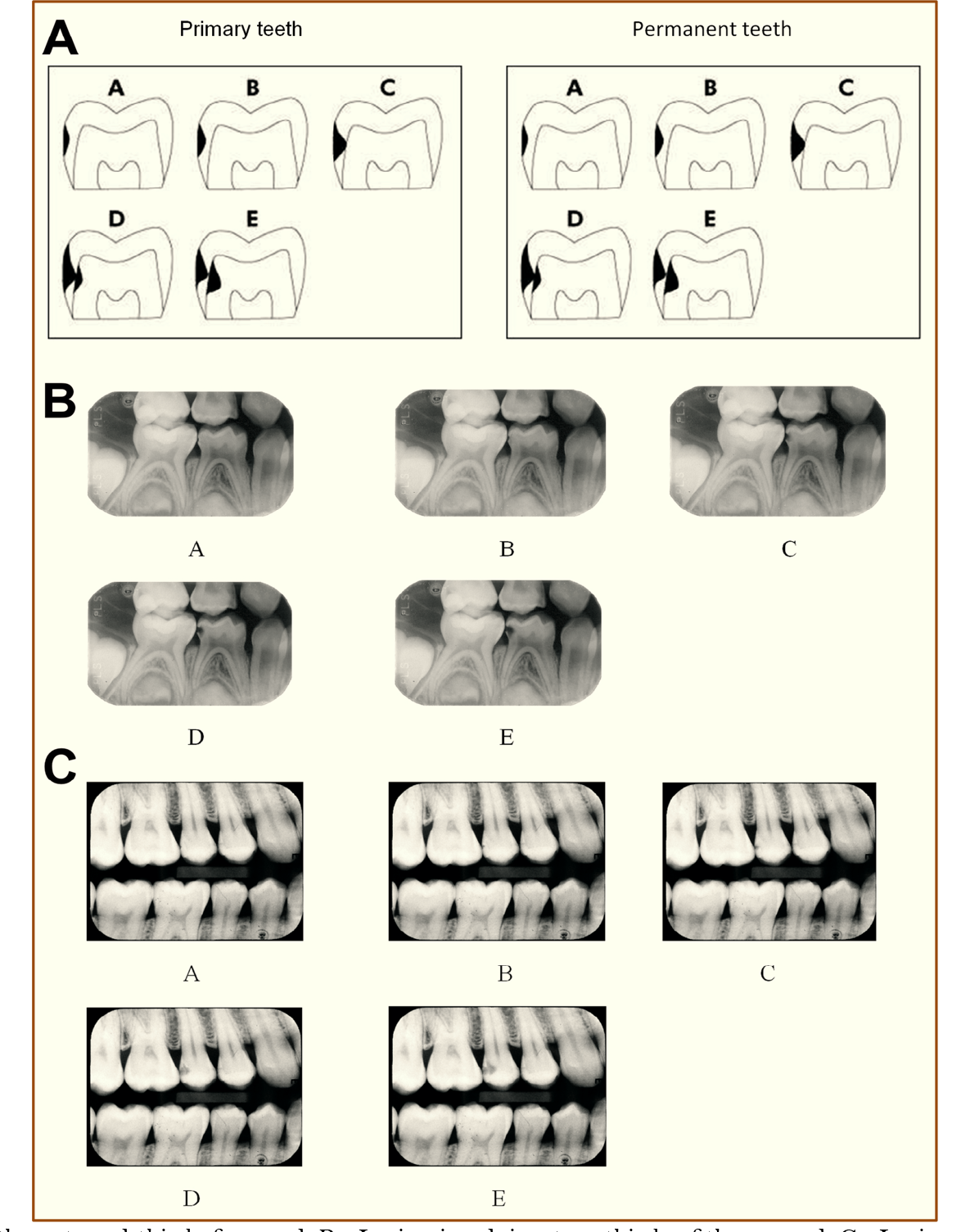 A - Lesion involving the external third of enamel. B - Lesion involving two-thirds of the enamel. C - Lesion at the enamel-dentinal junction level. D - Lesion involving the external third of dentine. E - Lesion involving two-thirds of the dentin.