Effect of Mouth Breathing on Sleep Quality and Oxygen Saturation in Pediatric Patients
ABSTRACT
Objective: To evaluate the effect of mouth breathing on sleep quality and oxygen saturation in pediatric patients aged 6-12 years.
Material and Methods: A cross-sectional observational study was done on 120 children recruited from the pediatric outpatient department. Based on clinical evaluation and parental history, participants were separated into 2 groups: mouth breathers (n=60) and nasal breathers (control group, n=60). Sleep quality was assessed using a validated Pediatric Sleep Questionnaire (PSQ), and overnight oxygen saturation was recorded with pulse oximetry. Data were analyzed using SPSS version 24.0, applying independent t-tests and Pearson’s correlation coefficient, with significance set at p<0.05.
Results: Children in the mouth-breathing group exhibited significantly poorer sleep quality, with mean PSQ scores of 6.8 ± 1.1 compared to 2.3 ± 0.9 in the nasal-breathing group (p<0.01). Mean overnight oxygen saturation was 93.6% ± 2.4 in mouth breathers versus 96.8% ± 1.8 in controls (p<0.01). A moderate positive correlation was observed between reduced oxygen saturation and deteriorated sleep quality (r = 0.64, p<0.01).
Conclusion: Mouth breathing in children is significantly associated with decreased sleep quality and reduced oxygen saturation during sleep. These findings highlight the importance of early identification and management of mouth breathing habits to mitigate their adverse effects on pediatric sleep and respiratory health.
Keywords:
Child; Mouth Breathing; Oxygen Saturation; Sleep Quality; Sleep Wake Disorders.
Introduction
Breathing is a fundamental physiological function essential for sustaining life, with nasal breathing considered the ideal route in children due to its multiple protective and functional advantages [1]. However, mouth breathing - a condition in which a child habitually breathes through the mouth rather than the nose - has been increasingly observed in pediatric populations. Though often overlooked by both parents and clinicians, mouth breathing can be a sign of underlying airway obstruction or anatomical abnormalities such as adenoid hypertrophy, allergic rhinitis, or a deviated nasal septum [2,3].
Mouth breathing not only alters craniofacial growth and oral health but also profoundly impacts sleep patterns and oxygenation. During sleep, mouth breathers are more likely to experience fragmented sleep, snoring, and even episodes of hypoxia due to compromised airflow dynamics [4,5]. These issues may go unnoticed in daily clinical practice, yet they can adversely affect a child's physical, emotional, and cognitive development. Sleep is essential for brain maturation, memory consolidation, and immune function in children; hence, disturbances in sleep architecture due to impaired breathing patterns should not be underestimated [6,7].
Previous studies have associated mouth breathing with sleep-disordered breathing (SDB), including conditions like obstructive sleep apnea (OSA). Decreased oxygen saturation during sleep, due to airway compromise, can trigger a cascade of physiological stress responses that further disrupt sleep quality [8,9]. Despite these concerning associations, there remains a paucity of literature specifically evaluating the simultaneous effects of sleep quality and nocturnal oxygen saturation in mouth-breathing children within a defined age group.
This study was done to evaluate the effect of mouth breathing on sleep quality and oxygen saturation in pediatric patients aged 6-12 years.
Material and Methods
Study Design and Ethical Clearance
This observational cross-sectional study was conducted in the Department of Paediatric and Preventive Dentistry, in collaboration with the Department of ENT, at a tertiary care centre over 6 months. Ethical clearance was obtained from the Institutional Review Board (IRB), Ref No: SDCH/ETHI/S/02-2023-24, and written informed consent was obtained from the parents or guardians of all participants.
Sample
Sample size was calculated for comparing two independent means (two groups of equal size) using the standard formula:
n per group = ((Zα/2+Zβ)2×2σ2)/∆2
where Z_{α/2} is the standard normal deviate for the two-sided significance level (1.96 for α = 0.05), Z_{β} is the deviate for desired power (0.84 for 80% power), σ is the assumed common standard deviation, and ∆ is the minimum clinically important difference between group means. For example, assuming σ ≈ = 1.0 (based on pilot data) and a clinically relevant difference ∆ = 1.0, the required sample per group is:
n = ((1.96 + 0.84)2 × 2 × 1.02) / 1.02 ≈ 16 per group.
If one wishes to detect a smaller difference (∆ = 0.5) with the same variance, the required sample size increases to: n ≈ ((2.8)2 × 2 × 1.02) / 0.52 ≈ 64 per group.
Thus, our chosen sample of 60 participants per group is adequate to detect small-to-moderate differences (≈0.5 SD) with near-80% power, and provides ample power to detect the larger differences observed in this study.
Data Collection
The study was conducted between April 2023 and October 2024. Participants were selected using a non-probability sampling method from the outpatient Department of Paediatric Dentistry at a tertiary care hospital. Children were chosen based on their breathing pattern, specifically mouth breathers and nasal breathers. The study population comprised 120 pediatric patients aged between 6 and 12 years who attended outpatient services.
Participants were divided into two equal groups (n=60 each): Group A (mouth breathers) and Group B (nasal breathers/control group), based on clinical history, ENT evaluation, and parental reporting. Inclusion criteria for Group A included children who demonstrated habitual mouth breathing during the day and at night for at least 6 months. Children with chronic illnesses, craniofacial syndromes, or neurological disorders were excluded from the study.
Sleep quality was assessed using a structured and validated Pediatric Sleep Questionnaire (PSQ), comprising 10 close-ended questions related to snoring, restlessness, waking episodes, daytime sleepiness, and other common symptoms of sleep disturbance. Each item was scored on a binary scale: 1 (Yes), 0 (No). A higher total score indicated poorer sleep quality. Scoring Interpretation: 0-3: Good sleep quality; 4-6: Mild to moderate disturbance; and 7-10: Poor sleep quality. The Pediatric Sleep Questionnaire (PSQ) was originally developed and validated by Chervin et al. [10].
A finger pulse oximeter (Advin Health Care, Advin Health Care Devices, Gujarat, India) was used to measure oxygen saturation levels in the children. The device was used only once per child during the assessment to ensure accuracy and minimize discomfort.
Overnight oxygen saturation was recorded using a fingertip pulse oximeter worn during sleep for a minimum of 6 continuous hours. The device recorded the mean SpO2 and minimum SpO2 levels. Data were recorded and cross-verified by caregivers.
Data Analysis
The collected data were analyzed using SPSS version 24.0 (IBM SPSS, Armonk, NY, USA). Descriptive statistics were calculated for all variables. The comparison between the two groups was made using the independent t-test for continuous variables and the Chi-square test for categorical variables. Pearson correlation was applied to determine the relationship between sleep quality score and oxygen saturation levels. A p-value of less than 0.05 was considered statistically significant.
Results
The mean age of participants in Group A was 8.9 ± 1.7 years, and in Group B, 9.1 ± 1.6 years, with no statistically significant difference (p = 0.62). The gender distribution was comparable between the groups, with 36 males (60%) and 24 females (40%) in Group A, and 34 males (56.6%) and 26 females (43.4%) in Group B (p = 0.72), indicating no significant gender bias.
Analysis of the Pediatric Sleep Questionnaire (PSQ) revealed significantly higher symptom scores in mouth breathers compared to nasal breathers. The mean total PSQ score for Group A was 6.8 ± 1.1, indicating poor sleep quality, while for Group B it was 2.3 ± 0.9, suggesting good sleep quality. This difference was statistically significant (p < 0.001). Specific symptoms such as loud snoring (82% vs. 10%), restless sleep (68% vs. 15%), daytime irritability (55% vs. 10%), and frequent night awakenings (60% vs. 8%) were notably more prevalent in Group A than in Group B, as shown in Table 1.
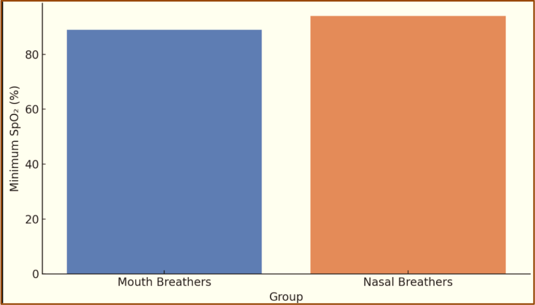
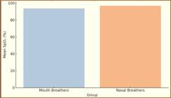
Oxygen saturation levels measured during overnight sleep also demonstrated significant differences between the groups. The mean SpO2 for mouth breathers was 93.6% ± 2.4, significantly lower than 96.8% ± 1.8 observed in nasal breathers (p< 0.001). Furthermore, the minimum recorded SpO2 during sleep dropped to 89% in some mouth-breathing children, whereas no values below 94% were observed in the control group.
A Pearson correlation analysis showed a moderate positive correlation (r = 0.64, p < 0.01) between lower oxygen saturation levels and higher PSQ scores, indicating that as oxygen saturation decreased, the severity of sleep disturbances increased.
Overall, the findings strongly support the hypothesis that mouth breathing significantly impairs sleep quality and reduces oxygen saturation during sleep in pediatric patients, reinforcing the need for early identification and intervention. Figure 1 shows higher PSQ scores in mouth breathers, indicating more severe sleep-related disturbances.

Mouth breathers had significantly lower average oxygen saturation during sleep compared to nasal breathers (Figure 2).
The lowest oxygen saturation levels were significantly lower in the mouth-breathing group, suggesting a potential risk of hypoxic episodes. Mouth breathers showed lower oxygen saturation and significantly higher PSQ scores than nasal breathers (Figure 3).

Discussion
Mouth breathing in children is often overlooked as a benign habit, yet growing evidence suggests it may have profound implications on sleep health and overall development [1,2]. It is frequently associated with upper airway obstruction caused by conditions such as adenoid hypertrophy, allergic rhinitis, or a deviated nasal septum. These anatomical or functional issues force children to rely on oral respiration, especially during sleep, leading to altered sleep architecture, fragmented rest, and intermittent hypoxia [3]. Disrupted sleep in early childhood is linked to behavioral problems, learning difficulties, and impaired growth [4]. Therefore, understanding the physiological impact of mouth breathing, particularly on sleep quality and oxygen saturation, is critical for early diagnosis and multidisciplinary management.
The present study aimed to investigate the impact of mouth breathing on sleep quality and oxygen saturation in pediatric patients. Our findings demonstrated a significant difference in both subjective (PSQ scores) and objective (SpO2 levels) sleep parameters between mouth-breathing and nasal-breathing children. These results reinforce the notion that mouth breathing is not merely a benign habit but a potential contributor to pediatric sleep-disordered breathing (SDB).
By comparing mouth-breathing children with age-matched nasal breathers, this research aims to provide valuable clinical insights into the importance of recognizing mouth breathing as a potential contributor to pediatric sleep and respiratory disturbances. The findings also support the development of timely interventions to prevent long-term consequences on child health and well-being.
The mechanism linking mouth breathing to impaired sleep and oxygen saturation likely involves upper airway obstruction, leading to altered breathing patterns, reduced nasal airflow, and increased respiratory effort, which disrupts sleep architecture and causes intermittent hypoxia [5,6].
The mean Pediatric Sleep Questionnaire (PSQ) score was markedly higher in the mouth-breathing group (6.8 ± 1.1) compared to nasal breathers (2.3 ± 0.9), indicating a statistically significant difference (p < 0.001). These findings align with the results of studies by Guilleminault et al. [11] and Abreu et al. [12], both of which emphasized the role of upper airway obstruction in disrupting sleep patterns and contributing to behavioral and developmental issues in children. Our study further supports their observations by demonstrating a clear link between mouth breathing and increased prevalence of symptoms such as loud snoring, restless sleep, and daytime irritability.
Objectively, oxygen saturation levels were significantly lower in mouth-breathing participants, with a mean SpO2 of 93.6% compared to 96.8% in the nasal-breathing group. This corresponds with findings from Mbam et al [13], who reported decreased nocturnal oxygen levels in children with adenotonsillar hypertrophy and mouth breathing. The minimum SpO2 in our study dropped to as low as 89% in some children, suggesting that mouth breathing may predispose pediatric patients to episodes of nocturnal hypoxemia, which can potentially affect cardiovascular and neurocognitive development.
Interestingly, a moderate positive correlation (r = 0.64, p < 0.01) was found between lower oxygen saturation and higher PSQ scores. This statistically significant relationship implies that deteriorating oxygen levels during sleep are directly associated with worsening sleep quality. This finding is consistent with the work of Sawatari et al. [14] who explored the correlation between pulse oximetry parameters and behavioral sleep disturbances in children and found similar trends.
It is noteworthy that gender and age distributions were statistically comparable between groups, which strengthens the internal validity of our study. This suggests that the differences in sleep quality and oxygen saturation were not confounded by demographic variables, further affirming the role of mouth breathing as a principal factor.
Although our study provides strong evidence linking mouth breathing with impaired sleep and lower oxygen saturation, it is not without limitations. The cross-sectional design restricts causality inference. Additionally, overnight oximetry, while non-invasive and child-friendly, is less detailed than full polysomnography, which remains the gold standard for sleep studies. Future longitudinal and interventional studies could better establish causal relationships and evaluate the benefits of therapeutic interventions, such as myofunctional therapy, nasal breathing retraining, or adenotonsillectomy.
Our findings underscore the critical need for early identification of mouth breathing in children, not only by ENT specialists and dentists but also by pediatricians and general practitioners. Prompt referral and intervention can significantly improve a child's sleep quality and overall development. Incorporating simple screening tools such as the PSQ into routine pediatric evaluations could be a cost-effective step toward improving pediatric health outcomes. The study's cross-sectional design and reliance on overnight oximetry rather than full polysomnography limit causal inference and detailed analysis of sleep architecture. Longitudinal polysomnographic studies and interventional trials targeting mouth-breathing habits could further elucidate causal pathways and therapeutic benefits.
Conclusion
Mouth breathing in pediatric patients is significantly associated with poor sleep quality and reduced oxygen saturation levels during sleep. Early detection and appropriate intervention are essential to prevent long-term developmental and health consequences.
-
Financial Support
None.
Data Availability
The data used to support the findings of this study can be made available upon request to the corresponding author.
References
-
[1] Trabalon M, Schaal B. It takes a mouth to eat and a nose to breathe: abnormal oral respiration affects neonates' oral competence and systemic adaptation. Int J Pediatr 2012; 2012:207605. https://doi.org/10.1155/2012/207605
» https://doi.org/10.1155/2012/207605 -
[2] Lin L, Zhao T, Qin D, Hua F, He H. The impact of mouth breathing on dentofacial development: A concise review. Front Public Health 2022; 10:929165. https://doi.org/10.3389/fpubh.2022.929165
» https://doi.org/10.3389/fpubh.2022.929165 -
[3] Pacheco MC, Casagrande CF, Teixeira LP, Finck NS, de Araújo MT. Guidelines proposal for clinical recognition of mouth breathing children. Dental Press J Orthod 2015; 20(4):39-44. https://doi.org/10.1590/2176-9451.20.4.039-044.oa
» https://doi.org/10.1590/2176-9451.20.4.039-044.oa -
[4] Pacheco MC, Fiorott BS, Finck NS, Araújo MT. Craniofacial changes and symptoms of sleep-disordered breathing in healthy children. Dental Press J Orthod 2015; 20(3):80-87. https://doi.org/10.1590/2176-9451.20.3.080-087.oar
» https://doi.org/10.1590/2176-9451.20.3.080-087.oar -
[5] Ikävalko T, Närhi M, Eloranta AM, Lintu N, Myllykangas R, Vierola A, et al. Predictors of sleep disordered breathing in children: the PANIC study. Eur J Orthod 2018; 40(3):268-272. https://doi.org/10.1093/ejo/cjx056
» https://doi.org/10.1093/ejo/cjx056 -
[6] Yang FN, Xie W, Wang Z. Effects of sleep duration on neurocognitive development in early adolescents in the USA: A propensity score matched, longitudinal, observational study. Lancet Child Adolesc Health 2022; 6(10):705-712. https://doi.org/10.1016/S2352-4642(22)00188-2
» https://doi.org/10.1016/S2352-4642(22)00188-2 -
[7] Liu J, Ji X, Pitt S, Wang G, Rovit E, Lipman T, et al. Childhood sleep: Physical, cognitive, and behavioral consequences and implications. World J Pediatr 2024; 20(2):122-132. https://doi.org/10.1007/s12519-022-00647-w
» https://doi.org/10.1007/s12519-022-00647-w -
[8] Abbasi A, Gupta SS, Sabharwal N, Meghrajani V, Sharma S, Kamholz S, et al. A comprehensive review of obstructive sleep apnea. Sleep Sci 2021; 14(2):142-154. https://doi.org/10.5935/1984-0063.20200056
» https://doi.org/10.5935/1984-0063.20200056 -
[9] Azarbarzin A, Labarca G, Kwon Y, Wellman A. Physiologic consequences of upper airway obstruction in sleep apnea. Chest 2024; 166(5):1209-1217. https://doi.org/10.1016/j.chest.2024.05.028
» https://doi.org/10.1016/j.chest.2024.05.028 -
[10] Chervin RD, Hedger K, Dillon JE, Pituch KJ. Pediatric sleep questionnaire (PSQ): Validity and reliability of scales for sleep-disordered breathing, snoring, sleepiness, and behavioral problems. Sleep Med 2000; 1(1):21-32. https://doi.org/10.1016/s1389-9457(99)00009-x
» https://doi.org/10.1016/s1389-9457(99)00009-x -
[11] Guilleminault C, Huang YS, Monteyrol PJ, Sato R, Quo S, Lin CH. Critical role of myofascial reeducation in pediatric sleep-disordered breathing. Sleep Med 2013; 14(6):518-525. https://doi.org/10.1016/j.sleep.2013.01.013
» https://doi.org/10.1016/j.sleep.2013.01.013 -
[12] Abreu AR, Stefanovski D, Patil SP, Siddharthan T, Chediak A, Wallace DM, et al. Neuromuscular electrical stimulation for obstructive sleep apnoea: Comparing adherence to active and sham therapy. ERJ Open Res 2023; 9(6):00474-2023. https://doi.org/10.1183/23120541.00474-2023
» https://doi.org/10.1183/23120541.00474-2023 -
[13] Mbam TT, Adeosun AA, Akinyemi OA, Nwaorgu OG. Comparing oxygen saturation of normal children with that of children with adenotonsillar hypertrophy. Indian J Otolaryngol Head Neck Surg 2014; 66(2):173-177. https://doi.org/10.1007/s12070-014-0701-y
» https://doi.org/10.1007/s12070-014-0701-y -
[14] Sawatari H, Rahmawati A, Moriyama N, Fujita K, Ohkusa T, Nao T, et al. Characteristics of sleep-disordered breathing in children with down syndrome - A comparison with typically developing children. Sleep Med X 2022; 4:100045. https://doi.org/10.1016/j.sleepx.2022.100045
» https://doi.org/10.1016/j.sleepx.2022.100045
Edited by
-
Academic Editor:
Alessandro Leite Cavalcanti