Energy Drink Consumption Causes Severe Dental Erosion: An in Vitro Study
ABSTRACT
Objective: To evaluate whether the consumption of energy drinks promotes dental erosion and assess the effectiveness of various treatments.
Materials and Methods: Forty specimens of bovine root dentin were prepared (4.00 mm × 4.00 mm × 3.00 mm in thickness). For erosion, the specimens were immersed in an energy drink (pH 3.24 at 10°C) in 1-minute cycles three times a day for 5 days. After the cycles, the specimens were divided into four groups (n = 10): NT, no treatment; FV, 5% fluoride varnish; L, laser Er,Cr:YSGG; and FV+L, 5% fluoride varnish followed by application of the Er,Cr:YSGG laser. The surface roughness and the wear profile of the dentin were analyzed by confocal laser scanning microscopy. Analysis of Variance (ANOVA) and Tukey post-hoc tests were performed for the surface roughness analysis. For volume loss, the Kruskal-Wallis test was used (α=0.05).
Results: There was no statistically significant difference between the groups in the control region (p>0.05) for surface roughness. In the experimental area, the NT (4.109 ± 0.184) and FV (3.850 ± 0.231) groups exhibited the highest surface roughness, with no significant difference between them. However, they showed a statistically significant difference from the L (2.058 ± 0.160) and FV+L (1.907 ± 0.179) groups (p<0.05), which also did not reveal any differences between themselves. The mean volume loss in all groups was approximately 20%, which remained after the treatments. Therefore, no statistical differences were found (p>0.05).
Conclusion: The consumption of energy drinks can lead to dental erosion, resulting in volume loss and increased roughness. None of the treatments could reverse the loss of volume. The irradiated groups achieved the best results in terms of surface roughness.
Keywords:
Energy Drinks; Tooth Erosion; Lasers; Fluoride
Introduction
Dental erosion is the loss of hard tissue, specifically enamel or dentin, caused by repeated exposure of a clean dental surface to non-bacterial acids. Dental enamel can be dissolved at a pH level below 5.5 and dentin at a pH level below 6.5. However, there is no consensus on the critical pH for dental erosion [1]. The development of dental erosion occurs through the continuous dissolution of enamel crystals, layer by layer, resulting in the permanent loss of enamel volume. However, the erosion at the dentin layer has not been completely elucidated. Several studies have demonstrated that acid exposure, combined with low salivary flow rates, leads to higher levels of dental surface dissolution [2,3], a significant contributor to dental sensitivity resulting from the exposure of dentinal tubules [4].
Dental erosion can be caused by exposure to extrinsic acids from food and drink, and intrinsic when the oral pH changes are triggered by hydrochloric acid present in the stomach that returns to the oral cavity through vomiting, regurgitation, or reflux, as in cases of anorexia, bulimia, and hiatus hernia [5].
Different beverages, such as sports drinks, energy drinks, carbonates, and mineral reloads, can cause dental erosion through their irreversible wear on dental surfaces [1,6], including the dentin layer [7], thereby increasing the roughness of dental and restoration surfaces. Dental biofilm adhesion and bacterial colonization are enhanced on rough surfaces, leading to increased dental caries, gingivitis, and periodontal disease. Dental surface irregularities facilitate the available bacterial area for adhesion, protecting the biofilm from the mechanisms of control by the oral microbiota, such as salivary flow, chewing, and swallowing [8].
The loss of dental structure and the consequent exposure of dentin are among the most common causes of dentin hypersensitivity, which can be explained by the hydrodynamic theory [9]. Treating dentin hypersensitivity involves removing or controlling etiological factors, such as acid erosion, salivary flow problems, and improper brushing techniques. The dentin permeability directly affects the patient's pain, so the therapy should be based on three possibilities: preventing dentin fluid movement, blocking pulp or dentin mechanoreceptors, or both simultaneously [10]. Different treatments for dentin hypersensitivity associated with dental erosion aim to obliterate the dentin tubules, reduce patient pain, and prevent the progression of the lesions. For these purposes, fluoride compounds like varnishes, toothpaste, mouth rinse and fluoride gel have been used, and the fluoride varnish, with the higher fluoride concentration, the most relevant product for the hypersensitivity treatment [11] because the formation of a protective layer of calcium fluoride at the surface of the dentin tubules which reduces the sensitivity temporarily [12], besides to reduce the erosion wear on the enamel [3].
In addition to fluoride compounds, various hypersensitive treatment methods, such as laser application, oxalates, adhesives, and restorations with resin composite or glass ionomer cement, have been recommended. The success depends on the oral environment's pH, the abrasive effects of brushing, and the presence of a smooth surface to prevent biofilm accumulation [13].
High-intensity lasers, such as Er,Cr:YSGG, can be used to treat dentin hypersensitivity because they work by melting dentin and obliterating the dentin tubules, thereby promoting a long-lasting response compared to other treatments [10,14]. The high-power laser associated with fluoride treatment can also increase fluoride deposition and incorporation into the dental substrate, thereby decreasing demineralization [3] and increasing the acid resistance of root dentin [15]. However, the Er,Cr:YSGG laser effect at the eroded root dentin is unknown [3].
Thus, the present study analyzed three different types of treatments for erosion caused by the Red Bull® energy drink, evaluating surface roughness and volume loss of bovine dentin root specimens. The present study hypothesized that the erosive challenges and various treatments would result in statistically significant differences in surface roughness and volume loss in bovine root dentin.
Material and Methods
Experimental Design
In the present study, 40 bovine root dentin specimens were used, which were eroded by an energy drink (Red Bull, Rauch Fruchtsäfte GmbH & Co OG, Rankweil, Vorarlberg, Austria) at a pH of 3.24 at 10°C (EN 1800 digital pH meter, Gehaka Brasil, São Paulo, SP, Brazil). The specimens were divided into four groups (n = 10): NT, no treatment; FV, 5% fluoride varnish (Duraphat, Colgate-Palmolive Company, São Bernardo do Campo, SP, Brazil); L, ErCr:YSGG laser; and FV+L, 5% fluoride varnish (Duraphat, Colgate-Palmolive Company, São Bernardo do Campo, SP, Brazil) followed by Er,Cr:YSGG laser application. The surface roughness and volume loss of dentin were quantitatively analyzed by confocal microscopy, comparing the experimental and control regions.
In the present study, the sample size was calculated to achieve a significance level of 5% and a power of the statistical test of at least 85%, thereby minimizing the chances of type I and type II errors.
Teeth Selection, Cleaning, and Specimen Preparation
Twenty bovine incisors, free from cracks and wear, were selected. The teeth were cleaned in the grinder (Tramontina Global, Guangzhou, China, ), rinsed in tap water, and stored in distilled and deionized water at 4°C, with the water changed daily for seven consecutive days.
The coronal portion of the bovine incisors was sectioned from the root using a diamond disk under refrigeration on the ISOMET 1000 cutting machine (Precision Saw Buehler, Illinois, USA). The first cut was made 1mm above the cementum-enamel junction. The second cut was performed in the mesiodistal direction, obtaining two halves (buccal and lingual). Each half was resected to obtain specimens with initial dimensions of 4.25 mm × 4.25 mm and a thickness of 3.00 mm. The specimens had their sides and thickness adjusted on the Arotec APL-4 polishing machine (Series 41042, Arotec S/A Indústria e Comércio, Cotia, SP, Brazil) using sandpaper #600 with water cooling until they reached the standard dimensions: 4.00 mm x 4.00 mm and 3.00 mm in thickness. Polishing on the outer surface of the specimens was not performed. Variations in size were allowed within a 10% tolerance, plus or minus.
Half of the surface of each specimen was covered with electrical tape. Two layers of red nail cosmetic nail polish and carving wax were applied to isolate it; after this procedure, the insulating tape was removed, and each specimen was left with half of its free surface protected by enamel and wax. The specimens were stored in distilled and deionized water at 4 °C until the erosion challenge. They were then randomly divided into four groups (n = 10), and each group received its respective treatment.
Erosive Challenge
The specimens were subjected to an erosive challenge using Red Bull energy drink (Red Bull, Rauch Fruchtsäfte GmbH & Co OG, Rankweil, Vorarlberg, Austria) with a pH of 3.24 at a temperature of 10 °C. All specimens were placed together with the drink in a becker for 1 minute on a magnetic stirrer that simulated the movement of liquid in the mouth (Model 221-1, ABC-Lab Produtos e Equipamentos Laboratório, Sao Bernardo do Campo, SP, Brazil). After this period, the erosive solution was discarded, and the specimens were washed in distilled and deionized water for 10 seconds. The specimens were stored in distilled water and placed in the oven at 37°C between cycles. This procedure was performed three times daily, with at least 2 hours' intervals between challenges, over five consecutive days. Erosion was verified using confocal laser scanning microscopy, as shown in Figure 1. After completing the erosive challenge, the specimens were stored at 4 °C in distilled water until the surface treatments [2, 7, 16].
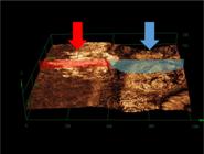
A representative image of volume loss. The red arrow indicates the control region (root dentin without exposure to the energy drink), and the blue arrow indicates the experimental region (exposed to treatments and the energy drink). There was a step between the two areas.
Surface Specimen Treatments
The NT group received no treatment, serving as the control group. In the FV group, fluoride varnish (5% sodium fluoride Duraphat, Colgate-Palmolive Company, São Bernardo do Campo, SP, Brazil) was applied to the surface of the specimens using a microbrush applicator (K G Sorensen Indústria e Comércio, Barueri, SP, Brazil). After 4 minutes of application, the excess was removed with a sterile gauze. Group L received the Er, Cr: YSGG laser application (Waterlase Millennium, Biolase Technologies Inc., San Clemente, USA), with the fiber-containing 600µm in diameter (tip model: ZipTip MZ6 3mm) in scan mode calibrated in the following parameters: 0.1W - 5Hz, without cooling, for 10 seconds [16,17]. The FVL Group received the initial treatment of 5% fluoride varnish (Duraphat) followed by the laser application as previously described.
Surface Roughness Analysis and Volume Loss
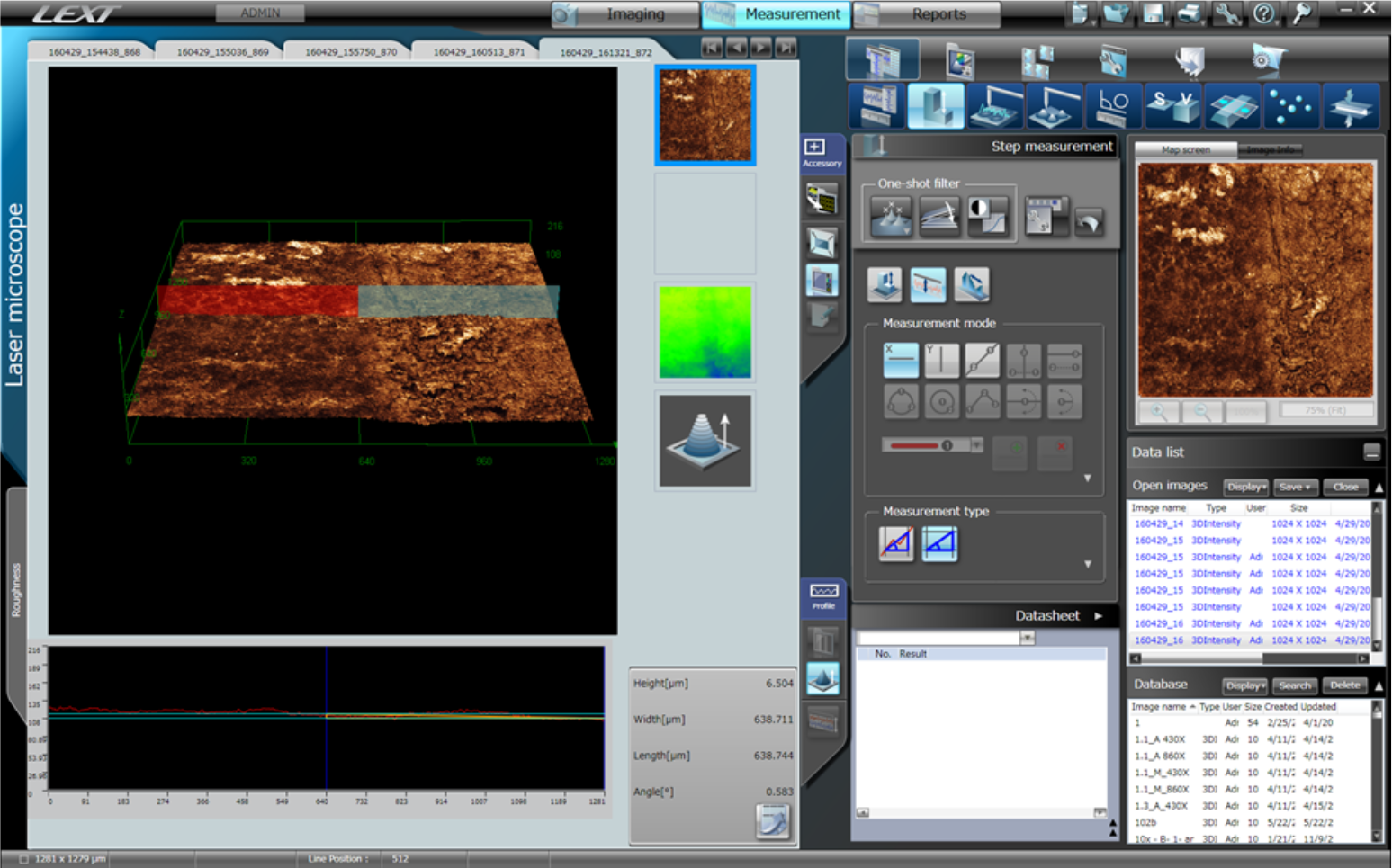
The specimens were immersed in an ultrasonic vial and shaken for 5 minutes, then positioned parallel to the LEXT confocal laser scanning microscope table (Olympus Corp., Tokyo, Japan) using a parallelometer. The central region of the specimens, measuring 1 mm x 1 mm, was selected for imaging using a 10x magnifying lens. The images were analyzed for surface roughness of the area (Ra) encompassing the healthy area (reference) and the eroded area [16,17]. The data in µm2 were acquired through a specific software (OLS4000, Olympus Corp., Tokyo, Japan).
The volume loss of the specimens (μm3) was calculated as the difference between the volumes of the control region and the experimental region, expressed as a percentage of volume loss (Figure 2). These analyses were performed after the specimens were subjected to the erosive challenge and the surface treatment.

Statistical Analysis
A surface roughness analysis was performed using a parametric statistical analysis of variance (ANOVA) test, which revealed significant differences in the means between the groups. Therefore, the Tukey post-test was performed, and all groups were compared to analyze their differences. For volume loss, the nonparametric Kruskall-Wallis test was used. All statistical tests employed a significance level of 5% (α = 0.05) using SPSS version 17.0.
Results
Table 1 shows that all evaluated groups had the same surface roughness value in the control region (p>0.05). In the experimental area, the NT group exhibited the highest surface roughness value, followed by the FV group, which also had a high roughness value, with no statistically significant difference between the two groups. Groups L and FV+L had lower roughness values with no statistically significant difference.
Mean values (standard deviation) of surface roughness in the area (μm2), considering the reference area (control region) and the experimental area.
Table 2 presents the average percentage value of volume loss and the standard deviation for the four analyzed groups. It was observed that there was no statistically significant difference in volume loss between the groups evaluated. In all groups assessed, the average dentin volume lost after five consecutive days of acid erosion with energy drinks was approximately 20%.
Discussion
This study evaluated the effect of various treatments on eroded bovine root dentin, examining the volume loss and surface roughness resulting from different treatments. The initial standardization of the examples was confirmed because the surface roughness of the control region showed no significant difference between the different analyzed groups (Table 1). Therefore, the research hypothesis was confirmed that exposure to erosive challenge and other treatments would result in statistically significant differences.
Bovine teeth were used because previous studies had shown that bovine and human dentine substrates have similar morphology [18,19]. Many beverages can have deleterious effects on enamel and dentin tissues, with dentin progressing more rapidly than enamel. This occurs because enamel has a higher mineral content, while dentin contains more water and organic matter. In turn, dentin erosion causes hypersensitivity because it alters the hydraulic fluid, with the exposed tubules directly stimulating the pulpal mega receptors and indirectly stimulating odontoblasts [10]. The sensitive, eroded dentin is permeable, and treatment that decreases dentin permeability would reduce fluid movement in the tubules, consequently promoting a decrease in pain from dentin hypersensitivity. Thus, fluoride compounds and lasers have been suggested to increase dentin resistance, seal the dentin, and decrease the diameter of dentin tubules [10, 16-20].
The Red Bull energy drink (pH = 3.24) was used because of its high erosive potential in root dentin [21], which was confirmed after the acid challenge performed in the present study, where it was possible to observe the high surface roughness (Table 1) and a medium volume loss of 20% in all groups analyzed (Table 2).
The NT group showed the highest surface roughness value, statistically equivalent to that of the FV group (Table 1), indicating that the 5% fluoride varnish applied alone was insufficient to decrease dentin surface roughness after the acid challenge. On the other hand, the L and FVL groups had lower surface roughness values (Table 1), which can be attributed to the Er,Cr:YSGG laser's ability to decrease surface roughness in the eroded dentin area. The authors revealed that when the Er,Cr:YSGG laser is used in ablative mode (with a power higher than 0.5 W), carbonization of the dentin surface occurs [22]. When a high-power laser is applied to dental structures, a higher ablated surface occurs in dentin because it contains more water than enamel [3]. In subablative mode, the heating caused by the Er,Cr:YSGG laser leads to chemical and crystallographic changes in dental tissues, thereby increasing acid resistance [3,17,23]. In the present study, a high-power laser was used in subablative mode (power of 0.1 W), which is indicated for treating erosion and dental hypersensitivity [10].
Despite the more remarkable results found with the Er,Cr:YSGG laser application in terms of surface roughness of the dentin surface (L and FV+L), the loss of dental volume was irreversible in all groups (Table 2). Authors have shown that the use of 5% fluoride varnish is essential to prevent dental demineralization, being able to increase the acid resistance of enamel and dentin, as well as reduce the progression of erosion wear [16] The association of Er,Cr:YSGG laser and 5% fluoride varnish increased the incorporation of fluoride ions into the dentin tissue, improving its resistance and avoiding significant volume losses in the face of acid challenges [16]. However, in the present study, the effect of the laser on improving the surface roughness after the eroded challenge was not observed in the mass loss results. This can be explained because, in the present study, the fluoride and laser protocol treatments were performed only after the erosive challenge, not before it. Thus, the results indicated that fluoride and high-power laser, associated or not in treatment protocols for eroded dentin, are effective in preventive approaches, preventing the loss of healthy dental structure [16] but not promoting the repair of eroded dentin. Fluoride compounds promote dental surface protection by forming a CaF2-like layer at eroded dentin areas, and their action can be improved using a high-power laser [3]. However, in the face of the aggressiveness of the eroded challenge performed in this study, this protection was unable to repair the volume mass loss.
The limitations of this study included the lack of different situations that teeth are subjected to in the oral cavity, such as changes in temperature and pH, as well as factors related to saliva and ion release between dental substrates [24-26].
Further studies with other treatments should be conducted to analyze additional physical and mechanical properties, evaluate the longevity of restorative techniques used on irradiated dentin, and investigate clinical studies and long-term effects. In light of the results obtained in this study, clinicians should advise their patients about the high erosive power of Red Bull energy drink and the consequent irreversible damage to dental structures, which cannot be restored by fluoride or high-power laser treatments.
Conclusion
The beverage used promoted permanent mass loss to the bovine dentin root. Additionally, the Er,Cr:YSGG, whether associated with or not 5% fluoride varnish, effectively decreased the surface roughness of the eroded dentin.
-
Financial Support
This study was partially financed by the Coordenação de Aperfeiçoamento de Pessoal de Nível Superior - Brasil (CAPES) - Finance Code 001, the Fundação de Amparo à Pesquisa do Estado de Minas Gerais (FAPEMIG), and the Programa Institucional de Apoio à Pesquisa (PAPE-UNIUBE).
Data Availability
The data supporting the findings of this study can be made available upon request to the corresponding author.
References
-
[1] Dionysopoulos D, Tolidis K, Strakas D, Sfeikos T. Evaluation of a clinical preventive treatment using Er,Cr:YSGG (2780 nm) laser on the susceptibility of enamel to erosive challenge. Lasers Med Sci 2019; 34(6):1089-1097. https://doi.org/10.1007/s10103-018-2679-2
» https://doi.org/10.1007/s10103-018-2679-2 -
[2] Ganss C, Lussi A. Diagnosis of erosive tooth wear. Monogr Oral Sci 2014; 25:22-31. https://doi.org/10.1159/000359935
» https://doi.org/10.1159/000359935 -
[3] Bezerra SJC, Trevisan LR, Viana IEL, Lopes RM, Pereira DL, Aranha ACC, et al. Er,Cr:YSGG laser associated with acidulated phosphate fluoride gel (1.23% F) for prevention and control of dentin erosion progression. Lasers Med Sci 2019; 34(3):449-455. https://doi.org/10.1007/s10103-018-2609-3
» https://doi.org/10.1007/s10103-018-2609-3 -
[4] Alcântara PM, Barroso NFF, Botelho AM, Douglas de Oliveira DW, Gonçalves PF, Flecha OD. Associated factors to cervical dentin hypersensitivity in adults: A transversal study. BMC Oral Health 2018;18(1):155. https://doi.org/10.1186/s12903-018-0616-1
» https://doi.org/10.1186/s12903-018-0616-1 -
[5] Huysmans MC, Chew HP, Ellwood RP. Clinical studies of dental erosion and erosive wear. Caries Res 2011; 45(Suppl 1):60-68. https://doi.org/10.1159/000325947
» https://doi.org/10.1159/000325947 -
[6] Luciano LCO, Ferreira MC, Paschoal MA. Prevalence and factors associated with dental erosion in individuals aged 12-30 years in a northeastern Brazilian city. Clin Cosmet Investig Dent 2017; 9:85-91. https://doi.org/10.2147/CCIDE.S144150
» https://doi.org/10.2147/CCIDE.S144150 -
[7] Zimmer S, Kirchner G, Bizhang M, Benedix M. Influence of various acidic beverages on tooth erosion. Evaluation by a new method. PLoS One 2015; 10(6):e0129462. https://doi.org/10.1371/journal.pone.0129462
» https://doi.org/10.1371/journal.pone.0129462 -
[8] Lepri CP, Palma-Dibb RG. Surface roughness and color change of a composite: influence of beverages and brushing. Dent Mater J 2012; 31(4):689-696. https://doi.org/10.4012/dmj.2012-063
» https://doi.org/10.4012/dmj.2012-063 - [9] Brännström M, Aström A. The hydrodynamics of the dentine; its possible relationship to dentinal pain. Int Dent J 1972; 22(2):219-227.
-
[10] Pourshahidi S, Ebrahimi H, Mansourian A, Mousavi Y, Kharazifard M. Comparison of Er,Cr:YSGG and diode laser effects on dentin hypersensitivity: A split-mouth randomized clinical trial. Clin Oral Investig 2019; 23(11):4051-4058. https://doi.org/10.1007/s00784-019-02841-z
» https://doi.org/10.1007/s00784-019-02841-z -
[11] Ozen T, Orhan K, Avsever H, Tunca YM, Ulker AE, Akyol M. Dentin hypersensitivity: A randomized clinical comparison of three different agents in a short-term treatment period. Oper Dent 2009; 34(4):392-398. https://doi.org/10.2341/08-118
» https://doi.org/10.2341/08-118 -
[12] Vieira A, Jager DH, Ruben JL, Huysmans MC. Inhibition of erosive wear by fluoride varnish. Caries Res 2007; 41(1):61-67. https://doi.org/10.1159/000096107
» https://doi.org/10.1159/000096107 -
[13] Terenzi M, Botan TG, Lopes de Oliveira GJP, Zandim-Barcelos DL, Sampaio JEC. Effectiveness of Clinpro XT in reducing dentin permeability and its resistance to acid challenges. Oral Health Prev Dent 2018; 16(4):339-344. https://doi.org/10.3290/j.ohpd.a40956
» https://doi.org/10.3290/j.ohpd.a40956 -
[14] Aranha AC, Eduardo Cde P. In vitro effects of Er,Cr:YSGG laser on dentine hypersensitivity. Dentine permeability and scanning electron microscopy analysis. Lasers Med Sci 2012; 27(4):827-834. https://doi.org/10.1007/s10103-011-0986-y
» https://doi.org/10.1007/s10103-011-0986-y -
[15] Geraldo-Martins VR, Lepri CP, Faraoni-Romano JJ, Palma-Dibb RG. The combined use of Er,Cr:YSGG laser and fluoride to prevent root dentin demineralization. J Appl Oral Sci 2014; 22(5):459-464. https://doi.org/10.1590/1678-775720130570
» https://doi.org/10.1590/1678-775720130570 -
[16] Arantes BF, de Oliveira Mendonça L, Palma-Dibb RG, Faraoni JJ, de Castro DT, Geraldo-Martins VR, et al. Influence of Er,Cr:YSGG laser, associated or not to desensitizing agents, in the prevention of acid erosion in bovine root dentin. Lasers Med Sci 2019; 34(5):893-900. https://doi.org/10.1007/s10103-018-2669-4
» https://doi.org/10.1007/s10103-018-2669-4 -
[17] Paiva GR, Dibb RGP, Faraoni JJ, Oliveira MAHM, Castro DT, Geraldo-Martins VR, et al. Influence of Er,Cr:YSGG laser on root dentin submitted to erosive and/or abrasive challenges. Braz Oral Res 2021; 35:e29. https://doi.org/10.1590/1807-3107bor-2021.vol35.0029
» https://doi.org/10.1590/1807-3107bor-2021.vol35.0029 -
[18] Wegehaupt F, Gries D, Wiegand A, Attin T. Is bovine dentine an appropriate substitute for human dentine in erosion/abrasion tests? J Oral Rehabil 2008; 35(5):390-394. https://doi.org/10.1111/j.1365-2842.2007.01843.x
» https://doi.org/10.1111/j.1365-2842.2007.01843.x -
[19] Hara AT, Queiroz CS, Paes Leme AF, Serra MC, Cury JA. Caries progression and inhibition in human and bovine root dentine in situ. Caries Res 2003; 37(5):339-344. https://doi.org/10.1159/000072165
» https://doi.org/10.1159/000072165 -
[20] Moeintaghavi A, Ahrari F, Nasrabadi N, Fallahrastegar A, Sarabadani J, Rajabian F. Low level laser therapy, Er,Cr:YSGG laser and fluoride varnish for treatment of dentin hypersensitivity after periodontal surgery: A randomized clinical trial. Lasers Med Sci 2021; 36(9):1949-1956. https://doi.org/10.1007/s10103-021-03310-4
» https://doi.org/10.1007/s10103-021-03310-4 -
[21] Caneppele TM, Jeronymo RD, Di Nicoló R, de Araújo MA, Soares LE. In Vitro assessment of dentin erosion after immersion in acidic beverages: Surface profile analysis and energy-dispersive X-ray fluorescence spectrometry study. Braz Dent J 2012; 23(4):373-378. https://doi.org/10.1590/s0103-64402012000400011
» https://doi.org/10.1590/s0103-64402012000400011 -
[22] Yilmaz HG, Bayindir H. Clinical and scanning electron microscopy evaluation of the Er,Cr:YSGG laser therapy for treating dentine hypersensitivity: Short-term, randomised, controlled study. J Oral Rehabil 2014; 41(5):392-398. https://doi.org/10.1111/joor.12156
» https://doi.org/10.1111/joor.12156 -
[23] Ramalho KM, Hsu CY, de Freitas PM, Aranha AC, Esteves-Oliveira M, Rocha RG, et al. Erbium lasers for the prevention of enamel and dentin demineralization: A literature review. Photomed Laser Surg 2015; 33(6):301-319. https://doi.org/10.1089/pho.2014.3874
» https://doi.org/10.1089/pho.2014.3874 -
[24] Buzalaf MA, Hannas AR, Kato MT. Saliva and dental erosion. J Appl Oral Sci 2012; 20(5):493-502. https://doi.org/10.1590/s1678-77572012000500001
» https://doi.org/10.1590/s1678-77572012000500001 -
[25] Scaramucci T, Borges AB, Lippert F, Frank NE, Hara AT. Sodium fluoride effect on erosion-abrasion under hyposalivatory simulating conditions. Arch Oral Biol 2013; 58(10):1457-1463. https://doi.org/10.1016/j.archoralbio.2013.06.004
» https://doi.org/10.1016/j.archoralbio.2013.06.004 -
[26] Steiger-Ronay V, Steingruber A, Becker K, Aykut-Yetkiner A, Wiedemeier DB, Attin T. Temperature-dependent erosivity of drinks in a model simulating oral fluid dynamics. J Dent 2018; 70:118-123. https://doi.org/10.1016/j.jdent.2018.01.002
» https://doi.org/10.1016/j.jdent.2018.01.002
-
Academic Editor:
Wilton Wilney Nascimento Padilha