Factors Associated with the Clinical Outcomes of Pharyngeal Teratoma (Epignathus) Cases: A Critical Review
ABSTRACT
Objective: To evaluate factors associated with the clinical outcomes of individuals with pharyngeal teratoma/epignathus.
Material and Methods: An integrative and systematic literature review was conducted using the PubMed, Scopus, Web of Science, and BVS databases, with the descriptors "Pharynx" and "Teratoma" or "Epignathus," selecting case reports and series published in English between 2014 and 2024. The search was performed in May 2024. Inclusion criteria followed the WHO (2022) recommendations, selecting cases of tumors with pharyngeal implantation that histologically presented the three embryonic germ layers. Statistical analysis was performed descriptively.
Results: 41 studies were selected, comprising 42 cases of epignathus. Cases were categorized based on clinical outcomes as survival (25 cases) or death (17 cases). The mean maternal age was 28.47 ± 6.30 years, with polyhydramnios being the most reported gestational complication (40.7%). There was a slight predominance of female cases (1.5:1). The oral portion of the pharynx was the most common anatomical site of tumor implantation (73.8%). The EXIT procedure was employed to prevent respiratory distress and improve neonatal survival (23.8%). Partial surgical excision of the tumors was directly associated with lesion recurrence (26.2%).
Conclusion: Identification of the lesion at ≤20 weeks of gestation, rapid tumor growth, predominance of immature tissues, intracranial involvement, and the presence of severe developmental abnormalities in the nervous and cardiocirculatory systems negatively impact the outcomes of epignathus cases.
Keywords:
Pharynx; Teratoma; Prognosis
Introduction
The term "teratoma" derives from the Greek word "teraton", meaning monstrosity. Rudolph Virchow made the first description of teratomas in 1863, who defined tumors formed by cellular proliferations originating from the three embryonic germ layers (ectoderm, endoderm, and mesoderm) [1-4]. Anatomically, teratomas can occur in either gonadal regions (testis and ovary) or extragonadal regions, including the sacrococcygeal, retroperitoneal, cervical, intracranial, mediastinal, and abdominal areas [5]. Teratomas are rare lesions, and their etiology remains unclear [2].
Sacrococcygeal teratomas are the most prevalent among extragonadal teratomas, whereas pharyngeal teratomas, also known as epignathus, have an estimated prevalence of 6.3% to 16% [4,5]. The term "epignathus" means "upon the jaw" and has been used synonymously with pharyngeal teratomas that protrude through the oral cavity [6]. Epignathus may implant singularly or multiply in the nasal, oral, and/or laryngeal portions of the pharynx, frequently being identified in the palate, basisphenoid, tongue, and tonsils [1,2,7].
Among the most accepted theories regarding the embryological origins of these tumors are: (1) proliferation of totipotent cells derived from the embryonic germ layers in the upper region of the stomodeum, where the palate develops; (2) proliferation in Rathke’s pouch, responsible for the development of the anterior lobe of the pituitary gland, located in the stomodeum roof anterior to the buccopharyngeal membrane; or (3) persistence of the craniopharyngeal canal, a sphenoid bone defect due to improper obliteration of Rathke’s pouch, connecting the nasopharynx to the skull base [7].
Histologically, teratomas can be classified as mature, immature, or mixed, depending on the degree of tissue differentiation present in the lesion [8,9]. Additionally, cellular morphological changes indicative of malignancy may be identified, allowing for classification as benign or malignant [2]. Prognostic parameters reported in the literature include lesion size, anatomical location, growth rate, and time of disease progression [4]. Given these factors, this study aims to evaluate parameters associated with the clinical outcomes of individuals with epignathus.
Material and Methods
Study Design
This study is a systematic literature review conducted in accordance with the PRISMA guidelines. The guiding research question was: "What parameters are associated with the clinical outcomes of individuals with epignathus?"
Bibliographic Search
The search for primary studies was conducted from May 1 to May 31, 2024, using the National Library of Medicine (PubMed), Scopus, Web of Science (WOS), and Virtual Health Library (BVS) databases. Keywords were obtained from Medical Subject Headings (MeSH) and combined using the Boolean operators "AND" and "OR": "Pharynx" AND "Teratoma" OR "Epignathus." The inclusion criteria encompassed case reports and case series of pharyngeal teratomas with histopathological characteristics consistent with WHO (2022) parameters, published in English between 2014 and 2024 [10]. Exclusion criteria included studies that did not involve humans, lacked clinical and/or histopathological information, had a distinct histopathological diagnosis, or did not address the research question. The search and selection flowchart is presented below (Figure 1).

The studies were compiled into an Excel® 2021 spreadsheet. Two researchers independently read the titles, removed duplicates, analyzed abstracts, and selected studies based on the inclusion criteria. After retrieving the full articles, they were reviewed to confirm adherence to the inclusion and exclusion criteria.
Data Collection and Analysis
For data collection, a structured instrument was developed to extract the following parameters: author, publication year, country of origin, journal, parental health history, parental consanguinity, type of fertilization, maternal age, prenatal care, development of polyhydramnios, time of diagnosis, type of delivery, individual’s sex, presence of airway obstruction, swallowing difficulties, diagnostic imaging, anatomical site of lesion implantation, lesion size, intracranial involvement, tissue maturation degree, identified tissues, immunohistochemical markers, treatment, recurrence, and outcome. Descriptive statistical analysis was conducted using IBM SPSS Statistics 20.0 (IBM Corp., Armonk, NY, USA).
Results
A total of 41 studies were selected, encompassing 42 cases of teratomas with pharyngeal implantation, of which 25 cases had a survival outcome and 17 resulted in death. The clinicopathological parameters related to parents, individuals, and lesions are described in Table 1. The studies were published in 36 different journals from 15 countries. The Asian continent reported the highest occurrence, with 27 cases, primarily from India (nine cases) and China (six cases), which were the leading countries reporting cases of epignathus.
Regarding parental parameters, few studies have addressed possible risk factors, such as age, consanguinity, medication use, and a history of previous illnesses. Maternal age ranged from 17 to 43 years, whereas paternal age was reported in only one case. Although prenatal care was highly prevalent, it was incomplete in many cases. Ultrasound was the most frequently performed imaging exam for diagnosing the lesions and other potential anomalies presented by the individuals, complemented by computed tomography and magnetic resonance imaging. Serum alpha-fetoprotein (AFP) levels were evaluated in seven cases, with levels above average in five of them. In four instances, post-surgical reduction to normal values was reported. Beta-human chorionic gonadotropin (β-HCG) levels were measured in two cases; however, their values were not disclosed.
The average birth weight of surviving individuals was 2.68 ± 0.60 kg, whereas for individuals with a fatal outcome, it was 1.61 ± 1.07 kg. The oral part of the pharynx was the primary site of tumor implantation. Imaging exams and autopsies evidenced intracranial involvement in three cases of epignathus with fatal outcomes, in which the lesions were implanted in the oral part of the pharynx. In two cases, the diagnosis occurred before the 20th week of gestation. One case diagnosed at the 23rd gestational week demonstrated the presence of a 2 mm canal on the right side of the sella turcica, connecting the intracranial tumor to the epignathus. Various developmental anomalies were identified in conjunction with pharyngeal teratomas, with cleft palate being the most prevalent (18 cases). Other craniofacial anomalies, such as macrostomia, bifid tongue, oral cavity duplication, and developmental and fusion abnormalities of facial bones, were also observed. Systemically, more severe anomalies were identified in the development of the cardiovascular system (ectopia cordis, diastematomyelia, arterial transposition, and significant vessel anomalies), nervous system (anencephaly, pituitary duplication, panhypopituitarism, and meningocele), respiratory system (pulmonary stenosis, nasal cavity bone and structural abnormalities), and endocrine system (central adrenal insufficiency), along with the development of neoplasms (lipoma, lymphangioma, and secondary tumors).
The presence of tissues from all three germ layers was evaluated in all cases. For the ectodermal layer, stratified keratinized squamous epithelium with skin appendages, glandular epithelium, nervous tissue, choroid plexus, and melanocytes were predominantly identified. The presence of connective tissue, adipose tissue, cartilage, renal glomeruli, striated skeletal muscle, smooth muscle tissue, bone tissue, and bone marrow characterized the mesodermal layer. Finally, the mature endodermal layer was identified by the presence of simple squamous and pseudostratified ciliated columnar epithelium, simple columnar epithelial tissue, and associated glands. The predominance of these differentiated components classified the teratomas as mature. The presence of focal areas of immature tissues was observed even in mature tumors, whereas the predominance of undifferentiated tissues determined the classification of tumors as immature teratomas. The formation of neuroectodermal tissue areas organizing into rosette-like structures was commonly observed in immature teratomas. No cases presented tissue alterations suggestive of malignancy, although many lesions exhibited aggressive behavior, characterized by rapid growth and recurrence. Tooth formation at different developmental stages was observed in seven cases. In one case, a rudimentary cardiac structure with autonomous activity and a feeding artery was identified.
Immunohistochemical analysis was performed in two cases of mature teratomas, identifying immunopositivity for glial fibrillary acidic protein (GFAP), oligodendrocyte transcription factor 2 (Olig-2), calretinin, NeuN, neuron-specific enolase (NSE), CD34, and Ki67 (1%), as well as the absence of immunoexpression for glioma tumor markers, isocitrate dehydrogenase 1 (IDH1), histone 3 lysine 27 to methionine (H3K27M) mutations, and p53.
Discussion
The first descriptions of congenital nasopharyngeal tumors are attributed to Ahlfield in 1875 [11]; however, the classification of these tumors has been complicated by the diversity of subtypes. The first attempt to classify head and neck teratomas is attributed to Dr. Arnold, derived from the classification system for teratoid malformations. At the end of the 19th century, they were categorized into dermoid cysts, teratoid cysts, true teratomas, and epignathus [3].
In 1940, Ewing classified congenital nasopharyngeal tumors as dermoids, when composed of ectoderm and mesoderm; teratomas, when composed of all three embryonic germ layers with a low degree of organization; and epignathus, when composed of all three germ layers with a high degree of organization and recognizable embryological structures [11,12]. Ehrich, in 1945, classified oropharyngeal tumors according to their site of origin as epiesphenoid, epipalatine, epurano, and epignathic tumors [3].
The WHO, when addressing head and neck tumors, defines teratomas as tumors derived from primordial germ cells originating from the three embryonic germ layers (ectoderm, mesoderm, and endoderm), which may be mature or immature depending on their degree of differentiation. The presence of malignancy associated with these lesions, as in gonadal lesions, occurs due to morphological cellular alterations [10]. Oosterhuis and Looijenga [13], while evaluating the embryological origin of germ cell tumors, highlight that the development of these tumors may result from the action of omnipotent, totipotent, and/or pluripotent cells after cellular reprogramming due to an intrinsic factor failure and the dysregulation of niche cells in controlling their latent developmental potential.
Epignathus has a low prevalence, accounting for less than 1% of congenital teratomas [14]. The literature indicates a female predilection (2.2:1) [15]. In the present study, we observed a slight female predominance (1.5:1); this lower proportion may be associated with the temporal framework employed in the study methodology or with more stringent criteria for diagnosing epignathus cases, as used for sample selection.
The presence and growth of teratomas in the pharynx may contribute to the development of embryological alterations, as well as obstructing the fetal airway and digestive tract. Consequently, difficulty or absence of fetal aspiration and/or swallowing contributes to an increase in amniotic fluid volume during pregnancy, characterizing polyhydramnios [16]. Polyhydramnios was recurrent in cases of large lesions where pregnancy was not interrupted due to difficulty in swallowing and aspirating amniotic fluid. Therapeutic amniocentesis is performed to reduce amniotic fluid volume, making it an elective procedure for managing polyhydramnios [17]. In cases with a fatal outcome, premature abortion may have contributed to the absence of polyhydramnios.
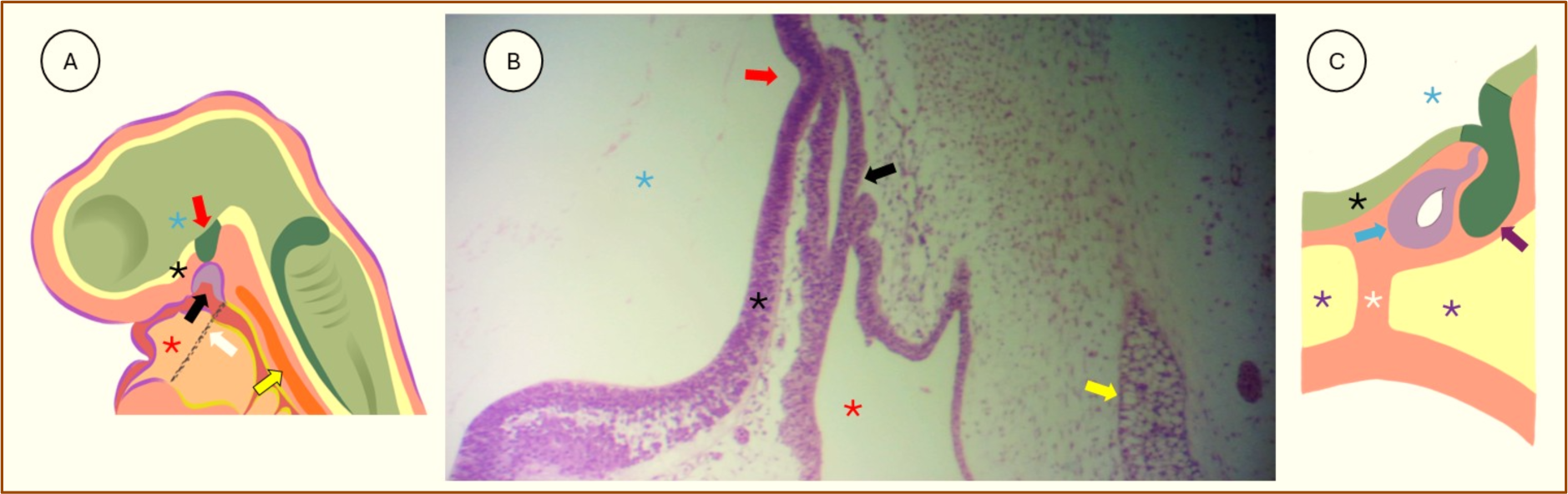
In the present study, we observed a higher prevalence of tumors implanted in the oral part of the pharynx, particularly in the palate, leading to fusion alteration of the palatal processes and subsequent cleft palate formation as the main developmental anomaly associated with epignathus, as reported in the cases described by Jadhav et al. [1], Kumar et al. [18], and Nguyen et al. [19]. Other different anatomical implantation sites were identified, highlighting the posterior third of the tongue, tonsils, and the roof of the pharynx. The main hypotheses associated with the embryological origin of these tumors relate to Rathke's pouch, the craniopharyngeal canal, or palatal development (Figure 2) [7]; however, these structures are located anterior to the buccopharyngeal membrane and, consequently, are covered solely by ectodermal tissue [20]. The different implantation sites of epignathus and the histological composition of the tumors, derived from tissues of all three germ layers, suggest an alternative embryological origin.
The developmental period of epignathus has a direct relationship with the direction of tumor growth and invasion of adjacent areas. In cases where tumors were identified up to the 22nd week of gestation, growth occurred from the oral cavity toward the vitelline cavity [21-23], whereas cases diagnosed after this period exhibited pharyngeal-directed growth and invasion of adjacent areas [8,24-26]. This characteristic may be related to airway morphogenesis and the rapid growth rate of tumors, which, when growing prematurely and aggressively, expand toward areas of greater space (oral and vitelline cavities) [27].

Embryological development of Rathke’s pouch and the craniopharyngeal canal. Figure 2A: Schematic representation of an embryo in the fourth week of intrauterine life, showing the formation of Rathke’s pouch (black arrow) lined by ectoderm in the roof of the primitive oral cavity or stomodeum (red asterisk), located anterior to the buccopharyngeal membrane (white arrow) and maintaining close contact with the diencephalic infundibulum (red arrow) within the neural tube (blue asterisk). Figure 2B: Low-magnification photomicrograph highlighting the development of Rathke’s pouch, which will give rise to the anterior lobe of the pituitary gland (blue arrow), and its close contact with the diencephalic infundibulum, which is internally lined by neuroepithelium (black asterisk) and will form the posterior lobe of the pituitary gland (purple arrow). Adjacent to these structures, ectomesenchyme provides structural support, and the notochord (indicated by the yellow arrow) is observed laterally. Chicken embryo (4 days), H&E stain, 40X magnification. Figure 2C: Schematic representation of the development of the craniopharyngeal canal and the pituitary lobes near the eighth week of intrauterine life. The fusion of the anterior and posterior lobes of the pituitary gland is observed, along with the path taken by the anterior lobe, forming the craniopharyngeal canal (white asterisk), which, in most cases, will be obliterated after complete ossification of the sphenoid bone (purple asterisk).
The developmental period of epignathus has a direct relationship with the direction of tumor growth and invasion of adjacent areas. In cases where tumors were identified up to the 22nd week of gestation, growth occurred from the oral cavity toward the vitelline cavity [21-23], whereas cases diagnosed after this period exhibited pharyngeal-directed growth and invasion of adjacent areas [8,24-26]. This characteristic may be related to airway morphogenesis and the rapid growth rate of tumors, which, when growing prematurely and aggressively, expand toward areas of greater space (oral and vitelline cavities) [27].
Evaluating parental health parameters and environmental conditions is an important method for assessing the etiology of congenital anomalies; however, few studies have reported parental health conditions. The history of previous abortion [9,28] and alcohol consumption during pregnancy [29] were the most frequently observed maternal health conditions. However, only Carvalho et al. [30] highlighted hypertension and diabetes as paternal health conditions, demonstrating the lack of data collection or availability regarding fathers. The absence of maternal exposure to drugs and radiation during pregnancy [2,31,32], as well as negative maternal serology for cytomegalovirus, toxoplasmosis, rubella, HIV, syphilis, and hepatitis B [2,33,34], represents critical clinical parameters in attempting to identify the etiology of epignathus. Parental consanguinity was not recognized as a risk factor for epignathus [2,19,22,30], although in vitro fertilization was reported [8,9].
We observed a higher prevalence of prenatal care, although in most cases, this care was limited to the early months of pregnancy and after complications arose. The International Society of Ultrasound in Obstetrics and Gynecology (ISUOG) emphasizes in its guidelines and clinical practice recommendations that first-trimester ultrasound examinations (up to the 14th gestational week) aim to estimate gestational age. After this period, they assess fetal growth parameters, including head and abdominal circumference. Second-trimester ultrasound is commonly performed between the 18th and 22nd gestational weeks to confirm gestational age and screen for possible developmental anomalies [35]. In the present study, most epignathus diagnoses occurred between the 22nd and 28th gestational weeks, which falls within the recommended second-trimester period for morphological ultrasound. Cases without adequate prenatal care were diagnosed after the 30th gestational week, increasing risks for both mother and fetus and reducing preparation time for potential interventions. ISUOG recommends additional diagnostic imaging, when necessary, in the third trimester [35]. The use of computed tomography and magnetic resonance imaging has been consistently reported as additional methods for assessing implantation, extension, and possible other associated and/or simultaneous alterations in epignathus. These examinations are necessary and should be planned by a multidisciplinary team according to the available therapeutic resources.
AFP is a glycoprotein belonging to the ABL gene family, which includes albumin and vitamin D-binding protein. It is predominantly expressed during the embryonic period in low amounts by the endoderm of the foregut, with its expression increasing in hepatocyte precursor cells as well as in the visceral endoderm of the yolk sac. After the embryonic period, AFP expression is suppressed, and its levels increase again in cases of liver regeneration and hepatocellular carcinoma [36]. AFP is used as a tumor marker for malignant recurrences of sacrococcygeal teratomas [37], whereas its measurement in epignathus has only been described sporadically in case reports.
Elevated AFP expression was observed in four cases with a favorable survival outcome [9, 19, 30, 38] and two cases with a fatal outcome [22,39]. In the four survival cases, AFP levels decreased after the initial surgical intervention, although tumor recurrence occurred in all four cases. In three of these cases, the initial approach involved partial tumor resection, followed by regrowth of the residual tumor. Two cases of epignathus with a favorable survival outcome did not exhibit alterations in AFP levels [40], which may be related to the presence of mature tissue components or the absence of hepatic components. Tunes et al. [34] reported a postoperative decrease in serum AFP levels, although the initial measurement was not provided. The measurement of β-HCG levels was reported in two cases, though their values were not disclosed [30,34].
Intracranial involvement of epignathus demonstrated the presence of tumors in the middle cranial fossa, causing alterations in the sella turcica of the sphenoid bone. This suggests a direct relationship with the development of Rathke’s pouch in these cases [41-43]. Epignathus with intracranial involvement or associated with hydrocephalus may complicate surgical intervention, leading to a worse prognosis [16]. In the present study, no cases of hydrocephalus associated with intracranial epignathus were observed. The presence of severe systemic abnormalities was associated with the fatal outcome of epignathus cases, notably including ectopia cordis [23], anencephaly [21], rudimentary cardiac structures [43], abnormalities of the great vessels, and other cardiovascular malformations [23,33].
The mean tumor size was slightly larger in cases with a fatal outcome; however, these tumors were notable for the premature termination of pregnancy, indicating an accelerated growth rate. The prenatal identification of congenital anomalies that may obstruct the airway or cause postnatal fetal distress due to respiratory obstruction necessitates the development of strategies to improve neonatal survival. In this regard, ex utero intrapartum treatment (EXIT) stands out. EXIT involves performing a cesarean section in which the fetus is partially delivered (head, neck, and torso) while maintaining placental support until a supportive procedure (intubation, tracheostomy, or lesion resection) is performed. After the procedure, the umbilical cord is clamped and subsequently cut [44,45].
The first successful EXIT procedure was performed in 1990 at Thomas Jefferson University Hospital in the United States for the treatment of a newborn with airway obstruction due to a congenital mass [46]. EXIT is a high-risk procedure for both mother and fetus, requiring a specialized multidisciplinary team. The procedure has been successful in most reported cases; however, difficulty in passing the endotracheal tube and the need for multiple intubation attempts are not uncommon [44,45]. Goto et al. [47] described a case of epignathus in which, after EXIT, a tracheostomy was attempted but proved difficult, followed by partial tumor resection. During the procedure, tracheal obstruction occurred, and the newborn did not survive. Postnatal intubation was frequently performed to ensure ventilatory support and manage possible complications.
Macroscopic evaluation of the tumors typically revealed solid lesions with cystic areas. The purely solid pattern was mainly described in lesions with a predominance of the immature areas. Routine histopathological evaluation using hematoxylin and eosin (H&E) staining remains the primary method for confirming these lesions. Immunohistochemical epignathus assessment can aid in establishing the diagnosis, particularly in identifying undifferentiated tissues, as well as mature tissue components that may sometimes be disorganized or isolated, as reported by Ge et al. [8] and Hu et al. [9]. In both cases, immunopositivity was observed for neural markers (GFAP, Olig-2, Calretinin, NeuN, and NSE), angiogenic markers (CD34, which identifies vascular endothelial cells), and markers of cellular proliferation (Ki-67).
Tumor recurrence was observed in most cases treated with partial surgical resection, whether due to lesion extension or anatomical implantation site, necessitating a second intervention for total resection and subsequent correction of deformities, such as cleft palate [8,9,23,25,33,38,49]. However, a few reports described a postoperative follow-up period longer than one year [8,30,48,49], highlighting the lack of long-term information regarding recurrence and associated comorbidities.
As limitations of this study, we emphasize the lack of information in case reports, which at times hindered the confirmation of lesion diagnoses. To minimize this limitation, we adopted stricter case selection criteria to ensure more reliable results.
Conclusion
Thus, epignathus is a rare lesion that poses a life-threatening risk to fetuses and neonates. The primary parameters associated with survival were complete prenatal care, smaller tumor size, access to life-support resources, and surgical intervention. Parameters linked to mortality included lesion identification at or before the 20th week, indicating premature tumor development and rapid growth potential. The degree of tissue maturation, with predominantly immature tumors indicating a worse prognosis, cannot be used as a predictive factor during pregnancy. Intracranial involvement and severe developmental anomalies of the nervous and cardiovascular systems negatively impact outcomes in epignathus cases.
Financial Support
-
None.
Data Availability
The data used to support the findings of this study can be made available upon request to the corresponding author.
References
-
[1] Jadhav SS, Korday CS, Malik S, Shah VK, Lad SK. Epignathus leading to fatal airway obstruction in a neonate. J Clin Diagn Res 2017; 11(1):SD04-SD05. https://doi.org/10.7860/JCDR/2017/24956.9283
» https://doi.org/10.7860/JCDR/2017/24956.9283 -
[2] Pellegrini V, Colasurdo F, Guerriero M. Epignathus with oropharynx destruction. Autops Case Rep 2021; 11:e2021293. https://doi.org/10.4322/acr.2021.293
» https://doi.org/10.4322/acr.2021.293 -
[3] Shah A, Latoo S, Ahmed I, Malik AH. Head and neck teratomas. J Maxillofac Oral Surg 2009; 8(1):60-63. https://doi.org/10.1007/s12663-009-0015-x
» https://doi.org/10.1007/s12663-009-0015-x -
[4] Simonini C, Strizek B, Berg C, Gembruch U, Mueller A, Heydweiller A, et al. Fetal teratomas - A retrospective observational single-center study. Prenat Diagn 2021; 41(3):301-307. https://doi.org/10.1002/pd.5872
» https://doi.org/10.1002/pd.5872 -
[5] Terenziani M, D'Angelo P, Inserra A, Boldrini R, Bisogno G, Babbo GL, et al. Mature and immature teratoma: A report from the second Italian pediatric study. Pediatr Blood Cancer 2015; 62(7):1202-1208. https://doi.org/10.1002/pbc.25423
» https://doi.org/10.1002/pbc.25423 -
[6] Joseph R, Abdoun O, Abdelrahim EM, Babiker AK, Mohammed AH. Congenital epignathus associated with a tricuspid incompetence. Cureus 2022; 14(7):e27135. https://doi.org/10.7759/cureus.27135
» https://doi.org/10.7759/cureus.27135 -
[7] Vandenhaute B, Leteurtre E, Lecomte-Houcke M, Pellerin P, Nuyts JP, Cuisset JM, et al. Epignathus teratoma: Report of three cases with a review of the literature. Cleft Palate Craniofac J 2000; 37(1):83-91. https://doi.org/10.1597/1545-1569_2000_037_0083_etrotc_2.3.co_2
» https://doi.org/10.1597/1545-1569_2000_037_0083_etrotc_2.3.co_2 -
[8] Ge S, Wang N, Shang W, Feng Y. Success in emergency treatment of neonatal giant teratoma with cleft palate: A case report. Cleft Palate Craniofac J 2023; 60(12):1659-1664. https://doi.org/10.1177/10556656221109753
» https://doi.org/10.1177/10556656221109753 -
[9] Hu X, Chu Y, Chen Y, Zhao M, Wang X, Xu L. Prenatal diagnosis of a giant epignathus in the second trimester and immediate successful management at birth: A case report. Maternal-Fetal Med 2023; 5(2):123-127. https://doi.org/10.1097/FM9.0000000000000116
» https://doi.org/10.1097/FM9.0000000000000116 - [10] World Health Organization. WHO Classification of Tumours Editorial Board. Head and neck tumours. 5th ed. Lyon (France): International Agency for Research on Cancer; 2022.
-
[11] Pavlin JE, O'Gorman A, Williams HB, Crépeau RJ, Shapiro RS. Epignathus: A report of two cases. Ann Plast Surg 1984; 13(5):452-456. https://doi.org/10.1097/00000637-198411000-00013
» https://doi.org/10.1097/00000637-198411000-00013 - [12] Ewing J. Neoplastic Diseases. Philadelphia: WB Saunders; 1940.
-
[13] Oosterhuis JW, Looijenga LHJ. Human germ cell tumours from a developmental perspective. Nat Rev Cancer 2019; 19(9):522-537. https://doi.org/10.1038/s41568-019-0178-9
» https://doi.org/10.1038/s41568-019-0178-9 -
[14] Sarioglu N, Wegner RD, Gasiorek-Wiens A, Entezami M, Schmock J, Hagen A, et al. Epignathus: Always a simple teratoma? Report of an exceptional case with two additional fetiforme bodies. Ultrasound Obstet Gynecol 2003; 21(4):397-403. https://doi.org/10.1002/uog.92
» https://doi.org/10.1002/uog.92 -
[15] Coppit GL 3rd, Perkins JA, Manning SC. Nasopharyngeal teratomas and dermoids: A review of the literature and case series. Int J Pediatr Otorhinolaryngol 2000; 52(3):219-227. https://doi.org/10.1016/s0165-5876(00)00288-3
» https://doi.org/10.1016/s0165-5876(00)00288-3 -
[16] Isaacs Jr H. Perinatal (fetal and neonatal) germ cell tumors. J Pediatr Surg 2004; 39(7):1003-1013. https://doi.org/10.1016/j.jpedsurg.2004.03.045
» https://doi.org/10.1016/j.jpedsurg.2004.03.045 - [17] Jindal A, Sharma M, Karena ZV, Chaudhary C. Amniocentesis. 2023. In: StatPearls. Treasure Island (FL): StatPearls Publishing; 2025.
-
[18] Kumar N, Ahluwalia C, Singla G, Singla S. Epiganthus presenting as respiratory distress in a male neonate: A rare case report. J Cancer Res Ther 2022; 18(4):1177-1179. https://doi.org/10.4103/jcrt.JCRT_888_19
» https://doi.org/10.4103/jcrt.JCRT_888_19 -
[19] Nguyen PTA, Lavisha P, Lynn KH, Jitendrakumar VS. Case report: Congenital palatal teratoma (epignathus) leading to neonatal airway obstruction. Int J Surg Case Rep 2023; 109:108518. https://doi.org/10.1016/j.ijscr.2023.108518
» https://doi.org/10.1016/j.ijscr.2023.108518 -
[20] Alatzoglou KS, Gregory LC, Dattani MT. Development of the Pituitary Gland. Compr Physiol 2020; 10(2):389-413. https://doi.org/10.1002/cphy.c150043
» https://doi.org/10.1002/cphy.c150043 -
[21] Morlino S, Castori M, Servadei F, Laino L, Silvestri E, Pediatric Craniofacial Malformation (PECRAM) Study Group, et al. Oropharyngeal teratoma, oral duplication, cervical diplomyelia and anencephaly in a 22-week fetus: A review of the craniofacial teratoma syndrome. Birth Defects Res A Clin Mol Teratol 2015; 103(6):554-566. https://doi.org/10.1002/bdra.23327
» https://doi.org/10.1002/bdra.23327 -
[22] Arora V, Bijarnia Mahay S, Rao S, Dimri N, Manocha A, Mansukhani C, et al. The fatal fetal tumor: A geneticist's perspective. J Matern Fetal Neonatal Med 2021; 34(6):1006-1008. https://doi.org/10.1080/14767058.2019.1622671
» https://doi.org/10.1080/14767058.2019.1622671 -
[23] Garofalo S, Guanà R, Suteu L, Alhellani H, Cortese MG, Lonati L, et al. Epignathus and thoracoabdominal ectopia cordis in a neonate. Pediatr Neonatol 2024; 65(4):410-411. https://doi.org/10.1016/j.pedneo.2024.03.005
» https://doi.org/10.1016/j.pedneo.2024.03.005 -
[24] Bayır Ö, Güneri EA, Dilek M, Özer E, Çakmakçı H, Erdağ TK. Nasopharyngeal mature teratoma in the newborn. Turk Pediatri Ars 2014; 49(3):257-260. https://doi.org/10.5152/tpa.2014.87
» https://doi.org/10.5152/tpa.2014.87 -
[25] Dakpé S, Demeer B, Cordonnier C, Devauchelle B. Emergency management of a congenital teratoma of the oral cavity at birth and three-year follow-up. Int J Oral Maxillofac Surg 2014; 43(4):433-436. https://doi.org/10.1016/j.ijom.2013.09.004
» https://doi.org/10.1016/j.ijom.2013.09.004 -
[26] Menezes Filho MP, Simão NM. Giant epignathus of the palate: A case report. J Bras Patol Med Lab 2015; 51(5):339-343. https://doi.org/10.5935/1676-2444.20150055
» https://doi.org/10.5935/1676-2444.20150055 -
[27] Schittny JC. Development of the lung. Cell Tissue Res 2017; 367(3):427-444. https://doi.org/10.1007/s00441-016-2545-0
» https://doi.org/10.1007/s00441-016-2545-0 -
[28] Kumar KM, Veligandla I, Lakshmi AR, Pandey V. Congenital giant teratoma arising from the hard palate: A rare clinical presentation. J Clin Diagn Res 2016; 10(7):ED03-ED04. https://doi.org/10.7860/JCDR/2016/18863.8083
» https://doi.org/10.7860/JCDR/2016/18863.8083 - [29] Akhtar R, Riffat M, Mehmood Z. Epignathus teratoma. J Coll Physicians Surg Pak 2016; 26(5):438-440.
-
[30] Carvalho CHP, Nonaka CFW, Elias CTV, Matheus RCS, Dias RMB, Souza LB, et al. Giant epignathus teratoma discovered at birth: A case report and 7-year follow-up. Braz Dent J 2017; 28(2):256-261. https://doi.org/10.1590/0103-6440201701368
» https://doi.org/10.1590/0103-6440201701368 - [31] Huang Y, Pan H. A fetus with a mass in the oral cavity: A rare case of oral immature teratoma. Int J Clin Exp Pathol 2017; 10(7):7890-7892.
-
[32] Zhang C, Yuan CL, Fang Q, Hu S. Teratoma of the tongue in a fetus: A case report and review of the literature. Pediatr Dev Pathol 2023; 26(1):86-90. https://doi.org/10.1177/10935266221132353
» https://doi.org/10.1177/10935266221132353 -
[33] Takagi MM, Bussamra LC, Araujo Júnior E, Drummond CL, Herbst SRS, Nardozza LMM, et al. Prenatal diagnosis of a large epignathus teratoma using two-dimensional and three-dimensional ultrasound: Correlation with pathological findings. Cleft Palate Craniofac J 2014; 51(3):350-353. https://doi.org/10.1597/12-222
» https://doi.org/10.1597/12-222 -
[34] Tunes RS, Cavalcanti GZ, Squarisi JMO, Patrocinio LG. Oral epignathus with maxilla duplication: Report of a rare case. Craniomaxillofac Trauma Reconstr 2019; 12(1):62-66. https://doi.org/10.1055/s-0038-1649497
» https://doi.org/10.1055/s-0038-1649497 -
[35] Salomon LJ, Alfirevic Z, Da Silva Costa F, Deter RL, Figueras F, Ghi T, et al. ISUOG Practice Guidelines: Ultrasound assessment of fetal biometry and growth. Ultrasound Obstet Gynecol 2019; 53(6):715-723. https://doi.org/10.1002/uog.20272
» https://doi.org/10.1002/uog.20272 -
[36] Jin L, Long L, Green MA, Spear BT. A região intensificadora da alfa-fetoproteína ativa os promotores da albumina e da alfa-fetoproteína durante o desenvolvimento do fígado. Dev Biol 2009; 336(2):294-300. https://doi.org/10.1016/j.ydbio.2009.09.026
» https://doi.org/10.1016/j.ydbio.2009.09.026 -
[37] van Heurn LJ, Knipscheer MM, Derikx JPM, van Heurn LWE. Diagnostic accuracy of serum alpha-fetoprotein levels in diagnosing recurrent sacrococcygeal teratoma: A systematic review. J Pediatr Surg 2020; 55(9):1732-1739. https://doi.org/10.1016/j.jpedsurg.2020.03.014
» https://doi.org/10.1016/j.jpedsurg.2020.03.014 -
[38] Jayagobi PA, Chandran S, Sriram B, Chang KT. Ex-utero Intrapartum Treatment (EXIT) Procedure for Giant Fetal Epignathus. Indian Pediatr 2015; 52(10):893-895. https://doi.org/10.1007/s13312-015-0740-9
» https://doi.org/10.1007/s13312-015-0740-9 -
[39] Faghfouri F, Bucourt M, Garel C, Benchimol M, Amarenco B, Soupre V, et al. Prenatal assessment of a fast-growing giant epignathus. Fetal Pediatr Pathol 2014; 33(1):55-59. https://doi.org/10.3109/15513815.2013.850134
» https://doi.org/10.3109/15513815.2013.850134 -
[40] Kadlub N, Touma J, Leboulanger N, Garel C, Soupre V, L’Herminé AC, et al. Head and neck teratoma: From diagnosis to treatment. J Craniomaxillofac Surg 2014; 42(8):1598-1603. https://doi.org/10.1016/j.jcms.2014.04.028
» https://doi.org/10.1016/j.jcms.2014.04.028 -
[41] Wang AC, Gu YQ, Zhou XY. Congenital giant epignathus with intracranial extension in a fetal. Chin Med J 2017; 130(19):2386-2387. https://doi.org/10.4103/0366-6999.215343
» https://doi.org/10.4103/0366-6999.215343 -
[42] Kirishima M, Yamada S, Shinya M, Onishi S, Goto Y, Kitazono I, et al. An autopsy case of epignathus (immature teratoma of the soft palate) with intracranial extension but without brain invasion: Case report and literature review. Diagn Pathol 2018; 13(1):99. https://doi.org/10.1186/s13000-018-0776-y
» https://doi.org/10.1186/s13000-018-0776-y -
[43] Tavares de Sousa M, Chaoui R, Herrmann J, Hecher K. Intrauterine laser coagulation of rapidly growing epignathus with autonomous cardiac activity. Ultrasound Obstet Gynecol 2024; 63(3):419-420. https://doi.org/10.1002/uog.27477
» https://doi.org/10.1002/uog.27477 -
[44] Mohammad S, Olutoye OA. Airway management for neonates requiring ex utero intrapartum treatment (EXIT). Paediatr Anaesth 2020; 30(3):248-256. https://doi.org/10.1111/pan.13818
» https://doi.org/10.1111/pan.13818 - [45] De Jong R, Fordham T. Ex utero Intrapartum Treatment (EXIT) Procedure. StatPearls. Treasure Island (FL): StatPearls Publishing; 2025.
- [46] Kelly MF, Berenholz L, Rizzo KA, Greco R, Wolfson P, Zwillenberg DA. Approach for oxygenation of the newborn with airway obstruction due to a cervical mass. Ann Otol Rhinol Laryngol 1990; 99(3 Pt 1):179-182.
-
[47] Goto S, Nakazawa A, Yoshizawa T, Aoki M, Hirabayashi T, Seino H, et al. Case report: A giant epignathus with a well-developed skeleton of head and neck. Int J Surg Pathol 2023; 31(7):1335-1339. https://doi.org/10.1177/10668969221133739
» https://doi.org/10.1177/10668969221133739 -
[48] Pang X, Kwon HJ, Shi B, Li C. A rare nasopharyngeal teratoma arising from the vomer. J Craniofac Surg 2016; 27(2):e165-166. https://doi.org/10.1097/SCS.0000000000002407
» https://doi.org/10.1097/SCS.0000000000002407 -
[49] Sugiyama M, Kanke K, Suenaga H, Isojima T, Fujishiro J, Hoshi K. Neonatal epignathus in the oral and pharyngeal regions: A case report. Oral Sci Int 2019; 16:40-46. https://doi.org/10.1002/osi2.1012
» https://doi.org/10.1002/osi2.1012
Edited by
-
Academic Editor:
Alidianne Fábia Cabral Cavalcanti