Microscopic Analysis of 3D-Printed Resin Composites for Definitive Restorations
ABSTRACT
Objective: To characterize the microscopic structure and surface properties of commercially available 3D-printed resin composites using Scanning Electron Microscopy (SEM) to assess their clinical applicability and durability.
Material and Methods: Four different 3D-printed resin composites were analyzed. Specimens were fabricated under controlled conditions, followed by standardized cleaning, post-curing, and polishing procedures. SEM imaging was performed at various magnifications to evaluate filler distribution, particle morphology, and surface integrity.
Results: The analysis revealed significant differences among the tested resins. Varseo Smile Crown Plus (Bego) exhibited the most homogeneous microstructure, with a high filler content, nanoscale particles, and strong polymer-filler integration, suggesting superior mechanical properties. Biocrown and Nanolabs resins showed intermediate levels of filler content and particle distribution. At the same time, Printax AA Temp had the lowest inorganic content and the highest incidence of structural defects, including layer discontinuities and cracks.
Conclusion: The structural and surface characteristics of 3D-printed resin composites vary significantly among different commercial brands. Varseo Smile Crown Plus demonstrated the most promising properties for clinical applications, while other materials showed limitations related to filler distribution and structural integrity. Further studies on mechanical performance and long-term stability are needed to optimize the application of these materials in definitive restorations.
Keywords:
Technology; Printing; Three-Dimensional; Microscopy; Electron; Scanning.
Introduction
The integration of digital systems has significantly transformed clinical dental practice, enabling precise and efficient workflows for rehabilitation procedures. Among these technological advancements, three-dimensional (3D) printing has emerged as a key innovation, allowing for in-office fabrication of diagnostic models, surgical guides, and even definitive restorations [1,2]. This process, known as stereolithography (SLA), utilizes light to solidify thin layers of a photosensitive liquid resin. The process occurs in a resin vat, where the light selectively cures the material by tracing the geometry of each layer on the liquid surface. Once a layer is cured, the build platform lowers slightly, allowing a new layer of resin to be exposed and solidified. This cycle repeats until the complete object is formed [3].
Over the years, continuous advancements, such as the development of more affordable LCD and DLP-based printers and user-friendly CAD/CAM software, have expanded its applications, making it increasingly accessible for in-office use [3]. This shift has not only optimized production efficiency but also introduced new material demands suited to digital workflows.
Similar to the evolution of conventional paste-based resin composites, 3D-printed resin composites have undergone advancements in filler technology. Initially, large filler particles were incorporated in paste-based composites, followed by progressively smaller ones, ultimately leading to the integration of nanoparticles to enhance mechanical and aesthetic properties. These nanoparticle-reinforced photopolymerizable resins were introduced to meet the demand for a universal restorative material suitable for both anterior and posterior teeth [4]. The application of nanotechnology in these formulations has resulted in mechanical properties comparable to microhybrid composites, while also improving polishability and achieving smoother surfaces, a characteristic typically associated with microparticle-based resins [5].
In contrast, 3D-printed resin composites have also evolved through the addition of fillers to enhance mechanical performance; however, standardization of the type, concentration, size, and surface treatment of these particles remains uncertain. Unlike conventional composites, where filler composition is well-established, the optimization of particle distribution and polymer matrix interactions in 3D-printed resins is still an ongoing area of research, with potential implications for their long-term clinical behavior [6].
The workflow for producing dental restorations using 3D printers and photopolymerizable resins can be summarized in three main steps: printing, post-print washing, and final curing. Post-print washing is a critical phase, as studies indicate that the most effective method involves immersing or rinsing the printed objects in an organic solvent, such as isopropyl alcohol (IPA), ethanol, or tripropylene glycol monomethyl ether (TPM) [7]. However, as highlighted by Liu et al. [7], the use of these solvents presents several drawbacks, including the potential degradation of mechanical properties when exposure is prolonged. Following the cleaning phase, final curing using violet light is essential to ensure the complete polymerization of the material and optimize its physical and mechanical properties. This step is particularly crucial for long-term mechanical strength and dimensional stability. Controlled exposure to violet light can also reduce the presence of unpolymerized residues on the surfaces of printed restorations, thereby enhancing their biocompatibility and surface quality.
Despite the rapid advancements in 3D printing technology and the increasing adoption of 3D-printed resin composites for definitive restorations, significant knowledge gaps remain regarding their structural and surface properties. Unlike traditional resin composites, which have well-characterized filler compositions and polymer matrices, the type, concentration, and surface treatment of fillers in 3D-printed resins are still not fully standardized. Additionally, post-processing variables, such as washing, curing, and finishing protocols, may significantly impact the final material properties.
To ensure the long-term clinical success of 3D-printed definitive restorations, it is essential to investigate how these materials behave at the microscopic level, particularly with respect to surface roughness and mechanical integrity [8]. Scanning Electron Microscopy (SEM) provides detailed evaluation of surface characteristics, enabling assessment of porosity, microdefects, and post-processing effects. Understanding these properties is crucial for optimizing printing parameters, post-processing techniques, and clinical protocols to enhance the reliability and performance of these materials.
Thus, this study aims to characterize 3D-printed resin composites by analyzing their microscopic structure and surface characteristics using SEM, providing insights into their clinical applicability and long-term durability.
Material and Methods
Calibration Parameters of 3D Printer and Resin
Four commercially available 3D printing resins with fillers were used (Biocrown, Nanolab 3D, Printax AA Temp, and Varseo Smile Crown Plus) (Table 1).
Initially, an exposure light calibration test was conducted. An object with known dimensions was printed with the four filler-containing 3D printing resins to validate the exposure time of violet light for each resin by assessing its dimensional accuracy through measurements of known dimensions using a digital caliper (Mitutoyo, Jundiaí, Brazil). The exposure time was adjusted based on the results of this preliminary calibration. Prior to printing, the resin was homogenized and preheated to 31°C for 40 minutes using a specialized device (Misturador de Resina, Slim3D, Curitiba, Brazil), which is capable of simultaneously heating and mixing the resin through controlled rotation. All prints, including the calibration test, were performed in a controlled environment. During the printing process, the resin temperature was maintained at 30°C using an external thermostat, as the printer itself does not have an integrated temperature control system. Temperature consistency was monitored with a portable thermometer placed inside the printer chamber.
Specimen Preparation
A rectangular plate (10mm width, 10mm height, and 2mm thickness) was digitally designed (Meshmixer, Autodesk Inc., San Rafael, CA, USA) and exported in STL format for the addition of standardized printing supports using a slicing software (Chitubox, ChiTu Systems, Guangdong Province, China). It was positioned parallel to the virtual printing platform in the slicing software and printing supports were added to the square bottom face (0.5 mm tips, 0.85 density and 3 mm height). This configuration was replicated for a total of 10 specimens (n=10) using the "copy" tool. The specimens (n=5) were printed using a Mono-LCD printer (Sonic Mini 8k, Phrozen 3D Printer, Hsinchu, Taiwan) following the calibration parameters described and the recommendations for each manufacturer of the four 3D printing resins. The printing process was conducted at a controlled temperature of 30°C, maintained using a thermostat and a portable thermometer placed inside the 3D printer.
After printing, all specimens were carefully detached from the platform and subjected to a cleaning and post-curing process. Isopropyl alcohol (Oficinallis Pharma, Jáu, SP, Brazil) was used as the cleaning agent for all tested resins. The specimens were immersed in the solution in a magnetic ultrasonic bath and, after 5 minutes, brushed with a soft toothbrush (Colgate Slim Soft Black, Colgate-Palmolive Company, São Paulo, SP, Brazil). After cleaning, the specimens were air-dried for 8 minutes using the "cooler" function in a wash-and-cure chamber (Wash & Cure, Phrozen, Hsinchu, Taiwan) to ensure complete evaporation of residual alcohol and avoid interference with material properties. The specimens were then post-cured under violet light (405nm) in the same chamber for 8 minutes. All samples were stored at 37ºC and 100% relative humidity for 24 hours. Polishing steps were performed using sandpaper to eliminate surface irregularities and standardize the specimens. The specimens were cleaned in an ultrasonic bath (UltraSonic 1440 Plus, Odontobrás Equipamentos Médicos e Odontológicos Ltda., Ribeirão Preto, SP, Brazil) for 15 minutes in distilled water and dried.
Scanning Electron Microscopy (SEM)
The specimens were subjected to scanning electron microscopy. They were fixed using carbon double-sided tape (marca), followed by a metal coating procedure, and then analyzed using a Sputter Coater, model K450 (Emitech/Quorum Technologies Ltd., Kent, UK), under a voltage of 15 kV and a current of 50 pA at magnifications of 35x abs 500x to observe the surface pattern of the material.
Results
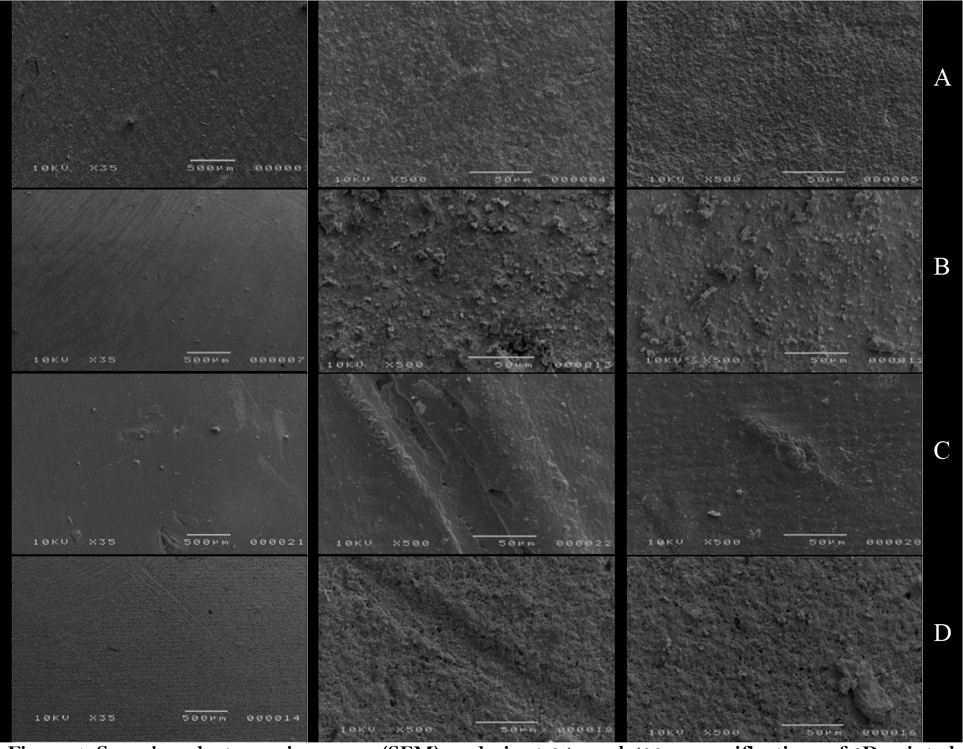
The qualitative analysis of the Varseo Smile Crown Plus 3D resin after SEM reveals a highly homogeneous distribution of filler particles within the organic matrix. This resin exhibited the highest filler particle content among all evaluated materials. The particles were well-defined and had the smallest average particle size, evenly distributed and did not form agglomerations. Additionally, the filler and organic matrix demonstrated good bonding. No visible printing defects, such as layer lines, bonding failures between layers, cracks, or material shrinkage, were observed (Figure 1A). Biocrown 3D resin displayed a low quantity of dispersed filler particles in the polymer matrix. The particles had an irregular surface and an average size and an average size higher than 1 µm. The filler distribution was also heterogeneous, with visible agglomerations. Printing defects such as distinct layer lines and adhesion failures between layers were observed, leading to cracks (Figure 1B). Nanolabs 3D resin exhibited an intermediate amount of inorganic particles (Figure 1D). However, the filler particles were significantly larger than Varseo Smile Crown Plus 3D resin, and a high degree of agglomeration was observed. Additionally, distinct printing lines were present, indicating layer discontinuities. Printax AA Temp presented an almost entirely polymeric matrix, with minimal inorganic content. The few filler particles present were irregular and poorly distributed. This resin exhibited the highest number of defects, including layer discontinuities, cracks, and bubbles and poor integration between organic and inorganic components was evident (Figure 1D).

Scanning electron microscopy (SEM) analysis at 35× and 500× magnifications of 3D-printed resin specimens. (A): Varseo Smile Crown Plus (Bego); (B): Biocrown (Makertech); (C): Printax AA Temp (Odontomega); (D): Nanolab 3D (Wilcos do Brasil).
Among the analyzed resins, Varseo Smile Crown Plus exhibited the most homogeneous microstructure, with a high filler content and uniform distribution of the smallest ceramic particles. Biocrown and Nanolabs resins showed an intermediate level of structural organization, with Biocrown exhibiting a lower filler content and Nanolabs displaying larger, more agglomerated particles. Printax AA Temp had the lowest inorganic content and the highest number of defects, indicating a less-reinforced polymeric matrix.
Discussion
Scanning Electron Microscopy (SEM) is widely used in dental materials research to analyze filler morphology, distribution, and the interface between inorganic particles and the polymer matrix, factors that directly impact mechanical behavior and clinical durability [9]. In this study, SEM revealed distinct microstructural features among the tested 3D-printed resins, highlighting differences in filler homogeneity, particle size, and surface defects. While SEM provides detailed topographic information, it has limitations, including limited depth of field, susceptibility to sample-preparation artifacts, and an inability to detect nanometric-scale roughness. Techniques such as Atomic Force Microscopy (AFM), Field Emission Gun SEM (FEG-SEM), and Energy Dispersive X-ray Spectroscopy (EDX) may complement SEM by providing higher resolution or compositional data [8,10]. These approaches can enhance understanding of how filler characteristics influence the structural integrity and clinical performance of 3D-printed composites.
The filler content in resin-based composites plays a critical role in determining their mechanical properties and clinical durability. In both conventional and 3D-printed systems, increased filler loading is typically associated with enhanced flexural strength, wear resistance, and reduced polymerization shrinkage [11,12]. In a recent study, Sasany et al. [13] demonstrated that 3D-printed resin composites exhibited greater nanoindentation creep than milled CAD/CAM blocks after storage in water and artificial saliva, highlighting their reduced dimensional stability over time. These findings underscore the influence of filler concentration and microstructural integrity on the material’s ability to resist long-term deformation under stress. In the present study, the Bego resin, which exhibited the highest filler content and most homogeneous distribution, is expected to show improved resistance to mechanical fatigue and creep over time. Conversely, Printax AA Temp, composed predominantly of an organic matrix and displaying low inorganic content and multiple structural defects, is likely more prone to deformation and clinical failure under load. While Biocrown and Nanolabs resins showed intermediate filler concentrations, the presence of irregular particle shapes and agglomerates may promote stress concentration, ultimately compromising their mechanical performance.
The size and morphology of filler particles also influence the physical and aesthetic properties of 3D-printed resins [14]. The SEM analysis showed that only Varseo Smile Crown Plus (Bego) contained nanoscale filler particles, whereas the other resins presented larger and more irregularly shaped particles. Smaller filler particles are associated with superior polishability, gloss retention, and lower wear rates, making the material more suitable for anterior and posterior restorations where both aesthetics and durability are critical. Additionally, larger filler particles are prone to detachment from the polymer matrix over time, creating microvoids that contribute to surface roughness and increased bacterial adhesion. The spherical shape of the ceramic particles in Bego resin further enhances its esthetic properties by facilitating more uniform light reflection, contributing to a more natural appearance. In contrast, the irregular morphology of the fillers in Biocrown and Nanolabs resins may compromise their optical properties and increase the risk of microstructural defects.
Çağlayan et al. [15] investigated the optical performance of additively and subtractively manufactured dental materials by evaluating parameters such as color stability (∆E00) and relative translucency (RTP) before and after repair procedures and thermocycling. Their results revealed that 3D-printed resin composites exhibited significantly higher color changes and greater variability in translucency compared to subtractively manufactured ceramics. These optical shifts were attributed to the intrinsic material structure and its interaction with the environment over time. Although the present study did not directly assess optical behavior, SEM analysis revealed marked differences in filler morphology and distribution among the tested 3D-printed resins. Notably, materials such as Printax AA Temp showed heterogeneous filler dispersion, interfacial voids, and surface irregularities, which may contribute to increased pigment absorption or accelerated optical degradation under clinical conditions. These findings reinforce the link between microstructure and esthetics as resins with agglomerates or porosity tend to allow greater fluid uptake and color instability over time. Therefore, assessing filler homogeneity and interfacial quality is not only relevant for predicting mechanical performance, but also for anticipating long-term esthetic outcomes.
Despite the mechanical benefits of increasing filler content in 3D-printed resin composites, its incorporation poses challenges related to viscosity, dispersion, and interfacial integrity. Higher filler loading elevates viscosity, hindering resin flow and potentially causing printing defects such as incomplete polymerization and interlayer adhesion failures [16]. Poor dispersion may also lead to agglomerates that act as stress concentrators. In this study, the layer discontinuities observed in Biocrown and Nanolabs may be attributed to inadequate filler dispersion, while the homogeneous structure of Bego resin suggests a balanced formulation that preserves printability. In addition, the chemical interaction between filler and matrix plays a critical role. Improperly silanized particles can weaken the structure, facilitating crack propagation, especially in the intraoral environment, characterized by fluctuations in moisture, temperature, and pH [17]. Zattera et al. found that well-bonded barium glass fillers improved flexural strength and stress distribution in 3D-printed resins [6], consistent with the strong filler-matrix interface observed in Bego.
In summary, the SEM analysis of the evaluated resins revealed significant qualitative differences in filler content, particle size, morphology, and distribution. Varseo Smile Crown Plus (Bego) demonstrated the most homogeneous structure, with a high filler load, nanoscale particle size, and a well-integrated polymer-filler interface. These characteristics suggest superior mechanical and esthetic performance, making it a promising material for both anterior and posterior restorations. In contrast, Biocrown and Nanolabs exhibited intermediate levels of organization, with Biocrown showing a lower filler content and Nanolabs presenting larger and more agglomerated particles. Printax AA Temp, with its predominantly polymeric composition, demonstrated the highest number of defects, indicating a less reinforced and structurally weaker material. However, it is important to note that filler characteristics are not the only determining factors in the clinical performance of 3D-printed resins. The monomer composition, degree of polymerization, and post-processing protocols [7] also play a fundamental role in defining the mechanical properties and long-term stability of these materials. Further investigations, including mechanical testing and long-term aging studies, are necessary to fully understand the implications of these microstructural differences on the clinical performance of 3D-printed resin-based composites.
Conclusion
Significant differences in filler content, particle size, distribution, and integration with the organic matrix were observed. Varseo Smile Crown Plus exhibited the most homogeneous microstructure, with a high filler load, nanoscale particles, and a well-integrated polymer-filler interface, suggesting superior mechanical and esthetic properties. In contrast, Biocrown and Nanolabs presented intermediate structural organization, while Printax AA Temp displayed the lowest inorganic content and the highest number of defects.
-
Financial Support
None.
Data Availability
The data used to support the findings of this study can be made available upon request to the corresponding author.
References
-
[1] Rekow ED. Digital dentistry: The new state of the art - Is it disruptive or destructive? Dent Mater 2020; 36(1):9-24. https://doi.org/10.1016/j.dental.2019.08.103
» https://doi.org/10.1016/j.dental.2019.08.103 -
[2] Whitaker M. The history of 3D printing in healthcare. Bull R Coll Surg Engl 2014; 96(7):228-229. https://doi.org/10.1308/147363514X13990346756481
» https://doi.org/10.1308/147363514X13990346756481 -
[3] Javaid M, Haleem A. Current status and applications of additive manufacturing in dentistry: A literature-based review. J Oral Biol Craniofac Res 2019; 9(3):179-185. https://doi.org/10.1016/j.jobcr.2019.04.004
» https://doi.org/10.1016/j.jobcr.2019.04.004 -
[4] Ferracane JL. Resin composite - State of the art. Dent Mater 2011; 27(1):29-38. https://doi.org/10.1016/j.dental.2010.10.020
» https://doi.org/10.1016/j.dental.2010.10.020 -
[5] Mitra SB, Wu D, Holmes BN. An application of nanotechnology in advanced dental materials. J Am Dent Assoc 2003; 134(10):1382-1390. https://doi.org/10.14219/jada.archive.2003.0054
» https://doi.org/10.14219/jada.archive.2003.0054 -
[6] Zattera ACA, Morganti FA, de Souza Balbinot G, Della Bona A, Collares FM. The influence of filler load in 3D printing resin-based composites. Dent Mater 2024; 40(7):1041-1046. https://doi.org/10.1016/j.dental.2024.05.016
» https://doi.org/10.1016/j.dental.2024.05.016 -
[7] LIU, Yunqi; JIN, Gan; LIM, Jung Hwa; KIM, Jong Eun. Effects of washing agents on the mechanical and biocompatibility properties of water washable 3D printing crown and bridge resin. Sci Rep 2024; 14(1):9909. https://doi.org/10.1038/s41598-024-60450-7
» https://doi.org/10.1038/s41598-024-60450-7 -
[8] Sandmair MN, Kleber C, Ströbele DA, von See C. AFM Analysis of a three-point flexure tested, 3d printing definitive restoration material for dentistry. J Funct Biomater 2023; 14(3):152. https://doi.org/10.3390/jfb14030152
» https://doi.org/10.3390/jfb14030152 -
[9] Hosoda H, Yamada T, Inokoshi S. SEM and elemental analysis of composite resins. J Prosthet Dent 1990; 64(6):669-76. https://doi.org/10.1016/0022-3913(90)90293-l
» https://doi.org/10.1016/0022-3913(90)90293-l -
[10] Rastelli AN, Jacomassi DP, Faloni AP, Queiroz TP, Rojas SS, Bernardi MI, et al. The filler content of the dental composite resins and their influence on different properties. Microsc Res Tech 2012; 75(6):758-765. https://doi.org/10.1002/jemt.21122
» https://doi.org/10.1002/jemt.21122 -
[11] Randolph LD, Palin WM, Leloup G, Leprince JG. Filler characteristics of modern dental resin composites and their influence on physico-mechanical properties. Dent Mater 2016; 32(12):1586-1599. https://doi.org/10.1016/j.dental.2016.09.034
» https://doi.org/10.1016/j.dental.2016.09.034 -
[12] Wang Z, Chiang MY. System compliance dictates the effect of composite filler content on polymerization shrinkage stress. Dent Mater 2016; 32(4):551-560. https://doi.org/10.1016/j.dental.2016.01.006
» https://doi.org/10.1016/j.dental.2016.01.006 -
[13] Sasany R, Jamjoom FZ, Uçar SM, Yilmaz B. Nanoindentation creep: The impact of water and artificial saliva storage on milled and 3D-printed resin composites. J Prosthodont 2024. https://doi.org/10.1111/jopr.13913 Epub ahead of print.
» https://doi.org/10.1111/jopr.13913 -
[14] Kim KH, Ong JL, Okuno O. The effect of filler loading and morphology on the mechanical properties of contemporary composites. J Prosthet Den 2002; 87(6):642-649. https://doi.org/10.1067/mpr.2002.125179
» https://doi.org/10.1067/mpr.2002.125179 -
[15] Çağlayan E, Sasany R, Bolat BN, Hartavi D, Batgerel OE, Ucar SM, et al. Optical properties of repaired additively manufactured resin composites and zirconia and subtractively manufactured ceramics: A comparative study of composite resins. BMC Oral Health 2025; 25(1):144. https://doi.org/10.1186/s12903-025-05538-w
» https://doi.org/10.1186/s12903-025-05538-w -
[16] Paral SK, Lin DZ, Cheng YL, Lin SC, Jeng JY. A review of critical issues in high-speed vat photopolymerization. Polymers 2023; 15(12):2716. https://doi.org/10.3390/polym15122716
» https://doi.org/10.3390/polym15122716 -
[17] Aydınoğlu A, Yoruç ABH. Effects of silane-modified fillers on properties of dental composite resin. Mater Sci Eng C Mater Biol Appl 2017; 79:382-389. https://doi.org/10.1016/j.msec.2017.04.151
» https://doi.org/10.1016/j.msec.2017.04.151
Edited by
-
Academic Editor:
Wilton Wilney Nascimento Padilha