
 A revision of the subgenera Tidops Chamberlin 1915 and Newportides Chamberlin 1921 (Scolopocryptopidae: Newportiinae: Newportia Gervais, 1847) with four new species from Northern Brazil
Abstract
This study presents a taxonomic review of the subgenera Tidops Chamberlin, 1915 and Newportides Chamberlin, 1921, and description of four new species: N. (Tidops) mapinguarisp. nov.,N. (Tidops) oxossisp. nov., N. (Newportides) anhangasp. nov., and N. (Newportides) exusp. nov., all from the Brazilian Amazonia. Additionally, new records of N. (Tidops) collaris Kraepelin, 1903, a key and a map of distribution for all known species of Newportia (Tidops) and Newportia (Newportides) are provided.
Keywords:
Centipede; Chilopoda; Scolopendromorpha; Neotropical Region; Taxonomy
INTRODUCTION
The genus Newportia is the most diverse in Scolopocryptopidae Pocock, 1896, with nearly 51 species described, which represents about 60% of all species currently known in the family. Newportia species can range from 05 to 94 mm in length, with color varying from yellow to reddish-brown (Edgecombe & Bonato, 2011; Tulande-M. et al., 2020), being distributed from northern Mexico, through the Caribbean, to as far south as Uruguay (Schileyko et al., 2020; Tulande-M. et al., 2020) (Fig. 1). Despite being the solely genus of the subfamily Newportiinae Pocock, 1896, no less than six sub-genera are presently recognized in Newportia: Newportia sensu stricto (40 species and 10 subspecies), NewportidesChamberlin, 1921, (three species), TidopsChamberlin, 1915 (four species), EctonocryptopsCrabill, 1977 (one species), EctonocryptoidesShelley & Mercurio, 2005 (two species) and Andeocryptops Tulande, Prado, Giménez & Chagas-Jr., 2020 (one species) (Schileyko et al., 2020; Tulande-M. et al., 2020).
Chamberlin (1915) proposed Tidops based on a species from Granada, T. simusChamberlin, 1915, arguing that Tidops differs from Newportia by the much shorter tarsungulum and by the strongly developed dental plate. Lately, Tidops was revised by Chagas-Jr. (2011), who recognized four species to Central and South America. In a phylogeny of Scolopendromorpha based on molecular data, Vahtera et al. (2013), recovered T. collarisKraepelin, 1903 as associated to a Newportia clade. As a result, Tidops was considered a subgenus of Newportia.Chamberlin (1921) proposed Newportides as a subgenus of Newportia, based on a single species from Guyana, N. (Newportides) unguifer Chamberlin, 1921, with claws (pretarsus) on the ultimate legs, a character absent in Newportia sensu stricto (Chamberlin, 1921). Newportides was latter synonymized with Newportia by Schileyko & Minelli (1999) but recognized as a subgenus of Newportia by Chagas-Jr. (2018).
This paper aims to provide an overview of the subgenera Tidops and Newportides, describing four new species (two of Tidops and two of Newportides)

Geographical distribution of NewportiaGervais, 1847. Newportia Gervais, 1847 in GBIF Secretariat (2021).
from Brazilian Amazon and providing updated diagnoses and identification keys to all currently known species of these taxa.
MATERIAL AND METHODS
The material herein reported is deposited in at the Museu Paraense Emílio Goeldi, Pará, Brazil (MPEG, curator: A.B. Bonaldo). Acronyms of other cited institutions: Museum of Comparative Zoology, Harvard University, Cambridge, Massachusetts, USA (MCZ); Museu de Zoologia da Universidade de São Paulo, São Paulo, Brazil (MZSP); Museo del Instituto de Zoología Agrícola Francisco Fernández Yépez, Maracay, Venezuela (MIZA); Zoologisches Museum Hamburg, Hamburg, Germany (ZMH) and Museu Nacional, Universidade Federal do Rio de Janeiro, Rio de Janeiro, Brazil (MNRJ).
The specimens were examined immersed in alcohol 80%. Visualization and descriptions were performed with the aid of a Leica MZ16 stereomicroscope. Measurements and photographs were performed with a M205A stereomicroscope coupled to a Leica MC170 HD digital camera. All measurements are expressed in millimeters (mm). Compound photographs were vectorized in Adobe Illustrator C.C. 2020. General morphology terminology follows Bonato et al. (2010), with modifications by Schileyko (2013) and Chagas-Jr. (2011, 2018). Distribution maps were created in QGIS 3.16. For the distribution map of the genus Newportia (Fig. 1), the records available on the biodiversity data platform Global Biodiversity Information Facility (GBIF, 2021) were downloaded and complemented with the new occurrences here recorded.
Abbreviations used in the descriptions: (AN): Antenna; (BA) basic articles (CP) cephalic plate; (PMS): posterior margin suture; (FC): forcipular coxosternite; (TAR): Tarsungula; (TP): tooth-plates; (PTP): processes of tronchanteroprefemur; (TE): Tergites; (PS): paramedian suture; (TS): transverse suture; (LS): lateral suture; (OLS): oblique lateral sutures; (MS): median suture (MTS): median tergite suture; (L): leg; (S): sternites; (LSS): lateral sulcus on the sternites; (PF): Pre-femur (F): Femur; (Ti): Tibia; (T1): Tarsus 1; (T2) Tarsus 2; (PT): pretarsus; (CXP): coxopleural process; (CX): coxopleura; (MLS): median longitudinal sulcus; (VSPU): ventral spinous processes of prefemur of ultimate legs; (MSPF): medial spinous processes of the ultimate femur; (CC): Coxosternal condyle; (LMK): longitudinal median keels.
RESULTS
Taxonomy
Order Scolopendromorpha Pocock, 1895 Family Scolopocryptopidae Pocock, 1896 Subfamily Newportiinae Pocock, 1896 Genus Newportia Gervais, 1847
Diagnosis (adapted from Schileyko et al., 2020; Tulande-M. et al., 2020): 23 pair of legs, cephalic plate without ocelli, prefemur of ultimate legs with more than one spinose process.
Key to subgenera of Newportia (amended from Tulande-M. et al., 2020 )
1. Tarsus of ultimate legs subclavate and bulbous 2
- Tarsus of ultimate legs non-subclavate and thin 3
2(1). Tarsus 2 of ultimate legs globose, small but well-developed (Schileyko et al., 2020: fig. 14)Ectonocryptops
- Tarsus 2 of ultimate legs reduced or rudimentary (Schileyko et al., 2020: fig. 15)Ectonocryptoides
3(1). Tarsus 2 of ultimate thin legs divided in minute secondary articles 4
- Tarsus 2 of ultimate thin legs consisting of a unique short thin article (Tulande-M. et al., 2020: fig. 3)Andeocryptops
4(3). Ultimate legs with well-developed claw-shaped pretarsus (Figs. 16, 32)Newportides
- Ultimate legs no claw-shaped 5
5(4). Forcipular tarsungula long; forcipular coxosternite without dentiform dentary plates (Schileyko et al., 2020: fig. 21)Newportia s. str.
- Forcipular tarsungula short; forcipular coxosternite presenting dentiform dentary plates (Figs. 36, 48)Tidops
Subgenus Newportides Chamberlin, 1921
Newportia (Newportides)Chamberlin, 1921: 9, Pl. 2, fig. 10; Attems, 1930: 283, fig. 382; Bücherl, 1940: 257; Bücherl, 1941b: 90. Bücherl, 1974: 128; González-Sponga, 1997: figs. 9-12; Schileyko & Minelli, 1999: 292, figs. 13b, c; Chagas-Jr., 2018: 155.
Type species: Newportia (Newportides) unguiferChamberlin, 1921, by original designation.
Diagnosis (amended from Chagas-Jr., 2018): Anterior margin of forcipular coxosternite bilobed, distinctly divided by a deep median diastema, without well-developed dental plates; tarsungula overlapping each other by at least ⅓ of their length when adducted. A pair of spiracles on the seventh body segment. Ultimate legs with tarsus 2 divided into 9-17 pseudosegments inconspicuous and well-developed claw-shaped pretarsus.
Included species: Newportia (Newportides) unguiferChamberlin, 1921; Newportia (Newportides) amazonicaBrölemann, 1904; Newportia (Newportides) pilosaGonzález-Sponga, 1997; Newportia (Newportides) anhanga sp. nov.; Newportia (Newportides) exu sp. nov. (Fig. 34).
Key to species of Newportia (Newportides)
1. Femur of ultimate legs with two short ventral spinose processes (Chagas-Jr., 2018: fig. 26)Newportia (Newportides) unguifer
- Femur of ultimate legs with a single large spinose process (Figs. 12, 28) 2
2(1). Walking legs without lateral spurs on tibia (Chagas-Jr., 2018: fig. 11)Newportia (Newportides) amazonica
- Walking legs with lateral spurs on tibia (Chagas-Jr., 2018, fig. 24) 3
3(2). Tarsus 2 entire (Chagas-Jr., 2018: fig. 47)Newportia (Newportides) pilosa
- Tarsus 2 with inconspicuous pseudosegments (Figs. 17, 33) 4
4(3). Prefemur of ultimate legs with four long ventral spinose processes (Figs. 27, 28)Newportia (Newportides) anhangasp. nov.
- Prefemur of ultimate legs with three long ventral spinose processes (Figs. 11, 12)Newportia (Newportides) exusp. nov.
Newportia (Newportides) unguifer Chamberlin, 1921
Newportia (Newportides) unguifer:Chamberlin, 1921: 9, Pl. 2, fig. 10 (holotype from Sand Hills, Labba Creek, Guiana, MCZ, not re-examined); Bücherl, 1974: 128; Chagas-Jr., 2018: 160.
Newportia unguifer:Attems, 1930: 283, fig. 382; Bücherl, 1940: 257; Bücherl, 1941b: 90; Schileyko & Minelli, 1999: 292, figs. 13b, c.
Newportia (Newportides) tetraspinaeGonzález-Sponga, 1997: Figs. 9-12; (synonymized with N. (N.) unguifer by Chagas-Jr., 2018: 160).
Diagnosis: Newportia (N.) unguifer closely resembles N. (N.) pilosa by the forcipular coxosternite with a short median suture, a very short distal denticle on the trochanter of the forcipular prefemur, and the first tergite with an anterior transverse suture and a complete paramedian suture. It can be distinguished by the presence of lateral and ventral tibial spurs on legs 3-18 (Chagas-Jr., 2018: fig. 24); legs 19 and 20 with ventral tibial spur; femur of the ultimate leg with two very short ventral spinose processes (Chagas-Jr., 2018: fig. 25). Additionally, the first tarsus of the ultimate leg is shorter than the tibia.
Distribution: Brazil, Venezuela, Honduras and Guiana.
Description: See Chagas-Jr. (2018: 159).
Newportia (Newportides) amazonica Brölemann, 1904
Newportia (Scolopendrides) amazonica:Brölemann, 1904: 69, Pl. 2, figs. 3, 4 (holotype from Manaus, Amazonas, Brazil, deposited in MZSP, not re-examined).
Newportia amazonica:Chamberlin, 1914: 165; Attems, 1930: 283, fig. 381; Bücherl, 1940: 257; Bücherl, 1941b: 90; Schileyko & Minelli, 1999: 272, figs. 4b, c; Schileyko, 2013: 51.
Newportia (Newportides) amazonica:Bücherl, 1974: 128; Chagas-Jr., 2018: 155.
Diagnosis: Newportia (N.) amazonica, shares similarities with N. (N.) unguifer, such as the slightly convex anterior margin of the forcipular coxosternite at the midpoint (Chagas-Jr., 2018: fig. 3); narrow and very short dentate plates on the forcipular coxosternite (Chagas-Jr., 2018: fig. 4); complete paramedian sutures on tergites 3-22 and; a marginally convex posterior margin on tergite 23. It differs in having legs 1-22 without lateral tibial spurs (Chagas-Jr., 2018: fig. 11); femur of the ultimate legs bearing a large spinose process (Chagas-Jr., 2018: figs. 12-14); first tarsus of the ultimate leg longer than the tibia, and distal half of the first tarsus divided into articles (Chagas-Jr., 2018: figs. 15-16).
Distribution: Brazil (State of Amazonas).
Description: See Chagas-Jr. (2018).
Newportia (Newportides) pilosa Gonzalez-Sponga, 1997
Newportia (Newportides) pilosaGonzález-Sponga, 1997: 40, figs. 13, 16 (syntype, Piar, Ed. Bolivar, Rio Caruay, Venezuela, deposited in MIZA, not re-examined); Chagas-Jr., 2018: 162.
Newportia (Newportides) guaiquinimensisGonzález-Sponga, 2000: 118, figs. 9a, d; (synonymized with N. (N.) pilosa by Chagas-Jr., 2018: 162).
Newportia amazonica:Schileyko & Stagl, 2004: 272.
Diagnosis: Newportia (N.) pilosa, shares similarities with N. (N.) exu sp. nov. by the incomplete longitudinal median grooves on sternites 2-21 and the presence of lateral spur on the tibiae of locomotor legs 1-20. It is distinguished by the first tarsus of the ultimate leg being equal in length to the tibia and longer than the basal part of tarsus 2; basal part of the first tarsus not divided into articles, with the distal part exhibiting shallow mosaic sutures (Chagas-Jr., 2018: figs. 46-47).
Distribution: Venezuela and Northern Brazil (State of Amazonas).

Newportia (Newportides) exusp. nov. (2) cephalic plate and tergite 1-3/dorsal view; (3) cephalic plate and tergite 1/dorsal view; (4-5) coxosternite forcip ular/ventral view; (6-7) sternites 1-3/dorsal view; Abbreviations: (BA) basic articles, (CP) cephalic plate, (TS) transverse suture, (TE) tergite, (LS) lateral suture, (MS) median suture, (PS) paramedian suture, (OLS) oblique lateral sutures, (TP) tooth-plates, (PTP) processes of tronchanteroprefemur, (CC) coxosternal condyle, (FC) forcip ular coxosternite, (PMS) posterior margin suture, (MTS) median tergite suture, (TAR) tarsungula, (CL) chitin-line and (LMK) longitudinal median keels. Scale: 0.5 mm.

Newportia (Newportides) exusp. nov. (8) sternites 21-23/ventral view; (9) sternites 3-4/ventral view; (10) sternites 22-23/vista ventral (11) coxo pleura and prefemur of the ultimate legs/lateral; (12) prefemur and femur of the ultimate legs/side view; (13) tarsus 2 of ultimate legs/side view; Abbreviations: (MLS) median longitudinal sulcus, (LSS) lateral sulcus on the sternites, (S) sternites, (CX) coxopleura, (CXP) coxopleural process, (VSPU) ventral spinous processes of prefemur of ultimate legs, (MSPF) medial spinous processes of the ultimate femur, (L) leg, (PF) prefemur, (F) femur, (Ti) tibia, (T1) tarsus 1, (T2) tarsus 2 e (PT) pre tarsus. Scale: 0.5 mm.
Description: See Chagas-Jr. (2018).
Newportia (Newportides) exu sp. nov. Figs. 2-17
Type material: Holotype: not sexed. Acampamento Mutum [02°36′11.2″S, 56°12′36.3″W], Juruti, Pará, Brazil, 05.VIII.2004, D. Santos-Souza, coll., (MPEG.CHI 000003). Paratype: not sexed, Acampamento Mutum [02°36′11.2″S, 56°12′36.3″W], Juruti, Pará, Brazil, 05.VIII.2004, D. Candiani, coll., (MPEG.CHI 000002).
Etymology: The epithet is a noun in apposition, as a reference to the Orisha Exu, a deity worshiped in religions of African origin.
Diagnosis: Newportia (N.) exu sp. nov. shares with N. (N.) anhanga sp. nov. a forcipular coxosternite with median sutures resembling fissures and short sutures on the posterior margin; legs 1-20 with lateral tibial spur (absent in legs 21-22). It can be recognized by the tergite 2 with short paramedian sutures, tergites 2-3 overlaping the posterior margin of the anterior segment, and tergites 5-21 possessing longitudinal median keels. Additionally, the prefemur of the ultimate legs in N. (N.) exu sp. nov. has three long ventral spinose processes, the tibia is shorter than tarsus 1 and longer than tarsus 2, and while tarsus 2 with pseudo-articles not well divided.
Description (Holotype): Total length 23.4 mm (ultimate legs excluded); length of head plate longitudinal: 1.3, transverse: 1.2; ultimate legs: total length right leg: 7.2; prefemur: 1.4; femur: 1.4; tibia: 1.3; tarsus 1: 1.6; tarsus 2: 1.5; total length left leg: 7.2; prefemur: 1.3; femur: 1.5; tibia: 1.3; tarsus 1: 1.6; tarsus 2: 1.5.
Color: body orange alternating smoothly to yellow posteriorly. Cephalic plate and forcipular coxosternum orange; antennae pale yellow with orange margins; legs yellow.
Antennae: short, reaching the anterior part of the second tergite, with 17 articles; first two basal and half of the third articles without bristles (Fig. 4), tenth article with long dispersed bristles, remaining articles with only short bristles.
Cephalic plate: longer than wide, with bristles and small grooves scattered on its surface, with a short median suture on the anterior margin and incomplete paramedian sutures on the posterior margin that reaches the middle of the plate (Figs. 2-3).
Forcipular coxosternum: with a short, fissure-like median suture on the anterior margin and short sutures on posterior margin (Figs. 4-5); tarsungulum with slightly

Newportia (Newportides) exusp. nov. (14-17) end legs/side view; Abbreviations: (Ti) tibia, (T1) tarsus 1, (T2) tarsus 2 e (PT) pretarsus. Scale: 0.3 mm.
convex anterior margin (Fig. 5); tooth-plates short and convex medially, not reaching the lateral margins of the coxosternum and slightly chitinized on the edges; distal denticle of forcipular trochanteroprefemur short and with an incomplete transverse suture (Figs. 4-5); forcipular femur with a small lateral projection (Fig. 4); coxosternal condyle with chitin lines reaching almost halfway the posterior margin of the coxosternum (Fig. 5).
Tergites: smooth, tergite 1 with an anterior transverse suture, an incomplete median suture and complete paramedian sutures (Figs. 2-3); tergite 2 smooth, complete paramedian sutures, short lateral sutures on the anterior margin and oblique lateral sutures on the posterior margin (Fig. 6); tergites 3-22 with complete paramedian sutures; tergites 5-20 with incomplete lateral longitudinal sutures; tergites 3-21 with longitudinal median keels; tergites 1-22 not rebordered; tergite 23 rebordered.
Sternites: smooth, sternite 2-21 incomplete medial longitudinal grooves; sternites 1-19 incomplete lateral sutures on the anterior margin (Figs. 8-9); sternites 1-19 incomplete lateral sutures on the anterior margin (Figs. 8-9); sternite 23 shorter than sternite 22, trapeziform, straight on the posterior margin, convex edges and lateral slits converging towards its middle (Fig. 10); Coxopleura almost entirely covered by a porous field, coxopleural process and a narrow area at the posterior margin of the coxopleura without pores, coxopleural process near the size of sternite 23 (Fig. 11).
Legs: walking legs 1-22 with few bristles along the surface; 1-20 with lateral tibial spurs, 21-22 without lateral spurs.
Ultimate legs: prefemur with three long ventral spinous processes (Figs. 11, 12); femur with a long ventral spinous process (Fig. 12); tibia smaller than tarsi 1 and 2 (Figs. 14, 15); basal part of tarsus 1 not divided into articles and tarsus 2 with pseudo-articles, not very well divided (Figs. 15, 17); pre-tarsal well-developed, claw-like (Fig. 16).
Distribution: Known only from the type locality, Juruti, in the state of Pará, Brazil.
Newportia (Newportides) anhanga sp. nov. Figs. 18-33
Type material: Holotype: not sexed. Rio Curuazinho, Estação Científica Ferreira Penna [01°44′15,5″S, 51°26′42,0″W], Floresta Nacional de Caxiuanã, Melgaço, Pará, Brazil, 14-15.VIII.1996, M. Zanuto, coll., (MPEG.CHI 000004). Paratypes: BRAZIL. Pará, Floresta Nacional de Caxiuanã, Estação Científica Ferreira Penna, Rio Curuazinho [01°41′32,75″S, 50°18′44,99″W], 09.IV.2002, A.B. Bonaldo & L. Montag, coll., (MPEG.CHI 000005). Rio Curuazinho, Estação Científica Ferreira Penna [01°44′15,5″S, 51°26′42,0″W], Floresta Nacional de Caxiuanã, Melgaço, Pará, Brazil, 14-15.VIII.1996, M. Zanuto, coll., (MPEG.CHI 000062).
Etymology: The epithet is a noun in apposition, in reference to Anhangá, the protective spirit of fauna and flora in Tupi-Guarani mythology.
Diagnosis: Newportia (N.) anhanga sp. nov. is similar to N. (N.) amazonica by the first tergite with an anterior transverse suture and a complete paramedian suture; tergites 1-22 without marginated while tergite 23 is marginated. However, it can be distinguished by tergite 2 having short lateral sutures and oblique lateral sutures on the posterior margin, tergites 2-3 overlapping the posterior margin of the anterior segment, and tergites 5-21 featuring longitudinal median keels. Additionally, the prefemur of the ultimate legs has four long ventral spinose processes, the tibia is shorter than tarsus 1 and longer than tarsus 2, and the distal half of tarsus 1 is divided into articles, the first article clearly visible on tarsus 2, while the remaining articles are weakly divided.
Description (Holotype): Total length 19.9 mm (ultimate legs excluded); length of head plate longitudinal: 1.3, transverse: 1.1; ultimate legs: total length 6.7 (left leg); prefemur: 1.6; femur: 1.3; tibia: 1.3; tarsus 1: 1.4; tarsus 2: 1.1.
Color: body orange. Cephalic plate yellowish and forcipular coxosternum orange; antennae yellow with orange edges; legs yellow.
Antennae: reaching the anterior part of the third tergite, with 17 articles; first two basal articles devoid of hairs, but with long bristles scattered on the surface, articles 3-9 with structures resembling median sutures (Fig. 18).
Cephalic plate: longer than wide, with a short median suture on the anterior border and incomplete paramedian sutures on the posterior border that reach the middle of the plate (Fig. 18).
Forcipular Coxosternum: with a short median suture on the anterior margin, with median fissure-like sutures, with short sutures on the posterior margin (Fig. 20); tarsungulum with convex anterior margin; tooth-plate wide, reaching edges of coxosternum; distal denticle of forcipular trochanteroprefemur short, with an incomplete transverse suture (Fig. 21); femur, tibia, and forcipular tarsus with lateral projections (Figs. 20-21); coxosternal condyle with short chitin lines (Fig. 21).
Tergites: straight; tergite 1 with anterior transverse suture and a complete paramedian suture; tergite 2 with short paramedian sutures, short lateral sutures, and oblique lateral sutures at the posterior margin (Figs. 18, 22), tergite 2-3 overlapping the posterior margin of the anterior segment (Fig. 19); tergite 3-22 with complete paramedian suture; tergite 5-20 with incomplete lateral longitudinal sutures; 5-21 with median longitudinal keels; tergites 1-22 not rebordered; tergite 23 rebordered.
Sternites: smooth; sternites 3-23 with incomplete medial longitudinal grooves (Fig. 23); sternites 4-20 with incomplete lateral sutures on anterior margin (Figs. 23-24); sternite 23 shorter than sternite 22, trapeziform, with posterior margin straight, convex edges and a short median longitudinal groove, almost imperceptible; coxopleura almost entirely covered by a porous field, coxopleural process and a narrow area at the posterior margin of the coxopleura without pores, coxopleural process approximately the size of the sternite 23; coxopleura processes long (Figs. 25-26).

Newportia (Newportides) ananhangasp. nov. (18) cephalic plate and tergite 1-3/dorsal view; (19) tergites 1-6/dorsal view; (20-21) coxosternite forcipular/ventral view; (22) tergites 1-3/dorsal view; (23) sternites 2-5/ventral view; Abbreviations: (CP) cephalic plate, (TE) tergite, (LMK) longitudinal median keels, (LS) lateral suture, (TAR) tarsungula, (TP) tooth-plates, (CC) coxosternal condyle, (FC) forcipular coxosternite, (PMS) posterior margin suture, (MS) median su ture, (CL) chitin-line, (OLS) oblique lateral sutures, (LSS) lateral sulcus on the sternites and (S) sternites. Scale: 0.5 mm.

Newportia (Newportides) ananhangasp. nov. (24) sternites 2-3/ventral view; (25) Coxopleura/side view; (26) sternites 22-23 and coxopleura/ven tral view; (27) ultimate legs/side view; (28) prefemur and femur of the ultimate legs/ventral view; (29) tarsus 2 of ultimate legs/side view; Abbreviations: (MLS) median longitudinal sulcus, (LSS) lateral sulcus on the sternites, (S) sternites, (CX) coxopleura, (CXP) coxopleural process, (PF) prefemur, (F) femur, (Ti) tibia, (T1) tarsus 1, (T2) tarsus 2, (VSPU) ventral spinous processes of prefemur of ultimate legs, (MSPF) medial spinous processes of the ultimate femur and (PT) pretarsus. Scale: 0.2 mm.
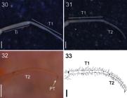
Newportia (Newportides) ananhangasp. nov. (30-33) end legs/side view; Abbreviations: (Ti) tibia, (T1) tarsus 1, (T2) tarsus 2 e (PT) pretarsus. Scale: 0.3 mm.
Legs: walking legs 1-22 with bristles; 1-20 with lateral tibial spurs, 21-22 without lateral spurs.
Ultimate legs: prefemur with four long ventral spinous processes (Figs. 27, 28); femur with a long ventral spinous process (Fig. 28); tibia smaller than tarsus 1 (Fig. 30), larger than tarsus 2; distal half of tarsus 1 divided into articles and tarsus 2 ranging from well-divided to few not-well-divided pseudo-articles (ca.12 “pseudo-articles”) with a well-developed claw-like pretarsus (Figs.29,31-33).
Distribution: Known only from the type locality, Estação Científica Ferreira Penna, Caxiuanã National Forest, Pará State, Brazil.
Subgenus Tidops Chamberlin, 1915
TidopsChamberlin, 1915: 495; Attems, 1930: 284; Bücherl, 1941a: 341; Schileyko & Minelli, 1999: 293; Schileyko, 2002: 483; Schileyko & Stagl, 2004: 63.
KartopsArchey, 1923: 113 (synonymized with Tidops by Chagas-Jr., 2011: 69).
Newportia (Tidops):Vahtera et al. (2013): 589.
Diagnosis (emended from Chagas-Jr., 2011): Anterior margin of coxosternite forcipular with two short, blunt projections; forcipular tarsungulum short, barely overlapping when adducted. Seventh body segment without spiracles. Ultimate legs with tarsus 2 divided into 6-23 variably distinct secondary articles, without claw-shaped pretarsus.
Type species: Tidops simusChamberlin, 1915.
Included species: Newportia (Tidops) simusChamberlin, 1915; Newportia (Tidops) balzanii Silvestri, 1895; Newportia (Tidops) collarisKraepelin, 1903; Newportia (Tidops) nisargani (Chagas-Jr., 2011); Newportia (Tidops) oxossi sp. nov. and Newportia (Tidops) mapinguari sp. nov. (Fig. 59).
Key to species of Newportia (Tidops)
1. Femur of ultimate legs without ventral and median spinous processes 2
- Femur of ultimate legs with ventral and median spinose processes 3
2(1). Prefemur of ultimate legs with three ventral spinous processes, femur and tibia with a small cylindrical process at the ventral corner of the distal end (Chagas-Jr., 2011, figs. 1, 3)Newportia (Tidops) balzanii
- Prefemur of ultimate legs with four large ventral processes, femur and tibia without cylindrical process (Chagas-Jr., 2011, figs. 16, 17 e 21)Newportia (Tidops) nisargani
3(1). Long or median sized coxopleural process 4
- Short coxopleural process 5
4(3). Femur of ultimate legs with one medial spinous process, tibia without cylindrical process (Chagas-Jr., 2011, figs. 5, 6)Newportia (Tidops) collaris
- Femur of ultimate legs without median spinous processes, tibia with a cylindrical process at the ventral corner of the distal endNewportia (Tidops) simus
5(3). Tarsus 2 of ultimate legs divided in 13 to 21 pseudoarticles (Figs. 55, 58)Newportia (Tidops) oxossisp. nov.
- Tarsus 2 of ultimate legs divided in 9 to 11(Figs. 44, 46)Newportia (Tidops) mapinguarisp. nov.
Newportia (Tidops) simus Chamberlin, 1915
Tidops simusChamberlin, 1915: 496 (Richmond Hill, Grenada holotype deposited at MCZ, not examined); Attems, 1930: 285; Schileyko & Minelli, 1999: 296; Schileyko & Stagl, 2004: 125; Chagas-Jr., 2011: 66.
Newportia (Tidops) simus:Vahtera et al., 2013: 589.
Diagnosis: Newportia (Tidops) simus resembles N. (T.) balzanii by the rounded distal margin of the dentiform plate, the presence of lateral tibial spurs, and by the ultimate leg prefemur with three ventral spiny processes and a cylindrical ventral process on the femur, tibia, and tarsus 1. It differs by the femur of the ultimate legs with two spiny processes, the femur and tibia of equal length, and the second tarsus divided into 9-11 “pseudo-articles”.
Distribution: Grenada and Brazil.
Description: See Chagas-Jr. (2011).
Newportia (Tidops) balzanii (Silvestri, 1895)
Newportia balzaniiSilvestri, 1895a: 767 (Holotype of Paraguay, Rio Apa, deposited in the ZMH, not examined). Silvestri, 1895b: 2. Kraepelin, 1903: 92. Attems, 1930: 277. Bücherl, 1941a: 337.
Tidops balzaniiSchileyko & Minelli, 1999: 293. Schileyko, 2002: 499; Chagas-Jr., 2011: 67.
Newportia (Tidops) balzanii:Vahtera et al. (2013): 589.
Diagnosis: Newportia (T.) balzanii shares with N. (T.) mapinguari sp. nov., a wider than long dentiform plate, with a rounded distal margin, the presence of lateral tibial spurs, and a cylindrical ventral process on the femur, tibia, and tarsus 1. It differs by the absence of spiny processes on the femur of the ultimate legs, the second tarsus having 11 or 12 “pseudo-articles”, and the tibia being equal to or larger than the femur.
Distribution: Rio Apa, Paraguay.
Description: See Chagas-Jr. (2011).
New records: Brazil: Piauí: Parque Nacional de Sete Cidades [04°08′8,2″S, 41°43′8,4″W]; Carvalho, L.S., Avelino, M.T.L. & Albuquerque, M.P. coll., (MPEG.CHI 20).
Newportia (Tidops) collaris Kraepelin, 1903
Newportia collarisKraepelin, 1903: 90 (Bas Carsopene holotype, deposited in ZMH, not examined).
Newportia bicegoiBrölemann, 1905: 67. Kraepelin, 1903: 93. Attems, 1930: 278. Bücherl, 1939: 298. Bücherl, 1941a: 337. Bücherl, 1974: 124. Schileyko & Minelli, 1999: 293 (synonymized by Schileyko & Minelli, 1999: 296).
Newportia bicegoi bicegoi:Attems, 1930: 278. Bücherl, 1939: 298. Bücherl, 1941a: 337. Bücherl, 1974: 124.
Newportia bicegoi collarisBrölemann, 1905: 67. Attems, 1930: 279. Bücherl, 1941b: 134. Bücherl, 1941a: 337. Bücherl, 1974: 124.
Tidops echinopusChamberlin, 1921: 4. Attems, 1930: 286. Bücherl, 1941a: 341. (synonymized by Schileyko & Minelli, 1999: 296).
Tidops collaris:Schileyko & Minelli, 1999: 296. Schileyko, 2002: 499. Schileyko e Stagl, 2004: 125. Edgecombe & Koch, 2009: fig. 1C, D, fig. 2A-E; Chagas-Jr., 2011: 69.
Kartops guianaeArchey, 1923: 114. Attems, 1930: 266. Bücherl, 1941a: 333. Bücherl, 1974: 128. (synonymized with N. (T.) collaris by Chagas-Jr., 2011: 69).
Newportia (Tidops) collaris:Vahtera et al., 2013): 589.
Diagnosis: Newportia (T.) collaris closely resembles N. (T.) nisargani by the straight distal margin of the dentiform plate and a coxopleural process of medium to long length. It differs by having the prefemur of the hind legs with four broad ventral spiny processes, the femur with two ventral spiny processes and one medial process, femur and tibia lacking a cylindrical process on the ventral corner of the distal end, femur being equal to or slightly larger than the tibia, and tarsus 2 with 21 “pseudo-articulations”.
Distribution: French Guiana, Guyana, Venezuela, Brazil and Paraguay.
Description: See Chagas-Jr. (2011).
New records: Brazil: Pará: Juruti, Sítio Barroso [02°28′10.5″S, 56°00′3.5″W], 11.VIII.2004, Candiani, D. coll., 1 index (MPEG.CHI 000008); 09.VIII.2004, 1 ind. (MPEG.CHI 000009); Acampamento Mutum [02°33′07.2″S, 56°13′06.2″W], 06.ii.2007, Lo-Man-Hung, N.F. Barreiros, J.A.P. coll., 1 index (MPEG.CHI 000010); 04-11.ix.2002, Bonaldo, A. coll., 1 index (MPEG.CHI 000011); Melgaço, Curuazinho River, Ferreira Penna Scientific Station, Caxiuanã National Forest [01°44′18″S, 51°27′48″W], 09.IV.2002, Bonaldo, A. coll., 1 index (MPEG.CHI 000012); 14-15.VIII.1996, Zanuto, M. coll., 1 index (MPEG.CHI 000013); Bonaldo, A. coll., 1 index (MPEG.CHI 000014); 10.IV.2004, Bonaldo, A. coll., 1 index (MPEG.CHI 000015); Juruti, Plateau do Rio Juruti [02°33′07.2″S, 56°13′06.2″W], 11-16.IX.2002, Bonaldo, A. coll., 1 index (MPEG.CHI 000016).
Newportia (Tidops) nisargani Chagas-Jr., 2011
Tidops nisargani,Chagas-Jr., 2011. (Holotype of Arraial d’Ajuda, Porto Seguro, Bahia, Brazil, deposited at the MNRJ, type material destroyed in the fire at the Museu Nacional/UFRJ).
Newportia (Tidops) nisarganiChagas-Jr., 2011 (Vahtera et al., 2013: 589).
Diagnosis: Newportia (T.) nisargani shares with a straight distal margin of the dentiform plate and a coxopleural process ranging from medium to long. It differs in having the prefemur of the ultimate legs with four broad ventral spiny processes, the femur with two ventral and one medial spiny process, and the femur and tibia lacking a cylindrical process in the ventral corner of the distal end. Additionally, the femur is equal to or slightly larger than the tibia, and the second tarsus has 21 “pseudo-articles”.
Description: See Chagas-Jr. (2011).
New records: Brazil: Pará: Pacajá, [03°45′05″S, 51°02′26″W], Ronildon coll., 1 index (MPEG.CHI 000022); [03°42′52″S, 51°03′13″W], Ronildon coll., 1 index (MPEG.CHI 000024); [03°43′54″S, 51°02′10″W], Ronildon coll., 1 index (MPEG.CHI 000025).

New Distribution Records for Newportia (Newportides). NewportiaGervais, 1847 in GBIF Secretariat (2021).
Distribution: Brazil (Amazonas, Pará and Bahia).
Newportia (Tidops) mapinguari sp. nov. Figs. 35-46
Type material: Holotype: not sexed. Caxiuanã National Forest [02°33′07.2″S, 51°29′00.7″W], Melgaço, Pará, Brazil, 09.VIII.2004, Barreiros, J.A.P. coll., (MPEG.CHI 000006).
Etymology: The specific epithet is a noun in opposition, referring to Mapinguari, a creature from Brazilian folklore, described as a red hairy giant having a cyclopean eye in the middle of his forehead and a gaping mouth on its abdomen.
Diagnosis: Newportia (T.) mapinguari sp. nov. reassembles N. (T.) simus by the distal margin of dentiform plate rounded; by the presence of lateral spurs on tibia; femur and tibia with similar length; ventral process cylindric along posterior legs on femur, tibia and tarsus 1. It differs by the pre-femur of the ultimate leg with four (right leg) and three (left leg) elongated spinose processes; right femur with two short ventral processes, the anterior one bigger than the posterior; left with a ventral spinose process and a median short; right tarsus 2 with ten articles and left tarsus 2 with six articles. See Table 1 for a character comparison of N. (T.) mappinguari with all remaining known species of the subgenus.
Description (Holotype)
Coloring: Antennae: composed of 17 articles, the first three devoid of hairs, but with some long bristles reaching the anterior margin of the eighth article; the other articles with thin, short bristles (Fig. 35).
Cephalic plate: smooth orange, overlapping the anterior border of the first tergite, with a short median suture on the anterior border and a pair of short paramedian sutures on its posterior border (Fig. 35).
Forcipular coxosternite: anterior margin of the coxosternite straight and narrow, with a small diamond-shaped elevation, having a small median suture protruding from its posterior margin (Fig. 36); forcipular trochanteroprefemur without distal denticle, with a median suture reaching the femur and forcipular tibia (Fig. 36); forcipular articles having long laterally rowed bristles and forcipular tibia with a lateral projection; tooth-plate dentiform, straight with lateral sides longer than medial lateral, anterior margins concave; tarsungula short and robust, apices not overlapping alone in closed position (Fig. 36).
Tergites: smooth, 1 with an anterior transverse suture at a very obtuse angle and paramedian sutures bifurcated anteriorly, forming a “W” just behind the anterior transverse suture, a median suture reaching the middle of the W groove, from which some lateral slits emerge reaching halfway of the tergite (Fig. 37). Tergite 2 with a pair of straight paramedian sutures on the anterior margin, and converging on the posterior margin; tergites 3-22 with complete paramedian sutures and incomplete lateral longitudinal sutures (Figs. 37-38); only the marginated tergite 23. The margin of tergite 23 medially truncated and concave on each side, without a median suture, but with depression on its lateral margin.
Sternites: smooth, longer than wide; with broad endosternite, delimited by a transverse groove of sternites 3-20, extending backward under the posterior margin of anterior sternite; sternites 2-22 with an incomplete longitudinal median sulcus; sternites 1-21 with incomplete lateral sutures on the anterior margin; sternite 23 much shorter than the previous one, with a slightly concave posterior margin and convex edges (Fig. 39); coxopleural process short, with an acute apex, without lateral spines, dorsal and ventral surface of the coxopleura without bristles. Numerous pores of moderate size, uniformly covering most of the coxopleura surface (Fig. 40).
Legs: walking legs hairy, resembling stiff spines, legs 1-21 with indivisible tarsus, 22 and 23 divided, legs 2-19 with lateral spurs on the tibia; and 4-21 with anterior accessory spines on the pretarsus.
Ultimate legs: right prefemur with four long ventral spinose processes and left with three; right femur with two small ventral spinose processes, the anterior being larger than the posterior; left femur with a small ventral and median spinose process; femur and tibia with the presence of a small cylindrical process in the ventral corner of the distal extremity; tarsus 1, short, with a small expansion at the ventral corner of the distal end; left tarsus 2 with six articles and right with ten articles (Figs. 41-46).
Distribution: Known only from the type locality.

Newportia (Tipods) mapinguarisp. nov. (35) cephalic plate and tergite 1-2 e part of tergite 3/dorsal view; (36) coxosternite forcipular and part of the sternites 1/ventral view; (37) tergites 1-4/dorsal view; (38) tergites 20-23/dorsal view; Abbreviations: (MS) median suture, (CP) cephalic plate, (PS) para median suture, (TS) transverse suture, (TAR) tarsungula, (TP) tooth-plates, (MS) median suture, (FC) forcipular coxosternite and (LS) lateral suture. Scale: 0.5 mm.

Newportia (Tipods) mapinguarisp. nov. (39) sternites 22-23 and coxopleural/side view; (40) coxopleura and prefemur of the ultimate legs; (41) prefemur and femur of the ultimate legs/ventral view; (42-46) end legs/side view; Abbreviations: (S) sternites, (L) leg, (CXP) coxopleural process, (CX) coxo pleura, (VSPU) ventral spinous processes of prefemur of ultimate legs, (PF) prefemur, (F) femur, (Ti) tibia, (T1) tarsus 1, (T2) tarsus 2 and (MSPF) medial spinous pro cesses of the ultimate femur. Scale: 0.5 mm.

Newportia (Tidops) oxossisp. nov. (47) cephalic plate and tergite 1-2/dorsal view; (48) coxosternite forcipular and sternites 1-2/ventral view; (49) tergite 1-3/dorsal view; (50) sternites 21-23/dorsal view; (51) sternites 12-14/ventral view; (52) sternites 22-23/ventral view; Abbreviations: (MS) median suture, (CP) cephalic plate, (PS) paramedian suture, (TAR) tarsungula, (TP) tooth-plates, (FC) forcipular coxosternite, (S) sternites, (TS) transverse suture, (LS) lateral suture, (TE) tergite, (MLS) median longitudinal sulcus, (CX) coxopleura and (CXP) coxopleural process. Scale: 0.5 mm.
Newportia (Tidops) oxossi sp. nov. Figs. 47-58
Type material: Holotype: not sexed. Curuazinho River [01°44′18″S, 51°27′48″W], Ferreira Penna Scientific Station, Caxiuanã National Forest, Melgaço, Pará, Brazil, 09.IV.2002, Bonaldo, A. coll., (MPEG.CHI 000007).
Etymology: The epithet is a noun in apposition, as a reference to the Orisha Oxóssi, a deity worshiped in religions of African origin as the lord of the forest and of all beings that inhabit it.
Diagnosis: Newportia (T.) oxossi sp. nov. resembles N. (T.) nisargani by having both femur and tibia lacking cylindric process in the ventral edge of distal extremity; tarsus 1 with a slight expansion in the ventral edge of distal extremity; tibia shorter than femur. It differs by the prefemur of the ultimate right leg with six ventral spinous processes and left with four; left femur with two ventral spinous processes and one median spine, and right femur with one median spine, tarsus 2 on thin legs (left) ca.13 articles and tarsus 2 (right) with ca.21 articles. See Table 1 for a character comparison of N. (T.) oxossi with all remaining known species of the subgenus.

Newportia (Tidops) oxossisp. nov. (53) coxopleural/vista lateral; (54-58) ultimate legs/side view; Abbreviations: (CX) coxopleura, (CXP) coxopleu ral process, (MSPF) medial spinous processes of the ultimate femur, (VSPU) ventral spinous processes of prefemur of ultimate legs, (PF) prefemur, (F) femur, (Ti) tib ia, (T1) tarsus 1, (T2) tarsus 2 and (LSF) lateral spine of femur of the ultimate legs. Scale: 0.5 mm.
Description (Holotype): Body Length: 29.38 (Without the last legs); longitudinal cephalic plate 1.9, transverse: 1.7; last legs: total length right leg: 7.8; prefemur: 1.4; femur: 1.4; tibia: 1.2; tarsus 1: 0.6; tarsus 2: 3.2; total length left leg: 7.8; prefemur: 1.6; femur: 1.5; tibia: 1.3; tarsus 1: 0.4; tarsus 2: 3.
Coloring: Yellowish body. Cephalic plate and coxosternum yellow forcipules; yellow antennae and legs.
Antennae: left antenna with eight articles and right with 12 articles, the first two and half of the third without hairs, but with some broad bristles reaching the anterior part of the seventh article, the others with short, fine bristles (Fig. 47).
Cephalic plate: smooth, overlapping the anterior margin of the first tergite, with bristles and small grooves along its surface, with a short median suture on the anterior part of the margin and a pair of short, almost invisible paramedian sutures on the posterior margin (Fig. 47).
Forcipular coxosternite: anterior margin of coxosternite straight and narrow; dentiform dental plate, straight with lateral sides longer than lateral mean with concave anterior margins; forcipular trochanteroprefemur without distal denticle, tibia and forcipular tarsus with a lateral projection; forcipular articles having long lateral row bristles and some spare bristles on the surface; tarsungula short and robust, apices not overlapping alone in closed position (Fig. 48).
Tergites: smooth; tergite 1 with an anterior transverse suture at a very obtuse angle and with paramedian sutures bifurcated anteriorly, forming a “W” just behind the anterior transverse suture (Fig. 49); tergite 2 with a pair of straight paramedian sutures on the anterior margin and converging on the posterior margin; tergites 3-22 with complete paramedian sutures; tergites 3-21 with incomplete lateral sutures on anterior margin (Fig. 49) only tergite 23 marginated; margin of tergite 23 medially truncated and concave on each side, without a median sulcus but with a shallow median depression (Fig. 50).

New Distribution Records for Newportia (Tidops). NewportiaGervais, 1847 in GBIF Secretariat (2021).
(Adapted from Chagas‑Jr., A. 2011). Comparative data for N. (T.) simus, N. (T.) balzanii, N. (T.) collaris, N. (T.) nisargani, N. (T.) oxossisp. nov. and N. (T.) mapinguarisp. nov.
Sternites: smooth; longer than wide; endosternite not very wide, sternites 2-21 with incomplete longitudinal median sulcus; sternites 1-21 with incomplete lateral longitudinal sutures (Fig. 51); sternite 23 a little shorter than the previous one, trapeziform, with a concave posterior margin (Fig. 52); coxopleural process short, converging forming almost a triangle with an acute apex, without lateral spines; lateral margins with small bristles; numerous moderately sized pores uniformly covering most of the coxopleura surface (Figs. 52,53).
Legs: walking legs with many bristles; tarsi 1-20 indivisible, 21-23 divided; legs 2-21 with lateral spurs on the tibia and 3-21 with accessory spines on the pretarsus.
Ultimate legs: right prefemur with six long ventral spinous processes and left with four; right femur with a median spine and left femur with two ventral spinous processes and a median spine; femur and tibia without cylindrical process at the ventral corner of the distal extremity, tarsus 1 with a small ventral expansion at the corner of the distal extremity; tarsus 2 (left) ca.13 articles and tarsus 2 (right) with ca.21 articles (Figs. 54-58).
Distribution: Known only from the type locality.
CONCLUSION
The genus Newportia exhibits a broad distribution in the Neotropical region, its diversity remains largely unexplored. Several knowledge gaps persist, encompassing aspects such as species richness, geographic distribution, evolutionary relationships, and ecological interactions. The subgenus Tidops is extensively distributed within the Neotropical region, reaching the highest diversity in the Amazon region, while Newportides is exclusively known from the Amazon region. In this paper, the known species richness of the subgenus Newportides is increased from three to five, while that of the subgenus Tidops increases from four to six known species.
ACKNOWLEDGMENTS:
This work is part of the M.Sc. dissertation of the first author, conducted under the supervision of the second author and co-supervision of the third author at the Programa de Pós-Graduação em Biodiversidade e Evolução, Museu Paraense Emílio Goeldi (MPEG). Ivan Brito thanks the Arachnology Laboratory (MPEG) for the support provided during the development of this research. The authors are also grateful to Fabián García, Paulo Pantoja, and Felipe Araújo for their support and valuable comments. This study was supported by the Coordenação de Aperfeiçoamento de Pessoal de Nível Superior (CAPES) through an M.Sc. scholarship (grant 88887.481301/2020 00 to Ivan Brito).
Data Availability:
All datasets generated during and/or analyzed during the current study are contained within the article.
REFERENCES
- Archey, G. 1923. A new genus of Chilopoda from British Guiana, and a new species of Wailamyctes from Auckland Island. Records of the Canterbury Museum, 2(3): 113-116.
- Attems, G. 1930. Scolopendromorpha. Das Tierreich, Berlim, 54: 1-308.
-
Bonato, L.; Edgecombe, G.D.; Lewis, J.G.; Minelli, A.; Pereira, L.A.; Shelley, R.M. & Zapparoli, M. 2010. A common terminology for the external anatomy of centipedes (Chilopoda). ZooKeys, 69: 17-51. https://doi.org/10.3897/zookeys.69.737.
» https://doi.org/10.3897/zookeys.69.737 -
Brölemann, H.W. 1904-1905. Myriapodes du Museu Paulista. IIe mémoire: Manaos. Revista do Museo Paulista, 6: 63-96. https://doi.org/10.5962/bhl.part.26467.
» https://doi.org/10.5962/bhl.part.26467 -
Bücherl, W. 1939-1940. Os quilópodos do Brasil. Memórias do Instituto Butantan, 13: 43-363. https://bibliotecadigital.butantan.gov.br/edicao/memorias-do-instituto-butantan-tomo-xiii-1939/58
» https://bibliotecadigital.butantan.gov.br/edicao/memorias-do-instituto-butantan-tomo-xiii-1939/58 - Bücherl, W. 1941a. Quilópodos novos da coleção do Museu Nacional do Rio de Janeiro. Memórias do Instituto Butantan, 15: 119-146.
- Bücherl, W. 1941b. Catálogo dos Quilópodos da zona neotrópica. Memórias do Instituto Butantan, 15: 251-372.
- Bücherl, W. 1974. Die Scolopendromorpha der Neotropischen Region. Symposia of the Zoological Society of London, 32: 99-133.
-
Chagas-Jr., A. 2011. A review of the centipede genus Tidops Chamberlin (Scolopendromorpha, Scolopocryptopidae, Newportiinae). International Journal of Myriapodology, 5: 63-82. https://doi.org/10.3897/ijm.5.1649.
» https://doi.org/10.3897/ijm.5.1649 -
Chagas-Jr., A. 2018. A review of Newportia species in the subgenus Newportides Chamberlin, 1921 (Scolopendromorpha, Scolopocryptopidae, Newportiinae). Zootaxa, 4403(1): 154-170. https://doi.org/10.11646/zootaxa.4403.1.9.
» https://doi.org/10.11646/zootaxa.4403.1.9 - Chamberlin, R.V. 1914. The Stanford Expedition to Brazil, 1911, John C. Branner, Director. The Chilopoda of Brazil. Bulletin of the Museum of Comparative Zoology, 58: 151-221.
- Chamberlin, R.V. 1915. New chilopods from Mexico and the West Indies. Bulletin of the Museum of Comparative Zoology at Harvard College, 59: 493-502.
- Chamberlin, R.V. 1921. Results of the Bryant Walker expeditions of the University of Michigan to Columbia 1913 and British Guiana 1914. Occasional Papers of the Museum of Zoology University of Michigan, 97: 1-28.
- Crabill, R.E. 1977. A new Cryptopid genus with key to the genera known to occur in North America including Mexico (Chilopoda: Scolopendromorpha: Cryptopidae). Proceedings of the Entomological Society of Washington, 79(3): 346-349.
-
Edgecombe, G.D. & Bonato, L. 2011. Chilopoda. In: Minelli, A. (Ed.). Treatise on Zoology - The Myriapoda: Treatise on Zoology - Anatomy, Taxonomy, Biology . Vol.1. Brill, Leiden. p. 392-407. https://doi.org/10.1163/9789004188266_002.
» https://doi.org/10.1163/9789004188266_002 - Edgecombe, G.D. & Koch M. 2009. The contribution of preoral chamber and foregut morphology to the phylogenetics of Scolopendromorpha (Chilopoda). Soil Organisms, 81(3): 295-318.
-
Global Biodiversity Information Facility (GBIF). 2021. GBIF backbone taxonomy. https://doi.org/10.15468/39omei. Access: 11/11/2021.
» https://doi.org/10.15468/39omei - Gervais, P. 1847. Myriapodes. In: Walckenaer, C.A. & Gervais, P. (Eds.). Histoire Naturelle des Insectes Aptères Paris, Librairie Encyclopédique de Roret. p. 210-333.
- González-Sponga, M.Á. 1997. Miriápodos de Venezuela. Siete nuevas especies del género Newportia (Chilopoda: Scolopendromorpha: Cryptopidae). Memoria de la Sociedad de Ciencias Naturales La Salle, 57(148): 33-47.
- González-Sponga, M.Á. 2000. Miriápodos de Venezuela. Diez nuevas especies del género Newportia (Chilopoda: Scolopendromorpha: Cryptopidae). Memoria de la Fundación La Salle de Ciencias Naturales, 60 (153): 103-122.
- Kraepelin, K. 1903. Revision der Scolopendriden. Mitteilungen aus dem naturhistorischen Museum in Hamburg, 20: 1-276.
-
Morrone, J.J. 2014. Biogeographical regionalisation of the Neotropical region. Zootaxa, 3782 (1): 1-110. https://doi.org/10.11646/zootaxa.3782.1.1.
» https://doi.org/10.11646/zootaxa.3782.1.1 - Pocock, R.I. 1896. Chilopoda and Diplopoda. In: Godman, F.D. & Salvin, O. (Eds.). Biologia Centrali-Americana. London, Taylor & Francis. p. 1-217.
- Schileyko, A. & Minelli, A. 1999 [1998]. On the genus Newportia Gervais, 1847 (Chilopoda: Scolopendromorpha: Newportiidae). Arthropoda Selecta, 7: 265-299.
- Schileyko, A. & Stagl, V. 2004. The collection of scolopendromorph centipedes (Chilopoda) in the Natural History Museum in Vienna: a critical re-evaluation of former taxonomic identifications. Annalen des Naturhistorisches Museum Wien, 105: 67-137.
- Schileyko, A.A. 2002. Scolopendromorpha, In: Adis, J. (Ed.). Amazonian arachnida and myriapoda. Identification keys to all classes, orders, families, some genera, and lists of known terrestrial species Sofia, Pensoft Publishers. p. 479-500.
-
Schileyko, A.A. 2013. A new species of Newportia Gervais, 1847 from Puerto Rico, with a revised key to the species of the genus (Chilopoda, Scolopendromorpha, Scolopocryptopidae). ZooKeys, 276: 39-54. https://doi.org/10.3897/zookeys.276.4876.
» https://doi.org/10.3897/zookeys.276.4876 -
Schileyko, A.A.; Vahtera, V. & Edgecombe, G.D. 2020. An overview of the extant genera and subgenera of the order Scolopendromorpha (Chilopoda): a new identification key and updated diagnoses. Zootaxa, 4825, 1: 1-64. https://doi.org/10.11646/zootaxa.4825.1.1.
» https://doi.org/10.11646/zootaxa.4825.1.1 -
Shelley, R.M. & Mercurio, R. 2005. Ectonocryptoides quadrimeropus, a new centipede genus and species from Jalisco, Mexico; proposal of Ectonocryptopinae, analysis of subfamilial relationships, and a key to subfamilies and genera of the Scolopocryptopidae (Scolopendromorpha). Zootaxa, 1094: 25-40. https://doi.org/10.11646/zootaxa.1094.1.2.
» https://doi.org/10.11646/zootaxa.1094.1.2 - Silvestri, F. 1895a. Chilopodi e diplopodi raccolti dal Capitano G. Bove e dal Prof. L. Balzan nell’America Meridionale. Annali del Museo Civico di Storia Naturale di Genova, 34: 764-783.
- Silvestri, F. 1895b. Viaggio deo dottor Alfredo Borelli nella Republica Argentina e nel Paraguay XIV. Chilopodi e Diplopodi. Bolletino dei Musei di Zoologia ed Anatomia Comparata, 10 (203): 1-12.
-
Tulande-M., E.; Prado, C.C.; Jiménez, S.G. & Chagas-Jr., A. 2020. A remarkable Newportia from the Colombian Andes, with the proposition of a new subgenus (Scolopendromorpha, Scolopocryptopidae, Newportiinae). Zootaxa, 4859 (2): 228-238. https://doi.org/10.11646/zootaxa.4859.2.3.
» https://doi.org/10.11646/zootaxa.4859.2.3 -
Vahtera, V.; Edgecombe, G.D. & Giribet, G. 2013. Phylogenetics of Scolopendromorph centipedes: can denser taxon sampling improve an artificial classification? Invertebrate Systematics, 27(5): 578-602. https://doi.org/10.1071/IS13035.
» https://doi.org/10.1071/IS13035
-
Ethical Statement:
Ethical approval was not applicable given the type of study conducted. No specimens were collected for this study, and all material examined corresponds to specimens previously deposited in the Chilopoda Collection of the Museu Paraense Emílio Goeldi.
-
AI Use:
Not applicable; no artificial intelligence tools were used in this study.
-
Funding:
This study was supported by the Conselho Nacional de Desenvolvimento Científico e Tecnológico (CNPq) through a Productivity Grant (PQ grant #307165/2022 1 to AB).
-
Published with the financial support of the “Programa de Apoio às Publicações Científicas Periódicas da Universidade de São Paulo”.
Edited by
-
Edited by:
Carlos José Einicker Lamas


