Genotoxicity of Imidacloprid on the forensically important species Chrysomya albiceps (Wiedemann, 1819) (Diptera: Calliphoridae), evaluated by Comet assay
Abstract
The increasing use of chemical pesticides for crop protection has led to irreversible DNA damage in many animal species, particularly soil-dwelling insects. In this study, we tested a protocol of Comet Assay to detect DNA damage in the hemolymph cells of Chrysomya albiceps larvae (Diptera: Calliphoridae) exposed to the neonicotinoid Imidacloprid (IMI). Third-instar larvae were fed on freeze-dried liver substrate rehydrated with one of the following treatments: (i) distilled water (negative control), (ii) imidacloprid (IMI) (0.4980 and 0.9360 mM), and (iii) cyclophosphamide 3.83 mM (positive control). We estimated the Damage Index (DI) and the Damage Frequency (DF) in 300 cells per treatment. According to the shape of the comets, the damage in cells exposed to biocides was significantly higher than the negative control. The DF was also consistently higher in the cells of intoxicated insects. The Comet assay in the haemolymph cells of C. albiceps larvae proved to be effective for detecting DNA damage, providing new evidence for genotoxicity. The increasing use of chemical pesticides for crop protection has led to irreversible DNA damage in many animal species, particularly in soil-dwelling insects. Third-instar larvae were fed freeze-dried liver substrate rehydrated with one of the following treatments: (i) distilled water (negative control), (ii) Imidacloprid (IMI) at concentrations of 0.4980 mM and 0.9360 mM, and (iii) cyclophosphamide at 3.83 mM (positive control). We estimated the Damage Index (DI) and Damage Frequency (DF) in 300 cells per treatment. According to the shape of the comets, DNA damage in cells exposed to biocides was significantly higher than in the negative control. The DF was also consistently higher in the cells of intoxicated insects. The Comet Assay in the hemolymph cells of C. albiceps larvae proved effective for detecting DNA damage, providing new evidence of the genotoxic effects of IMI on a necrophagous insect species. We discuss the advantages and limitations of the protocol in the context of environmental forensic entomotoxicology.
Keywords:
Entomotoxicology; Blowflies; Forensic entomology; Neoticotinoids
INTRODUCTION
Exposure to toxicants can alter the development, feeding and behaviour of necrophagous insects, which could influence carrion decomposition and their use in forensic entomology. Several compounds interfere with larval development, which can produce errors in estimating the minimum post-mortem interval (min PMI) using methods strictly based on the insect life cycle (Estrada et al., 2009). Larvae fed on contaminated carcasses can also exhibit modified mobility and dispersal, which can influence the likelihood of sampling entomological at the scene of the crime (Jales et al., 2021).
Such questions nurtured the development of forensic entomotoxicology, a branch of forensic entomology that validates the use of insects as: (i) an indirect source of toxicological evidence in the absence of direct matrices, such as blood, urine, soil or water, and (ii) Indiscriminate pesticide use has sparked new insights into environmental crimes (Goff & Lord, 2001).
The use of broad-spectrum insecticides for controlling insects of medical, veterinary, and agricultural importance has stirred novel perspectives on environmental crimes, so that a branch of forensic entomology has emerged, that is, environmental forensic entomotoxicology (EFF). EFF employs insects as indicators of environmental pollution, bioaccumulation of heavy metals in trophic webs, and deleterious effects on non-target organisms (Hodecek, 2020). For example, drug absorption, distribution, and excretion may occur at different rates in biological systems, resulting in a quantity of toxicant in an insect that may differ from its concentration in the environment (Goff & Lord, 2001).
Neonicotinoid pesticides, such as imidacloprid (C₉H₁₀ClN₅O₂), are used worldwide and have raised concern due to their adverse effects on non-target insects, particularly on soil-dwelling larvae of Diptera and Coleoptera (Wang et al., 2023). Imidacloprid can simulate the action of acetylcholine and interfere with the nervous activity of insects, thus resulting in death due to long-term over-excitation (Li et al., 2012). When applied in the field, only a small part reaches its biological targets; most particles remain in the soil or the water until degradation or bioaccumulation. Over 90% of neonicotinoids could move off-site and persist in non-target environmental media (Cavallaro et al., 2023).
Dung is a protein source and breeding site for forensically important species of Muscidae, Sarcophagidae, and Calliphoridae (Diptera). Chrysomya albiceps (Wiedemann, 1819) (Calliphoridae) was a dominant species in cattle dung in Spain, and occurred in urban areas, rainforests, dry forests, and agro-ecosystems (Vasconcelos et al., 2015, 2016; Carmo et al., 2021), the Amazon Forest (Souza & Guimarães, 2022), and Europe (Hodecek et al., 2024). It is also one of the most frequent species found in human cadavers in many countries (Oliveira & Vasconcelos, 2010). C. albiceps is a model species in forensic entomotoxicology because a diversity of toxic substances, such as malathion and parathion, can be detected and quantified in its tissues (Gosselin et al., 2011).
However, empirical data on the damage caused by xenobiotics, such as insecticides, at the cellular level are scarce. Sub-lethal and sub-organismal level effects (e.g., small turnover rate for proteins, DNA fragmentation, etc.) can influence energy metabolism, fitness, and reproductive success, which leads to population-level effects (Jha, 2008). Genotoxic compounds alter the structure and the information content of DNA, and interfere, temporarily or not, directly or indirectly, in normal processes in the cells (Jha, 2008; Phillips & Arlt, 2009; Gajski et al., 2019; Menz et al., 2023). In the case of soil-dwelling insects, exposure to biocides released into the environment may result in unrepaired DNA strand breaks, leading to mutations and changes in normal cell processes (Phillips & Arlt, 2009; Chatterjee & Walker, 2017).
The Comet assay (CE), or single-cell gel electrophoresis (SCGE), is a simple method for measuring DNA strand breaks in eukaryotic cells because it is a sensitive, rapid and relatively inexpensive technique, amenable to any species (Jha, 2008; Chatterjee & Walker, 2017). It combines agarose electrophoresis with fluorescence microscopy to observe and quantify DNA strand breaks at the level of single cells. Cells with damaged DNA exhibit increased migration of the chromosomal DNA from the nucleus in an electric field, and the migration pattern has a typical ‘comet’ shape, consisting of a head and a tail. The bulk DNA, also called nucleoid (“head”), moves from the cathode to the anode during electrophoresis more slowly than the short, broken DNA fragments (“tail”), resembling a comet (Hartmann et al., 2003; Møller et al., 2020; Collins et al., 2023).
The Comet assay has advantages in toxicological research because various cell types can be used without prior knowledge of their karyotype or genome structure (Jha, 2008; Lapuente et al., 2015). Since its pioneering use as a model to detect DNA damage using the comet assay (Gaivão & Sierra, 2014), Drosophila melanogaster Meigen, 1830 (Diptera: Drosophilidae) has been exposed to xenobiotics, natural radiation, and urban pollution (Augustyniak et al., 2016; Verçosa et al., 2017; Santana et al., 2018). The Comet assay has also been used to evaluate the fate of insecticides in the environment using dung beetles (Coleoptera: Scarabaeidae) as models (Cavallaro et al., 2023). This information can help to understand cellular responses to detrimental factors and to predict negative effects at both the organism and population levels.
Given the growing importance of environmental forensic entomotoxicology, we aimed to: (i) test a protocol for using the comet assay using Chrysomya albiceps larvae as a target model; (ii) detect genotoxic effects, measured by DNA damage, caused by imidacloprid in haemolymph cells, and (iii) assess the validity of the comet assay as a technique to be used in toxicological and forensic protocols.
MATERIAL AND METHODS
We used C. albiceps larvae from a standardized laboratory colony under controlled conditions of temperature (24 ± 2℃), relative humidity (70% ± 5%), and photoperiod (12:12 light: dark). The colony has been established for at least five generations from samples collected in rainforest fragments in Recife (08°22′54″S, 34°56′53″W), Northeastern Brazil.
Because there are no published protocols for the Comet assay using larvae of C. albiceps, we based our protocol on techniques developed for D. melanogaster (Verçosa et al., 2017; Amorim et al., 2020). We carried out pilot tests using different sample sizes (5, 15, and 35 larvae) for collecting haemolymph cells of C. albiceps larvae to provide sufficient biological material. The most consistent quality and quantity of haemocytes was obtained from a sample of 35 larvae at 3rd instar of homogeneous size and weight.
For the experimental set-up, the larvae were allowed to feed for 24 h on a treatment medium composed of freeze-dried bovine liver (10 g), on which we administered 20 mL of one of the following treatments: (i) distilled water (negative control); (ii) imidacloprid (IMI) (0.4980 and 0.9360 mM) (Bayer Evidence® 700 WG, 700 g/kg, 70% m/m); and (iii) cyclophosphamide 3.83 mM (positive control). All chemicals were diluted in distilled water. The choice of IMI concentrations followed the recommendations of Sousa et al. (2019) and Frantzios et al. (2008), who verified genotoxic effect of the compounds on D. melanogaster. Cyclophosphamid has been used in comet assays as a positive control due to its potent genotoxicity to insect cells, so that the maximum damage can be visually defined.
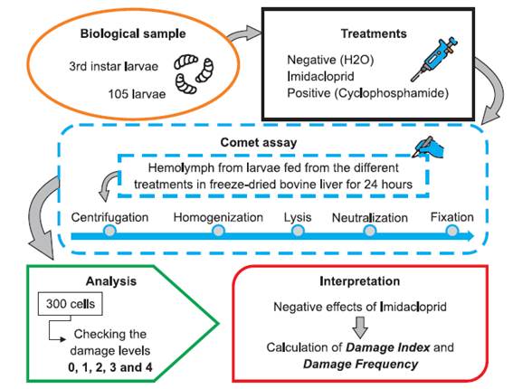
We then proceeded with the comet assay, as summarized in Fig. 1. Haemocytes from larvae fed on each treatment were extracted from a pool of 35 individuals per replicate (three replicates in total). The larvae were transferred to a well of an excavated plate containing 150 µL of EDTA to prevent haemolymph clotting. The cell pool (0.5 mL) was transferred to 1.5 mL microtubes and centrifuged at 3,000 rpm for 3 min. Then, 100 µL of the supernatant was discarded, and 100 µL of new EDTA was added, and the procedure was repeated three times. Cuticle breakdown of 35 larvae resulted in 60 µL of haemolymph per replicate, which were homogenized with a 0.5% low-melting agarose solution (LM agarose) at 37℃, in the dark.

Steps of the comet assay in haemolymph cells of Chrysomya albiceps larvae exposed to concentrations of Imidacloprid (IMI).
The homogenized material was applied to four histological slides, previously washed and bathed in standard 1.5% agarose (diluted in Phosphate-Buffered Saline pH 7.4) and dried at 24℃ for 48 h. The slides were covered with coverslips and kept at 4℃ for 10 min. They were then immersed in a lysis solution (2.5 M NaCl; 100 mM EDTA; 1 M NaOH; 1% Tris pH 10; 1% Triton X-100 and 10% DMSO) at 4℃ for 48 h. Subsequently, the slides were aligned in a 40 cm electrophoresis vat filled with buffer (1 M NaOH; 200 mM EDTA pH > 13) and immersed at 4℃ for 20 min. The slides were subjected to an electrophoretic run at 40 V and 300 mA for 20 min. The slides were transferred to a neutralization solution (0.4 M Tris-HCl, pH 7.5) for 15 min, fixed in ethanol for 5 min, air-dried and stored at 4℃ until observation under microscope.
To visualize the nucleoids the material was stained with 50 µL of GelRed (Biotium) diluted in purified water (1:500) and observed under a fluorescence microscope Zeiss-Imager M2, equipped with the Alexa-Fluor 546 filter, under 400× magnification. Comets were classified according to Ostling & Johanson (1984) e Møller et al. (2023) (Fig. 2). It consists of a grade of the integrity of the “head” and the length of the “tail” of nucleoids, assigning comparative values from 0 (intact), 1 (minimal), 2 (intermediate), 3 (intense) and class 4 (maximum damage). One hundred cells were observed for each replicate, totaling 300 nucleoids per treatment.
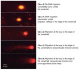
Representative adapted images of DNA damage observed using the Comet assay grading system described by Ostling & Johanson (1984) and Møller et al. (2023).
The Damage Index (DI) was calculated as follows: DI = 0x(N₀) + 1x(N₁) + 2x(N₂) + 3x(N₃) + 4x(N₄), so that it ranges from a minimum of zero - in case no DNA damage is observed in any cell (level 0) - to 400, which corresponds to all cells (N = 100) under the maximum DNA damage (level 4). The Damage Frequency (DF%) = [(NT - N₀).100] / NT, where N₁, N₂, N₃, N₄ represent the total number of damages 0, 1, 2, 3, and 4, respectively; NT is the total number of damages; and N₀, the total number of zero damages. The Damage Frequency varied from 0 to 100%.
Because visual interpretation of the comet shape could be subjective, all categorizations from 0 to 4 were performed in a double-blind manner. Two people analyzed each slide, and only fully matched diagnoses were considered. For the sake of simplicity, we refer to concentrations of imidacloprid as “low” and “high” to facilitate a preliminary interpretation of dose-response. The mean values of DI and DF% in the treatments were compared using one-way ANOVA followed by the Bonferroni post-test, which provides a conservative adjustment for multiple comparisons and reduces the likelihood of errors when evaluating differences between treatment means, using the Stata 14.2 program, with 5% significance level.
RESULTS
When data from all treatments were combined, we designed a visual scale from 0 to 4, based on the increasing level of DNA damage in the haemolymph cells of C. albiceps larvae (Fig. 3). Based on this calibration, the levels of cellular damage in our study were classified into one of five categories: 0, 1, 2, 3, or 4.

Images of GelRed-stained nucleoids obtained from Chrysomya albiceps larvae. Class zero (0) represents the absence of genetic damage and classes 1 to 4 indicate increasing DNA damage (increase in the comet’s tail and decrease in its head).
IMI induced severe DNA damage in the haemolymph cells at the concentration of 0.0468 mM, ranking a large proportion of cells into classes 3 and 4 (25.67% and 42.0%, respectively). However, no clear dose-response relationship was observed between the two concentrations tested, suggesting a possible non-linear effect, as the higher concentration tested (0.0936 mM) showed a reduced effect in class 3 (20.0%) and class 4 (31.3%). In the negative control, 89.7% of cells remained undamaged (class 0), with maximum damage level (class 4) recorded in only 2.3%, confirming a low baseline genotoxicity for comparison with chemicals (Table 1).
Percentage of cells under each level of damage and standard deviation (SD) in haemolymph cells of Chrysomya albiceps larvae subjected to treatments with distilled water (Negative control); concentrations of Imidacloprid (IMI) and Cyclophosphamide (Positive control). Damage levels: zero (0) represents no apparent genetic damage and 1 to 4 are increasing levels of genetic damage.
Both IMI concentrations induced high levels of DNA damage in the haemolymph cells of C. albiceps larvae. The Damage Frequency in the cells exceeded 78.0% in both treatments, in sharp contrast with the negative control (± 10%), indicating a significant genotoxic effect at both concentrations (P < 0.001). The Damage Index was also considerable for both treatments (approximately 270 for 0.0468 mM and 220 for 0.0936 mM) (Fig. 4a).

Mean Values (± SD) of Damage Index (a) and the Damage Frequency (b) after exposure of Chrysomya albiceps larvae to the treatments with distilled water (negative control), Imidacloprid (IMI), and cyclophosphamide (positive control). In (b) different letters indicate statistically significant differences.
The Damage Index showed that the negative control group differed significantly from all others (P < 0.001). The 0.0468 mM IMI treatment differed significantly from the 0.0936 mM IMI treatment (P < 0.001), but its effect did not differ markedly from the positive control (P = 0.142). Despite the intense genotoxic response observed at both IMI concentrations, the higher dose (0.0936 mM) exhibited a slight reduction in the Damage Index, suggesting a possible non-monotoxic effect. Notably, the 0.0468 mM concentration induced damage comparable to the positive control, while the higher concentration differed statistically from it (P < 0.001), indicating that intermediate concentrations may cause greater genetic damage (Fig. 4b).
DISCUSSION
Toxicants can cause deleterious effects at multiple levels of biological organization, ranging from populations across generations to individual molecules within a living cell. Here, we assessed the validity of the comet assay (CE) to detect DNA damage caused by a commonly used biocide in the haemolymph cells of C. albiceps larvae, the most widely used insect species in forensic entomology. The findings can be incorporated into basic (e.g., carrion ecology) and applied entomology, as exemplified by environmental forensic entomotoxicology.
Blow fly larvae develop in the necrobiome, a patchy and ephemeral ecosystem represented by the interactions between the biota and the decomposing organic animal matter in terrestrial environments (Benbow & Pechal, 2019). High residues of Imidacloprid have been detected at different concentrations in many matrices, including soil and freshwater resources, and have increased dramatically over the last 20 years (He et al., 2024). Nevertheless, CE protocols have prioritised non-insects to detect genotoxic effects of imidacloprid, such as the freshwater crustacean Ceriodaphnia dubia Richard, 1894 (Raby et al., 2018).
Among terrestrial invertebrates, the CE was more effective than micronucleus tests in detecting DNA damage caused by IMI in the earthworm Eisenia fetida (Zang et al., 2000). Comet assay evidenced that Apis spp. populations from agricultural areas exposed to pesticides presented more DNA fragmentation than pesticide-free areas (Hayat et al., 2019). Necrophagous insects, as a trophic group of tremendous ecosystem services, face magnified risks in areas under intensive agriculture, especially in countries with lenient regulation of pesticide registration and application, which is the case of Brazil. This is particularly threatening because C. albiceps is an important pollinator of crops in the semi-arid region in Brazil (Carmo et al., 2021).
A landmark achievement occurred when the Organization for Economic Cooperation and Development (OECD) adopted a comet assay guideline for in vivo testing of DNA strand breaks in animals (Møller et al., 2020). To our knowledge, this is the first attempt to adjust the CE for forensic entomotoxicology and proved to be effective for detecting genotoxic damage in C. albiceps larvae exposed to toxicants. Thus, we propose criteria that display explicitly advantages and weak points for the conditions tested, based on our results and on the available literature (Hartmann et al., 2003; Møller et al., 2020; Collins et al., 2023; Nugnes et al., 2023) (Table 2).
The advantages include its reliability, ease of use, relatively low cost, fast results, reduced ethical restraints (as opposed to in vivo experiments), and the adaptability to techniques and equipment available in molecular biology laboratories. Additionally, the comet assay can be used for almost all living organisms and for a variety of cell types (Møller et al., 2020). In this study, the negative and positive controls successfully calibrated the scope of DNA damage, and the scale of damage, from 0 to 4, was consistent. CE detects DNA strand breaks and alkali-labile sites (e.g., apurinic/apyrimidinic sites), alkylated and oxidized nucleobases, DNA-DNA crosslinks, UV-induced cyclobutane pyrimidine dimers, among others (Collins et al., 2023). Despite its sensitivity, further tests are needed to discover the exact nature of the DNA damage.
Variability in DNA damage may reflect individual or physiological differences among larvae, affecting the consistency of comet tail measurements. Standardizing larval age, exposure conditions, and sample handling in future assays can help minimize this variability and improve reliability. Several sources of variability exist, including animal-to-animal, cell-to-cell, or even slide-to-slide, which must be accounted for in the design of experiments and in the interpretation of results (Hartmann et al., 2003). We need to improve some procedures to avoid the formation of additional DNA damage during the processing of samples, especially in the standardization of oral administration, the incorporation of quantitative data (e.g., amount of DNA in the tail), and in the use of statistical analyses.
Møller et al. (2014) alert that it is important to guarantee that the replicates are not nested or a set of subsamples. To achieve that, our experimental unit consisted of 35 larvae, from different rearing cages and from different generations, to warrant independent observations. Since no data is available for C. albiceps, we tested two dosages. Although this number is insufficient to demonstrate a clear dose-dependent response, it represents the minimum required under current protocols (Hartmann et al., 2003).
The ecotoxicological consequences of genomic instability and its correlation with DNA breaks measured by the Comet Assay are greatly overlooked (Jha, 2008). DNA strand breaks can be repaired by a series of mechanisms, so that the damage represented by the comet does not necessarily result in cell death. To gain ecological relevance, a mechanistic association between genotoxic stress and effects at higher biological levels should be confirmed. DNA damage in insect populations can lead to reduced survival, impaired reproduction, and developmental abnormalities, which may ultimately affect population growth and stability (Lapuente et al., 2015).
A recent review by Collins et al. (2023) lists only D. melanogaster as the insect species targeted for CE. We expand this spectrum by testing C. albiceps larvae. The species can be used as a sentinel to detect the fate of xenobiotics in terrestrial environments. They can respond to drugs, toxicants, hormones and biocides by altering their development and behaviour (Chophi et al., 2019). We add a novel approach for the use of C. albiceps in forensic entomology, which can be applied to other necrophagous species.
CONCLUSION
This innovative study combines a well-established method for detecting DNA strand breaks with a novel application strategy using the necrophagous fly C. albiceps. The approach stands out not only for its sensitivity and laboratory applicability but also for paving the way towards new experimental models for insects. Although widely validated in other organisms, this work proposes C. albiceps larvae as a promising sentinel species for the genotoxic assessment of xenobiotics in overlooked terrestrial ecosystems.
DATA AVAILABILITY:
All data generated or analysed during this study are included in this published article.
Acknowledgments:
We thank Fundação de Amparo à Ciência e Tecnologia do Estado de Pernambuco (FACEPE), Conselho Nacional de Desenvolvimento Científico (CNPq) and Coordenação de Aperfeiçoamento de Pessoal de Nível Superior (CAPES) for scholarships, the colleagues at the Laboratory of Insects of Forensic Importance (UFPE) for technical support, and Dr. Trevor Williams for revising the English version. SDV is a recipient of a productivity grant from CNPq.
REFERENCES
-
Amorim, E.M.; Santana, S.L.; Silva, A.S.; Aquino, N.C.; Silveira, E.R.; Ximenes, R.M. & Rohde, C. 2020. Genotoxic assessment of the dry decoction of Myracrodruon urundeuva Allemão (Anacardiaceae) leaves in somatic cells of Drosophila melanogaster by the Comet and SMART assays. Environmental and Molecular Mutagenesis, 61(3): 329-237. https://doi.org/10.1002/em.22332.
» https://doi.org/10.1002/em.22332 -
Augustyniak, M.; Gladysz, M. & Dziewięcka, M. 2016. The Comet assay in insects-Status, prospects and benefits for science. Mutation Research / Reviews in Mutation Research, 767: 67-76. https://doi.org/10.1016/j.mrrev.2015.09.001.
» https://doi.org/10.1016/j.mrrev.2015.09.001 - Benbow, M.E. & Pechal, J.L. 2019. Forensic entomology and the microbiome. In: Byrd, J.H. & Tomberlin, J.K. (Eds.). Forensic entomology 3. ed. CRC Press. p. 499-517.
-
Carmo, R.F.R.; Barbosa, T.M.; Torris, A.F.; Torris, A.F.; Bezerra, M.S. & Vasconcelos, S.D. 2021. Diversity of sarcosaprophagous Diptera (Calliphoridae, Sarcophagidae) in organic and conventional mango plantations in the Brazilian semi-arid region. Revista Brasileira de Entomologia, 65(1): 1-5, e20200108. https://doi.org/10.1590/1806-9665-RBENT-2020-0108.
» https://doi.org/10.1590/1806-9665-RBENT-2020-0108 -
Cavallaro, M.C.; Hladik, M.L.; Hittson, S.; Middleton, G. & Hoback, W.W. 2023. Comparative toxicity of two neonicotinoid insecticides at environmentally relevant concentrations to telecoprid dung beetles. Scientific Reports, 13: 1-10, 8537. https://doi.org/10.1038/s41598-023-35262-w.
» https://doi.org/10.1038/s41598-023-35262-w -
Chatterjee, N. & Walker, G.C. 2017. Mechanisms of DNA damage, repair and mutagenesis. Environmental and Molecular Mutagenesis, 58(5): 235-263. https://doi.org/10.1002/em.22087.
» https://doi.org/10.1002/em.22087 -
Chophi, R.; Sharma, S.; Sharma, S. & Singh, R. 2019. Forensic entomotoxicology: current concepts, trends and challenges. Journal of Forensic and Legal Medicine, 67: 28-36. https://doi.org/10.1016/j.jflm.2019.07.010.
» https://doi.org/10.1016/j.jflm.2019.07.010 -
Collins, A.; Møller, P.; Gajski, G.; Vodenková, S.; Abdulwahed, A.; Anderson, D.; Bankoglu, EE.; Bonassi, S.; Boutet-Robinet, E.; Brunborg, G.; Chao, C.; Cooke, MS.; Costa, C.; Costa, S.; Dhawan, A.; de Lapuente, J.; Del Bo’, C.; Dubus, J.; Dusinska, M.; Duthie, S.J.; El Yamani, N.; Engelward, B.; Gaivão, I.; Giovannelli, L.; Godschalk, R.; Guilherme, S.; Gutzkow, K.B.; Habas, K.; Hernández, A.; Herrero, O.; Isidori, M.; Jha, A.N.; Knasmüller, S.; Kooter, I.M.; Koppen, G.; Kruszewski, M.; Ladeira, C.; Laffon, B.; Larramendy, M.; Le Hégarat, L.; Lewies, A.; Lewinska, A.; Liwszyc, GE.; López de Cerain, A.; Manjanatha, M.; Marcos, R.; Milić, M.; Moraes de Andrade, V.; Moretti, M.; Muruzabal, D.; Novak, M.; Oliveira, R.; Olsen, A.K.; Owiti, N.; Pacheco, M.; Pandey, A.K.; Pfuhler, S.; Pourrut, B.; Reisinger, K.; Rojas, E.; Rundén-Pran, E.; Sanz-Serrano, J.; Shaposhnikov, S.; Sipinen, V.; Smeets, K.; Stopper, H.; Teixeira, J.P.; Valdiglesias, V.; Valverde, M.; van Acker, F.; van Schooten, F.J.; Vasquez, M.; Wentzel, J.F.; Wnuk, M.; Wouters, A.; Žegura, B.; Zikmund, T.; Langie, S.A.S. & Azqueta, A. 2023. Measuring DNA modifications with the comet assay: a compendium of protocols. Nature Protocols, 18(3): 929-989. https://doi.org/10.1038/s41596-022-00754-y.
» https://doi.org/10.1038/s41596-022-00754-y -
Estrada, D.A.; Grella, M.D.; Thyssen, P.J. & Linhares, AX. 2009. Chrysomya albiceps Wiedemann (Diptera: Calliphoridae) developmental rate on artificial diet with animal tissues for forensic purpose. Neotropical Entomology, 38: 203-207. https://doi.org/10.1590/S1519-566X2009000200006.
» https://doi.org/10.1590/S1519-566X2009000200006 -
Frantzios, G.; Paptsiki, K.; Sidiropoulou, B.; Lazaridis, I.; Theophilidis, G. & Mavragani-Tsipidou, P. 2008. Evaluation of insecticidal and genotoxic effects of imidacloprid and acetochlor in Drosophila melanogaster Journal of Applied Entomology, 132(7): 583-590. https://doi.org/10.1111/j.1439-0418.2008.01269.x.
» https://doi.org/10.1111/j.1439-0418.2008.01269.x -
Gaivão, I. & Sierra, L.M. 2014. Drosophila comet assay: insights, uses, and future perspectives. Frontiers in Genetics, 5: 1-8. https://doi.org/10.3389/fgene.2014.00304.
» https://doi.org/10.3389/fgene.2014.00304 -
Gajski, G.; Žegura, B.; Ladeira, C.; Pourrut, B.; Del Bo’, C.; Novak, M.; Sramkova, M.; Milić, M.; Gutzkow, KB.; Costa, S.; Dusinska, M.; Brunborg, G. & Collins, A. 2019. The comet assay in animal models: From bugs to whales - (Part 1 Vertebrates). Mutation Research / Reviews in Mutation Research, 779: 82-113. https://doi.org/10.1016/j.mrrev.2019.02.003.
» https://doi.org/10.1016/j.mrrev.2019.02.003 -
Gosselin M, Wille SM, Fernandez M del M, Di Fazio V, Samyn N, De Boeck G, Bourel B. 2011. Entomotoxicology, experimental set-up and interpretation for forensic toxicologists. Forensic Science International, 208(1-3): 1-9. https://doi.org/10.1016/j.forsciint.2010.12.015.
» https://doi.org/10.1016/j.forsciint.2010.12.015 - Goff, M.L. & Lord, W.D. 2001. Entomotoxicology: Insects as toxicological indicators and the impact of drugs and toxins on insect development. In: Byrd, J.H. & Castner, J.L. (Eds.). Forensic entomology, the utility of arthropods in legal investigations CRC. p. 331-341.
-
Hartmann, A.; Agurell, E.; Beevers, C.; Brendler-Schwaab, S.; Burlinson, B.; Clay, P.; Collins, A.; Smith, A.; Speit, G.; Thybaud, V. & Tice, R.R. 2003. Recommendations for conducting the in vivo alkaline Comet assay. Mutagenesis, 18(1): 45-51. https://doi.org/10.1093/mutage/18.1.45.
» https://doi.org/10.1093/mutage/18.1.45 -
Hayat, K.; Afzal, M.; Aqueel, M.A.; Ali, S.; Saeed, M.F.; Qureshi, A.K.; Ullah, M.I.; Khan, Q.M.; Naseem, M.T.; Ashfaq, U. & Damalas, C.A. 2019. Insecticide toxic effects and blood biochemical alterations in occupationally exposed individuals in Punjab, Pakistan. Science of the Total Environment, 655: 102-111. https://doi.org/10.1016/j.scitotenv.2018.11.175.
» https://doi.org/10.1016/j.scitotenv.2018.11.175 -
He, F.; Wan, J.; Huo, C.; Li, X.; Cui, Z.; Li, Y.; Liu, R. & Zong, W. 2024. New strategies for evaluating imidacloprid-induced biological consequences targeted to Eisenia fetida species and the corresponding mechanisms of its toxicity. Journal of Environmental Management, 349: 1-14. https://doi.org/10.1016/j.jenvman.2023.119456.
» https://doi.org/10.1016/j.jenvman.2023.119456 -
Hodecek, J. 2020. Revisiting the concept of entomotoxicology. Forensic Science International: Synergy, 2: 282-286. https://doi.org/10.1016/j.fsisyn.2020.09.003.
» https://doi.org/10.1016/j.fsisyn.2020.09.003 -
Hodecek, J.; Fumagalli, L. & Jakubec, P. 2024. All insects matter: a review of 160 entomology cases from 1993 to 2007 in Switzerland - part I (Diptera). Journal of Medical Entomology, 61(2): 400-409. https://doi.org/10.1093/jme/tjad164.
» https://doi.org/10.1093/jme/tjad164 -
Jales, J.T.; Barbosa, T.M.; Soares, V.P. & Gama, R.A. 2021. Effect of Terbufos (Organophosphate) on the cadaveric colonization process: implications for postmortem interval calculation. Journal of Medical Entomology, 58(3): 1056-1063. https://doi.org/10.1093/jme/tjaa284.
» https://doi.org/10.1093/jme/tjaa284 -
Jha, A.N. 2008. Ecotoxicological applications and significance of the comet assay. Mutagenesis, 23(3): 207-221. https://doi.org/10.1093/mutage/gen014.
» https://doi.org/10.1093/mutage/gen014 -
Lapuente, J.; Lourenço, J.; Mendo, S.A.; Borràs, M.; Martins, M.G.; Costa, P.M. & Pacheco, M. 2015. The Comet Assay and its applications in the field of ecotoxicology: a mature tool that continues to expand its perspectives. Frontiers in Genetics, 6: 1-20. https://doi.org/10.3389/fgene.2015.00180.
» https://doi.org/10.3389/fgene.2015.00180 -
Li, Q.; Kong, X.; Xiao, Z.; Zhang, L.; Wang, F.; Zhang, H.; Li, Y. & Wang, Y. 2012. Structural determinants of imidacloprid-based nicotinic acetylcholine receptor inhibitors identified using 3D-QSAR, docking and molecular dynamics. Journal of Molecular Modeling, 18: 2279-2289. https://doi.org/10.1007/s00894-011-1293-z.
» https://doi.org/10.1007/s00894-011-1293-z -
Menz, J.; Götz, M.E.; Gündel, U.; Gürtler, R.; Herrmann, K.; Hessel-Pras, S.; Kneuer, C.; Kolrep, F.; Nitzsche, D.; Pabel, U.; Sachse, B.; Schmeisser, S.; Schumacher, D.M.; Schwerdtle, T.; Tralau, T.; Zellmer, S. & Schäfer, B. 2023. Genotoxicity assessment: opportunities, challenges and perspectives for quantitative evaluations of dose-response data. Archives of Toxicology, 97: 2303-2328. https://doi.org/10.1007/s00204-023-03553-w.
» https://doi.org/10.1007/s00204-023-03553-w -
Møller, P.; Azqueta, A.; Boutet-Robinet, E.; Koppen, G.; Bonassi, S.; Milić, M.; Gajski, G.; Costa, S.; Teixeira, J.P.; Pereira, C.C.; Dusinska, M.; Godschalk, R.; Brunborg, G.; Gutzkow, K.B.; Giovannelli, L.; Cooke, M.S.; Richling, E.; Laffon, B.; Valdiglesias, V.; Basaran, N.; Del Bo’, C.; Zegura, B.; Novak, M.; Stopper, H.; Vodicka, P.; Vodenkova, S.; Moraes de Andrade, V.; Sramkova, M.; Gabelova, A.; Collins, A. & Langie, S.A.S. 2020. Minimum Information for Reporting on the Comet Assay (MIRCA): recommendations for describing comet assay procedures and results. Nature Protocols, 15(12): 3817-3826. https://doi.org/10.1038/s41596-020-0398-1.
» https://doi.org/10.1038/s41596-020-0398-1 -
Møller, P.; Azqueta, A.; Sanz-Serrano, J.; Bakuradze, T.; Richling, E.; Bankoglu, E.E.; Stopper, H.; Bastos, V.C.; Langie, S.A.S.; Jensen, A.; Scavone, F.; Giovannelli, L.; Wojewódzka, M.; Kruszewski, M.; Valdiglesias, V.; Laffon, B.; Costa, C.; Costa, S.; Teixeira, J.P.; Marino, M.; Del Bo’, C.; Riso, P.; Zheng, C.; Shaposhnikov, S. & Collins, A. 2023. Visual comet scoring revisited: a guide to scoring comet assay slides and obtaining reliable results. Mutagenesis, 38(5): 253-263. https://doi.org/10.1093/mutage/gead015.
» https://doi.org/10.1093/mutage/gead015 -
Møller, P.; Loft, S.; Ersson, C.; Koppen, G.; Dusinska, M. & Collins, A. 2014. On the search for an intelligible comet assay descriptor. Frontiers in Genetics, 5: 217. https://doi.org/10.3389/fgene.2014.00217.
» https://doi.org/10.3389/fgene.2014.00217 -
Nugnes, R.; Russo, C.; Orlo, E.; Lavorgna, M. & Isidori, M. 2023. Imidacloprid: Comparative toxicity, DNA damage, ROS production and risk assessment for aquatic non-target organisms. Environmental Pollution, 316(2): 1-6. https://doi.org/10.1016/j.envpol.2022.120682.
» https://doi.org/10.1016/j.envpol.2022.120682 -
Oliveira, D.L. & Vasconcelos, S.D. 2010. Insects (Diptera) associated with cadavers at the Institute of Legal Medicine in Pernambuco, Brazil and its implications for forensic entomology. Forensic Science International, 198(1-3): 97-102. https://doi.org/10.1016/j.forsciint.2010.01.011.
» https://doi.org/10.1016/j.forsciint.2010.01.011 -
Ostling, O. & Johanson, K. 1984. Microelectrophoretic study of radiation-induced DNA damages in individual mammalian cells. Biochemical and Biophysical Research Communications, 123: 291-298. https://doi.org/10.1016/0006-291x(84)90411-x.
» https://doi.org/10.1016/0006-291x(84)90411-x - Phillips, D.H. & Arlt, V.M. 2009. Genotoxicity: damage to DNA and its consequences. In: Luch, A. (Ed.). Molecular, Clinical and Environmental Toxicology, v. 3 Environmental toxicology. Springer. p. 87-110. (Experientiae Supplementum, 101)
-
Raby, M.; Zhao, X.; Hao, C.; Poirier, D.G. & Sibley, P.K. 2018. Relative chronic sensitivity of neonicotinoid insecticides to Ceriodaphnia dubia and Daphnia magna Ecotoxicology and Environmental Safety, 163: 238-244. https://doi.org/10.1016/j.ecoenv.2018.07.086.
» https://doi.org/10.1016/j.ecoenv.2018.07.086 -
Santana, S.L.; Verçosa, C.J.; Castro, I.F.A.; Amorim, E.M.; Silva, A.S.; Bastos, T.M.R.; Neto, L.J.S.; Santos, T.O.; França, E.J. & Rohde, C. 2018. Drosophila melanogaster as model organism for monitoring and analyzing genotoxicity associated with city air pollution. Environmental Science and Pollution Research, 25: 32409-32417. https://doi.org/10.1007/s11356-018-3186-5.
» https://doi.org/10.1007/s11356-018-3186-5 -
Sousa, F.A.; Morais, C.R.; Vieira, J.S.; Maranho, L.S.; Machado, F.L.; Pereira, S.; Barbosa, L.C.; Coelho, H.E.; Campos, C.F. & Bonetti, A.M. 2019. Genotoxicity and carcinogenicity of ivermectin and amoxicillin in vivo systems. Environmental Toxicology and Pharmacology, 70: 1-9. https://doi.org/10.1016/j.etap.2019.103196.
» https://doi.org/10.1016/j.etap.2019.103196 -
Souza, R.B. & Guimarães, J.R. 2022. Effects of Avermectins on the environment based on its toxicity to plants and soil invertebrates - a review. Water, Air, & Soil Pollution, 233(7): 1-23. https://doi.org/10.1007/s11270-022-05744-0.
» https://doi.org/10.1007/s11270-022-05744-0 -
Vasconcelos, S.D.; Barbosa, T.M. & Oliveira, T.P.B. 2015. Diversity of forensically-important dipteran species in different environments in northeastern Brazil, with notes on the attractiveness of animal baits. Florida Entomologist, 98(2): 770-775. https://doi.org/10.1653/024.098.0256.
» https://doi.org/10.1653/024.098.0256 -
Vasconcelos, S.D.; Salgado, R.L.; Barbosa, T.M. & Souza, J.R.B. 2016. Diptera of medico-legal importance associated with pig carrion in a tropical dry forest. Journal of Medical Entomology, 53(5): 1131-1139. https://doi.org/10.1093/jme/tjw093.
» https://doi.org/10.1093/jme/tjw093 -
Verçosa, C.J.; Moraes Filho, A.V.; Castro, I.F.A.; Santos, R.G.; Cunha, K.S.; Silva, D.M.; Garcia, A.C.L.; Navoni, J.A.; Amaral, V.S. & Rohde, C. 2017. Validation of Comet assay in Oregon-R and Wild type strains of Drosophila melanogaster exposed to a natural radioactive environment in Brazilian semiarid region. Ecotoxicology and Environmental Safety, 141: 148-153. https://doi.org/10.1016/j.ecoenv.2017.03.024.
» https://doi.org/10.1016/j.ecoenv.2017.03.024 -
Wang, L.; Zhang, Z.F.; Liu, L.Y.; Zhu, F.J. & Ma, W.L. 2023. National-scale monitoring of historic used organochlorine pesticides (OCPs) and current used pesticides (CUPs) in Chinese surface soil: old topic and new story. Journal of Hazardous Materials, 443(part B): 1-13. https://doi.org/10.1016/j.jhazmat.2022.130285.
» https://doi.org/10.1016/j.jhazmat.2022.130285 -
Zang, Y.; Zhong, Y.; Luo, Y. & Kong, Z.M. 2000. Genotoxicity of two novel pesticides for the earthworm, Eisenia fetida Environmental Pollution, 108(2): 271-278. https://doi.org/10.1016/S0269-7491(99)00191-8.
» https://doi.org/10.1016/S0269-7491(99)00191-8