Major diseases in edible red algae Pyropia under aquaculture: Infectious agents and procedure, detection, influencing factors, prevention, and treatment
ABSTRACT
The red algae, Pyropia has been one of the major edible marine algae in East Asian countries especially in China, Japan, and South Korea for several hundred years and it has recently become a global food ingredient. Cultivating methods have been developed along Pyropia’s unique life cycle and to improve harvest yield and product quality. Various red algal diseases are caused by oomycetes, bacteria, viruses, or diatoms. Outbreaks of red-rot disease, Olpidiopsis disease, green-spot disease, and diatom-related diseases such as diatom felt and diatom blooms have been reported as major concerns in Pyropia aquaculture, as they bring serious damage to sea farms by inhibiting crop growth, destroying thalli of Pyropia, and/or exhausting nutrients. In this study, we review the causative agents, infection or impacting processes, detection methods, influencing factors, prevention strategies, and treatments for these four major diseases, namely red-rot disease, Olpidiopsis disease, green-spot disease, and diatom-related disease, and discuss remaining knowledge gaps and related or additional issues.
Keywords:
Pyropia; Red algae; Seaweed cultivation; Pathogen; Algal disease
INTRODUCTION
Dried red algae are mainly popular in the East Asian food market, and global consumption of this marine plant food has recently increased. Edible red algae is a healthy food ingredient providing large amounts of protein, ash, vitamins, and carbohydrates (Noda, 1993). It also has a unique flavor and texture when dried for serving. In the red algae cultivation industry, the global yield of production from aquaculture has increased gradually every year, growing from about 1.6 million tons to 2.8 million tons from 2011 to 2021. Moreover, as of 2021, there were three East Asian countries responsible for the global production of red algae, namely China, South Korea, and Japan, producing 71%, 20%, and 9%, respectively (Figure 1.) (FAO, 2024). In these countries, certain red algae are a major type of edible seaweed. It comprises a large portion of the total amount of cultivated seaweed in each country, representing 8% in China in 2015 and 72% and 32% in Japan and South Korea, respectively, in 2022 (FAO, 2024; MAFF, 2024; MOF, 2024). For cultivating, the red algae genus Pyropia is mainly selected.

Edible red algae production by main countries including China, Japan, and South Korea in million tonnes (FAO 2024).
Pyropia yezoensis is the primary species cultivated commercially in Japan and South Korea, accounting for almost 70% of total production in South Korea (Cho et al., 2020; Nagano et al., 2021). Moreover, Pyropia haitanensis provides for approximately 70% of the total annual production of Pyropia species in China (He et al., 2018). These species have a haploid-diploid heteromorphic life cycle composed of haploid macroscopic leafy gametophytes (thallus) and diploid microscopic filamentous sporophytes (conchocelis; Brodie and Irvine, 2003; Blouin et al., 2011). Diploid carpospores formed by fertilization of haploid male and female gametes on the thallus germinate to filamentous conchocelis, which, in turn, generates conchosporangia, releasing conchospores; these conchospores form the gametophytic thallus (Blouin et al., 2011; Mikami et al., 2012; Takahashi and Mikami, 2017). Over a long period of time, cultivating methods suitable for unique life cycle of Pyropia have been developed.
Pyropia has been cultivated for several hundred years in East Asia and modern culturing methods have enhanced production. Old Korean books assert that is unknown how gim (dried red algae in Korean) first came to be cultivated; however, the ‘Seopkkoj-i’ method, which involves placing cut branches of trees in mudflats, began in the late 15th century in Korea (Bae, 1991). The modern farming method, the most commonly used farming method in East Asia currently, involves two major steps. The first step is rearing the conchocelis in an indoor facility, and the second involves setting the conchospores seeded nets into seawater when the seasonal sea temperature is lower, typically in autumn. The methods have developed from fixed to floating nets as the culturing area expands from inshore to the open sea; they are typically categorized as floating, semi-floating, or fixed net methods depending on sea farm locations. Sometimes, seeded nets are frozen for multiple cropping and controlling Pyropia diseases. By developing modern farming methods, challenges have been addressed; for example, it is possible to select and culture high-quality varieties of gim, control seedling time, extend the cultivating area (Hwang and Park, 2020), and increase production. The culturing method is still being developed with the aim of overcoming the remaining challenges that disturb Pyropia’s health.
The major challenge for red algae farms relates to disease occurrence and consequent issues in harvesting. When Pyropia is infected, the disease directly or indirectly impacts both the quality and quantity of harvested crop. Diseases can inhibit the growth of thalli of red algae, destroy thalli, and drop them from nets, resulting in serious economic damage to sea farms. The most studied disease-red-rot disease, caused by the oomycete Pythium porphyrae-is known to cause a 15-20% loss in production and 20-30% reduction in selling prices in South Korea (Park et al., 2000). In Japan and China, red-rot disease led to 64% and 25-30% crop loss in 1993 and 2003, respectively (Park et al., 2001; Gachon et al., 2010). In January 2018, Pyropia yezoensis production in Haizhou Bay was estimated to decrease by 50% (Yan et al. 2019). At the same time, there are several major diseases beyond red-rot disease. Olpidiopsis disease occurs via the oomycete Olpidiopsis porphyrae, and led to the loss of 24.5% in terms of the total sales of sea farms (Arasaki, 1960; Ding and Ma, 2005; Kim et al., 2014). Green-spot disease is caused by gram-negative bacteria or chloroplast virus and brings damage as serious as that associated with oomycete disease (Fujita et al., 1972; Fujita, 1990; Kim et al., 2016; Nakao et al., 1972). The study indicated that 10.7% of total sales were lost because of green-spot disease; however, the pathogen has rarely been studied at this point (Kim et al. 2014). Diatom felt brought by epiphytic diatoms does not result in production loss, but negatively impacts quality due to Pyropia growth, pigments, and total protein levels (Fujita, 1990; Lee and Kim, 1989; Ohgai, 1986; Patil et al., 2024). As a result, there is a lower price per weight, at about one-third of the normal price (Kim et al., 2014). Massive diatom blooms can cause production loss and inferior quality of Pyropia; it has been putatively estimated that such blooms reduce the price by approximately two-thirds compared with healthy specimens (Yamaguchi et al., 2014).
Despite the economic importance of Pyropia, relatively little research has focused on the diseases that can cause significant damage to their production. In this review, we categorize and provide details about causative agents, infection or impacting processes, detection methods, influencing factors, and controlling methods for each disease by reviewing and analyzing previous studies on the four main red algal diseases including red-rot disease, Olpidiopsis disease, green-spot disease, and diatom-related disease (Figure 2). Since characteristics and factors related to the habitat environment and life cycle of Pyropia severely impact the occurrence and intensity of microbes, this review includes details on the environmental parameters and biology of Pyropia available from previous studies. At the end of the review, knowledge gaps and contents that are related to the topic but that did not fit perfectly into the sections on the specific diseases are further discussed.

Causative agents, detection methods, influencing factors, prevention, and treatment strategies for damage to Pyropia in culture
RED-ROT DISEASE
CAUSATIVE AGENT AND INFECTION
Red-rot disease, the most studied red algal disease, was first reported in 1947 (Arasaki, 1947), and its pathogen, parasitic oomycete, Pythium porphyrae, was isolated in 1977 (Takahashi, 1977). Muñoz et al. (2024) study provided evidence of Pythium porphyrae infection in red algae species of Porphyra in Chile and the Southeastern Pacific. Another Pythium species, Pythium chondricola, was first isolated from decaying Chondrus crispus, a species of red algae in the Netherlands, and it was also found in infected Pyropia yezoensis thalli in South Korea (De Cock, 1986; Lee et al., 2015). In China, Pythium chondricola was isolated and identified from infected Pyropia yezoensis in which red-rot disease symptoms appeared (Qiu et al., 2019). Pythium porphyrae and Pythium chondricola are morphologically very similar. Moreover, in the phylogenetic analysis, they were found to be highly similar and could not be distinguished by genetic species delimitation analysis using the sequences of cytochrome oxidase subunit 1 and internal transcribed spacer of the rDNA (Diehl et al., 2017; Nguyen et al., 2022). Due to the difficulty in distinguishing between the two pathogens, two names have been employed interchangeably for the pathogen found in Pyropia cultures in East Asia (Wen et al., 2023). However, the two species should be regarded as distinct, and moreover, a method to distinguish the two species using polymerase chain reaction restriction fragment length polymorphism (PCR-RELP) analysis was developed (Lee et al., 2015, 2017; Lee & Lee, 2020; Qiu et al., 2019). Additionally, a fluorescent quantitative polymerase chain reaction (qPCR) method was developed to accurately quantify the abundance of both species (Liu et al., 2024). A deuteromycete fungus, an Alternaria sp. known as a major pathogen affecting land plants, was isolated from Pyropia yezoensis with red-rot disease symptoms, showing the same characteristic histopathology (Mo et al., 2016).
Random swimming biflagellate zoospores of Pythium sp. reach and adhere to the surface of host thallus. Then, their flagella are detached following cyst induction within 15-20 minutes of exposure. Soon after, the cysts germinate and form appressoria to penetrate the surface of the thallus; the internal mycelia proliferate and infect most of the host cells within 24-48 hours (Uppalapati et al., 2001). The eucarpic mycelial thallus extends from one organism to the other through the host cell wall (Fujita, 1990). Some parts of the thallus turn into zoosporangia, forming zoospores in the asexual cycle, or fertilizing oogonium and antheridium located near diatom blooms, forming oospores in the sexual cycle, which might depend on the surrounding nutritional condition of Pythium porphyrae (Kim, 2015).
For the attachment of zoospores, encystment and formation of appressorium, porphyran-a sulfated polysaccharide-is required (Uppalapati and Fujita, 2000). For zoosporangia formation and zoospore release, external calcium is required although zoospores show less motility when there is a higher concentration of calcium ions. Moreover, increased chelating calcium ions inhibit germination of zoospores and the formation of appressoria (Addepalli and Fujita, 2002). While the role of calcium and porphyran is well understood in Pythium porphyrae, the molecular mechanisms underlying its pathogenicity remain less explored. In terms of associations with pathogenicity-related genes in Pythium porphyrae, like in other phytopathogenic Pythium species, the RxLR effector-one of the major oomycete effectors-has not been detected, but crinklers, glycoside hydrolases, cellulose-binding elicitor lectin-like proteins, and elicitins have been detected (Badis et al., 2020).
Despite these findings, the full host range of Pythium porphyrae or Pythium chondricola has not been investigated yet. According to several related studies, gametophytes belonging to species of the genera Pyropia, Neoporphyra, Calidia (redefined from Porphyra [Yang et al., 2020b]), and Pyropia could be infected within 48 hours, but conchocelis are not infected. In addition, there is no infection in other red algae species, including Bangia sp. 1, Bangia sp. 2, and species from the genera Clymene, Champia, and Bostrychia, as well as in green algal species from the genus Ulva (Diehl et al., 2017). In another study, the zoospores were found to be capable of infecting only Pyropia yezoensis and Bangia atropurpurea thalli, and despite attaching and encysting other red algae species, their mycelia do not proliferate. Attachment and encystment have been rarely detected on red algae Kappaphycus striatum, brown algae species, and green algae species (Uppalapati and Fujita, 2000). The outbreak of red-rot disease usually starts at a river mouth (Kawamura et al., 2005; Klochkova et al., 2017) and how Pythium porphyrae survives during summer when cultivating nets are removed is unclear. Oospores of Pythium porphyrae have been detected for two months in sterile seawater at 14-29 ℃ (Yokoo et al., 1999). Among 10 sampling sites in the Ariake Sea in Japan, at one site, Pythium porphyrae was detected in the seafloor sediment by polymerase chain reaction (PCR) on two different dates, and Pythium porphyrae might oversummer during cultivation off-seasons as oospores and survive in seafloor sediments (Kawamura et al., 2005). Also, hyphae of Pythium porphyrae could infect 8 of 11 land plant seedlings and grow better in lower salinity than in seawater. Based on this, terrestrial runoff might be an additional source of red-rot disease in sea farms (Klochkova et al., 2017).
DETECTION
At the infected site, red spots appear, and the spots become bigger over time. The periphery of the spot is always red, but the color of the center of the spot changes to green. As the disease progresses, pathogens penetrate the thallus at the infected site; within 2-3 weeks, Pythium porphyrae destroys the entire crop.
Several methods have been established to detect Pythium porphyrae from infected thalli or seawater. In the 1990s, methods using monoclonal antibodies were developed; however, they could not identify free zoospores but only germinated zoospores and germ-tubes (Amano et al., 1995, 1996). A method using polyclonal antibodies against the surface components of zoospores and encysted zoospores was then developed, but less pathogens could be identified with time (Addepalli and Fujita, 2001).
With PCR, which is widely used for detecting pathogens due to its high sensitivity and rapid detection capability, Pythium porphyrae could be detected from infected or dried thalli. Table 1 summarizes primer sequences used in each study. First, the nuclear internal transcribed spacer (ITS) region was used to verify the taxonomy because the ITS region differs among fungal species (Table 1) (Park et al., 2001). However, fungal DNA barcoding studies revealed that the ITS region lacks sufficient taxonomic resolution to distinguish closely related Pythium species (Robideau et al., 2011). Later, Pythium chondricola was identified as another red-rot disease pathogen, prompting the development of PCR-based methods to differentiate Pythium porphyrae and Pythium chondricola. A method combining ITS and mitochondrial cox1 gene analysis was introduced (Lee et al., 2015), followed by a cox2-based PCR method with high specificity for Pythium species (Lee et al., 2017). For precise and rapid identification, a PCR-RFLP method was developed, utilizing the cox2 region with AgsI and KpnI restriction enzymes and the ribosomal RNA large subunit (LSU) region with NlaIII (Lee and Lee, 2022). Furthermore, to monitor the abundance of Pythium porphyrae and Pythium chondricola, qPCR method was established, demonstrating high sensitivity by detecting as few as 100 Pythium porphyrae and 10 Pythium chondricola zoospores per mL in a 200 mL solution (Liu et al., 2024).
INFLUENCING FACTORS
Water temperature, pH, and salinity are known factors that affect red-rot disease outbreaks. A Pythium sp. isolated from South Korea and three Pythium spp. from Japan were found to grow at 5-30 ℃, pH 5-8.5, in seawater concentrations from 0% to 100%. The optimal conditions were 20 ℃ at pH 7.5 for the Pythium spp. isolates from Japan and pH 8 for the Pythium sp. isolate from South Korea. Regarding optimal seawater concentration, it was 50% for two different isolates from Japan and 80% for two different isolates from South Korea and Japan (Park et al., 2000). Pythium chondricola isolated from Jiangsu Province in China could grow at 8-31 ℃, pH 5-9, salinity 0-45‰, and the optimal conditions were 22-25 ℃, pH 7-8, salinity 20‰ (Qiu et al., 2019). Another red-rot disease pathogen, Alternaria sp., could grow at 8-36 ℃, salinity 5-50‰, and the optimal condition was at 24 ℃, salinity 24‰ (Mo et al., 2016).
Usually, outbreaks occur at the beginning and end of the cultivation season in open water due to the high temperature of seawater at these times. In field investigations conducted in China, several Pyropia diseases occurred simultaneously, including red-rot disease, and they were usually evident in October and November (Ding and Ma, 2005). In Japan, red-rot disease occurs during late October, early December, February, and March (Fujita, 1990). In the Ariake Sea, the main cultivating area in Japan, red-rot disease was found every year from 1986 to 2006 in both autumn nets and frozen nets, and the damage became severe with high sea temperatures over 18 ℃ or a lot of rainfall (Fujitake et al., 2009). In the three cultivating seasons of 2012 to 2015 in South Korea, the average infection rate was below 5% from December to March, increasing by an average of 15% in April (Moon, 2015).
Compositions of bacterial communities with uninfected and infected Pyropia yezoensis thalli and adjacent seawater of thalli have been found to differ. Operational taxonomic units (OTUs) assigned to genera Flavirhabdus and Sulfitobacter were increased in diseased thalli; in contrast, members of the family Rhodobacteraceae were enriched in healthy thalli (Yan et al., 2019). Moreover, 2 OTUs from Pyropia yezoensis and 12 OTUs from adjacent seawater were found to be potential indicators for the health status of thalli (Yan et al., 2019). In another study on the microbiome of Pyropia yezoensis, the SAR86 and OM43 clades, which belong to the health-related cluster in a co-occurrence analysis, exhibited similar trends to Yan et al. (2019) study (Bae et al., 2024).
PREVENTION AND TREATMENT
The main methods for controlling red-rot disease are exposing nets to air and acid treatment. Exposing nets to air is a traditional method that is known to be effective for treating red-rot disease given Pyropia species’ tolerance to desiccation. However, according to a recent study, net exposure to air is not an effective way to treat red-rot disease since Pythium spp. are protected from desiccation by host cells (Kim et al., 2014). Acid treatment is a regular method known as another effective measure to control red-rot disease; its cost accounts for 12% of total production cost (Kim et al., 2014). Hydrochloric acid is an inorganic acid, which is cheaper than organic acid; however, its allowed concentration was limited in the cultivation of red algae in Japan in 1984 and in South Korea in 1994 due to its harmfulness to the ecosystem. Therefore, developing an effective treatment agent with high organic acid content has been studied, and organic acids-including lactic acid, malic acid, citric acid, gluconic acid, tartaric acid, and lactic acid-could kill Pythium porphyrae at pH 3.0-5.0, whereas other organic acids could do so at pH 2.0 (Akizuki et al., 2007). For eradication and prevention of Pythium porphyrae, one of the performed methods is freezing seeded nets since Pythium porphyrae has been found to be not resistant to freezing temperature (Fujita and Migita, 1980). Another developed method is treating histidine, as thalli infected with red-rot disease recovered when histidine was treated at a concentration of 10-100 ppm, and histidine-treated thalli were not reinfected for at least 2 weeks (Noda et al., 1979). A method using calcium salts was developed to treat oomycete pathogens and, among them, treatment with 100 mM calcium propionate (CP) for 30 seconds was found to significantly lower the infection rate of Pythium porphyrae (14.3%) compared to the control group (>95%) after two days. This confirmed that the infection rate was lower when treated with CP compared to acid treatment even in experimental farms (Kim et al., 2023). A recent study has investigated the use of probiotics to control Pythium porphyrae using two strains of Pseudoalteromonas piscicida (P3 and P6), both of which demonstrated significant inhibitory effects. Strain P3 exhibited inhibition rates ranging from 42.81% to 74.35%, while strain P6 achieved rates ranging from 60.33% to 91.11% (Weng et al., 2024).
OLPIDIOPSIS DISEASE
CAUSATIVE AGENT AND INFECTION
First reported in 1960, Olpidiopsis disease is caused by the oomycete of Olpidiopsis, which grows intracellularly (Arasaki, 1960; Migita, 1969). In 2008, a detailed taxonomical study of an Olpidiopsis sp. was conducted, including its morphology, host specificity, and phylogenetic analysis, and the species was named Olpidiopsis porphyrae (Sekimoto et al., 2008). Olpidiopsis pyropiae, isolated from a sea farm in South Korea, showed a sequence of the small subunit (SSU) ribosomal RNA gene that was 90.4% identical to that of Olpidiopsis porphyrae (Klochkova et al., 2016).
In Olpidiopsis disease, an encysted zoospore attaches to the surface of the host thallus, penetrates the cell wall with its formed germ-tube and enters the host cell. It forms a thallus within the host cell, grows from uninucleate to multinucleate and contains several mitochondrial, dense-inclusion bodies. Occasionally, vegetative thalli fuse with a bigger thallus when the host cell decays. Then, the thalli transfer to zoosporangia, finally releasing sub-apically biflagellate zoospores through a discharge tube (Klochkova et al., 2016; Sekimoto et al., 2008). Monosaccharide inhibition experiments suggested that lectin-carbohydrate interaction might be involved during recognition and attachment of Olpidiopsis sp. to the host cell (Klochkova et al., 2012).
Olpidiopsis species infect young thalli more seriously than adult thalli (Fujita, 1990). The parasitic ratio is three times higher for young thalli with a length of 0.5-1 cm than for grown thalli with a length of 8-10 cm, and it is twice as high at the upper part of the thallus than at the basal part (Migita, 1969). Laboratory infection of Olpidiopsis porphyrae was not observed in 3 brown, 2 green, and 21 red algae, but it was found in 7 red algae belonging to the genera Pyropia and Bangia including Pyropia yezoensis, Pyropia tenera, Bangia fuscopurpurea, and Bangia gloiopeltidicola. Unlike Pythium porphyrae, Olpidiopsis could severely infect both conchosporangia and gametophytes, with a weaker infection strength on conchocelis (Sekimoto et al., 2008).
DETECTION
At the early stage of infection, it is hard to distinguish thalli with Olpidiopsis disease from healthy thalli. The infected site turns pale green or green, numerous holes form on the thalli, and the thalli are ultimately destroyed.
Before developing PCR detection, the only method for detecting and identifying the severity of Olpidiopsis disease from Pyropia or Neoporphyra thalli was using a microscope. Then, a PCR method targeting the 18S rRNA gene was developed for early detection of zoospores from seawater, and PCR zoospores could be detected a month earlier than they could be via microscope (Table 1) (Yokoo et al., 2005).
INFLUENCING FACTORS
Olpidiopsis spp. can grow at 5-30 ℃ at a chlorinity of 7.8-18.7‰, with an optimal condition of 15-20 ℃ and the organism can be resistant to desiccation (Migita, 1969).
On a sea farm in Jiangsu, China, Olpidiopsis disease typically occurred with other Pyropia diseases in October and November (Ding and Ma, 2005). In Japan, the outbreak of the disease was usually reported in early November and mid-December (Fujita, 1990). In the Ariake Sea, Olpidiopsis disease was found often from autumn nets and frozen nets every year in the period of 1986-2006, and disease appeared faster and caused worse damage when the temperature of seawater was over 23 °C (Fujitake et al., 2009). In South Korea, Olpidiopsis disease has been detected frequently on farms, and it has caused serious damage in this context. In the three cultivating seasons from 2012 to 2015, the average infection rate was 8% from December to March, increasing to an average of 26% in April (Moon, 2015). In addition, during the cultivating season from 2008 to 2009 in Seocheon, South Korea, the incidence rate of Olpidiopsis disease increased from 8.8% in December to 24.8% in March (Lee et al., 2012).
TREATMENT
A method was developed to treat oomycetes including Pythium porphyrae and Olpidiopsis pyropiae, and treatment with 100 mM calcium propionate (CP) for 30 seconds significantly reduced the infection rate of Olpidiopsis pyropiae to 20.17% compared to the control group (>95%) after two days (Kim et al., 2023). One of the prevention methods performed is freezing nets before transferring to the cultivating sea farm; however, Olpidiopsis disease was still found in frozen nets stored at −20 °C for 20 days (Ding and Ma, 2005). Other methods, including acid treatment and exposing nets to air, could not eradicate Olpidiopsis spp. (Fujita, 1990; Kim et al., 2014).
GREEN-SPOT DISEASE
CAUSATIVE AGENT AND INFECTION
the outbreak of a peculiar red algal disease occurred in Japan in 1968, and it was suggested that the disease should be named green-spot disease (Saito et al., 1972). Several different pathogens, bacteria, and viruses were considered causative agents of green-spot disease. In total, five Gram-negative bacteria, three Pseudomonas spp. (strains N9, W1, and W2), and two Vibrio spp. (strains V7 and Y1) were isolated from infected thalli, and green-spot disease-like symptoms occurred except for plasmoptysis in in vitro infection (Nakao et al., 1972). However, further validation of these species as pathogens is needed, as the symptoms differ from those described by Saito et al. (1972) and there are no published strains or reported genomes available for them. In China, two bacterial species, a Pseudoalteromonas sp. and a Vibrio sp., were isolated from each infected Pyropia yezoensis and Pyropia haitanensis (Han et al. 2015; Yong et al., 2002). In South Korea, a novel chloroplast virus, Pyropia-infecting virus 1 (PyroV1), was isolated from infected thalli and was predicted to be a double-strand RNA virus (Kim et al., 2016).
According to a green-spot disease report from 1972, in the initial stage of infection, host cells were contracted and aggregated in the center of the lesion; what caused the contraction and aggregation of cells was unclear and might be a physical or chemical stimulus. Adjacent healthy cells impacted by infected cells swelled, expelled the protoplasm, and contracted. Bacteria were found between the cell wall and plasma membrane of the shrunken cell (Saito et al., 1972).
In the initial stage of PyroV1 infection, transport vesicles develop, and virus particles appear around enlarged pyrenoids in the host cell. Virus particles increase throughout the chloroplast when transport vesicles merge and enlarge, while other organelles degenerate. Finally, the chloroplast, containing several hundreds of virus particles, is found to be swollen and collapsed (Kim et al., 2016). PyroV1 can infect only gametophytes of the species Pyropia tenera, Pyropia yezoensis, and Pyropia dentata, but not the conchocelis of these species or the blades of Pyropia plicata and Porphyra lucasii (Kim et al., 2016).
DETECTION
Green spot disease is characterized by green lesions, with symptoms that vary depending on the pathogens. According to a 1972 report that first described green spot disease, the symptoms begin with the appearance of small rust-red or pink spots, which develop into green foci. These spots eventually grow into large circular lesions in the inner part of the thallus and semicircular green-affected areas along the edge (Saito et al., 1972). In Saito’s study, no pathogen was identified. Additionally, similar symptoms caused by Pseudoalteromonas marina were reported, in which red spots turn gray-green and the lesions expand (Li et al., 2018). In contrast, Nakao et al. (1972) described different symptoms caused by Vibrio and Pseudomonas spp., noting that small green or dark dead spots initially appeared on the edge of the thallus, gradually expanding to form semicircular lesions. Green-spot disease caused by virus has significantly different symptoms. Chains of green spots usually appear from the wounded area of the thallus, and spots grow to peripheral pinkish green lesions, with the whole thallus decaying in a day or two (Kim et al., 2016).
Detecting pathogens of green-spot disease has rarely been studied. Associated pathogenic bacteria, Pseudoalteromonas marina detection with PCR method has been developed targeting the dnaA gene (encoding chromosome replication initiator protein) and dnaN gene (encoding the β sliding clamp of DNA polymerase Ⅲ protein) (Yang et al., 2020a) (Table 1).
INFLUENCING FACTORS
In an artificial infection experiment with Pseudoalteromonas marina, the thallus of Pyropia yezoensis was most severely infected at 22 °C, considering the temperature range of 8-22 °C. In the seawater, specific gravity ranges from 1.019-1.025 (corresponding to seawater salinity of 24.5-32.7), and it was found that the infection spread most rapidly and widely at a specific gravity of 1.022 (Li et al., 2018). In in vitro infection of Pseudomonas sp. N9 strain, the maceration rate of Pyropia yezoensis tissue was higher at a chlorinity of 5.0‰ compared with 7.5-15‰ and pH 8.4 compared with pH 7.8-8.2. In the Vibrio sp. V7 strain, the infection rate was higher at 20 °C than at 5-15 °C (Nakao et al., 1972).
In China, green-spot disease appeared from November to December, bringing whole-farm damage within a week (Mou, 2012). In Japan, green-spot disease appeared in mid-November to January in 1968 (Saito et al., 1972). In addition, during the cultivating season from 2008 to 2009 in Seocheon, South Korea, the incidence rates of green-spot disease were around 20% in November and December, with the rate decreasing to 2% in February (Lee et al., 2012). In the three cultivating seasons of 2012 to 2015, the average infection rate was 11% in December, and this decreased gradually to 2% in April (Moon, 2015).
TREATMENT
No effective method for treating green-spot disease exists. Methods for treating other diseases, including acid wash and exposing nets to air, are not effective measures for treating green-spot disease (Kim et al., 2014).
DIATOM FELT & DIATOM BLOOMS
CAUSATIVE AGENT AND DAMAGE PROCESS
Diatom felt does not cause production loss, but it does lower the quality and price of Pyropia due to discoloration. Nutrient depletion and pathogenic infection are known major factors that induce discoloration in Pyropia (Cho et al., 2020). Diverse diatoms attached to the cultivating nets or the surface of thalli impact the growth of Pyropia by shading light and intercepting nutrients, ultimately bleaching the thalli (Lee and Kim, 1989; Ohgai, 1986). Benthic diatom significantly hinders growth, pigments, and total protein levels of Pyropia haitanensis, impacting the economic value of produced red algae (Patil et al., 2024).
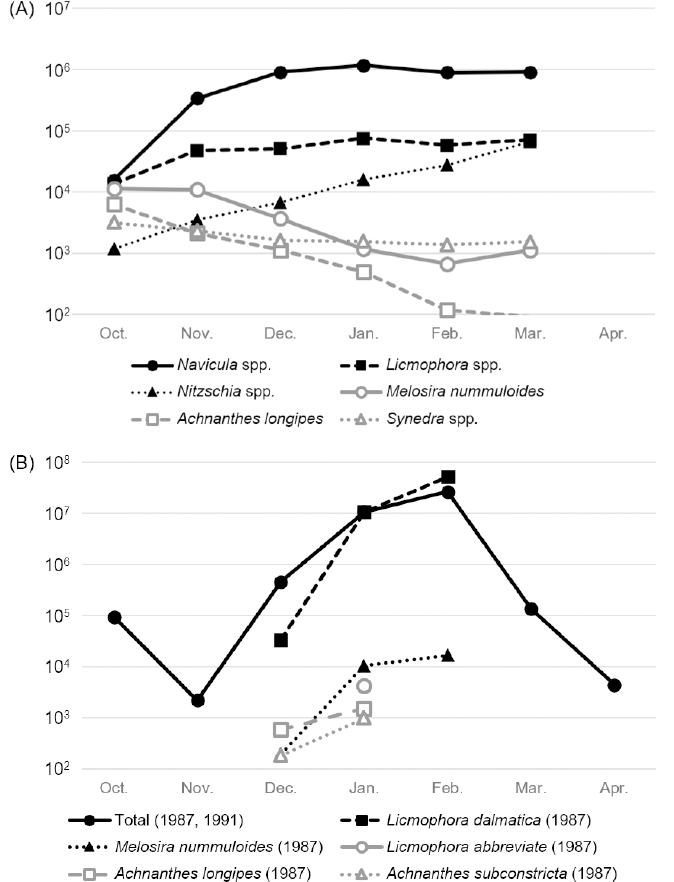
Dominant species of diatoms affecting Pyropia cultivation vary by region and season. During the cultivating season from 1982 to 1983 in Japan, Navicula spp. were the most dominant species attaching to cultivating nets, followed by Licmophora spp., Nitzschia spp., Melosira nummuloides, Achnanthes longipes, and Synedra spp., and the most dominant species attaching to thalli were Licmophora spp. (Ohgai, 1986). From February 1989 to March 1990, in the western coastal region in South Korea, 40 species of diatoms were identified on thalli of Pyropia and ambient seawater. The most dominant species were found in the following order: Licmophora dalmatica, Licmophora abbreviata, Melosira nummuloides, Paralia sulcata, Achnanthes javanica var. subconstricta, Grammatophora oceanica, Navicula sp., Synedra sp., Pinnularia sp., Fragilaria straitula, and Cocconeis scutellum var. parva (Kim et al., 1991). In the three cultivation seasons from 2012 to 2015, the pennate diatoms Fragilaria spp., Licmophora flabellata, tube-dwelling Navicula spp., and the centric diatom Melosira moniliformis were identified to cause severe contamination, and the severest damage by diatom felt occurred in December and January by the Licmophora and Navicula spp. (Kim et al., 2014; Moon, 2015). Not only epiphytic diatoms but also bloom-forming diatoms can impact Pyropia cultivation. Eucampia zodiacus Ehrenberg, a harmful bloom-forming species, was not considered to damage Pyropia spp. cultivation; however, since the mid-1990s, its blooms have been reported annually in the Seto Inland Sea in Japan when the seawater temperature is low (Nishikawa and Yamaguchi, 2006). Cell densities of Eucampia zodiacus were the highest from mid-February to early April, with decreasing nutrient concentration over this time (Nishikawa et al., 2007). Another harmful bloom-forming species, Asteroplanus karianus, has been reported and is known to bleach Pyropia every winter in the Ariake Sea in Japan since 2008 (Yamaguchi et al., 2014).
The changes in the proportion and detected cell numbers of diatoms exhibit patterns, which are shown in Figure 3. From 1982 to 1983 in Japan, the most dominant species, Navicula spp., occupied from 30% of the total cell number of diatoms in October to 87% in March; the second most dominant species, Licmophora spp., occupied from 27% in October to 7% in March (Figure 3A; Ohgai, 1986). From December 1987 to February 1988, the total standing crop (cells/gram of dry weight of thallus) varied from 36,000 to 52,000,000 depending on the collecting time and site, and Licmophora dalmatica was the most identified epiphytic diatom on blades, occupying over 93% of the total standing crop, followed by Melosira nummuloides, Licmophora abbreviata, and Achnanthes longipes (Figure 3B).

Monthly change of predominant diatoms in the cultivating red algae. (A) Average cell numbers of each diatom in the Shimonoseki and Onoda coastal areas in Japan from October 1982 to March 1983 (Ohgai 1986). (B) Average standing crop (cells/gram of dry-weight of thallus) of epiphytic diatoms in South Korea from December 1987 to February 1988 and from February 1989 to March 1990 (Lee and Kim 1989, Kim et al 1991).
Licmophora species are triangular in shape (thin fan shape), forming a cluster and attaching to the host with a long mucilaginous tube. They cover the surface of the thalli, block sunlight and nutrients, and, finally, result in an inferior quality of crops (Kim et al., 2014; Ohgai, 1986). Navicula species are boat-shaped and tube-dwelling diatoms, forming a mucilaginous tube and living within it. Mucilaginous tubes attach to and occupy cultivation nets, inhibiting the growth of Pyropia or Neoporphyra species by shading light and consuming periphery nutrients (Moon, 2015). Melosira nummuloides are centric diatoms; they are circular in shape and form a cluster of a long chain with mucilage pads. Achnanthes longipes are linear and concave in the middle and slightly curved in the girdle view (Lee et al., 2013). Melosira nummuloides and Achnanthes longipes attach to and occupy cultivation nets, inhibiting the growth of crops in the early stage of cultivation (Ohgai, 1986). Synedra gracilis has a long and thin shape with a slightly convex center; it exhibits mucilaginous pores at the end part valve side, which attach to thalli (Ohgai, 1986). In Eucampia zodiacus, rectangular cells form coil chains and engage in competitive consumption of nutrients, especially nitrogen, leading to Pyropia bleaching (Lee and Lee, 2012; Nishikawa et al., 2007). Anvil-shaped cells, usually numbering up to eight, form circular, asterisk-like colonies and exhaust dissolved inorganic nitrogen and dissolved inorganic phosphorus from seawater during massive blooms, causing discoloration of Pyropia or Neoporphyra species due to nutritional deficiency (Crawford and Gardner, 1997; Yamaguchi et al., 2014).
DETECTION
Diatom felt could be easily distinguished with the brown epiphytes suffused over the thalli and with its unpleasant and earthy odor (Kim et al., 2014). The visible morphological features can be confirmed by using light, epifluorescence, or electron microscopy, which are typical methods for the further identification of diatoms.
INFLUENCING FACTORS
The growth of epiphytic diatoms is highly related to temperature and light. The optimal growth conditions of the dominant species are 10-20 °C, chlorinity of 15-22‰, and pH of 6.5-9.0 for Navicula directa prox. and 15-25 °C, chlorinity of 15-16.5‰, and pH of 7.5-9.0 for Licmophora abbreviata (Ohgai, 1986). Eucampia zodiacus was found to grow at 7-30 °C with salinity of 10‰, with its optimal ranges of temperature and salinity being 20-25 °C and 20-30‰, respectively, which is different from the cultivating field situation of blooms that lower seawater temperature below optimal level to grow and occurred in the winter and spring (Nishikawa, 2002). Asteroplanus karianus could grow well at a temperature range of 15-25 °C and salinity of 15-33‰ (Shikata et al., 2015).
Diatom felt was the most common disease of Pyropia in the cultivating season from 2008 to 2009, and its incidence rate gradually increased from 3.8% in November to 64.3% in March (Lee et al., 2012). The cell numbers of Navicula spp. and Licmophora spp., which were relatively low in October and highest in January, tended to increase according to the decrease of seawater temperature; in contrast, the cell numbers of Melosira nummuloides, Achnanthes spp., and Synedra spp., which were highest in October and gradually lowered to March, tended to decrease according to the decrease of seawater temperature (Figure 3A; Ohgai, 1986). Since the 1990s, massive blooms of Eucampia zodiacus have been reported, despite low seawater temperatures, and cell densities of Eucampia zodiacus have been highest from mid-February to early April (Nishikawa et al., 2007). Since 2008, Asteroplanus karianus has formed massive winter blooms annually from December to January (Yamaguchi et al., 2014).
PREVENTION AND TREATMENT
Diatoms eradication from the thallus is challenging, however, acid wash, a regular method for controlling red algal diseases, was suggested as an effective treatment method. Epiphytic diatom species Navicula sp. lost their photosynthetic ability after 20-second pH shock with 8% HCl (Kang and Kim, 2022). Exposing nets to air and washing nets with a jet of seawater have been suggested to reduce the damage caused by diatoms (Ohgai, 1986). Eradicating diatoms from the surface of thalli via treatment with hydrochloric acid was reported to increase eight times the amount of dropped epiphytic diatoms higher than control (Moon, 2015).
DISCUSSION
This article reviewed the main red algal diseases, causative agents, symptoms, detection methods, optimal growth condition, and treatment. Table 2 summarizes the information reviewed. For the disease, it was found that continuous studies and reports on the four main Pyropia diseases were rarely conducted, although multiple diseases have occurred simultaneously and continuously. In total, two to four diseases have been observed at the same time during the beginning and ending stages of the open sea cultivation (Ding and Ma, 2005; Lee et al., 2012). Moreover, the incidence rate of diseases in the sampled thalli varied from 37.8% in November to 80.6% in March in the cultivating season from 2008 to 2009 (Lee et al., 2012). In South Korea, from 2008 to 2009, red-rot disease outbreak was not found but the incidence rates for other diseases varied. It ranged from 8.8% in December to 32.8% in February for Olpidiopsis disease, from 20.3% in November to 1.6% in February for green-spot disease, and from 3.8% in November to 64.3% in March for diatom felt (Lee et al., 2012). The infection rate of red-rot disease in the three cultivating seasons from 2012 to 2015 was from 3.4% in December to 1.1% in February and 15% in April, lower than the infection rate of Olpidiopsis disease (Moon, 2015). In South Korea, red-rot disease has been detected frequently in farms, but it is not the most common disease in the country. Seasonal and regional variance for the disease occurrence was also found, as depicted in Table 3. High variance across habitats and disease types suggests more site- and disease-specific studies are required in both the laboratory and field.
Red-rot disease caused by Pythium spp. is the only well-studied disease among the Pyropia diseases, which is part of the major diseases. The causative agents, detection methods, influencing factors, and treatment methods have been thoroughly studied. Nevertheless, knowledge gaps persist regarding the genome of Pythium spp. and development of alternative and effective treatment methods. Traditional treatment methods are still only partially effective or not effective in eradicating causative agents. Acid treatment, known to be the most effective method for controlling red-rot disease at present, represents about 12% of the total cost of operating sea farms in South Korea (Kim et al., 2014). Moreover, it has been found that Pythium porphyrae mycelia tolerance to acid increases at pH 4 compared with pH 8 (Hwang et al., 2009). This suggests that Pythium porphyrae acquires tolerance to acid and can adapt to pH change (Hwang et al., 2009). Freezing nets, another method known for preventing infection, is partially effective since red-rot disease and Olpidiopsis disease were not effectively eliminated from thalli attached to the nets frozen at -20 °C (Ding and Ma, 2005).
Olpidiopsis disease was first reported several decades ago; however, a detailed taxonomical study of the causative agent was only conducted recently. The life cycle, infection mechanism, and genome of Olpidiopsis spp. are largely unknown. Detection methods using PCR have been developed. However, treatment methods such as acid wash and desiccation are ineffective in controlling Olpidiopsis spp.. Thus, developing an effective treatment method is required. Studies applying or developing the currently available methods, namely acid treatment and net freeze, have been performed. In one study, via the process of drying, freezing, and thawing nets, infection rates of Pythium porphyrae and Olpidiopsis porphyrae were reduced by 0.37 times and 0.3 times, respectively (Park, 2020). In addition, the expression of the heat shock protein gene was improved, being specific to oomycete infection and the resistance of Pyropia spp. to oomycetes (Park, 2020). Calcium salt was a suggested candidate for an alternative treatment agent because it is effective against Pythium spp. on land plants. Particularly, CP inhibits the growth of mycelium and even prevents infection of Pythium porphyrae and Olpidiopsis (Kim et al., 2023; Moon, 2015). CP was able to inhibit Olpidiopsis sp. right after treatment of infected thalli; however, the effectiveness did not last over 24 hours (Moon, 2015).
Although outbreaks of green-spot disease were continuously reported, information is limited on the causative agents, infection process, pathogen detection, and prevention and treatment methods. Several bacteria or viruses are known to cause green-spot disease, and the chloroplast virus, PyroV1, is one of the suggested causative agents that is assumed to be the dsRNA virus (Kim et al., 2016). Besides, symptoms of infection by bacteria are different from those associated with infection by the virus. Symptoms appearing on thalli infected by green-spot disease are similar to those of anagusare disease (shot hole disease) and anaaki disease. For anagusare disease, at the initial stage, numerous holes with greenish peripheries appear at the tip of blade, and they subsequently spread over the blade. Compared with anagusare disease, at the initial stage of anaaki disease, several needle-tip holes are formed in the middle of the thallus, with these holes growing larger (Fujita, 1990; Tsuchiya, 1984). The causative agent of anagusare disease is unknown, and it is only presumed to occur by physical and mechanical irritation in the estuary. Outbreaks of this disease have rarely been reported since the 1950s (Fujita, 1990).
In terms of anaaki disease, the biggest difference between its symptoms and those of green-spot disease is the presence of host cell plasmoptysis in green-spot disease (Fujita, 1990; Tsuchiya, 1984). During infection of anaaki disease, the causative agent may lyse the intercellular matrix, resulting in cells becoming detached and the holes growing larger (Fujita, 1990; Tsuchiya, 1984). Gram-negative bacteria forming yellow colonies and decomposing agar were suggested as the causative agents of anaaki disease in 1984, and Flavobacterium sp. LAD-1 was isolated from infected thalli (Sunairi et al., 1995; Tsuchiya, 1984). To treat anaaki disease, the method of net exposure to air was recommended, and further infection is likely prevented by acid treatment (Fujita, 1990; Sunairi et al., 1995).
Diatom felt, the most common disease from 2008 to 2009 in South Korea, can cause damage comparable to oomycete diseases. It has been reported that diatoms could induce low quality crops via drying processes (Kim et al., 2014). Moreover, massive bloom-forming diatoms could cause a lower yield of red algae and discolored Pyropia spp. by inhibiting nutrient uptake from seawater. Until the 1990s, most species of diatom blooms affecting Pyropia cultivation in Japan have been known to be related to Coscinodiscus wailesii. However, the species resulting in blooms have been widened to Thalassiosira diporocyclus, Eucampia zodiacus, and Asteroplanus karianus (Miyahara et al., 1996; Nagai et al., 1995; Nishikawa, 2002; Yamaguchi et al., 2014). Among the traditional treatment methods, the acid treatment was suggested as an effective method to eradicate epiphytic diatom (Kang and Kim, 2022). A study reported that the treatment with sodium bicarbonate and sodium percarbonate is effective for inhibiting the attachment of diatoms (Moon, 2015).
Pyropia cultivation started hundreds of years ago and the occurrence of various related diseases has been reported for the last decades. Disease outbreaks during cultivation have increased with the global production of red algae and climate change. In this review, we found the specific gaps in knowledge on infectious agents and procedure, detection, prevention, and treatment of Pyropia. Most of the red algal diseases have rarely been studied, except the red-rot disease that is relatively well studied. More studies seem necessary to better understand the other major diseases, including Olpidiopsis disease, green-spot disease, and diatom-related diseases. Similarly, methods of early detection and effective treatment have not yet been developed for them. Edible seaweed has been highlighted as a sustainable crop providing healthy and nutritious food, and its market has the potential to increase. Further studies are fundamental to ensure stable and quality production of the edible red algae species Pyropia.
ACKNOWLEDGMENTS
We thank Ji-Sook Kim and Hea-Won Kim, who helped the project operations.
REFERENCES
- Addepalli, M. K. & Fujita, Y. 2001. Serological detection of red rot disease initiation stages of microbial pathogen, Pythium porphyrae (Oomycota) on Porphyra yezoensis. Journal of Applied Phycology, 13, 219-25.
-
Addepalli, M. K. & Fujita, Y. 2002. Regulatory role of external calcium on Pythium porphyrae (Oomycota) zoospore release, development and infection in causing red rot disease of Porphyra yezoensis (Rhodophyta). FEMS Microbiology Letters, 211(2), 253-7. DOI: https://doi.org/10.1111/j.1574-6968.2002.tb11233.x
» https://doi.org/10.1111/j.1574-6968.2002.tb11233.x -
Akizuki, A., Tabata, M. & Kawamura, Y. 2007. Disinfectant effects of lactic acid on Pythium porphyrae as acid treatment agent. Aquaculture Science, 55(3), 325-30. DOI: https://doi.org/10.11233/aquaculturesci1953.55.325
» https://doi.org/10.11233/aquaculturesci1953.55.325 - Amano, H., Sakaguchi, K., Maegawa, M. & Noda, H. 1996. The use of a monoclonal antibody for the detection of fungal parasite, Pythium sp., the causative organism of red rot disease, in seawater from Porphyra cultivation farms. Fisheries science, 62(4), 556-60.
-
Amano, H., Suginaga, R., Arashima, K. & Noda, H. 1995. Immunological detection of the fungal parasite, Pythium sp.; the causative organism of red rot disease in Porphyra yezoensis. Journal of Applied Phycology , 7, 53-8. DOI: https://doi.org/10.1007/BF00003550
» https://doi.org/10.1007/BF00003550 - Arasaki, S. 1947. Studies on the wasting disease of the cultured lavers (Porphyra tenera). Bulletin of the Japanese Society for the Science of Fish, 13, 74-90.
- Arasaki, S. 1960. A chytridean parasaite on the Porphyra. Nippon Suisan Gakkaishi, 26, 543-8.
- Badis, Y., Han, J.W., Klochkova, T.A., Gachon, C.M.M. & Kim, G.H. 2020. The gene repertoire of Pythium porphyrae (Oomycota) suggests an adapted plant pathogen tackling red algae. Algae, 35, 133-44.
-
Bae, H.-J., Bang, Y.-J., Jeong, J.-A., Kim, W.-K., Lee, H., Moon, S. Y. & Jeong, T.-Y. 2024. Differential surface microbial community and thalli metabolome as early indicators of disease in red algae Pyropia yezoensis. Aquaculture International, 32, 1-18. DOI: https://doi.org/10.1007/s10499-024-01600-6
» https://doi.org/10.1007/s10499-024-01600-6 - Bae, S.-H. 1991. The origin and development process of laver culture industry in South Korea-1. Laver culture history till the end of Chosun dynasty. South Korean Journal of Fisheries and Aquatic Sciences, 24(3), 153-66.
-
Blouin, N. A., Brodie, J. A., Grossman, A. C., Xu, P. & Brawley, S. H. 2011. Porphyra: a marine crop shaped by stress. Trends in Plant Science, 16(1), 29-37. DOI: https://doi.org/10.1016/j.tplants.2010.10.004
» https://doi.org/10.1016/j.tplants.2010.10.004 - Brodie, J & Irvine, L. M. 2003 Seaweeds of the British Isles Volume 1 Rhodophyta Part 3B Bangiophycidae. Natural History Museum, London.
-
Cho, K., Heo, J., Lee, S. M., Han, J., Hong, H. D., Jeon, H., Hwang, H.J., Baek, K. & Han, J. W. 2020. Discoloration in the marine red algae Pyropia: causative factors and exploiting the biotechnological potential of a waste resource. Reviews in Aquaculture, 13(2), 822-835. DOI: https://doi.org/10.1111/raq.12500
» https://doi.org/10.1111/raq.12500 -
Crawford, R. M. & Gardner, C. 1997. The transfer of Asterionellopsis kariana to the new genus Asteroplanus (Bacillariophyceae), with reference to the fine structure. Nova Hedwigia, 47-57. DOI: https://doi.org/10.1127/nova.hedwigia/65/1997/47
» https://doi.org/10.1127/nova.hedwigia/65/1997/47 - De Cock, A. W. A. M. 1986. Marine Pythiaceae from decaying seaweeds in the Netherlands. Mycotaxon, 25(1), 101-10.
- Diehl, N., Kim, G. H. & Zuccarello, G. C. 2017. A pathogen of New Zealand Pyropia plicata (Bangiales, Rhodophyta), Pythium porphyrae (Oomycota). Algae, 32, 29-39.
-
Ding, H. & Ma, J. 2005. Simultaneous infection by red rot and chytrid diseases in Porphyra yezoensis Ueda. Journal of Applied Phycology , 17, 51-6. DOI: https://doi.org/10.1007/s10811-005-5523-6
» https://doi.org/10.1007/s10811-005-5523-6 -
FAO 2024. FAO Fisheries and Aquaculture - Global Statistical Collections. In: FAO Fisheries and Aquaculture Division [online]. Available from: Available from: https://www.fao.org/fishery/en/statistics Access date: 2024 Jun. 13.
» https://www.fao.org/fishery/en/statistics - Fujita, Y., Zenitani, B., Nakao, Y. & Matsubara, T. 1972. Bacteriological studies on diseases of cultured laver-II. Bacteria associated with diseased laver. Bulletin of the Japanese Society of Scientific Fisheries, 38(6), 565-569.
- Fujita, Y. & Migita, S. 1980. Death of parasitic Pythium porphyrae by drying and freeze-preservation of red rot infected thalli of Porphyra yezoensis. Bulletin of the Faculty of Fisheries, Nagasaki University, 49, 11-16.
- Fujita, Y. 1990. Diseases of cultivated Porphyra in Japan. Introduction to applied phycology. 177-90.
- Fujitake, F., Kuno, K. & Igata, K. 2009. Occurrence of red rot disease and chytrid blight disease in nori [Porphyra] culture farms of Saga [Japan] Prefecture in the inner part of Ariake Sea. Bulletin of Saga Prefectural Ariake Fisheries Research and Development Center (Japan).
-
Gachon, C. M. M., Sime-Ngando, T., Strittmatter, M., Chambouvet, A. & Kim, G. H. 2010. Algal diseases: spotlight on a black box. Trends in Plant Science , 15(11), 633-40. DOI: https://doi.org/10.1016/j.tplants.2010.08.005
» https://doi.org/10.1016/j.tplants.2010.08.005 - Han, X., Mao, Y., Li, J., Li, G., Li, C., Liu, L. & Mo, Z. 2015. Identification and pathogenecity of a novel pathogen causing green rot disease in Porphyra haitanensis. Journal of Fisheries of China, 39, 1721-9.
- He, P. M., Zhang, Z. Y., Zhang, X. C., & Ma, J. H. 2018. Seaweed cultivation. Sciences Academic Press.
- Hwang, E. K. & Park, C. S. 2020. Seaweed cultivation and utilization of South Korea. Algae, 35, 107-21.
-
Hwang, E. K., Park, C. S. & Kakinuma, M. 2009. Physicochemical responses of Pythium porphyrae (Oomycota), the causative organism of red rot disease in Porphyra to acidification. Aquaculture Research, 40(15), 1777-84. DOI: https://doi.org/10.1111/j.1365-2109.2009.02284.x
» https://doi.org/10.1111/j.1365-2109.2009.02284.x -
Kang, E. J. & Kim, J.-H. 2022. Development of an efficiency criterion for the removal of pest organisms (ulvoid green algae and diatoms) from Neopyropia aquaculture using the acid wash (pH shock) method. Aquaculture, 548, 737677. DOI: https://doi.org/10.1016/j.aquaculture.2021.737677
» https://doi.org/10.1016/j.aquaculture.2021.737677 -
Kawamura, Y., Yokoo, K., Tojo, M. & Hishiike, M. 2005. Distribution of Pythium porphyrae, the causal agent of red rot disease of Porphyrae spp., in the Ariake Sea, Japan. Plant Disease, 89, 1041-7. DOI: https://doi.org/10.1094/PD-89-1041
» https://doi.org/10.1094/PD-89-1041 -
Kim, G. H., Klochkova, T. A. & Im, S. H. 2016. Chloroplast virus causes green-spot disease in cultivated Pyropia of South Korea. Algal Research. 17, 293-99. DOI: https://doi.org/10.1016/j.algal.2016.05.023
» https://doi.org/10.1016/j.algal.2016.05.023 -
Kim, G. H., Moon, K.-H., Kim, J.-Y., Shim, J. & Klochkova, T. A. 2014. A revaluation of algal diseases in South Korean Pyropia (Porphyra) sea farms and their economic impact. Algae. 29(4), 249-65. DOI: https://doi.org/10.4490/algae.2014.29.4.249
» https://doi.org/10.4490/algae.2014.29.4.249 - Kim, J. 2015. Life history and infection mechanism of Pythium porphyrae, a causative agent of red-rot disease in Pyropia spp. Master dissertation, Kongju National University, Kongju, 60pp. (In South Korean with English abstract)
- Kim, J.-R., Shin, Y.-K., Lee, G.-H. & Lee, W.-H. 1991. A Study on the relationships between the epiphytic microbes and the blight of Porphyra species from the coastal waters of the Yellow Sea, South Korea 1. Species composition and standing stocks of epiphytic diatom and ambient water phytoplankton. South Korean Journal of Fisheries and Aquatic Sciences , 24, 79-88.
- Kim, Y.T., Kim, R., Shim, E., Park, H., Klochkova, T.A., Kim, G.H., Kim, Y.T., Kim, R., Shim, E. & Park, H. 2023. Control of oomycete pathogens during Pyropia farming and processing using calcium propionate. Algae, 38(1), 71-80.
-
Klochkova, T. A., Jung, S. & Kim, G. H. 2017. Host range and salinity tolerance of Pythium porphyrae may indicate its terrestrial origin. Journal of Applied Phycology , 29, 371-9. DOI: https://doi.org/10.1007/s10811-016-0947-8
» https://doi.org/10.1007/s10811-016-0947-8 -
Klochkova, T. A., Shim, J. B., Hwang, M. S. & Kim, G. H. 2012. Host-parasite interactions and host species susceptibility of the marine oomycete parasite, Olpidiopsis sp., from South Korea that infects red algae. Journal of Applied Phycology , 24(1), 135-44. DOI: https://doi.org/10.1007/s10811-011-9661-8
» https://doi.org/10.1007/s10811-011-9661-8 -
Klochkova, T. A., Shin, Y. J., Moon, K.-H., Motomura, T. & Kim, G. H. 2016. New species of unicellular obligate parasite, Olpidiopsis pyropiae sp. nov., that plagues Pyropia sea farms in South Korea. Journal of Applied Phycology , 28(1), 73-83. DOI: https://doi.org/10.1007/s10811-015-0595-4
» https://doi.org/10.1007/s10811-015-0595-4 - Lee, J. M. & Lee, J. H. 2012. Morphological study of the genus Eucampia (Bacillariophyceae) in South Korean coastal waters. Algae, 27, 235-47.
-
Lee, S. J., Park, S. W., Lee, J. H. & Kim, Y. S. 2012. Diseases of the cultivated Porphyra at Seocheon area. Journal of fish pathology, 25(3), 249-56. DOI: https://doi.org/10.7847/jfp.2012.25.3.249
» https://doi.org/10.7847/jfp.2012.25.3.249 -
Lee, S. D., Park, J. S. & Lee, J. H. 2013. Taxonomic study of the genus Achnanthes (Bacillariophyta) in South Korean coastal waters. Journal of Ecology and Environment, 36(4), 391-406. DOI: https://doi.org/10.5141/ecoenv.2013.391
» https://doi.org/10.5141/ecoenv.2013.391 -
Lee, S. J. & Lee, S. R. 2022. Rapid Detection of Red Rot Disease Pathogens (Pythium chondricola and P. porphyrae) in Pyropia yezoensis (Rhodophyta) with PCR-RFLP. Plant Disease, 106, 30-33. DOI: https://doi.org/10.1094/PDIS-07-21-1494-SC
» https://doi.org/10.1094/PDIS-07-21-1494-SC - Lee, S. J., Hwang, M. S., Park, M., Baek, J. M., Ha, D.-S., Lee, J. E. & Lee, S.-R. 2015. Molecular identification of the algal pathogen Pythium chondricola (Oomycetes) from Pyropia yezoensis (Rhodophyta) using ITS and cox1 markers. Algae, 30, 217-22.
- Lee, S. J., Jee, B. Y., Son, M.-H. & Lee, S.-R. 2017. Infection and cox2 sequence of Pythium chondricola (Oomycetes) causing red rot disease in Pyropia yezoensis (Rhodophyta) in South Korea. Algae, 32, 155-60.
- Lee, W. H. & Kim, J. R. 1989. A quantitative study on the epiphytic diatoms of Porphyra species from the coastal waters of the Yellow Sea. South Korea. Marine Development Research Kunsan National University, 1, 51-66.
-
Li, J., Mou, Z. J., Yang, H. C., Mao, Y. X., Yan, Y. W. & Mo, Z. L. 2018. Isolation and identification the pathogen of Pyropia yezoensis green spot disease. Progress in Fishery Science, 40(4), 140-146. DOI: https://doi.org/10.19663/j.issn2095-9869.20180710002
» https://doi.org/10.19663/j.issn2095-9869.20180710002 - Liu, J., Xia, S., Yang, H., Mo, Z., Li, J., & Yan, Y. 2024. Quantitative detection of Pythium porphyrae and Pythium chondricola (Oomycota), the causative agents of red rot disease in Pyropia farms in China. Algae, 39(3), 177-186.
-
MAFF 2024. Fisheries Census. Available from: Available from: https://www.e-stat.go.jp Access date: 2024 Jun. 13.
» https://www.e-stat.go.jp - Migita, S. 1969. Olpidiopsis disease of culture Porphyra. Bulletin of the Faculty of Fisheries , Nagasaki University, 28, 131-45.
- Mikami, K., Li, L. & Takahashi, M. 2012. Monospore-based asexual life cycle in Porphyra yezoensis. Frontiers in Physiological and Molecular Biological Research, 15-37.
-
Miyahara, K., Nagai, S., Itakura, S., Yamamoto, K., Fujisawa, K., Iwamoto, T., Yoshimatsu, S., Matsuoka, S., Yuasa, A., Makino, K., Hori, Y., Nagata, S., Nagasaki, K., Yamaguchi, M., Honjo, T. 1996. First Record of a Bloom of Thalassiosira diporocyclus in the Eastern Seto Inland Sea. Fisheries science , 62(6), 878-82. DOI: https://doi.org/10.2331/fishsci.62.878
» https://doi.org/10.2331/fishsci.62.878 -
Mo, Z., Li, S., Kong, F., Tang, X. & Mao, Y. 2016. Characterization of a novel fungal disease that infects the gametophyte of Pyropia yezoensis (Bangiales, Rhodophyta). Journal of Applied Phycology , 28, 395-404. DOI: https://doi.org/10.1007/s10811-015-0539-z
» https://doi.org/10.1007/s10811-015-0539-z -
MOF 2024. Fisheries statistics. Available from: Available from: https://www.mof.go.kr/statPortal Access date: 2024 Jun. 13.
» https://www.mof.go.kr/statPortal - Moon, K.-H. 2015. Studies on algal diseases in Pyropia farms in South Korea. (Ph.D. dissertation). Kongju: Kongju National University.
- Mou, Z. 2012. Etiological Study on Pyropia/Porphyra yezoensis and Screening of Agarase-producing Bacteria. (Master dissertation). Qingdao: Ocean University of China.
-
Muñoz, L., Patiño, D. J. & Murúa, P. 2024. Natural biocontrol of a Porphyra sp. pest on farmed Gracilaria chilensis by a pythiosis outbreak. Journal of Applied Phycology , 36, 2029-37. DOI: https://doi.org/10.1007/s10811-024-03228-8
» https://doi.org/10.1007/s10811-024-03228-8 -
Nagai, S., Hori, Y., Manabe, T. & Imai, I. 1995. Morphology and Rejuvenation of Coscinodiscus wailesii GRAN (Bacillariophyceae) Resting Cells Found in Bottom Sediments of Harima-Nada, Seto Inland Sea, Japan. NIPPON SUISAN GAKKAISHI, 61(2), 179-85. DOI: https://doi.org/10.2331/suisan.61.179
» https://doi.org/10.2331/suisan.61.179 -
Nagano, Y., Kimura, K., Kobayashi, G. & Kawamura, Y. 2021. Genomic diversity of 39 samples of Pyropia species grown in Japan. Plos one, 16(6), e0252207. DOI: https://doi.org/10.1371/journal.pone.0252207
» https://doi.org/10.1371/journal.pone.0252207 -
Nakao, Y., Onohara, T., Zenitani, B., Fujita, Y. & MATSUBAR, T. 1972. BACTERIOLOGICAL STUDIES ON DISEASES OF CULTURED LAVER. 1. GREEN SPOT ROTTING-LIKE DETERIORATION OF LAVER FROND BY BACTERIA, IN-VITRO. Bulletin of the Japanese Society of Scientific Fisheries , 38(6), 561. DOI: https://doi.org/10.2331/suisan.38.561
» https://doi.org/10.2331/suisan.38.561 -
Nguyen, H. D. T., Dodge, A., Dadej, K., Rintoul, T. L., Ponomareva, E., Martin, F. N., De Cock, A. W. A. M., Lévesque, C. A., Redhead, S. A. & Spies, C. F. J. 2022. Whole genome sequencing and phylogenomic analysis show support for the splitting of genus Pythium. Mycologia, 114(3), 501-515. DOI: https://doi.org/10.1080/00275514.2022.2045116
» https://doi.org/10.1080/00275514.2022.2045116 - Nishikawa, T. 2002. Effects of temperature, salinity and irradiance on the growth of the diatom Eucampia zodiacus caused bleaching of seaweed Porphyra isolated from Harima-Nada, Seto Inland Sea, Japan. NIPPON SUISAN GAKKAISHI , 68, 356-61.
-
Nishikawa, T., Hori, Y., Tanida, K. & Imai, I. 2007. Population dynamics of the harmful diatom Eucampia zodiacus Ehrenberg causing bleachings of Porphyra thalli in aquaculture in Harima-Nada, the Seto Inland Sea, Japan. Harmful Algae, 6(6), 763-73. DOI: https://doi.org/10.1016/j.hal.2007.04.005
» https://doi.org/10.1016/j.hal.2007.04.005 - Nishikawa, T. & Yamaguchi, M. 2006. Effect of temperature on light-limited growth of the harmful diatom Eucampia zodiacus Ehrenberg, a causative organism in the discoloration of Porphyra thalli. Harmful Algae , 5, 141-7.
- Noda, H., Amano, H., Ohta, F. & Horiguchi, Y. 1979. The effects of amino acids in curing and preventing “akagusare”, red rot disease, of the laver Porphyra spp. Bulletin of the Japanese Society of Scientific Fisheries , 45(9), 1155-1162.
-
Noda, H. 1993. Health benefits and nutritional properties of nori. Journal of Applied Phycology , 5, 255-8. DOI: https://doi.org/10.1007/BF00004027
» https://doi.org/10.1007/BF00004027 - Ohgai, M. 1986. Studies on the morphology and ecology of epiphytic and epilithic diatoms in Nori cultivation grounds. Journal of the Shimonoseki University of Fisheries, 34, 2 (In Japanese with English abstract)
-
Park, C., Sakaguchi, K., Kakinuma, M. & Amano, H. 2000. Comparison of the morphological and physiological features of the red rot disease fungus Pythium sp. isolated from Porphyra yezoensis from South Korea and Japan. Fisheries science , 66(2000), 261-9. DOI: https://doi.org/10.1046/j.1444-2906.2000.00043.x
» https://doi.org/10.1046/j.1444-2906.2000.00043.x - Park, C. S., Kakinuma, M. & Amano, H. 2001. Detection of the red rot disease fungi Pythium spp. by polymerase chain reaction. Fisheries science , 67, 197-9.
- Park, J.H. 2020. Priming of resistance against oomycete pathogens through desiccation and freezing in Pyropia yezoensis (Rhodophyta). (Master dissertation). Kongju: Kongju National University.
-
Patil, V., Sun, L., Mohite, V., Liang, J., Wang, D., Gao, Y. & Chen, C. 2024. Effect of benthic and planktonic diatoms on the growth and biochemical composition of the commercial macroalga Pyropia haitanensis. Marine Pollution Bulletin, 203, 116411. DOI: https://doi.org/10.1016/j.marpolbul.2024.116411
» https://doi.org/10.1016/j.marpolbul.2024.116411 -
Qiu, L., Mao, Y., Tang, L., Tang, X. & Mo, Z. 2019. Characterization of Pythium chondricola associated with red rot disease of Pyropia yezoensis (Ueda) (Bangiales, Rhodophyta) from Lianyungang, China. Journal of Oceanology and Limnology, 37(2), 1102-12. DOI: https://doi.org/10.1007/s00343-019-8075-3
» https://doi.org/10.1007/s00343-019-8075-3 -
Robideau, G. P., De Cock, A. W., Coffey, M. D., Voglmayr, H., Brouwer, H., Bala, K., Chitty, D. W., Desaulniers, N., Eggertson, Q. A., Gachon, C. M. M., Hu, C.-H., Küpper, F. C., Rintoul, T. L., Sarhan, E., Verstappen, E. C. P., Zhang, Y., Bonants, P. J. M., Ristaino, J. B., & Lévesque, C. A. 2011. DNA barcoding of oomycetes with cytochrome c oxidase subunit I and internal transcribed spacer. Molecular ecology resources, 11(6), 1002-1011. DOI: https://doi.org/10.1111/j.1755-0998.2011.03041.x
» https://doi.org/10.1111/j.1755-0998.2011.03041.x -
Saito, Y., Matsusato, T. & Yoshikawa, K. 1972. On the symptoms of “green spot” and “crape” in nori (Porphyra) culture. Bulletin of Nansei National Fisheries Research Institute, 5, 1-9. DOI: https://doi.org/10.1007/s00343-019-9045-5
» https://doi.org/10.1007/s00343-019-9045-5 -
Sekimoto, S., Yokoo, K., Kawamura, Y. & Honda, D. 2008. Taxonomy, molecular phylogeny, and ultrastructural morphology of Olpidiopsis porphyrae sp. nov. (Oomycetes, straminipiles), a unicellular obligate endoparasite of Bangia and Porphyra spp. (Bangiales, Rhodophyta). Mycological Research, 112(3), 361-74. DOI: https://doi.org/10.1016/j.mycres.2007.11.002
» https://doi.org/10.1016/j.mycres.2007.11.002 -
Shikata, T., Matsubara, T., Yoshida, M., Sakamoto, S. & Yamaguchi, M. 2015. Effects of temperature, salinity, and photosynthetic photon flux density on the growth of the harmful diatom Asteroplanus karianus in the Ariake Sea, Japan. Fisheries science , 81, 1063-9. DOI: https://doi.org/10.1007/s12562-015-0930-3
» https://doi.org/10.1007/s12562-015-0930-3 -
Sunairi, M., Tsuchiya, H., Tsuchiya, T., Omura, Y., Koyanagi, Y., Ozawa, M., Iwabuchi, N. et al. 1995. Isolation of a bacterium that causes anaaki disease of the red algae Porphyra yezoensis. Journal of Applied Bacteriology, 79(2), 225-9. DOI: https://doi.org/10.1111/j.1365-2672.1995.tb00939.x
» https://doi.org/10.1111/j.1365-2672.1995.tb00939.x - Takahashi, M. 1977. Pythium porphyrae Takahashi et Sasaki, sp. nov. causing red rot of marine red algae Porphyra spp. Transactions of the Mycological Society of Japan, 18(3), 279-85.
-
Takahashi, M. & Mikami, K. 2017. Oxidative stress promotes asexual reproduction and apogamy in the red seaweed Pyropia yezoensis. Frontiers in Plant Science, 8, 62. DOI: https://doi.org/10.3389/fpls.2017.00062
» https://doi.org/10.3389/fpls.2017.00062 - Tsuchiya, H. 1984. Nori no kasho `anaaki-sho’ ni tsuite (About the disease of Porphyra, tentatively named ’anaaki disease’). Chiba Prefectural Fisheries Experimental Station Research Report, 67-71)
-
Uppalapati, S. R. & Fujita, Y. 2000. Carbohydrate regulation of attachment, encystment, and appressorium formation by Pythium porphyrae (Oomycota) zoospores on Porphyra yezoensis (Rhodophyta). Journal of Phycology, 36(2), 359-66. DOI: https://doi.org/10.1046/j.1529-8817.2000.99099.x
» https://doi.org/10.1046/j.1529-8817.2000.99099.x -
Uppalapati, S. R., Kerwin, J. L. & Fujita, Y. 2001. Epifluorescence and scanning electron microscopy of host-pathogen interactions between Pythium porphyrae (Peronosporales, Oomycota) and Porphyra yezoensis (Bangiales, Rhodophyta). Botanica Marina, 44(2), 139-145. DOI: https://doi.org/10.1515/BOT.2001.019
» https://doi.org/10.1515/BOT.2001.019 - Wen, X., Zuccarello, G. C., Klochkova, T. A. & Kim, G. H. 2023. Oomycete pathogens, red algal defense mechanisms and control measures. Algae, 38(4), 203-215.
-
Weng, P., Yang, H., Mo, Z., Zhang, W., Yan, Y., Rong, X. & Li, J. 2024. Application and evaluation of probiotics against red rot disease in Pyropia. Aquaculture, 578, 740050. DOI: https://doi.org/10.1016/j.aquaculture.2023.740050
» https://doi.org/10.1016/j.aquaculture.2023.740050 -
Yamaguchi, H., Minamida, M., Matsubara, T. & Okamura, K. 2014. Novel blooms of the diatom Asteroplanus karianus deplete nutrients from Ariake Sea coastal waters. Marine Ecology Progress Series, 517, 51-60. DOI: https://doi.org/10.3354/meps11014
» https://doi.org/10.3354/meps11014 -
Yan, Y.-W., Yang, H.-C., Tang, L., Li, J., Mao, Y.-X. & Mo, Z.-L. 2019. Compositional shifts of bacterial communities associated with Pyropia yezoensis and surrounding seawater co-occurring with red rot disease. Frontiers in Microbiology, 10, 1666. DOI: https://doi.org/10.3389/fmicb.2019.01666
» https://doi.org/10.3389/fmicb.2019.01666 -
Yang, H., Yan, Y., Li, J., Tang, L., Mao, Y. & Mo, Z. 2020a. Development of a PCR method for detection of Pseudoalteromonas marina associated with green spot disease in Pyropia yezoensis. Journal of Oceanology and Limnology , 38, 168-76. DOI: https://doi.org/10.1007/s00343-019-9045-5
» https://doi.org/10.1007/s00343-019-9045-5 -
Yang, L-E., Deng, Y-Y., Xu, G-P., Russell, S., Lu, Q-Q. & Brodie, J. 2020b. Redefining Pyropia (Bangiales, Rhodophyta): four new genera, resurrection of Porphyrella and description of Calidia pseudolobata sp. nov. from China. Journal of Phycology , 56(4), 862-79. DOI: https://doi.org/10.1111/jpy.12992
» https://doi.org/10.1111/jpy.12992 - Yokoo, K., Kawamura, Y. & Tojo, M. 1999. Oospore survival of Pythium porphyrae, in sterile sediments. Bulletin of Saga Prefectural Ariake Fisheries Experimental Station, 19, 1-7.
- Yokoo, K., Sekimoto, S., Kawamura, Y. & Honda, D. 2005. The early detection of Olpidiopsis sp. (Oomycetes, Chromista) which causes damage to nori [laver] cultivation using a PCR. Bulletin of the Japanese Society of Scientific Fisheries (Japan).
- Yong, Y., Jiahai, M., Pu, X., Qihuan, S. & Hanqing, W. 2002. Pseudoalteromonas citrea, the causative agent of green-spot disease of Porphyrae yezoensis. Zhongguo Shui Chan ke xue= Journal of Fishery Sciences of China, 9, 353-8.
-
DATA AVAILABILITY STATEMENT
All data are available from the corresponding author upon reasonable request.
-
SUPPLEMENTARY MATERIAL
There is no supplementary material for this article.
-
FUNDING
This study was funded by the National Research Foundation of South Korea (NRF) grant funded by the South Korea government (MSIT) [2021R1C1C1006155]; and the Hankuk University of Foreign Studies Research Fund under Grant [20211441001].
All data are available from the corresponding author upon reasonable request.