Poly(lactic acid) Films Reinforced with TiO2 and ZnO Nanoparticles: Thermal, Mechanical, Optical, and Phytotoxicity Assessment
Abstract
Poly(lactic acid) (PLA)-based films containing 0.5 and 1.5 wt% of TiO2 or ZnO nanoparticles were prepared by solvent casting and evaluated in terms of thermal, mechanical, optical, structural, and ecotoxicological performance, targeting food packaging applications. X-ray diffraction and scanning electron microscopy revealed that low nanoparticle contents promoted partial crystalline ordering and relatively homogeneous dispersion, whereas higher loadings led to agglomeration and reduced crystallinity. Thermogravimetric analysis showed that the incorporation of 0.5 wt% TiO2 increased the onset decomposition temperature of PLA from 303.3 to 322.8 °C. In contrast, higher nanoparticle contents and all ZnO-containing films exhibited reduced thermal stability and lower glass transition temperatures, with Tg decreasing from 51.3 °C (PLA) to 44.0 °C for PLA15ZnO. Nanoindentation demonstrated a significant increase in nanohardness for all nanocomposites, reaching improvements of up to ~27% compared to neat PLA. Optical analysis showed effective UV-shielding below 400 nm and preservation of visible-light transmittance above ~85% for films containing 0.5 wt% ZnO, whereas higher TiO2 contents induced pronounced opacity and color changes. Germination assays using lettuce and cucumber indicated concentration-dependent phytotoxic effects for TiO2, while ZnO showed neutral or slightly positive effects at low concentrations.
Keywords:
Poly(lactic acid); TiO2NPs. ZnONPs; Food Packaging; Phytotoxicity
1. Introduction
Food packaging plays a fundamental role in protecting food against several forms of contamination and deterioration, such as those caused by biological, chemical, and/or physical agents. In addition to preserving food products, packaging extends their shelf life by maintaining quality, ensuring consumer safety, and reducing losses along the production chain1–3. However, the polymers traditionally used in the production of these materials are of petrochemical origin and pose significant environmental challenges due to their non-biodegradability, leading to their accumulation in the environment and overloading landfills4. Another relevant impact associated with the inadequate disposal of these plastic materials is the formation of microplastics, which are fragments capable of bioaccumulating mainly in aquatic ecosystems5,6. Furthermore, recent scientific evidence has demonstrated the presence of these particles in human tissues, such as the placenta and lungs, raising important concerns regarding their potential deleterious effects on human health7,8. Therefore, the development of sustainable and environmentally friendly food packaging has become inevitable9–11.
Poly(lactic acid) (PLA), a biodegradable and compostable polyester derived from renewable resources12,13, emerges as a promising alternative to petrochemical-based polymers used in the food packaging sector9–11,14. An important advantage of this polyester is its contribution to reducing carbon dioxide (CO2) emissions15, one of the main greenhouse gases responsible for global warming. This reduction occurs because the biomass cultivated for its production absorbs CO2 during growth16. However, despite the environmental benefits associated with PLA, its application in food packaging is limited by its relatively low mechanical strength and thermal stability, as well as its poor gas and moisture barrier properties9,14,17,18. To overcome these limitations, the incorporation of additives into the PLA matrix, particularly nanoparticles, has emerged as a promising strategy.
The incorporation of titanium dioxide (TiO2NPs) and zinc oxide (ZnONPs) nanoparticles into PLA has been widely investigated due to their ability to enhance mechanical properties19–24, provide UV-blocking17–23,25, improve gas and moisture barrier performance17,18,20, and deliver antimicrobial activity17,19–21,23,25–27 to the material, thereby extending the shelf life of fresh foods. These nanoparticles are biocompatible and, generally, regarded as safe for use in food-contact materials28, reinforcing their potential for packaging applications. However, these investigations have predominantly focused on material performance, with limited attention given to the potential biological or environmental implications of nanoparticle incorporation.
Addition of TiO2NPs and ZnONPs to PLA may enhance the intrinsic biodegradability of this polymer29–31, thereby increasing the potential release of nanoparticles into soil and water. At low concentrations, both nanoparticles can promote plant growth, as observed for lettuce32, rice33, apples34, and cucumber35. In contrast, exposure of plants and soil to high concentrations of TiO2NPs has been associated with loss of nutritional quality32, reduced lettuce seedling growth36, decreased chloroplast number and pigment content in mulberry seedlings37, and the upregulation of antioxidant enzymes, indicating a plant response to counteract oxidative stress38. For ZnONPs, increasing concentration has also been reported to induce toxic effects, including oxidative stress in plants39,40, and disruption of microbial communities, leading to reduced microbial biomass and essential enzymatic activities related to soil health41.
Although TiO2NPs and ZnONPs have been widely investigated as reinforcing agents in PLA-based materials, most studies primarily focus on property enhancement, with limited consideration of the potential biological and environmental implications associated with nanoparticle incorporation. In particular, systematic investigations that simultaneously correlate thermal, mechanical, and optical performance with preliminary ecotoxicological responses, as a function of nanoparticle type and concentration, remain scarce for PLA films intended for food packaging applications. This study aimed to investigate the influence of TiO2NP and ZnONP on the thermal, mechanical, and optical properties of PLA-based films prepared by solvent casting, as well as to evaluate their ecotoxicological effects using lettuce and cucumber seed germination assays, targeting potential food packaging applications. The films were evaluated for thermal stability through thermogravimetric analysis (TGA), mechanical behavior by Nanoindentation, and their influence on color perception against different backgrounds using CIELab color space analysis. The crystalline structure and ordering of the films were investigated by X-ray diffraction (XRD), while the morphology, dispersion of nanoparticles within the polymer matrix, and elemental composition were analyzed by scanning electron microscopy (SEM) coupled with energy-dispersive X-ray spectroscopy (EDX). In addition, the films were characterized with respect to their chemical structure in terms of functional groups by Fourier transform infrared spectroscopy (FTIR) and thermal events by Differential Scanning Calorimetry (DSC). Finally, the potential environmental impact of these nanoparticles was assessed through germination tests of lettuce (Lactuca sativa L.) and cucumber (Cucumis sativus L.), evaluating seed germination percentage (SG%), dry biomass content in roots and shoots of germinated seedlings, and the translocation of metal (Ti and Zn) into plant tissues. In this way, the study aims to provide an integrated perspective on the application potential of these nanocomposites, considering both technological performance and their interaction with plant organisms.
2. Materials and Methods
2.1. Materials
The following materials were used in this study: PLA (4060D, about 12% of D-lactide, NatureWorks LLC) with 100,357 g/mol of average weight molar mass (Mw), and polydispersion of 2.01 measured by gel permeation chromatography (GPC) (Shimadzu Co., UFLC), chloroform (CHCl3) (Sigma-Aldrich®), TiO2NPs (<100 nm, Sigma-Aldrich®), and ZnONPs (<50 nm, Sigma-Aldrich®).
2.2. Methods
2.2.1. Preparation of PLA-based films
Firstly, TiO2NPs, and ZnONPs were individually dispersed in 30 mL of CHCl3 using an ultrasonic bath (SolidSteel, SSBu3,8L) at 40 W for 30 min, at a concentration of 0.5 wt% or 1.5 wt% relative to the PLA mass (1.5 g). PLA was added to the suspensions and the systems were kept under magnetic stirring for 1h. After complete dissolution, the solutions were transferred to Petri dishes (150 mm in diameter), which were wrapped with polyvinyl chloride (PVC) film, and allowed to dry for 7 days in a fume hood, forming composite films. Subsequently, the films were dried in an oven (SolidSteel, SSE40L) at 60 °C for 48 h. A neat PLA film was also prepared using the same procedure. All films were assigned sample codes for identification, as shown in Table 1.
2.2.2. Thermogravimetric analysis
The onset degradation temperature (Tonset), the temperature corresponding to the maximum degradation rate (Tmax), and the total weight loss of the polymeric films were determined by TGA using a thermogravimetric analyzer (TA Instruments, Q500). Analyses were performed over a temperature range of 27 – 700°C at a heating rate of 10°C/min under a nitrogen (N2) atmosphere (50 mL/min). Approximately 5 mg of each sample was placed in platinum crucibles for measurement.
2.2.3. Differential scanning calorimetry
The glass transition temperature (Tg) of the polymeric films was determined by DSC, using calorimeter (TA Instruments, Q1000) following the procedure: heating the sample from 10 to 120°C at 10°C/min (first heating cycle), holding at 120°C for 1 min, cooling to 10°C at 30°C/min, and reheating to 120°C at 20°C/min (second heating cycle). This protocol was designed to erase the thermal history of the materials and allow clear detection of the glass transition. Approximately 10 mg of each sample was used, and all analyses were conducted under N2 atmosphere (50 mL/min).
2.2.4. Fourier transform infrared spectroscopy
Chemical characterization of the films was performed by FTIR in a spectrometer (PerkinElmer, Frontier) equipped with an attenuated total reflectance (ATR) accessory (Pike Technologies, MIRacleTM). Samples were placed directly onto the ZnSe crystal and gently pressed to ensure good contact. Spectra were collected in the mid-infrared region (4000 – 400 cm-1) at a resolution of 4 cm-1, averaging 60 scans per sample.
2.2.5. X-ray Diffraction
XRD analyses were performed on the samples in the form of films using a diffractometer (Rigaku, ULTIMA IV). The measurements were carried out at 25 °C, with a scanning step of 0.05° s−1, over a 2θ range from 2° to 60°. The diffractometer was equipped with a CuKα radiation source, with a wavelength of 0.15418 nm.
The degree of crystallinity () of samples analyzed by XRD was calculated using Equation 142, where Ac is the area under the crystalline peaks and Aɑ is the area under the amorphous halo, obtained using the tools available in OriginPro® software (version 9.5).
2.2.6 Scanning Electron Microscopy and energy-dispersive X-ray spectroscopy analysis
For morphological analysis, the samples were initially frozen in an ultrafreezer at −72 °C for 96 h and subsequently fractured manually using metallic tweezers in order to expose the internal surface of the films. The fractured surfaces were then mounted on carbon conductive tape and coated with a thin silver layer using a sputter coater (Bal-Tec, model SDC 005), operated at a current of 30 mA for 250 s.
Morphological characterization was carried out using a SEM microscope (HITACHI, TM3000) operating at an accelerating voltage of 15 kV. In addition, EDX elemental mapping was performed using an EDX detector coupled to the SEM to evaluate the spatial distribution of Ti and Zn within the polymer matrix, providing complementary information on the dispersion and possible agglomeration of the nanoparticles in the PLA films.
2.2.7. Nanohardness by nanoindentation
Nanoindentation tests were performed using nanoindenter (Shimadzu Co., DUH-W211S) equipped with a pyramidal diamond tip. A constant loading rate of 0.01 mgf/s was applied. The analysis was conducted in triplicate on specimens with a thickness of 0.104 ± 0.03 mm.
2.2.8. Color analysis in the CIELab space using CIEDE2000 color-difference formula
Color analysis in the CIELab space provides a quantitative and standardized method for evaluating visual changes in materials. This model describes color using the three independent parameters, lightness (), red-green (), and yellow-blue () axes.
Color measurements were performed using a portable digital colorimeter (ARTBULL, WR-10QC) against colored cardstock backgrounds (yellow, white, orange, green, and red) chosen to simulate different food product categories. These backgrounds served as substrates for baseline CIELab color readings. Subsequently, the neat PLA film and the composite films were placed over each background, and new measurements were taken to evaluate the influence of the materials on the parameters , , and . All measurements were performed in triplicate.
The color difference () between the background and the system formed by the polymeric film over the background was calculated according to the CIEDE2000 formula43–45, presented in Equation 2.
where , , , , , , and were obtained through the computational procedure proposed by Sharma, Wu, and Dalal43 and described in the supplementary material, while the parameters , , and were assigned values of 2, 1, and 1, respectively, in accordance with Cinko and Becerir45.
2.2.9. Transmittance
Optical transmittance of neat PLA and PLA-based composite films was measured using a UV–Vis spectrophotometer (U-2450, Hitachi High-Technologies, Tokyo, Japan) operating in the wavelength range from 200 to 900 nm. The measurements were performed at room temperature using the instrument baseline correction procedure with no sample in the optical path. The films were fixed in a dedicated film holder to ensure proper alignment, flatness, and reproducible positioning during analysis.
Qualitative transparency was additionally evaluated by photographic analysis. The films were photographed under standardized artificial lighting conditions, maintaining a fixed distance of 10 cm between the camera and the samples. Images were acquired using a Samsung Galaxy A54 smartphone. This standardized setup ensured consistent illumination and imaging geometry, allowing reliable visual comparison of transparency among the films.
2.2.10. Germination test
Germination assays were carried out using lettuce (Lactuca sativa L.) and cucumber (Cucumis sativus L.) seeds exposed to aqueous suspensions of TiO2NPs, and ZnONPs at concentrations of 1, 2, and 3 mg/mL. Ten seeds of each vegetable were treated with one of the nanoparticle suspensions and placed on filter paper in a germination chamber under controlled conditions (12 h light/12 h dark cycle, 50% relative humidity). This setup resulted in 18 treatment groups and 2 control groups (untreated seeds) for each replicate, and the test was conducted in triplicate. Germination was monitored for 14 days, and the number of germinated seeds in each group was recorded. The results of the germination test were reported as seed germination percentage (SG%), obtained by Equation 3.
For dry biomass determination, seedlings were dried in a vacuum oven (30 L, LGI Scientific, LGI-VO-6030B) at 40°C until constant weight, in order to eliminate the influence of residual water on the estimated mass of seeds and shoots. Elemental composition was subsequently determined by energy-dispersive X-ray spectroscopy (EDX) using a Shimadzu EDX-720 apparatus, operated under vacuum for 320 s.
2.2.11. Statistical analysis
The results of nanohardness, color Analysis (CIELab color parameters and ), and germination (dry biomass) were statistically tested to evaluate differences among sample means using analysis of variance (ANOVA), followed by Tukey’s multiple comparison test. Differences were considered significant at p < 0.05. Statistical analyses were performed using OriginPro® software.
3. Results and Discussion
3.1. Thermal stability of PLA based films
Figure 1 shows the TGA thermograms of PLA and the composites films containing TiO2NPs and ZnONPs. All curves exhibit a major, single-step mass loss event between 250°C and 400°C, corresponding to the thermal decomposition of the PLA matrix. Additionally, a secondary mass loss is observed around 100°C, attributed to residual CHCl3 that remained in the films due to interaction with the PLA and was not fully removed during the drying steps46, 47.

Table 2 presents the Tonset and Tmax values, along with the total weight loss of the materials. The PLA sample exhibits a degradation profile consistent with values reported in the literature48, with Tonset and Tmax of 303.3ºC and 327.6°C, respectively. Incorporation of ZnONPs into PLA matrix significantly reduced the thermal stability of composite films, as evidenced by a shift in Tonset to lower temperatures. The composite containing 0.5 wt% ZnONPs showed the most pronounced reduction in thermal stability, with a Tonset of 248.1°C. These results suggest that ZnONPs may have acted as catalysts for PLA thermal decomposition49–51. The more pronounced reduction in thermal stability observed for PLA05ZnO may be attributed to the better dispersion of the nanoparticles within the polymer matrix, which results in a larger exposed surface area and, consequently, a higher number of active catalytic sites, thus promoting the thermal decomposition of PLA.
In contrast, the addition of TiO2NPs at lowest concentration (0.5 wt%) enhanced thermal stability of the composite, shifting Tonset to 322.8°C, which is higher than the value observed for PLA. However, increasing the concentration of TiO2NPs negatively affected the material’s thermal stability, resulting in a slight reduction in Tonset compared to PLA. This decrease in stability may be attributed to nanoparticle agglomeration at higher concentrations, which creates localized heat accumulation zones and disrupts the uniform dispersion required for optimal reinforcement52,53.
3.2. Influence of TiO2NP and ZnONP on the mobility of PLA segments chains
To evaluate the influence of TiO2NPs and ZnONPs addition on the mobility of PLA segments chains, the films produced in this study were subjected to DSC analysis. Second heating cycle curves, performed after the thermal history of the materials had been erased, are shown in Figure 2. The Tg values obtained from these curves wesdf’re: 51.3°C for PLA, 51.2°C for PLA05TiO2, 47.8°C for PLA15TiO2, 49.2°C for PLA05ZnO, and 44.0°C for PLA15ZnO. Based on the Tg values of the analyzed samples, a decreasing trend in this temperature is observed upon the addition of TiO2NPs and ZnONPs to the PLA matrix. This effect became more pronounced with higher nanoparticle concentrations. Furthermore, ZnONPs had a more significant impact on lowering Tg compared to TiO2NPs.

The observed decrease in Tg indicates enhanced segmental mobility of the PLA chains in the amorphous phase, which may be associated with microstructural changes induced by the presence of the nanoparticles. At higher concentrations, these nanoparticles tend to agglomerate, creating heterogeneous regions that disrupt the continuity of the polymer network and reduce chain entanglement54. Such structural disruptions facilitate segmental mobility, thereby lowering the glass transition temperature. The reduction in Tg also suggests weak or negligible interaction between the polymer matrix and the nanoparticles. According to Huang et al.55, the presence of nanoparticles introduces an interphase region where polymer chains exhibit dynamics distinct from those of the bulk. When the interaction strength at the polymer–particle interface is low, the interphase retains excess free volume and enhanced chain mobility, both of which contribute to a decrease in Tg55. A complementary interpretation, as proposed by Marom56, views well-dispersed nanocomposites with poor interfacial bonding as solid solutions, in which nanoparticles act analogously to solute molecules that disrupt the structural homogeneity of the polymer matrix. In this context, Tg depression follows a colligative-like behavior, becoming more pronounced with increasing nanoparticle concentration due to the greater number of interfacial regions exhibiting higher segmental freedom56.
In systems with stronger interfacial interactions, an increase in Tg would be expected due to restricted chain mobility near the nanoparticle surface, as can be observed by experimental and computational scientific studies57–59. These findings from DSC analysis align with the TGA results, which showed a decrease in thermal stability at higher nanoparticle concentrations, particularly for ZnONP, supporting the interpretation that both structural disruption and catalytic effects contribute to reduced thermal resistance.
From a practical aspect, simultaneous reduction in Tg and thermal stability indicates an overall decrease in the thermal resistance of the material, which could limit its application in high-temperature packaging. However, in applications where enhanced flexibility or degradability is desirable, such modifications may offer advantages.
3.3. FTIR spectral analysis
The infrared spectra obtained for the PLA and composite films are presented in Figure 3. All spectra exhibit characteristic absorption bands of the polymeric matrix, as commonly reported in the literature24,60–62. No absorption bands indicative of interactions between the nanoparticles and the PLA are observed. The main absorption bands identified in the spectra correspond to the asymmetric and symmetric stretching vibrations of the –CH3 groups (2995 and 2944 cm−1, respectively), the stretching vibration of the C=O bond from the carbonyl group (1746 cm−1), the angular deformation of the –CH3 group (1451 cm−1), the C–H bending of CH and CH3 (1381 and 1358 cm−1), and the C=O bending (1268 cm-1), as well as the stretching vibrations of the C–O–C bonds (1180 to 1081 cm−1). Additional absorptions at 866 and 752 cm−1 are attributed to C–H stretching and bending vibrations, respectively.

3.4. Crystalline structure of PLA based films
XRD analysis results of the polymeric films are presented as diffractograms in Figure 4. The curve obtained for the PLA film exhibits a broad amorphous halo in the 2θ range between 5° and 35°, which is characteristic of materials with a high amorphous fraction. In addition, the presence of a crystalline peak at 2θ = 16.6° is observed, attributed to the (110/200) plane of the α crystalline phase of PLA63, as well as lower-intensity peaks at 2θ equal to 13.9° and 25.4°. Presence of these peaks indicates that the film casting method promoted crystalline ordering within the polymer matrix, even for the PLA grade used, which has a low intrinsic crystallization ability due to its D-lactide isomer content.

XRD diffractograms of PLA and composites films containing TiO2NP and ZnONP at 0.5 or 1.5 wt%.
Analysis of the diffractograms obtained for the composite films reveals the absence of crystalline peaks associated with the nanoparticles used, which is often related to well-dispersion of these additives within the polymer matrix, as indicated by the absence of distinct crystalline domains in the analyzed region. However, the incorporation of these additives resulted in an intensification of the PLA crystalline peaks for the composites containing TiO2NPs (0.5 and 1.5 wt%) and ZnONPs (0.5 wt%). This behavior suggests that, at least at low loadings, the nanoparticles acted as heterogeneous nucleating agents, promoting local organization of the polymer chains and enhancing the crystalline ordering induced by the casting process. Table 3 presents the calculated from the diffractograms of the analyzed samples. The increased from 13.2% for neat PLA to 42.9% for PLA05TiO2 and 25.9% for PLA05ZnO. In contrast, increasing the inorganic filler content reduced the crystallinity to 25.5% for the TiO2NP composite and to 7.9% for the ZnONP composite. This reduction in crystallinity with increasing nanoparticle content in the polymer films can be attributed to nanoparticle agglomeration, which decreases the effectiveness of the nucleating action of the additive. Importantly, regardless of composition, all diffractograms exhibited an amorphous halo, indicating that the obtained materials are semicrystalline, with different degrees of induced ordering.
3.5. Morphology of composite films
The morphology of the nanoparticles and the fracture surfaces of the PLA-based films were investigated by SEM in order to evaluate particle shape, aggregation state, and dispersion within the polymer matrix. Figure 5 presents the micrographs obtained for the TiO2NPs and ZnONPs. These micrographs revealed marked morphological differences between the two nanoparticles. TiO2NPs consist predominantly of near-spherical nanometric primary particles, strongly agglomerated into compact micrometric clusters. This behavior is associated with their high surface energy and strong particle–particle interactions, particularly in the absence of surface functionalization. In contrast, ZnONPs exhibited a predominantly anisotropic morphology, characterized by rod-like structures with nanometric diameters and submicrometric lengths. These nanorods formed less compact and more open agglomerates, reflecting weaker packing efficiency and reduced interparticle cohesion compared to TiO2NPs.

These intrinsic morphological characteristics of the nanoparticles played a decisive role in determining the microstructural organization of the PLA-based films. Figure 6 shows micrographs of fractured areas and EDX results of the polymeric films. Fracture surface analysis provides direct insight into the microstructural arrangement of the PLA matrix and the distribution of embedded nanoparticles, which are critical for understanding the structure–property relationships of the films. The fracture surface of neat PLA exhibited a smooth and homogeneous appearance, typical of a predominantly amorphous polymer processed by solvent casting, with no evidence of phase separation, void formation, or microstructural defects.

SEM micrographs and EDX analysis showing the effect of nanoparticle content on the microstructure of PLA-based nanocomposite films.
Upon nanoparticle incorporation, clear changes in fracture morphology were observed. Films containing 0.5 wt% of TiO2NPs or ZnONPs displayed moderately rougher fracture surfaces compared to neat PLA, indicating the presence of inorganic domains embedded within the polymer matrix. At this nanoparticle loading, dispersion appeared relatively homogeneous, particularly for ZnONP-containing films, consistent with the increased crystallinity observed by XRD. This suggests that, when well distributed, nanoparticles can act as heterogeneous nucleation sites during film formation, promoting local ordering of the PLA chains.
At higher nanoparticle loading (1.5 wt%), the fracture surfaces became markedly more heterogeneous. Irregular regions and clustered domains were observed, especially in films containing TiO2NPs, reflecting the strong aggregation tendency previously identified in the nanoparticle morphology analysis. Such agglomeration reduces the effective polymer–particle interfacial area and locally disrupts polymer packing, which is consistent with the reduction in crystallinity detected by XRD for these samples. In contrast, films containing ZnONPs at the same concentration exhibited comparatively less compact agglomerates, in agreement with the rod-like morphology of the nanoparticles, which limits dense packing and favors a more distributed inorganic phase within the polymer matrix.
EDX spectroscopy further corroborated the SEM observations by confirming the presence and spatial distribution of Ti and Zn within the PLA films. For nanocomposites containing 0.5 wt% nanoparticles, elemental mapping revealed a relatively uniform distribution of Ti and Zn throughout the analyzed areas. Conversely, films containing 1.5 wt% nanoparticles exhibited localized regions of higher elemental intensity, corresponding to agglomerated domains identified in the SEM micrographs. These results demonstrate that nanoparticle morphology and concentration are key parameters governing the microstructural homogeneity of PLA-based nanocomposites.
Overall, the combined SEM and EDX analyses provide a consistent microstructural framework that supports the crystallinity trends observed by XRD and establishes a morphological basis for interpreting the mechanical, optical, and environmental responses of the films discussed in the subsequent sections.
3.6. Nanohardness of the PLA based films
Nanohardness is a measure of a material's resistance to permanent plastic deformation at the nanometric scale. A progressive enhancement in nanohardness was observed with the incorporation of TiO2NPs and ZnONPs into the PLA matrix, as evidenced by the nanoindentation results shown in Figure 7. PLA film exhibited a nanohardness of 192.07 ± 3.33 MPa, while addition of 0.5 wt% and 1.5 wt% TiO2NPs increased this value to 222.73 ± 6.24 MPa and 243.97 ± 10.40 MPa, respectively. The values of nanohardness of 0.5 wt% and 1.5 wt% ZnONPs were 215.63 ± 3.12 MPa and 237.20 ± 9,27 MPa, respectively. The maximum nanohardness value was achieved by the PLA15TiO2 sample, representing an increase of approximately 27% compared to the PLA film.

Nanohardness results (means ± sd) from the nanoindentation test for PLA films and composites containing 0.5 and 1.5 wt% of TiO2NP and ZnONP. Different letters indicate statistically significant differences among the means according to Tukey’s test (p < 0.05).
The statistical analysis of the results, performed using ANOVA, identified a significant difference (p = 3.86×10⁻⁵) among the mean nanohardness values of the polymeric films. Tukey’s test allowed the identification of specific differences between samples. This test revealed that all nanocomposites exhibited significantly higher nanohardness than neat PLA (p < 0.05 for all comparisons). Therefore, it can be concluded that both nanoparticles acted as mechanical reinforcements for the material. Evaluation of the effect of nanoparticle concentration showed a significant increase in hardness when the filler content was raised from 0.5 to 1.5 wt%, for both TiO2NPs (p = 0.0287) and ZnONPs (p = 0.02627), indicating that higher nanoparticle loading promoted more effective reinforcement of the PLA matrix. Finally, the comparison between the two oxides at equivalent concentrations showed no significant differences. At 0.5 wt% (PLA05TiO2 vs. PLA05ZnO, p = 0.74102) and 1.5 wt% (PLA15TiO2 vs. PLA15ZnO, p = 0.77125), the nanocomposites exhibited similar mechanical performance, indicating that both fillers provided comparable reinforcement under the studied conditions. Such reinforcement in nanohardness translates into higher resistance to handling and potential abuse during storage and distribution, which is a critical requirement for food packaging films.
The standard deviation (sd) observed in nanohardness measurements may reflect local mechanical heterogeneity, potentially associated with the distribution of nanoparticles within the polymer matrix60,64, given the nanometric scale of the indenter tip and the localized nature of the test. Thus, the higher standard deviations recorded for the PLA15TiO2 and PLA15ZnO films relative to those with 0.5 wt% nanoparticle content suggest that increasing filler concentration impaired their uniform distribution within the polymer matrix.
3.7. CIELab colorimetric characterization
The CIELab color parameters for PLA films and their composites containing TiO2NPs and ZnONPs, measured against different backgrounds, are presented in Table 4. Overall, along the axis, the polymeric films increased the perceived brightness of the colored backgrounds, with the most pronounced differences observed for the green, orange, and red backgrounds. This effect was more evident in the composite films containing TiO2NPs, particularly at the higher concentration (1.5 wt%), which produced significant increases in lightness. On the white and yellow backgrounds, the changes induced by the polymeric films were less pronounced, limited to subtle yet still significant differences in some cases, such as PLA15TiO2 on the yellow background. The increase in perceived brightness observed for certain backgrounds can be attributed to light scattering promoted by the nanoparticles, particularly TiO2NPs, which have high refractive indices of 2.49 and 2.90 for the anatase and rutile phases, respectively65.
CIELab color parameters , , and , and (mean ± sd) for PLA, PLA05TiO2, PLA15TiO2, PLA05ZnO, and PLA15ZnO over different background colors (I. green, II. orange, III. red, IV. white, V. yellow). For the same background and same color parameter (, , and ) or , different letters indicate statistically significant differences among the means according to Tukey’s test (p < 0.05).
Chromatic coordinates and further confirm the influence of the nanoparticles on the alteration of color perception, which in a packaging context may lead to undesired changes in how the food product appears to consumers. For the yellow, orange, and red backgrounds, corresponding to warm colors, the addition of TiO2NPs progressively reduced the values of and parameters as nanoparticle concentration increased. This indicates attenuation of color intensity, meaning a desaturation effect on the perceived color. Films containing ZnONPs displayed a similar behavior, although less pronounced, suggesting a lower ability to modify chromaticity compared to TiO2NPs. An exception was observed for the yellow background, where ZnONP containing films exhibited stronger desaturation along the axis than TiO2NP containing films. On the green background, the modifications were even more evident: values became less negative, while values underwent marked reductions, in some cases even changing signs. As a result, the composites, particularly those containing TiO2NPs, induced a perceptible shift toward bluish hues. On the white background, chromatic alterations were minimal, as both PLA and the composites showed and values very close to those of the background.
Analysis of revealed that the presence of the polymeric films altered the perceived color of the backgrounds, except for the white background, where values remained below 1.5, corresponding to visually negligible differences66. For the more saturated backgrounds, it became evident that the incorporation of nanoparticles into the polymeric films promoted perceptible color changes, which became more pronounced as nanoparticle concentration increased. Composites containing 1.5 wt% TiO2NPs exhibited the highest values, reaching 11.62 ± 0.65 and 18.28 ± 1.00 for the yellow and green backgrounds, respectively. Values above 6.0 for these composites indicate color variations that are easily detectable by the naked eye66. Composites containing ZnONPs also increased values, although to a lesser extent. These findings highlight that TiO2NPs exert a stronger visual impact on the optical properties of the films compared to ZnONPs, particularly on backgrounds with higher chromatic saturation. Thus, the pronounced shifts, especially with 1.5 wt% TiO2, may negatively affect consumer perception of packaged foods, as background color alterations can distort the visual appearance of products on shelf.
3.8. Optical transmittance and transparency of PLA-based films
Figure 8(a) shows the UV–Vis transmittance spectra of neat PLA and PLA-based films reinforced with TiO2NPs and ZnONPs. Neat PLA exhibited high transparency in the visible region, with transmittance values above approximately 85% for wavelengths longer than 400 nm, in agreement with its predominantly amorphous structure and reported optical behavior.

(a) UV–Vis transmittance spectra of neat PLA and PLA-based composite films containing TiO2NPs and ZnONPs at 0.5 and 1.5 wt% in the wavelength range from 200 to 900 nm. (b) Photographs of the corresponding films placed over a uniform background, illustrating qualitative transparency differences as a function of nanoparticle type and content.
The incorporation of both nanoparticles led to a marked reduction in transmittance in the ultraviolet region (λ < 400 nm), demonstrating an effective UV-blocking effect. This behavior is attributed to the intrinsic optical absorption and scattering characteristics of TiO2 and ZnO nanoparticles. ZnONPs-containing films at 0.5 wt% maintained transmittance levels close to neat PLA in the visible range, whereas TiO2NPs caused a more pronounced decrease, particularly at 1.5 wt%.
At higher nanoparticle loadings, the reduction in visible-light transmittance became more evident, especially for TiO2NP composites. This effect is associated with increased light scattering caused by nanoparticle agglomeration, as previously identified by SEM and inferred from the reduction in crystalline ordering observed by XRD. The higher refractive index of TiO2 compared to ZnO further amplifies this optical scattering effect.67
The qualitative transparency assessment shown in Figure 8(b) corroborates the UV–Vis spectroscopic results. While neat PLA and PLA05ZnO films retained high visible transparency, films containing higher nanoparticle contents, particularly PLA15TiO2, exhibited noticeable opacity.
From a packaging perspective, these results indicate that both TiO2NPs and ZnONPs effectively confer UV-shielding functionality to PLA films. However, ZnONPs allow better preservation of visible transparency at low concentrations, whereas higher TiO2NP contents, although advantageous for UV protection, may compromise consumer perception due to reduced transparency. These findings are consistent with the colorimetric changes observed in the CIELab analysis and reinforce the need to balance optical performance with visual appearance in food packaging applications.
3.9. Influence of TiO2NP and ZnONP on lettuce. and cucumber germination
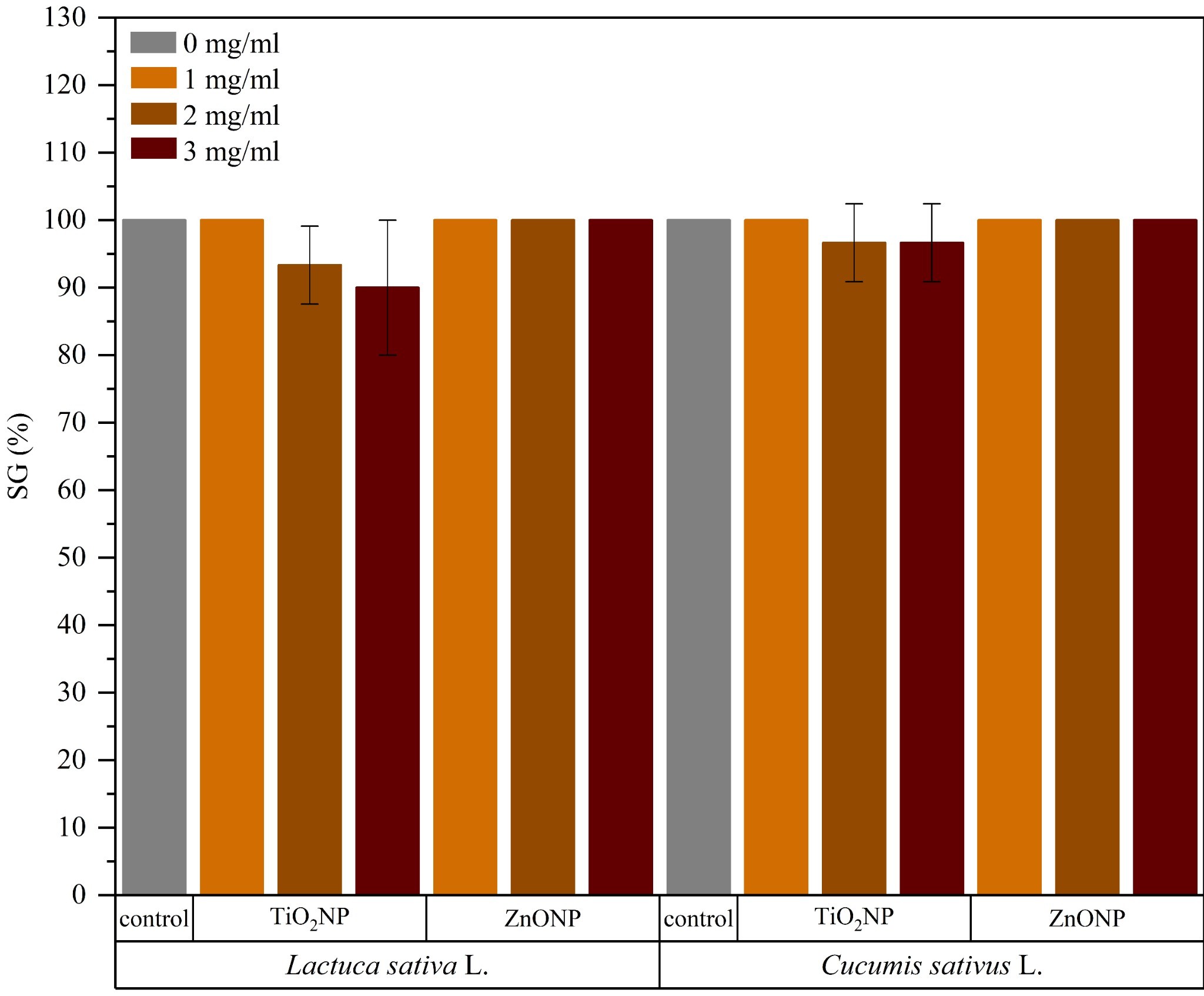
Germination tests were conducted to assess the potential phytotoxic effects of TiO2NPs and ZnONPs on the early development of lettuce. and cucumber seeds. The germination results after 14 days of testing are presented in Figure 9. At the end of the germination cycle, nearly all seed groups reached 100% germination, except for seed groups exposed to TiO2NP suspensions at concentrations of 2 and 3 mg/mL. Lettuce seeds exposed to these suspensions achieved SG% values of 93.33 ± 5.77% and 90.00 ± 10.00%, respectively. For cucumber seeds, SG% values were 96.67 ± 5.77% for both groups. Therefore, it can be stated that the nanoparticles did not exhibit phytotoxic behavior toward the tested seeds, as all groups showed SG% values above 83%68.

Germination test results in terms of SG% (mean ± sd) after 14 days of germination of Lactuca sativa L. and Cucumis sativus L. seeds exposed to suspensions of TiO2NPs and ZnONPs (1, 2, and 3 mg/ml). The control group was treated with distilled water. For the same seed, different letters indicate statistically significant differences among the means according to Tukey’s test (p < 0.05).
Another approach to assess the impact of nanoparticles on the growth of lettuce and cucumber consisted of analyzing the dry biomass content in the roots and shoots of seedlings obtained from germinated seeds. The results of this analysis are presented in Figure 10. For lettuce, seeds treated with ZnONP suspensions showed no significant differences compared to the control group (seeds treated with distilled water), with dry biomass values close to that of control (0.36 ± 0.02 g). In contrast, exposure to TiO2NPs resulted in dose-dependent reductions in dry biomass content, with the group treated with 3 mg/mL showing significantly lower values compared to the control (p = 0.00061). In the case of cucumber, the control group exhibited a higher average dry biomass content than lettuce, reaching 0.57 ± 0.02 g. Exposure to 1 mg/mL TiO2NP suspension did not promote significant difference in dry biomass compared to the control group. However, increasing nanoparticle concentration led to reductions in dry biomass, with a statistically significant decrease observed for seeds exposed to 3 mg/mL TiO2NPs (p = 0.02). Regarding ZnONPs, the group treated with 1 mg/mL showed slightly higher dry biomass than the control group, although without statistical significance. The groups treated with 2 and 3 mg/mL suspensions displayed a reduction trend, with a significant difference compared to the control only for the highest nanoparticle concentration (p = 0.31).

Dry biomass content (mean ± sd) of germinated seedlings after 30 days of testing. For the same seed (Lactuca sativa L. or Cucumis sativus L.), different letters indicate statistically significant differences among the means according to Tukey’s test (p < 0.05).
Damage to seedling development, observed through the reduction in dry biomass content in seeds exposed to TiO2NPs, can be attributed to the absorption of these nanoparticles by the roots, which inhibits the uptake of water and nutrients32. Furthermore, as observed for cucumber seeds exposed to low concentrations of ZnONPs, nanoparticles may promote seedling development; however, increasing concentrations or accumulation lead to metal-induced toxicity69, favoring oxidative stress 70,71.
To further investigate phytotoxicity of the nanoparticles, nutritional quality of cucumber seedlings obtained after 30 days of germination was assessed by measuring the mass content of Na, K, Ca, Mg, Zn, Fe, and Ti using EDX. Due to the small biomass yield of lettuce seedlings, EDX analysis was not feasible for this vegetable. Figure 11 presents the results of this analysis for the control group and for the groups exposed to TiO2NP and ZnONP suspensions at concentrations of 1, 2, and 3 mg/mL. The results show that seeds treated with TiO2NPs exhibited a progressive reduction in the mass content of these elements in the seedlings. In the group treated with the highest nanoparticle concentration, Na decreased from 13% in the control to 7%, K from 9% to 6%, Ca from 14% to 10%, Mg from 13% to 9%, Zn from 17% to 12%, and Fe from 18% to 11%. Consistent with this reduction, an increasing accumulation of Ti was observed, absent in the control group but reaching 7% in the seedlings treated with 3 mg/mL TiO2NPs. These findings indicate that TiO2NP exposure compromised the uptake of essential nutrients and promoted the translocation of Ti into plant tissues, as previously reported in studies with tomato69, lettuce71, and fennel flower72.

Elemental mass content (Na, K, Ca, Mg, Zn, Fe, and Ti) in cucumber seedlings obtained after 30 days of germination of seeds exposed to suspensions of TiO2NPs and ZnONPs (1, 2, and 3 mg/ml). The seeds present in the control group were exposed to distilled water.
For the groups treated with ZnONPs, an increase in Zn content was observed, rising from 17% in the control to 25% in seedlings exposed to 3 mg/mL of the nanoparticle. In contrast, K and Fe levels showed a decreasing trend (from 9% and 18% in the control to 5% and 11%, respectively, in the group treated with 3 mg/mL), while Ca and Mg varied only slightly. These results suggest that at lower concentrations ZnONPs may act as a supplemental source of Zn2+, but at higher concentrations it tends to impair the balanced uptake of other essential nutrients. Thus, the observed reductions in germination and biomass, together with altered nutrient uptake in seedlings, indicate that nanoparticle release from PLA composites could pose ecological risks at the disposal stage of packaging materials. These findings highlight the importance of coupling functional performance with end-of-life safety considerations in the development of sustainable food packaging.
4. Conclusions
This study demonstrated that the incorporation of TiO2 and ZnO nanoparticles significantly influences the thermal, structural, mechanical, optical, and ecotoxicological behavior of PLA-based films, with effects strongly dependent on nanoparticle type and concentration. Low nanoparticle loadings promoted partial crystalline ordering and relatively homogeneous dispersion, while higher contents led to agglomeration and reduced structural organization. From a performance standpoint, both oxides acted as effective mechanical reinforcements, whereas their influence on thermal and optical properties differed markedly, with TiO2 enhancing UV-shielding but inducing greater opacity, and ZnO better preserving visible transparency at low concentrations.
From an application perspective, the results highlight a clear trade-off between functional enhancement and visual appearance, as well as potential environmental safety concerns. Germination assays indicated concentration-dependent phytotoxic effects for TiO2, while ZnO showed neutral or mildly beneficial responses at low concentrations, although higher loadings impaired seedling development. These findings emphasize that the design of PLA-based nanocomposite films for food packaging should balance mechanical performance, optical characteristics, and end-of-life environmental impact. Future studies should address nanoparticle migration and long-term stability to support the safe and sustainable application of these materials.
Supplementary Material
The following online material is available for this article:
Supplementary Material
5. Acknowledgments
This work was supported by Fundação Carlos Chagas Filho de Amparo à Pesquisa do Estado do Rio de Janeiro (grants E26/210.045/2024 [290330], E26/211.665/2021 [269591], and 260003/013261/2024 [204.550/2024]), Coordenação de Aperfeiçoamento de Pessoal de Nível Superior, and Conselho Nacional de Desenvolvimento Científico e Tecnológico.
-
Data Availability
All data supporting the findings of this study have been published within the article itself.
6. References
-
1 Tan C, Han F, Zhang S, Li P, Shang N. Novel bio-based materials and applications in antimicrobial food packaging: recent advances and future trends. Int J Mol Sci. 2021;22(18):9663. http://doi.org/10.3390/ijms22189663
» http://doi.org/10.3390/ijms22189663 -
2 Ahmed S, Sameen DE, Lu R, Li R, Dai J, Qin W, et al. Research progress on antimicrobial materials for food packaging. Crit Rev Food Sci Nutr. 2020;62(11):3088-102. http://doi.org/10.1080/10408398.2020.1863327
» http://doi.org/10.1080/10408398.2020.1863327 -
3 Joshi NC, Negi PB, Gururani P. A review on metal/metal oxide nanoparticles in food processing and packaging. Food Sci Biotechnol. 2024;33(6):1307-22. http://doi.org/10.1007/s10068-023-01500-0
» http://doi.org/10.1007/s10068-023-01500-0 -
4 Ncube LK, Ude AU, Ogunmuyiwa EN, Zulkifli R, Beas IN. An overview of plastic waste generation and management in food packaging industries. Recycling. 2021;6(1):12. http://doi.org/10.3390/recycling6010012
» http://doi.org/10.3390/recycling6010012 -
5 Coyle R, Hardiman G, Driscoll KO. Microplastics in the marine environment: a review of their sources, distribution processes, uptake and exchange in ecosystems. CSCEE. 2020;2:100010. http://doi.org/10.1016/j.cscee.2020.100010
» http://doi.org/10.1016/j.cscee.2020.100010 -
6 Vázquez OA, Rahman MS. An ecotoxicological approach to microplastics on terrestrial and aquatic organisms: a systematic review in assessment, monitoring and biological impact. Environ Toxicol Pharmacol. 2021;84:103615. http://doi.org/10.1016/j.etap.2021.103615
» http://doi.org/10.1016/j.etap.2021.103615 -
7 Ragusa A, Svelato A, Santacroce C, Catalano P, Notarstefano V, Carnevali O, et al. Plasticenta: first evidence of microplastics in human placenta. Environ Int. 2021;146:106274. http://doi.org/10.1016/j.envint.2020.106274
» http://doi.org/10.1016/j.envint.2020.106274 -
8 Jenner LC, Rotchell JM, Bennett RT, Cowen M, Tentzeris V, Sadofsky LR. Detection of microplastics in human lung tissue using μFTIR spectroscopy. Sci Total Environ. 2022;831:154907. http://doi.org/10.1016/j.scitotenv.2022.154907
» http://doi.org/10.1016/j.scitotenv.2022.154907 -
9 Dirpan A, Ainani AF, Djalal M. A review on biopolymer-based biodegradable film for food packaging: trends over the last decade and future research. Polymers. 2023;15(13):2781. http://doi.org/10.3390/polym15132781
» http://doi.org/10.3390/polym15132781 -
10 Verma SK, Prasad A, Sonika S, Katiyar V. State of art review on sustainable biodegradable polymers with a market overview for sustainability packaging. Materials Today Sustainability. 2024;26:100776. http://doi.org/10.1016/j.mtsust.2024.100776
» http://doi.org/10.1016/j.mtsust.2024.100776 -
11 Muthu A, Nguyen DHH, Neji C, Törős G, Ferroudj A, Atieh R, et al. Nanomaterials for smart and sustainable food packaging: nano-sensing mechanisms, and regulatory perspectives. Foods. 2025;14(15):2657. http://doi.org/10.3390/foods14152657
» http://doi.org/10.3390/foods14152657 -
12 De Albuquerque TL, Marques Júnior JE, De Queiroz LP, Ricardo ADS, Rocha MVP. Polylactic acid production from biotechnological routes: A review. Int J Biol Macromol. 2021;186:933-51. http://doi.org/10.1016/j.ijbiomac.2021.07.074
» http://doi.org/10.1016/j.ijbiomac.2021.07.074 -
13 Righetti GIC, Faedi F, Famulari A. Embracing sustainability: the world of bio-based polymers in a mini review. Polymers. 2024;16(7):950. http://doi.org/10.3390/polym16070950
» http://doi.org/10.3390/polym16070950 -
14 Mulla MZ, Rahman MRT, Marcos B, Tiwari B, Pathania S. Poly Lactic Acid (PLA) nanocomposites: effect of inorganic nanoparticles reinforcement on its performance and food packaging applications. Molecules. 2021;26(7):1967. http://doi.org/10.3390/molecules26071967
» http://doi.org/10.3390/molecules26071967 -
15 Freeland B, McCarthy E, Balakrishnan R, Fahy S, Boland A, Rochfort KD, et al. A review of polylactic acid as a replacement material for single-use laboratory components. Materials. 2022;15(9):2989. http://doi.org/10.3390/ma15092989
» http://doi.org/10.3390/ma15092989 -
16 Vink ETH, Rábago KR, Glassner DA, Gruber PR. Applications of life cycle assessment to NatureWorksTM polylactide (PLA) production. Polym Degrad Stabil. 2003;80(3):403-19. http://doi.org/10.1016/S0141-3910(02)00372-5
» http://doi.org/10.1016/S0141-3910(02)00372-5 -
17 Arfat YA, Ahmed J, Al Hazza A, Jacob H, Joseph A. Comparative effects of untreated and 3-methacryloxypropyltrimethoxysilane treated ZnO nanoparticle reinforcement on properties of polylactide-based nanocomposite films. Int J Biol Macromol. 2017;101:1041-50. http://doi.org/10.1016/j.ijbiomac.2017.03.176
» http://doi.org/10.1016/j.ijbiomac.2017.03.176 -
18 Baek N, Kim YT, Marcy JE, Duncan SE, O’Keefe SF. Physical properties of nanocomposite polylactic acid films prepared with oleic acid modified titanium dioxide. Food Packag Shelf Life. 2018;17:30-8. http://doi.org/10.1016/j.fpsl.2018.05.004
» http://doi.org/10.1016/j.fpsl.2018.05.004 -
19 Huang Y, Wang T, Zhao X, Wang X, Zhou L, Yang Y, et al. Poly(lactic acid)/graphene oxide-ZnO nanocomposite films with good mechanical, dynamic mechanical, anti-UV and antibacterial properties: the poly(lactic acid)/graphene oxide-ZnO nanocomposites. J Chem Technol Biotechnol. 2015;90(9):1677-84. http://doi.org/10.1002/jctb.4476
» http://doi.org/10.1002/jctb.4476 -
20 Shankar S, Wang LF, Rhim JW. Incorporation of zinc oxide nanoparticles improved the mechanical, water vapor barrier, UV-light barrier, and antibacterial properties of PLA-based nanocomposite films. Mater Sci Eng C. 2018;93:289-98. http://doi.org/10.1016/j.msec.2018.08.002
» http://doi.org/10.1016/j.msec.2018.08.002 -
21 Tajdari A, Babaei A, Goudarzi A, Partovi R. Preparation and study on the optical, mechanical, and antibacterial properties of polylactic acid/ZnO/TiO2 shared nanocomposites. J Plast Film Sheeting. 2020;36(3):285-311. http://doi.org/10.1177/8756087919900365
» http://doi.org/10.1177/8756087919900365 -
22 Zhang Z, Wang Y, Li T, Ma P, Zhang X, Xia B, et al. High-performance polylactic acid materials enabled by TiO2 –polydopamine hybrid nanoparticles. Ind Eng Chem Res. 2021;60(10):3999-4008. http://doi.org/10.1021/acs.iecr.0c06238
» http://doi.org/10.1021/acs.iecr.0c06238 -
23 Zhang R, Lan W, Ji T, Sameen DE, Ahmed S, Qin W, et al. Development of polylactic acid/ZnO composite membranes prepared by ultrasonication and electrospinning for food packaging. Lebensm Wiss Technol. 2021;135:110072. http://doi.org/10.1016/j.lwt.2020.110072
» http://doi.org/10.1016/j.lwt.2020.110072 -
24 Khalil AM, El-Sayed SM, Youssef AM. Valorization of polylactic acid bionanocomposites enriched with CuO-TiO2 for packaging applications. Biomass Convers Biorefin. 2025;15(3):3485-94. http://doi.org/10.1007/s13399-023-05152-2
» http://doi.org/10.1007/s13399-023-05152-2 -
25 Kim I, Viswanathan K, Kasi G, Sadeghi K, Thanakkasaranee S, Seo J. Poly(Lactic Acid)/ZnO bionanocomposite films with positively charged ZnO as potential antimicrobial food packaging materials. Polymers. 2019;11(9):1427. http://doi.org/10.3390/polym11091427
» http://doi.org/10.3390/polym11091427 -
26 Li W, Li L, Zhang H, Yuan M, Qin Y. Evaluation of PLA nanocomposite films on physicochemical and microbiological properties of refrigerated cottage cheese. J Food Process Preserv. 2018;42(1):e13362. http://doi.org/10.1111/jfpp.13362
» http://doi.org/10.1111/jfpp.13362 -
27 Akshaykranth A, Jayarambabu N, Kumar A, Venkatappa Rao T, Kumar RR, Srinivasa Rao L. Novel nanocomposite polylactic acid films with Curcumin-ZnO: structural, thermal, optical and antibacterial properties. CRGSC. 2022;5:100332. http://doi.org/10.1016/j.crgsc.2022.100332
» http://doi.org/10.1016/j.crgsc.2022.100332 -
28 Herrera-Rivera MDR, Torres-Arellanes SP, Cortés-Martínez CI, Navarro-Ibarra DC, Hernández-Sánchez L, Solis-Pomar F, et al. Nanotechnology in food packaging materials: role and application of nanoparticles. RSC Advances. 2024;14(30):21832-58. http://doi.org/10.1039/D4RA03711A
» http://doi.org/10.1039/D4RA03711A -
29 Wang YY, Yu HY, Yang L, Abdalkarim SYH, Chen WL. Enhancing long-term biodegradability and UV-shielding performances of transparent polylactic acid nanocomposite films by adding cellulose nanocrystal-zinc oxide hybrids. Int J Biol Macromol. 2019;141:893-905. http://doi.org/10.1016/j.ijbiomac.2019.09.062
» http://doi.org/10.1016/j.ijbiomac.2019.09.062 -
30 Luo Y, Lin Z, Guo G. Biodegradation assessment of poly (lactic acid) filled with functionalized titania nanoparticles (PLA/TiO2) under compost conditions. Nanoscale Res Lett. 2019;14(1):56. http://doi.org/10.1186/s11671-019-2891-4
» http://doi.org/10.1186/s11671-019-2891-4 -
31 Goñi-Ciaurriz L, Durán A, Peñas FJ, Vélaz I. Enhanced biodegradation of polylactic acid/cellulose acetate nanocomposite films reinforced with TiO2 and β-cyclodextrin for sustainable food packaging applications. Int J Biol Macromol. 2025;332:148711. http://doi.org/10.1016/j.ijbiomac.2025.148711
» http://doi.org/10.1016/j.ijbiomac.2025.148711 -
32 Hu J, Wu X, Wu F, Chen W, Zhang X, White JC, et al. TiO2 nanoparticle exposure on lettuce (Lactuca sativa L.): dose-dependent deterioration of nutritional quality. Environ Sci Nano. 2020;7(2):501-13. http://doi.org/10.1039/C9EN01215J
» http://doi.org/10.1039/C9EN01215J -
33 Afzal S, Singh NK. Effect of zinc and iron oxide nanoparticles on plant physiology, seed quality and microbial community structure in a rice-soil-microbial ecosystem. Environ Pollut. 2022;314:120224. http://doi.org/10.1016/j.envpol.2022.120224
» http://doi.org/10.1016/j.envpol.2022.120224 -
34 Soliman S, Wang Y, Samaan MSF. Titanium dioxide nanoparticles alleviate phosphorus deficiency stress in apple plants. Sci Rep. 2025;15(1):24443. http://doi.org/10.1038/s41598-025-07987-3
» http://doi.org/10.1038/s41598-025-07987-3 -
35 Ijaz R, Yasin A, Keerio AA, Maroof T, Fatima E, Ali R. Field assessment of foliar applied micronutrients impact on growth and yield of okra (Abelmoschus esculentus L.). Plant Animal. 2025;4(4):117-22. http://doi.org/10.71454/PA.004.04.0151
» http://doi.org/10.71454/PA.004.04.0151 -
36 Zamora-Ledezma E, Aragundi GLL, Guamán Marquines WS, Macías Pro MA, García Díaz JV, Pacheco Gil HA, et al. Phytotoxic effects and agricultural potential of nanofertilizers: a study using zeolite, zinc oxide, and titanium dioxide under controlled conditions. J Xenobiot. 2025;15(4):123. http://doi.org/10.3390/jox15040123
» http://doi.org/10.3390/jox15040123 -
37 Yu D, Lu Q, Wei Y, Hou D, Yin X, Cai K, et al. Combined analysis of transcriptomics and metabolomics on the cumulative effect of nano-titanium dioxide on mulberry seedlings. Front Plant Sci. 2023;14:1175012. http://doi.org/10.3389/fpls.2023.1175012
» http://doi.org/10.3389/fpls.2023.1175012 -
38 Chahardoli A, Karimi N, Sharifan H. Elucidating the phytotoxic endpoints of sub-chronic exposure to titanium dioxide nanoparticles in Endemic Persian Dracocephalum species. Chemosphere. 2025;370:143853. http://doi.org/10.1016/j.chemosphere.2024.143853
» http://doi.org/10.1016/j.chemosphere.2024.143853 -
39 Zoufan P, Baroonian M, Zargar B. ZnO nanoparticles-induced oxidative stress in Chenopodium murale L, Zn uptake, and accumulation under hydroponic culture. Environ Sci Pollut Res Int. 2020;27(10):11066-78. http://doi.org/10.1007/s11356-020-07735-2
» http://doi.org/10.1007/s11356-020-07735-2 -
40 Voloshina M, Rajput V, Minkina T, Vechkanov E, Mandzhieva S, Mazarji M, et al. zinc oxide nanoparticles: physiological and biochemical responses in Barley (Hordeum vulgare L.). Plants. 2022;11(20):2759. http://doi.org/10.3390/plants11202759
» http://doi.org/10.3390/plants11202759 -
41 Verma Y, Singh SK, Jatav HS, Rajput VD, Minkina T. Interaction of zinc oxide nanoparticles with soil: insights into the chemical and biological properties. Environ Geochem Health. 2022;44(1):221-34. http://doi.org/10.1007/s10653-021-00929-8
» http://doi.org/10.1007/s10653-021-00929-8 -
42 Shen X, Hu W, Russell TP. Measuring the degree of crystallinity in semicrystalline regioregular Poly(3-hexylthiophene). Macromolecules. 2016;49(12):4501-9. http://doi.org/10.1021/acs.macromol.6b00799
» http://doi.org/10.1021/acs.macromol.6b00799 -
43 Sharma G, Wu W, Dalal EN. The CIEDE2000 color-difference formula: implementation notes, supplementary test data, and mathematical observations. Color Res Appl. 2004;30(1):21-30. http://doi.org/10.1002/col.20070
» http://doi.org/10.1002/col.20070 -
44 Kim A, Kim H-S, Park S-O. Measuring of the perceptibility and acceptability in various color quality measures. J Opt Soc Korea. 2011;15(3):310-7. http://doi.org/10.3807/JOSK.2011.15.3.310
» http://doi.org/10.3807/JOSK.2011.15.3.310 -
45 Cinko UO, Becerir B. Computing characteristics of color difference formulas for regular coordinate changes in CIELAB color space. Text Res J. 2024;95(11–12):1387-408. http://doi.org/10.1177/00405175241278025
» http://doi.org/10.1177/00405175241278025 -
46 Teske M, Arbeiter D, Schober K, Eickner T, Grabow N. Systemic analysis about residual chloroform in PLLA films. Curr Dir Biomed Eng. 2016;2(1):49-52. http://doi.org/10.1515/cdbme-2016-0014
» http://doi.org/10.1515/cdbme-2016-0014 -
47 Checchetto R, Rigotti D, Pegoretti A, Miotello A. Chloroform desorption from poly(lactic acid) nanocomposites: a thermal desorption spectroscopy study. Pure Appl Chem. 2019;92(3):391-8. http://doi.org/10.1515/pac-2018-1216
» http://doi.org/10.1515/pac-2018-1216 -
48 Farah S, Anderson DG, Langer R. Physical and mechanical properties of PLA, and their functions in widespread applications: a comprehensive review. Adv Drug Deliv Rev. 2016;107:367-92. http://doi.org/10.1016/j.addr.2016.06.012
» http://doi.org/10.1016/j.addr.2016.06.012 -
49 Wang XJ, Huang Z, Wei MY, Lu T, Nong DD, Zhao JX, et al. Catalytic effect of nanosized ZnO and TiO2 on thermal degradation of poly(lactic acid) and isoconversional kinetic analysis. Thermochim Acta. 2019;672:14-24. http://doi.org/10.1016/j.tca.2018.12.008
» http://doi.org/10.1016/j.tca.2018.12.008 -
50 Tarani E, Pušnik Črešnar K, Zemljič LF, Chrissafis K, Papageorgiou GZ, Lambropoulou D, et al. Cold crystallization kinetics and thermal degradation of PLA composites with metal oxide nanofillers. Appl Sci. 2021;11(7):3004. http://doi.org/10.3390/app11073004
» http://doi.org/10.3390/app11073004 -
51 Yan YF, Liang XB, Feng YL, Shi LF, Chen RP, Guo JZ, et al. Manipulation of crystallization nucleation and thermal degradation of PLA films by multi-morphologies CNC-ZnO nanoparticles. Carbohydr Polym. 2023;320:121251. http://doi.org/10.1016/j.carbpol.2023.121251
» http://doi.org/10.1016/j.carbpol.2023.121251 -
52 Zare Y. Study of nanoparticles aggregation/agglomeration in polymer particulate nanocomposites by mechanical properties. Compos, Part A Appl Sci Manuf. 2016;84:158-64. http://doi.org/10.1016/j.compositesa.2016.01.020
» http://doi.org/10.1016/j.compositesa.2016.01.020 -
53 Lee J, Kim JH, Wang H, Shin H. Development of multiscale analysis method for predicting thermo-mechanical properties of polymeric nanocomposites containing clustered nanoparticles. Funct. Compos. Struct. 2023;5(2):025003. http://doi.org/10.1088/2631-6331/accc6b
» http://doi.org/10.1088/2631-6331/accc6b -
54 Qiao R, Deng H, Putz KW, Brinson LC. Effect of particle agglomeration and interphase on the glass transition temperature of polymer nanocomposites. J Polym Sci, B, Polym Phys. 2011;49(10):740-8. http://doi.org/10.1002/polb.22236
» http://doi.org/10.1002/polb.22236 - 55 Huang J, Zhou J, Liu M. Interphase in polymer nanocomposites. JACS. 2022;2(2):280-91.
-
56 Marom G. Why do nanoparticles (CNTs) reduce the glass transition temperature of nanocomposites? J. Compos. Sci. 2023;7(3):114. http://doi.org/10.3390/jcs7030114
» http://doi.org/10.3390/jcs7030114 -
57 Panigrahi R, Chakraborty S, Ye J, Lim GS, Lim FCH, Yam JKH, et al. Elucidating the Role of Interfacial Hydrogen Bonds on Glass Transition Temperature Change in a Poly(Vinyl Alcohol)/SiO2 Polymer‐Nanocomposite by Noncovalent Interaction Characterization and Atomistic Molecular Dynamics Simulations. Macromol Rapid Commun. 2020;41(21):2000240. http://doi.org/10.1002/marc.202000240
» http://doi.org/10.1002/marc.202000240 -
58 Pathak AK, Dhakate SR. Validation of experimental results for graphene oxide‐epoxy polymer nanocomposite through computational analysis. J Polym Sci. 2020;59(1):84-99. http://doi.org/10.1002/pol.20200442
» http://doi.org/10.1002/pol.20200442 -
59 Young WW, Saez JP, Katsumata R. Rationalizing the composition dependence of glass transition temperatures in amorphous polymer/POSS composites. ACS Macro Lett. 2021;10(11):1404-9. http://doi.org/10.1021/acsmacrolett.1c00597
» http://doi.org/10.1021/acsmacrolett.1c00597 -
60 Rocha ACDS, Pinheiro MVDS, Menezes LRD, Silva EOD. Core-shell nanoparticles based on zirconia covered with silver as an advantageous perspective for obtaining antimicrobial nanocomposites with good mechanical properties and less cytotoxicity. J Mech Behav Biomed Mater. 2021;123:104726. http://doi.org/10.1016/j.jmbbm.2021.104726
» http://doi.org/10.1016/j.jmbbm.2021.104726 -
61 Moldovan A, Cuc S, Prodan D, Rusu M, Popa D, Taut AC, et al. Development and characterization of polylactic acid (PLA)-based nanocomposites used for food packaging. Polymers. 2023;15(13):2855. http://doi.org/10.3390/polym15132855
» http://doi.org/10.3390/polym15132855 -
62 Alpdogan C, Birtane H, Cagrı Mehmetoglu A, Kaya Y, Beyler Cigil A, Sahinbaskan T. Sustainable Active Packaging Films From PLA and PBAT With Thyme Oil: Extending the Shelf Life of Cherry Tomatoes. J Appl Polym Sci. 2025;142(42):e57583. http://doi.org/10.1002/app.57583
» http://doi.org/10.1002/app.57583 -
63 Molinari G, Parlanti P, Aliotta L, Lazzeri A, Gemmi M. TEM morphological analysis of biopolymers: The case of Poly (Lactic Acid) (PLA). Mater Today Commun. 2024;38:107868. http://doi.org/10.1016/j.mtcomm.2023.107868
» http://doi.org/10.1016/j.mtcomm.2023.107868 -
64 Mota RCAG, De Menezes LR, Da Silva EO. Poly(lactic acid) polymers containing silver and titanium dioxide nanoparticles to be used as scaffolds for bioengineering. J Mater Res. 2021;36(2):406-19. http://doi.org/10.1557/s43578-020-00038-9
» http://doi.org/10.1557/s43578-020-00038-9 -
65 Hanaor DAH, Sorrell CC. Review of the anatase to rutile phase transformation. J Mater Sci. 2010;46(4):855-74. http://doi.org/10.1007/s10853-010-5113-0
» http://doi.org/10.1007/s10853-010-5113-0 -
66 Yang Y, Ming J, Yu N. Color Image Quality Assessment Based on CIEDE2000. Adv Multimedia. 2012;2012:1-6. http://doi.org/10.1155/2012/273723
» http://doi.org/10.1155/2012/273723 -
67 de Menezes LR, Cavalcante MP, Vaz JLMR, Silva PSRC, Tavares MIB. Obtention of higher refractive index and transparent polymeric nanocomposite systems with small amounts of fillers for lenses application. J Compos Mater. 2020;55(5):675-86. http://doi.org/10.1177/0021998320957070
» http://doi.org/10.1177/0021998320957070 -
68 Luo Y, Liang J, Zeng G, Chen M, Mo D, Li G, et al. Seed germination test for toxicity evaluation of compost: its roles, problems and prospects. Waste Manag. 2018;71:109-14. http://doi.org/10.1016/j.wasman.2017.09.023
» http://doi.org/10.1016/j.wasman.2017.09.023 -
69 Raliya R, Nair R, Chavalmane S, Wang WN, Biswas P. Mechanistic evaluation of translocation and physiological impact of titanium dioxide and zinc oxide nanoparticles on the tomato (Solanum lycopersicum L.) plant. Metallomics. 2015;7(12):1584-94. http://doi.org/10.1039/C5MT00168D
» http://doi.org/10.1039/C5MT00168D -
70 Wang X, Yang X, Chen S, Li Q, Wang W, Hou C, et al. Zinc oxide nanoparticles affect biomass accumulation and photosynthesis in arabidopsis. Front Plant Sci. 2016;6:1243. http://doi.org/10.3389/fpls.2015.01243
» http://doi.org/10.3389/fpls.2015.01243 -
71 Caser M, Percivalle NM, Cauda V. The application of micro- and nano-sized zinc oxide particles differently triggers seed germination in Ocimum basilicum L., Lactuca sativa L., and Lepidium sativum L. under controlled conditions. Horticulturae. 2024;10(6):575. http://doi.org/10.3390/horticulturae10060575
» http://doi.org/10.3390/horticulturae10060575 -
72 Chahardoli A, Sharifan H, Karimi N, Kakavand SN. Uptake, translocation, phytotoxicity, and hormetic effects of titanium dioxide nanoparticles (TiO2NPs) in Nigella arvensis L. Sci Total Environ. 2022;806:151222. http://doi.org/10.1016/j.scitotenv.2021.151222
» http://doi.org/10.1016/j.scitotenv.2021.151222
Edited by
-
Associate Editor:
Elisabete Frollini.
-
Editor-in-Chief:
Luiz Antonio Pessan.
All data supporting the findings of this study have been published within the article itself.