Topography, Corrosion Resistance and Bioactivity of a Cobalt Titanium Dioxide Coating on a Titanium Surface Via Plasma Electrolytic Oxidation
Abstract
Titanium and titanium alloys are widely used to treat orthopedic and dental deficiencies and restorations because of their excellent biocompatibility and mechanical properties. However, they have some drawbacks, such as an inherent biological inertness that prevents them from adhering to cells or tissues, resulting in weak bone induction and angiogenesis abilities. Improving the biological properties of titanium and titanium alloy implants to increase osteogenesis and angiogenesis remains a significant challenge in the field of biomaterials. In addition, the continuous release of various metal ions in the microenvironment caused by body fluid corrosion can also lead to ultimate failure in implanting. Therefore, improving the corrosion resistance of both titanium and titanium alloys is an urgent task. In this study, we prepared cobalt titanium dioxide (Co-TiO2) coatings on medical titanium surfaces using micro arc oxidation. The surface characteristics, chemical composition, and structure of the coatings were analyzed by scanning electron microscopy (SEM), atomic force microscopy (AFM), X-ray diffraction (XRD) and X-ray photoelectron spectroscopy (XPS). The corrosion resistance of the coatings was evaluated using an electrochemical workstation. These results confirmed that plasma electrolytic oxidation technology can be used to prepare cobalt titanium dioxide coatings in cobalt-containing electrolyte solutions successfully. The coating has a porous structure and a rough surface, and cobalt is successfully doped onto the coating surface. In addition, the cobalt titanium dioxide coating improved the corrosion resistance of titanium. In vitro experiments have shown that cobalt titanium dioxide coating can promote the adhesion and proliferation of MC3T3-E1 cells and has good biological activity. Conclusion: Cobalt titanium dioxide coatings can be prepared on medical titanium surfaces through plasma electrolytic oxidation. These coatings have good surface morphology and biological activity, improve the corrosion resistance of medical titanium, and have good clinical application prospects.
Keywords:
Titanium implant; Bioinertness; Surface treatment; Coating; Corrosion resistance; Biocompatibility
1. Introduction
The materials currently used for orthopedic implants primarily include medical metal materials, polymer materials, and inorganic nonmetallic materials. Medical metal materials are widely used in the clinical treatment of bone defects because of their good mechanical properties, good ductility, and high hardness. At present, common medical metal materials in clinical practice include titanium and titanium alloys, cobalt chromium alloys, magnesium alloys, and stainless steel. Among these materials, titanium and titanium alloys have been used clinically for decades because of their low density, good mechanical properties, and biocompatibility. However, many challenges, including weak bone integration performance, still exist for titanium and titanium alloys. Owing to the lack of biological activity of titanium metal implants, it is difficult to form chemical bonds with surrounding bone tissue after implantation, resulting in poor osteogenic induction. Poor antibacterial adhesion: The surface of titanium metal implants does not have antibacterial adhesion properties, making it easy for bacteria to adhere to their surface and increasing the risk of infection1. In addition, the weak antioxidant capacity of titanium metal implants can cause immune suppression in the human body after implantation, easily producing a large amount of reactive oxygen species (ROS) at the implantation site, which can seriously affect bone integration and even cause inflammation and other problems2,3. The bottlenecks associated with titanium metal implants further limit their clinical efficacy. Therefore, surface modification is needed to improve the bone integration, antibacterial adhesion, and antioxidant properties of titanium metal implants.
In recent years, researchers have developed innovative methods for the surface modification of multiple implant materials, including methods from fields such as physics, chemistry, and biochemistry. The main methods of physical modification include grinding and polishing, sandblasting, mechanical deformation, physical vapor deposition, plasma spraying, ion implantation, magnetron sputtering, and plasma spraying4-7. Owing to the advantages of low cost and fast deposition rate, plasma modification technology is in mainstream use for the surface modification of biomaterials. Plasma spraying has many advantages, including a fast deposition rate, thick sediment, and low cost. Owing to its ease of use, plasma spraying is the preferred method for manufacturing calcium phosphate coatings on biomaterials. The most commonly used spraying material is hydroxyapatite (HA), which can promote bone integration after direct contact with surrounding tissues after implantation. The HA coating prepared through plasma spraying of the titanium surface undergoes osseointegration with surrounding tissues and generates new bone8. However, the poor mechanical properties of the HA coating may lead to fracture and delamination, making it prone to structural changes. Moreover, its bonding strength with the substrate is also poor9.
Plasma electrolytic oxidation (PEO), also known as micro-arc oxidation (MAO), is an electrochemical surface modification technique applied to metals or alloys such as Ti, Zr, Nb, Ta, Al, and Mg (which are collectively referred to as valve metals). By placing the material in an electrolyte and applying a high voltage to break it down, micro-arc discharge is generated on the surface, causing in situ growth of the oxide ceramic layer. This in situ-grown ceramic layer has high bonding strength with the substrate. By adjusting the electrical parameters and formulation during the reaction process, the coating structure, roughness, hydrophilicity, and coating composition can be functionally designed to enhance the material's wear resistance, corrosion resistance, biocompatibility, and other properties10,11.
Cobalt (Co) is an important component of vitamin B12 molecules and is crucial for the formation of red blood cells. Cobalt also plays important roles in bone growth and maintenance, energy conversion, DNA synthesis, and immune regulation12. Cobalt not only affects the metabolism of proteins, fats, and sugars but also plays crucial roles in the synthesis of hemoglobin. In addition, cobalt may help improve immune function, thereby helping resist infections. Research has shown that there is a synergistic effect between cobalt and elements such as zinc, copper, and manganese, which can promote the absorption of zinc and enhance its biological activity, thereby having a positive impact on growth and development13. Cobalt deficiency can hinder the synthesis of vitamin B12, leading to megaloblastic anemia as well as ulcers and inflammation in the mouth and tongue. The DNA synthesis phase and late stage of bone marrow cell growth are prolonged due to cobalt deficiency, thereby increasing the risk of disease.
Because plasma electrolytic oxidation can improve the biological activity of titanium and cobalt has many biological activities, this study uses plasma electrolytic oxidation technology to introduce cobalt into the surface of titanium. Scanning electron microscopy, energy dispersive spectroscopy, atomic force microscopy, and X-ray diffraction were used to evaluate the surface properties of the film layer, and the corrosion performance of the material was evaluated using an electrochemical workstation. The frictional properties of cobalt-doped biocoatings were also studied. On this basis, the biocompatibility and biological activity of the coating were evaluated through in vitro cell experiments. This study provides a new method for improving the biological activity of titanium and lays a theoretical foundation for the clinical application of cobalt-doped coatings on titanium surfaces.
The fabrication of coatings with excellent morphological characteristics and good biological activity on the surface of titanium via surface modification, thereby achieving the integration of the surface topography/biomedical function of medical titanium implants, is currently an important topic in titanium implant study. The key to achieving the integration of the surface topography and biomedical function of medical metal materials lies in the fabrication of target materials for clinical applications. To maintain the performance of titanium implants, the dual purpose of mechanical bearing capacity and biomedical function is achieved by introducing bioactive substances into specific structures. In this study, we attempt to use plasma electrolytic oxidation to introduce the bone-inducing active element cobalt into titanium implants and alter the micro/nanomorphology of the titanium surface (cobalt titanium dioxide coating). Owing to the unique surface morphology of plasma electrolytic oxidation, they could provide a good environment for cell growth and improve the biological activity of titanium implants. The slow release of active substances is expected to promote new bone formation. The cobalt titanium dioxide coating is expected to address problems involving complications related to titanium implants and to lay a theoretical and practical foundation for the study and development of implants.
2. Materials and Methods
2.1. Preparation of the Co-TiO2
In this study, commercial titanium sheets (Baoji INT Titanium Materials Co. Ltd., Baoji, China) were cut and processed into circular pieces with a diameter of 15 mm and a thickness of 1.0 mm using a CNC machine. They were then polished with 100-1000 # silicon carbide sandpaper and washed in acetone, anhydrous ethanol, and deionized water for 10 minutes each. After drying, they were set aside for later use. Experimental grouping: The control group was pure titanium (Ti group) and titanium dioxide coating (TiO2 coating group) , and the experimental group was cobalt titanium dioxide coating (Co-TiO2 coating group).
For the preparation of the TiO2 coating: 0.2 mol/L calcium acetate and 0.02 mol/L sodium glycerophosphate were dissolved in deionized water, 0.01 mol/L EDTA, and 0.01 mol/L glycerol were added, where EDTA and glycerol were used to stabilize the electrolyte solution and reduce the arc voltage. And the mixture was allowed to stand at room temperature for 1 hour.
For the preparation of the Co-TiO2 coating: 0.03 mol/L cobalt gluconate was added to the above mentioned electrolyte mixture, and plasma oxidation was performed under the same parameters.In this study, all chemical reagents (analytical purity) were procured from Shanghai Macklin Biochemical Co., Ltd.
The plasma electrolytic oxidation parameters are as follows: pulse frequency of 800 Hz; duty cycle of 30%; and duration of 5 minutes. A stainless steel plate was used as the cathode, and a titanium plate was used as the anode for plasma electrolytic oxidation. After plasma electrolytic oxidation treatment, residual electrolyte remains on the surface of the titanium alloy sample, which must be washed clean with distilled water. After cleaning, the samples were air dried and stored with identification numbers to prepare for subsequent membrane composition analysis and performance testing.
2.2. Evaluation of coating surface properties
Scanning electron microscopy (SEM; JEOL JSM-6700F, Japan) was used to observe the surface morphology of the sample, atomic force microscopy (AFM; Asylum Research, USA) was used to evaluate the roughness of the sample, X-ray diffraction (XRD, D/MAX2500V, Rigaku, Japan) was used to analyze the phase composition of the sample, and energy dispersive spectroscopy (EDS) and X-ray photoelectron spectroscopy (XPS) were used to analyze the elemental composition and and chemical state of the sample.
2.3. Corrosion resistance performance
The corrosion resistance performance was evaluated using an electrochemical testing system (Shanghai Chenhua Instrument Co. Ltd., Shanghai, China) with a Tafel polarization curve and a Nyquist curve. A three-electrode system was adopted: a saturated calomel electrode (SCE) was used as the reference electrode (RE), a platinum electrode was used as the auxiliary electrode (CE), and the sample was used as the working electrode. The corrosion solution was 0.9 wt.% NaCl, and the potential scanning range during the polarization curve measurements was -7501 to 500 mV, with a scanning speed of 1 mV/s.
2.4. In vitro cell research
2.4.1. Cell culture
In this study, the MC3T3-E1 osteogenic precursor cell line was used for in vitro cell experiments. The cells were cultivated in α-MEM (Life Technologies, USA) containing 10% fetal bovine serum in a 37 °C cell culture incubator containing 5% CO2 until the third generation was reached, after which cell experiments were conducted.
2.4.2 Live/Dead Staining
The cell viability staining method was used to evaluate cell compatibility. MC3T3-E1 cells at a density of 2 × 10⁴ cells/well were inoculated into the Ti control group or cobalt titanium dioxide coating group, cultured for 48 hours, and transferred to a new 24-well plate. Then, 50 μL of Live/Dead staining agent was added dropwise, the samples were incubated in the dark at 37 °C in a cell culture incubator, and the samples were observed under a fluorescence microscope.
2.4.3 EdU staining
After the cells on the surfaces of the Ti control group and cobalt titanium dioxide coating group were cultured for 2 days, 200 μL of 50 μM EdU (Beyotime, Shanghai, China) medium was added, and the mixture was incubated for 2 hours. Then, 100 µL of cell fixative was added to each well and incubated at room temperature for 30 minutes. Following incubation with 100 μL of glycine, 200 μL of 1X Apollo® staining reaction solution was added to each well, followed by incubation with 200 μL of Hoechst 3342 reaction solution and observation under a fluorescence microscope.
2.5 Statistical analysis
All of the data are expressed as the means ± standard deviations and were analyzed via SPSS 17.0. One-way ANOVA and the SNK test were used to compare the differences, and p < 0.05 indicated a statistically significant difference.
3. Results and Discussion
3.1 Surface morphology of coatings
Figure 1 shows the scanning electron microscopy morphology of the Ti control group and the cobalt titanium dioxide coating group. The surface of the Ti control group had sandpaper scratches and was not smooth. The surface of the cobalt titanium dioxide coating was covered with micropores of varying sizes, resulting in a “volcanic crater” shape. These micropores are interconnected, with some small micropores embedded within larger micropores. In addition, the thickness of the cobalt titanium dioxide coating was approximately 5-8 μm.

Figure 2 shows the AFM morphology of the Ti control group and the cobalt titanium dioxide coating group. The surface of pure Ti is relatively flat, with no obvious ups or downs. The cobalt titanium dioxide coating has obvious irregularities, similar to the morphology of the scanning electron microscope mentioned above, and it has the characteristic appearance of a “volcano”. This increase and decrease primarily contributes to the increase in the surface roughness of titanium.

Figure 3 shows the EDS image of the cobalt titanium dioxide coating. The cobalt titanium dioxide coating is composed of cobalt, calcium, phosphorus, oxygen, and titanium. It is evident that cobalt, calcium, and phosphorus come from electrolyte solutions, whereas titanium comes from the titanium matrix.

Figure 4 shows a map of the cobalt titanium dioxide coating. The mapping diagram clearly presents the elemental composition and distribution characteristics of the cobalt titanium dioxide coating in various colors. The cobalt titanium dioxide coating is primarily composed of cobalt, calcium, phosphorus, oxygen, and titanium, and these elements are uniformly distributed in the cobalt titanium dioxide coating.

Figure 5 shows the XRD phase of the Ti control group and the cobalt titanium dioxide coating group. The cobalt titanium dioxide coating is primarily composed of rutile TiO2, rutile TiO2, and titanium, and no cobalt, calcium, or phosphorus phases were found, which may be related to the low contents of these elements in the film layer.

Figure 6 shows the XPS diagram of Co-TiO2, and Figure 6 (a) shows the full X-ray photoelectron spectroscopy of the Co-TiO2 microporous coating. In addition to the characteristic peaks of titanium, oxygen, calcium and phosphorus, there were characteristic peaks of Co, and the peaks of Co 2p corresponded to CoO. The peak in the Ti2p spectrum corresponded to titanium dioxide, and the P-O bond between P2p and PO43- was consistent, thus indicating that P exists in the form of PO43-. The peaks of Ca 2p were located at 350.5 eV and 346.7 eV, and Ca 2p and P 2p existed as Ca3(PO4)2.

(a) XPS full spectrum for Co-TiO2 and (b) Ti 2p, (c) Ca 2p, (d) Co 2p, (e) P 2p, (f) O 1s XPS data.
Figure 7 shows the corrosion resistances of each group of samples. Figure 7 (a) shows the bode-impedance curves. Compared with Ti and TiO2, the low-frequency impedance value of Co-TiO2 is significantly increased, indicating that Co-TiO2 has fewer surface defects and does not suffer serious damage in SBF solution, indicating strong corrosion resistance. Figure 7 (b) shows the bode-phase curves, and the Co-TiO2surface exhibits typical pure capacitive behavior response characteristics. Figure 7 (c) shows the dynamic point polarization curves of each group of samples. The corrosion corrosion potential (0.597V) of the Co-TiO2 sample was lower than that of Ti (0.326V) and TiO2 (0.479V). According to the polarization curves, compared with that of the Ti and TiO2, the corrosion current density (Icorr) of Co-TiO2 decreased, whereas the corrosion potential (Ecorr) increased, indicating that Co-TiO2 improved the corrosion resistance of Ti. Figure 7 (d) shows the Nyquist curves of each group of samples. The Nyquist curves of Ti, TiO2 and Co-TiO2 are clearly both arc shaped, but the radii of the arcs are different. Generally, the larger the radius of the capacitance arc there is, the lower the corrosion rate of the coating. The capacitance arc diameter of Co-TiO2 is larger than that of the Ti and TiO2, indicating that the Co-TiO2 has better corrosion resistance performance.

Figure 8 shows the staining of live/dead MC3T3-E1 cells after 48 hours of cultivation in the Ti control group and cobalt titanium dioxide coating group. The staining of live/dead cells can reflect the cell compatibility of a material. Both the Ti control group and the cobalt titanium dioxide coating group presented live green cells on the surface, with only scattered dead red cells, indicating good cell compatibility in both groups of samples.

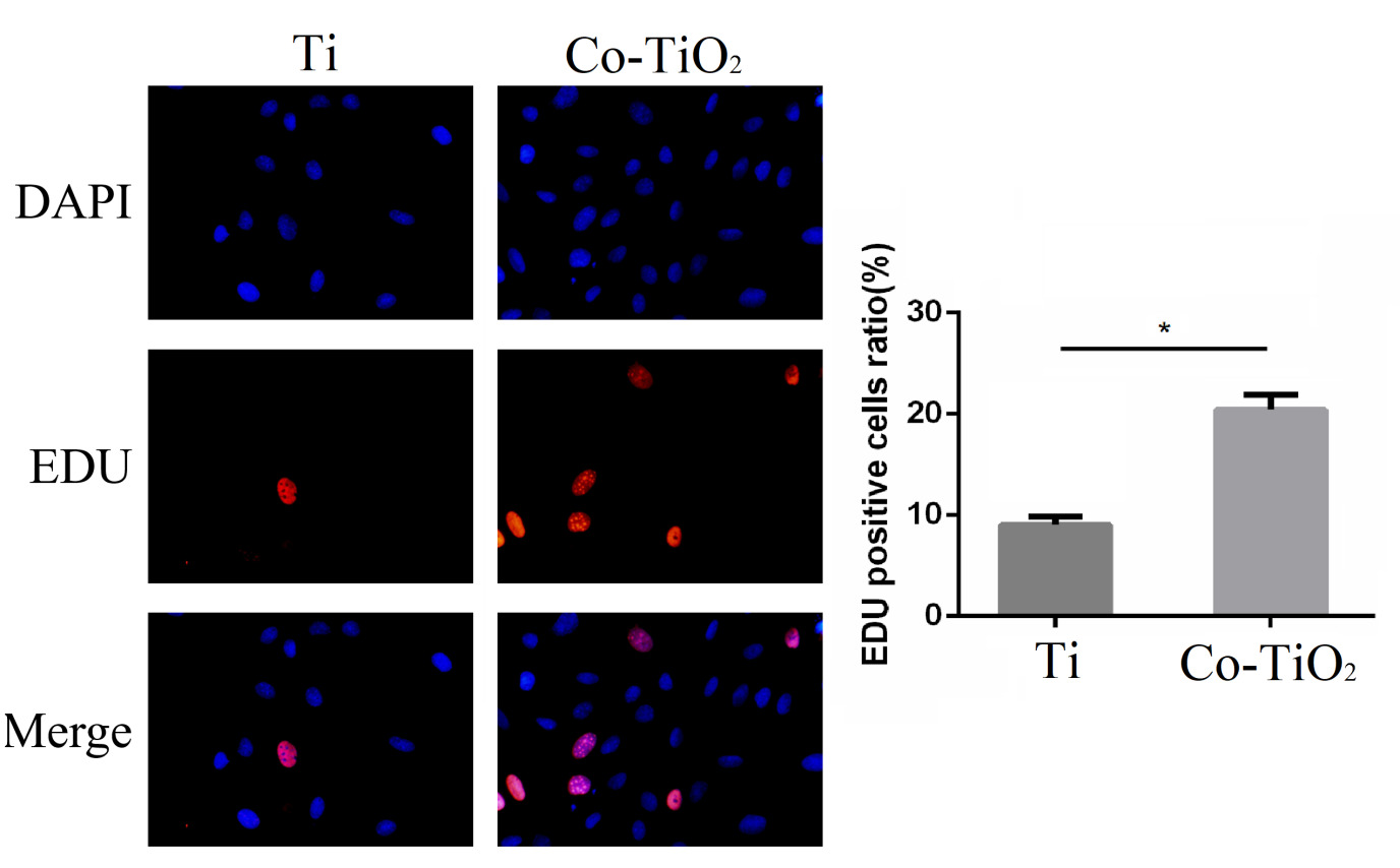
Figure 9 shows the EdU staining results of MC3T3-E1 cells in the Ti control group and cobalt titanium dioxide coating group. EdU staining can vividly reflect the proliferation of cells on the surface of materials. The cells labeled with red fluorescence were EdU-positive, indicating that the cells were in the proliferation phase. The greater the number of cells labeled with red fluorescence is, the more strongly the material can promote cell proliferation. Compared with the Ti group, the cobalt titanium dioxide coating group presented a significant increase in the number of EdU-positive cells, indicating that the cobalt titanium dioxide coating promoted cell proliferation.

EdU staining of Ti and Co-TiO2. The data are presented as the means±SDs, n=6; *p<0.05 compared with the Ti group.
4. Discussion
Titanium and its alloys have excellent properties, such as good tissue compatibility, light weight, and suitable mechanical properties, and they have been widely used in clinical bone defect treatment and dental implants. However, the frequent migration of titanium implants and the resulting implant loosening indicate poor osseointegration between the titanium implants and surrounding bone tissue, ultimately leading to a short implant lifespan and even implant failure14. Therefore, it is crucial to activate and functionalize the physical and chemical properties of titanium implant surfaces, including their composition and morphology, to enhance their biological activity and biocompatibility.
Plasma electrolytic oxidation is the in situ growth of ceramic film layers that are composed primarily of matrix metal oxides on the surface of titanium and its alloys through the combination of an electrolyte and its corresponding electrical parameters and relies on the instantaneous high temperature and high pressure generated by arc discharge. plasma electrolytic oxidation can be used to prepare micro/nanoporous coatings with bioactive elements on titanium surfaces, which were applied to surface modifications of oral implants at the end of the last century. Porous coatings prepared by plasma electrolytic oxidation can significantly improve the biological activity of titanium surfaces, increase the contact area with body tissues, and rapidly promote osseointegration. Compared with other surface modification techniques, plasma electrolytic oxidation involves in situ growth of a ceramic film layer on a substrate, which is firmly bonded, dense and uniform, not limited by the geometric shape of the workpiece, and has advantages such as a stable and reliable process, simple equipment, and convenient operation. plasma electrolytic oxidation can be used to adjust the pore size of a coating by adjusting the parameters15; for example, adjusting the time parameter can be used to adjust the pore size of the coating in a stable manner. The numerous parameters of plasma electrolytic oxidation significantly affect the overall performance of coatings, among which the time factor is usually directly related to the pore diameter and coating crystal structure.
By analyzing the PEO process, the formation of plasma electrolytic oxidation coatings can be divided into four stages: the anodic oxidation stage has a lower voltage and has not reached the critical breakdown voltage. At this stage, a very thin insulating oxide film forms on the metal surface. During the spark discharge stage, the voltage gradually increases to the critical breakdown voltage, the oxide film breaks down, and small white sparks appear on the metal surface. During this stage, the current rapidly increases, and the coating begins to thicken. Micro-arc discharge stage: As the coating thickness continues to increase, the corresponding voltage also begins to increase. There are some moving orange-red micro-arcs with diameters ranging from a few micrometers to tens of micrometers on the weak areas of the coating surface. The temperature of the micro-arc is very high, and when it stays on the surface of the coating, it melts the oxide in the remaining area, causing a phase transition of the oxide. When the micro-arc moves, the oxide at the previously retained site sinters under the rapid cooling of the electrolyte, resulting in an increase in the coating thickness. Arc discharge stage: At this stage, the micro-arc size is large and no longer moves, remaining on the surface of the coating for continuous discharge and forming large pits of several tens of micrometers16.
The corrosion of titanium implants inside the body is currently an urgent problem that needs to be addressed, as the implants are easily corroded after long-term exposure to body fluids17. In this study, the corrosion resistance of the cobalt-doped coatings was significantly better than that of the titanium-doped coatings, indicating that the cobalt-doped coatings formed after plasma electrolytic oxidation improved the corrosion resistance of the titanium. As titanium dioxide is a ceramic, the greatest advantage of ceramics is their excellent corrosion resistance. Because plasma electrolytic oxidation is carried out at high temperatures, the coating is directly formed on the titanium surface during the high-temperature sintering process, ensuring the tightness and integrity of the bonding between the coating and titanium substrate. More importantly, although the surface of cobalt-doped coatings has a micro/nanoporous structure, the micropores do not directly communicate with the titanium substrate but reach only the middle of the coating, and the closer the micropores are to the deeper part, the finer the pore size. Therefore, micropores do not damage the integrity of the ceramic. In addition, after plasma electrolytic oxidation, the increase in thickness of the titanium surface coating also reduces the contact between titanium and body fluids, improving its corrosion resistance18. Therefore, the corrosion resistance of the cobalt-doped coating after plasma electrolytic oxidation has improved.
The general view is that a rough surface increases the initial stability of the implant19, as it increases the contact area between the bone and the implant and significantly accelerates bone integration. In this study, we measured the roughness of a cobalt titanium dioxide coating with a profiler. The results showed that the roughness of the cobalt titanium dioxide coating was higher than that of the control group, and the difference was significant. The study showed that after plasma electrolytic oxidation of pure titanium, an oxide coating with a microporous structure was formed on the surface. The microporous structure with different pore sizes presented similar “volcano” shapes. The microporous structure increased the roughness of the titanium surface, increased the contact area and enabled osseointegration of the implants. Further study showed that the microstructures of the titanium oxide coatings were closely related to the plasma electrolytic oxidation voltages and oxidation times used. The sizes of the micropores on the surface of the oxide coating, the thickness of the oxide coating, and the surface roughness all increased with increasing voltage and longer action times.
The surface morphology is an important factor affecting the biological activity of an implant, and a good surface morphology favors cell adhesion and proliferation20. In this study, we found that there were many irregular pores distributed on the surface of the cobalt titanium dioxide coating, and most appeared as volcanic crater protrusions with many small and flat pores scattered around them. Increased magnification indicated that many nanoscale pores were scattered around the large pores. At the initial stage of arc striking, when the sample surface was just starting to exhibit breakdown discharge, the thin and uniform oxide film enabled uniform discharge on the oxide film surface, and many small holes were formed on the sample surface after breakdown. Larger volcanic pores were formed in the middle and later stages of the reaction. At this time, a series of thermal oxidation and plasma oxidation reactions occurred on the surface of the sample to generate high-temperature molten oxides on the surface. These molten substances were ejected from the arc channel by the arc light, and when encountering colder solutions around them, they solidified on the solid areas around the arc light. After the reaction was complete, the solution cooled. Subsequently, the oxide protrusions formed by condensation of these molten substances and discharge channels with varying pore sizes were left behind. Further research showed that the cobalt titanium dioxide coating had a porous structure with interconnected pores, which was conducive to cell adhesion and proliferation.
The adhesion of cells on titanium surfaces is the basis of cell proliferation and reflects the biological activity of titanium, affecting the proliferation and differentiation of cells on titanium surfaces. In this study, compared with that in the control group, the number of adherent cells on the surface of the cobalt-doped coatings increased, and the difference was statistically significant. We attribute this result to the rough surface of the cobalt-doped coating. The porous structure of rough surfaces is more conducive to cell adhesion and extension; however, rough surfaces have a larger surface area, providing more spatial sites for integrin binding, which is beneficial for cell adhesion21.
Multiple factors on the surface of titanium affect its biological activity, including surface morphology, roughness, surface energy, chemical composition, and hydrophilicity22. The surface morphology and chemical composition of titanium are the main factors affecting cell proliferation on its surface23. The good cell adhesion and proliferation on the surface of titanium means that it can have more cells and more bone mass at the same time. In this study, compared with those of the control group, the adhesion and proliferation of cells on the surface of the cobalt-doped coatings increased, and the difference was statistically significant. This significance is attributed to the porous structure of the cobalt-doped coatings, which increases the roughness of the titanium. In addition, cobalt has good biological activity and can promote cell adhesion, extension, and proliferation on its surface.
In summary, the study utilised plasma electrolytic oxidation to achieve a porous morphology and structure on the surface of titanium, as well as the incorporation of bioactive substances, which is of great significance in solving the clinical limitations of titanium. The microporous structure resulting from plasma electrolytic oxidation contributes to the adhesion and spreading of MC3T3-E1 cells. Moreover, the addition of bioactive cobalt endows titanium with bioactive functions. However, as a biomaterial, the cytotoxicity or immune response of the material needs to be considered. In this study, we found that the cobalt titanium dioxide coating has good cell compatibility, which may be related to the low cobalt content in the cobalt titanium dioxide coating. Cobalt may cause toxicity or immune reactions at high doses, so studies on the additional dose of cobalt and the physiological toxicity of implants should also be conducted. By studying the effects of different doses on cellular toxicity, a safe range of element concentrations can be explored.
5. Conclusion
In this study, a cobalt titanium dioxide coating was prepared on a titanium surface with plasma electrolytic oxidation, and the cobalt titanium dioxide coating exhibited a porous micro/nanostructure with cobalt uniformly doped on the coating surface. This coating increased the roughness of the titanium surface and provided good corrosion resistance. More importantly, the cobalt titanium dioxide coating promoted the adhesion, extension, and proliferation of MC3T3-E1 cells and exhibited good biological activity and clinical application prospects. Our study provides a new method for improving the biological activity of titanium implants by studying the preparation and performance of cobalt titanium dioxide coatings on titanium surfaces, which has potential application prospects.
6. Acknowledgments
This work was supported by Nantong University Special Research Fund for Clinical Medicine (2024JZ051) and Research start-up fund for talent introduction (2025RC024).
7. Data Availability
Data available on request from the authors.
8. References
-
1 Geetha M, Singh AK, Asokamani R, Gogia AK. Ti based biomaterials, the ultimate choice for orthopaedic implants - A review. Prog Mater Sci. 2009;54(3):397-425. https://doi.org/10.1016/j.pmatsci.2008.06.004
» https://doi.org/10.1016/j.pmatsci.2008.06.004 -
2 Abdel-Hady Gepreel M, Niinomi M. Biocompatibility of Ti-alloys for long-term implantation. J Mech Behav Biomed Mater. 2013;20:407-15. https://doi.org/10.1016/j.jmbbm.2012.11.014 PMid:23507261.
» https://doi.org/10.1016/j.jmbbm.2012.11.014 -
3 Tschernitschek H, Borchers L, Geurtsen W. Nonalloyed titanium as a bioinert metal: a review. J Prosthet Dent. 2006;96(1):12. https://doi.org/10.1016/j.prosdent.2006.02.020
» https://doi.org/10.1016/j.prosdent.2006.02.020 -
4 Maher S, Mazinani A, Barati MR, Losic D. Engineered titanium implants for localized drug delivery: recent advances and perspectives of Titania nanotubes arrays. Expert Opin Drug Deliv. 2018;15(10):1021-37. https://doi.org/10.1080/17425247.2018.1517743 PMid:30259776.
» https://doi.org/10.1080/17425247.2018.1517743 -
5 Le Guéhennec L, Soueidan A, Layrolle P, Amouriq Y. Surface treatments of titanium dental implants for rapid osseointegration. Dent Mater. 2007;23(7):844-54. https://doi.org/10.1016/j.dental.2006.06.025 PMid:16904738.
» https://doi.org/10.1016/j.dental.2006.06.025 -
6 Stepanovska J, Matejka R, Rosina J, Bacakova L, Kolarova H. Treatments for enhancing the biocompatibility of titanium implants. Biomedical Papers. 2020;164(1):23-33. https://doi.org/10.5507/bp.2019.062 PMid:31907491.
» https://doi.org/10.5507/bp.2019.062 -
7 Zhang K, Zhang B, Huang C, Gao S, Li B, Cao R, et al. Biocompatibility and antibacterial properties of pure titanium surfaces coated with yttrium-doped hydroxyapatite. J Mech Behav Biomed Mater. 2019;100:103363. https://doi.org/10.1016/j.jmbbm.2019.07.021 PMid:31415945.
» https://doi.org/10.1016/j.jmbbm.2019.07.021 -
8 Khor KA, Gu YW, Pan D, Cheang P. Microstructure and mechanical properties of plasma sprayed HA/YSZ/ Ti-6Al-4V composite coatings. Biomaterials. 2004;25(18):4009-17. https://doi.org/10.1016/j.biomaterials.2003.10.089 PMid:15046891.
» https://doi.org/10.1016/j.biomaterials.2003.10.089 -
9 Shen Y, Fang K, Xiang Y, Xu K, Yu L, Chen J, et al. Improvement in osteogenesis, vascularization, and corrosion resistance of titanium with silicon-nitride doped plasma electrolytic oxidation coatings. Front Bioeng Biotechnol. 2022;10:1023032. https://doi.org/10.3389/fbioe.2022.1023032 PMid:36324887.
» https://doi.org/10.3389/fbioe.2022.1023032 -
10 Cimenoglu H, Gunyuz M, Kose GT, Baydogan M, Uǧurlu F, Sener C. plasma electrolytic oxidation of Ti6Al4V and Ti6Al7Nb alloys for biomedical applications. Mater Charact. 2011;62(3):304-11. https://doi.org/10.1016/j.matchar.2011.01.002
» https://doi.org/10.1016/j.matchar.2011.01.002 -
11 Pan X, Li Y, Abdullah AO, Wang W, Qi M, Liu Y. Micro/nano-hierarchical structured TiO2 coating on titanium by plasma electrolytic oxidation enhances osteoblast adhesion and differentiation. R Soc Open Sci. 2019;6(4):182031. https://doi.org/10.1098/rsos.182031 PMid:31183132.
» https://doi.org/10.1098/rsos.182031 -
12 Yang X, Zhang C, Zhang T, Xiao J. Cobalt-doped Ti surface promotes immunomodulation. Biomed Mater. 2022;17(2):025003. https://doi.org/10.1088/1748-605X/ac4612 PMid:34942605.
» https://doi.org/10.1088/1748-605X/ac4612 -
13 Kulanthaivel S, Roy B, Agarwal T, Giri S, Pramanik K, Pal K, et al. Cobalt doped proangiogenic hydroxyapatite for bone tissue engineering application. Mater Sci Eng C. 2016;58:648-58. https://doi.org/10.1016/j.msec.2015.08.052 PMid:26478356.
» https://doi.org/10.1016/j.msec.2015.08.052 -
14 Kaur M, Singh K. Review on titanium and titanium based alloys as biomaterials for orthopaedic applications. Mater Sci Eng C Mater Biol Appl. 2019;102:844-62. https://doi.org/10.1016/j.msec.2019.04.064 PMid:31147056.
» https://doi.org/10.1016/j.msec.2019.04.064 -
15 Li Y, Wang W, Liu H, Lei J, Zhang J, Zhou H, et al. Formation and in vitro/in vivo performance of “cortex-like” micro/nano-structured TiO2 coatings on titanium by plasma electrolytic oxidation. Mater Sci Eng C. 2018;87:90-103. https://doi.org/10.1016/j.msec.2018.02.023 PMid:29549953.
» https://doi.org/10.1016/j.msec.2018.02.023 - 16 Xiao GX. Review on plasma electrolytic oxidation. Surf Technol. 2007;36(4):66-8.
-
17 Ghodrati H, Goodarzi A, Golrokhian M, Fattahi F, Anzabi RM, Mohammadikhah M, et al. A narrative review of recent developments in osseointegration and anti-corrosion of titanium dental implants with nano surface. Bone Rep. 2025;25:101846. https://doi.org/10.1016/j.bonr.2025.101846 PMid:40454244.
» https://doi.org/10.1016/j.bonr.2025.101846 -
18 Sikdar S, Menezes PV, Maccione R, Jacob T, Menezes PL. Plasma Electrolytic Oxidation (PEO) process-processing, properties, and applications. Nanomaterials (Basel). 2021;11(6):1375. https://doi.org/10.3390/nano11061375 PMid:34067483.
» https://doi.org/10.3390/nano11061375 -
19 De Bruyn H, Christiaens V, Doornewaard R, Jacobsson M, Cosyn J, Jacquet W, et al. Implant surface roughness and patient factors on long-term peri-implant bone loss. Periodontol 2000. 2017;73(1):218-27. https://doi.org/10.1111/prd.12177 PMid:28000269.
» https://doi.org/10.1111/prd.12177 -
20 Barfeie A, Wilson J, Rees J. Implant surface characteristics and their effect on osseointegration. Br Dent J. 2015;218(5):E9. https://doi.org/10.1038/sj.bdj.2015.171 PMid:25766196.
» https://doi.org/10.1038/sj.bdj.2015.171 -
21 Elias CN, Oshida Y, Lima JHC, Muller CA. Relationship between surface properties (roughness, wettability and morphology) of titanium and dental implant removal torque. J Mech Behav Biomed Mater. 2008;1(3):234-42. https://doi.org/10.1016/j.jmbbm.2007.12.002 PMid:19627788.
» https://doi.org/10.1016/j.jmbbm.2007.12.002 -
22 Rupp F, Liang L, Geis-Gerstorfer J, Scheideler L, Hüttig F. Surface characteristics of dental implants: a review. Dent Mater. 2018;34(1):40-57. https://doi.org/10.1016/j.dental.2017.09.007 PMid:29029850.
» https://doi.org/10.1016/j.dental.2017.09.007 -
23 Manivasagam VK, Sabino RM, Kantam P, Popat KC. Surface modification strategies to improve titanium hemocompatibility: a comprehensive review. Mater Adv. 2021;2(18):5824-42. https://doi.org/10.1039/D1MA00367D PMid:34671743.
» https://doi.org/10.1039/D1MA00367D
Edited by
-
Associate Editor:
Aloisio Klein.
-
Editor-in-Chief:
Luiz Antonio Pessan.