 Effects of exposure to Rickettsia bellii on reservoir competence of guinea pigs for Rickettsia rickettsii
Abstract
BACKGROUND Amblyomma sculptum is the main vector implicated in the transmission of Rickettsia rickettsii in southeastern Brazil, where capybaras are known to be the main amplifying hosts of this pathogen. Interestingly, many areas inhabited by large populations of capybaras and A. sculptum remain free of R. rickettsii, suggesting that other drivers may be involved in the pathogen perpetuation. A. sculptum frequently occurs in sympatry with Amblyomma dubitatum ticks, sharing the same hosts with them. Considering that A. dubitatum ticks are frequently infected with Rickettsia bellii, a possible interference of R. bellii in the reservoir competence of capybaras for R. rickettsii could explain the heterogeneous distribution of R. rickettsii in A. sculptum populations in southeastern Brazil.
OBJECTIVES The present study aimed to evaluate the effect of primary exposure to R. bellii-infected A. dubitatum on the reservoir competence for R. rickettsii using guinea pigs as experimental models.
METHODS Three guinea pigs were infested with R. bellii-infected A. dubitatum (Group GB), four guinea pigs with uninfected A. dubitatum (Group GD), and three guinea pigs were not exposed to A. dubitatum (Group GC). After infestation with A. dubitatum, all guinea pigs were exposed to one single R. rickettsii-infected A. sculptum female and then were infested with uninfected A. sculptum larvae. After ecdysis, nymphs were tested for detection of spotted fever group rickettsiae.
FINDINGS Five out of six guinea pigs from GD and GC groups died, while one out of three guinea pigs in the GB group had a fatal outcome. R. rickettsii infection rate among ticks fed on animals from GB group was 21% (17/80), significantly lower than the rate of 54% (60/111) recorded in ticks fed on animals from GD and GC groups.
MAIN CONCLUSIONS Prior exposure of guinea pigs to R. bellii-infected A. dubitatum ticks reduced their reservoir competence for R. rickettsii.
Key words:
Amblyomma dubitatum
;
Amblyomma sculptum
; spotted fever; capybara; rickettsiosis
INTRODUCTION
Rocky Mountain spotted fever, also known as Brazilian spotted fever (BSF), is caused by the bacterium Rickettsia rickettsii. BSF is the most relevant tick-borne disease in Brazil, where Amblyomma sculptum is the main vector implicated in the transmission of this pathogen to humans.1234
In spite of being a competent vector, A. sculptum is partially refractory to R. rickettsii infection and less efficient than other tick species in maintaining R. rickettsii by transstadial perpetuation and transovarial transmission.567 These features suggest that vertical transmission alone is insufficient to perpetuate R. rickettsii infection over successive tick generations; thus, horizontal transmission through an amplifying vertebrate host is necessary.8 Indeed, mathematical models have predicted that after a 90% reduction in amplifying hosts birth rate, R. rickettsii infection tends to disappear within two years from A. sculptum populations.9
Capybaras (Hydrochoerus hydrochaeris) are efficient hosts for all A. sculptum feeding stages and act as amplifying hosts of R. rickettsii for this tick species.1011 Since the 1980’s, this rodent population has been experiencing rapid growth in southeastern Brazil,121314 and this has been associated with spatial expansion of BSF occurrence as well as higher incidence rates.1815 However, it is noteworthy that many areas with high capybara and A. sculptum densities remain free of R. rickettsii in southeastern Brazil,161718 indicating that other factors may be involved in the maintenance of the pathogen in vector populations.
Besides being hosts for A. sculptum, capybaras are also primary hosts of Amblyomma dubitatum1918 and these two ticks occur in sympatry in many areas across southeastern Brazil.31820212223 Interestingly, natural infection of A. dubitatum with R. rickettsii has never been reported, while different studies have described Rickettsia bellii infection in A. dubitatum populations, often presenting high infection frequencies.212425262728293031323334
Amblyomma sculptum and A. dubitatum were the only ticks found to infest capybaras in anthropised landscapes in southeastern Brazil.1835 Nevertheless, Luz et al.18 noticed that A. sculptum greatly outnumbered A. dubitatum in BSF-endemic areas, while in BSF-non-endemic areas, abundances of these two species were similar, suggesting a negative correlation between R. rickettsii infection and higher A. dubitatum/A. sculptum abundance ratio.
Spotted fever group (SFG) Rickettsia are antigenically related bacteria and exhibit strong cross-reactivity in serological tests. In spite of not being part of SFG, R. bellii also presents antigenic cross-reactivity with SFG Rickettsia.36 Considering cross-reactivity between these two bacteria, it would be expected that host immune responses may affect the natural dynamics of these antigenically related organisms. Indeed, several experimental studies have shown that infection with a Rickettsia species elicits immune response in hosts reducing or preventing illness and fatal outcomes by a second infection with a pathogenic Rickettsia, besides reducing transmission of the pathogen to susceptible ticks.3738394041
Since A. sculptum is not highly efficient in perpetuating R. rickettsii infection across generations, amplifying hosts are essential for the pathogen maintenance in vector populations. Therefore, it is plausible that reducing the reservoir competence (ability to transmit an infection to arthropod vectors) of capybaras would affect R. rickettsii perpetuation. Considering that capybaras are primary hosts for A. dubitatum, and these ticks are frequently infected with R. bellii, one can hypothesise that exposure of capybaras to R. bellii elicit an immunological response capable of reducing the reservoir competence of capybaras for R. rickettsii. Thereby, this study aimed to evaluate if exposure of amplifying hosts to R. bellii-infected A. dubitatum ticks could reduce or preclude R. rickettsii transmission to A. sculptum ticks, using guinea pigs as experimental models.
MATERIALS AND METHODS
Animals and ticks
Animals and ticks - All animals used along the study were kept in the same room, with temperature (23ºC), ventilation and photoperiod (12/12) control. They were maintained in individual cages during the whole experiment, preventing physical contact between them and were fed with a commercial guinea pig pellet diet, fresh grass and water ad libitum. Ticks were held in an incubator at 25ºC, 85-90% relative humidity, and total scotophase for off-host developmental stages.
Ticks used in this study derived from colonies maintained in our laboratory by feeding on tick-naïve guinea pigs (Cavia porcellus) or tick-naïve rabbits (Oryctolagus cuniculus). All the colonies derived from host-seeking adult ticks collected in 2022 in São Paulo municipality, State of São Paulo (-23.557, -46.718). Two colonies of A. dubitatum were used, one naturally infected with R. bellii and one naturally uninfected, as previously reported.42 For A. sculptum, also two colonies were used, however, one infected with R. rickettsii and one uninfected. For establishing the R. rickettsii-infected colony, guinea pig organs (liver and lung) infected with R. rickettsii strain Itu3 stored at -80ºC were thawed at room temperature, crushed with sterile phosphate buffered saline (PBS), and the resultant homogenate was inoculated intraperitoneally in guinea pigs, as previously described.57 One day after inoculation, A. sculptum larvae were allowed to feed on guinea pigs inside feeding chambers (white cotton sleeves) glued to the animals shaved back. Sleeves were daily opened and detached engorged larvae were removed and taken to an incubator for ecdysis. Thereafter, ticks were fed on tick-naïve guinea pigs.
In order to assess rickettsial infection status of the colonies, flat nymphs and adults were individually tested by different conventional polymerase chain reaction (PCR) protocols. To that end, genomic DNA was extracted from the ticks using the phenol-chloroform-guanidine-isothiocyanate method, as described elsewhere.43 All samples were initially submitted to a PCR reaction using primers CS-78 and CS-323, targeting a 401 bp fragment of the citrate synthase gene (gltA), common in all representatives of the genus Rickettsia.24 DNA samples positive for infection were further tested by two additional protocols: the first one using primers Rr190.7044 and Rr190.701,45 which amplifies a 631 bp fragment of the rickettsial 190-kDa outer membrane protein gene (ompA) in most of the SFG rickettsiae; and the second one, a R. bellii-specific assay, targeting a 338 bp fragment of the gltA gene, as previously described.46
From the R. bellii-infected colony, 100% (48/48) of the A. dubitatum individuals were shown to be positive for R. bellii and negative for other Rickettsia species, while 100% (50/50) of uninfected A. dubitatum ticks and 100% (33/33) of uninfected A. sculptum ticks were negative for any rickettsial infection. On the other hand, 67% (22/33) of A. sculptum ticks exposed to R. rickettsii in the larval stage were shown to be infected. Ticks from these four colonies were used in the present study.
Tick infestations
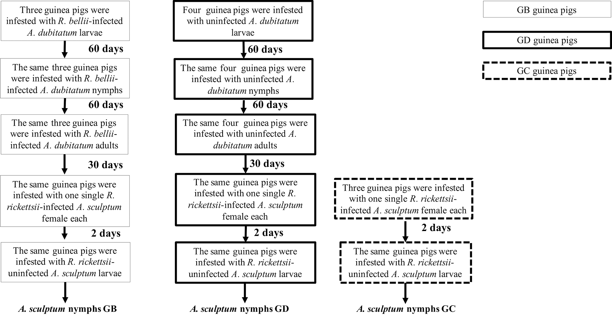
Tick infestations - Infestation design is summarised in Fig. 1. Ten tick-naïve guinea pigs were allocated in three experimental groups: three guinea pigs previously exposed to R. bellii-infected A. dubitatum ticks (group GB); four guinea pigs previously exposed to R. bellii-uninfected A. dubitatum ticks (group GD); and three guinea pigs that were not exposed to A. dubitatum ticks (group GC). Guinea pigs from GB and GD groups were initially exposed to R. bellii-infected and -uninfected larvae, respectively. Approximately 60 and 120 days after the larval infestation, the same animals were exposed to A. dubitatum nymphs and adults, respectively. Guinea pigs from group GC were kept free of ticks along this period.
Diagram illustrating experimental procedures with guinea pigs.
Groups GB and GD were composed by three and four guinea pigs, respectively, each of them infested by Amblyomma dubitatum larvae, nymphs and adults at 60-day intervals. GB served as hosts for A. dubitatum ticks infected by Rickettsia bellii, whereas GD served as hosts for uninfected A. dubitatum. Thirty days later, all animals were exposed to Rickettsia rickettsii-infected Amblyomma sculptum females (one infected female per guinea pig), and two days later, by uninfected larvae of A. sculptum. The GC group, composed of three guinea pigs never exposed to A. dubitatum ticks, were submitted to the same protocols of infestation by A. sculptum females and larvae.
Thirty days after infestation with A. dubitatum adults (GB and GD), guinea pigs from all three groups (GB, GD and GC) were infested by A. sculptum ticks. Two feeding chambers were glued to each animal’s back. In the first chamber, one single R. rickettsii-infected A. sculptum female was allowed to feed for two days. In order to infest guinea pigs only with R. rickettsii-infected females, previous to the infestation, females had one leg cut off and submitted to DNA extraction by the boiling method.26 The respective DNA samples were tested by a quantitative polymerase chain reaction (qPCR) protocol (described below). Only females that yielded a positive result in this qPCR protocol were allowed to feed. After two days of feeding, females were detached from the host and submitted to the same qPCR protocol in order to confirm the infection status. One day after the removal of females, uninfected A. sculptum larvae (approximately 500 - 1000 larvae/animal) were released in the second chamber and were allowed to feed until repletion. Feeding chambers were opened daily and engorged detached larvae were taken to an incubator for ecdysis. All the procedures were done simultaneously for the three experimental groups.
All animals were monitored for 21 days after each infestation and had their rectal temperature daily measured. Guinea pigs were considered febrile when rectal temperature was > 39.5ºC.47 In all infestations, approximately 500 µL of blood was collected from the lateral saphenous vein or from the tarsal vein from each guinea pig48 0 and 21 day(s) after infestations (DAI). Additionally, febrile animals had blood samples collected daily during the febrile period. Blood samples collected at 0 and 21 DAI were centrifuged, and the respective serum samples were tested for the presence of antibodies against R. rickettsii and R. bellii, as described below, while blood samples collected during the febrile period were stored at -20ºC for molecular analysis, as described below.
Serological analyses
Serological analyses - Guinea pig serum samples were individually tested by the indirect immunofluorescence assay (IFA) using a commercial fluorescein isothiocyanate-labelled goat anti-guinea pig IgG (Sigma Diagnostics, Saint Louis, MO) diluted 1:400 and crude antigens derived from R. rickettsii strain Taiaçu49 and R. bellii strain Mogi.49 Each serum was diluted in two-fold increments with PBS from 1:64 to endpoint titre, as previously described.16 In each slide, a serum previously shown to be non-reactive (negative control) and a known reactive serum (positive control) from a previous study in our laboratory7 were tested at the 1:64 dilution.
Rickettsial detection
Rickettsial detection - After the last infestation with A. dubitatum ticks, two males from the R. bellii-infected colony, previous fed on each guinea pig from group GB, were dissected and had their salivary glands removed and washed in PBS, as described by Benatti et al.50 One fragment of the salivary gland was imprinted in a glass slide by gently touching the tissue to the slide in a vertical up-and-down motion. The imprints were left to air dry and then were stained by the Giménez staining.51 Imprints were evaluated under an optical microscope at a 1000x magnification. The second salivary gland fragment was submitted to DNA extraction using the phenol-chloroform-guanidine-isothiocyanate method.43 DNA samples were tested by a R. bellii specific PCR assay.46
DNA samples from A. sculptum females used in the infestations were tested by a Taqman qPCR assay targeting a fragment of the citrate synthase gene (gltA), using the primers CS-552 and CS-624 and an internal fluorogenic probe as described by Soares et al.6 Each reaction had a positive (Rickettsia vini DNA) and a negative control (molecular-grade water).
Blood samples from febrile guinea pigs were submitted to DNA extraction using the DNeasy Blood & Tissue kit (Qiagen, Germany), according to manufacturer instructions. For R. rickettsii detection, a probe-based qPCR assay was performed to amplify a gene encoding hypothetical protein A1G_04230 using primers RRi6_F and RRi6_R and an internal fluorogenic probe described by Kato et al.53 Reactions were performed in a 7500 Real Time PCR Systems apparatus (Applied BioSystems, Foster City, CA). For each reaction, 2.5 μL of DNA template was added to 10 μL of the GoTaq Probe qPCR Master Mix (Promega, Madison, WI), 1.8 μL of each primer at 10 μM, 0.8 μL of probe at 10 μM and molecular-grade water to a final volume of 20 μL. qPCR cycling conditions were 1 cycle of 2 min at 95ºC, followed by 45 cycles of 15 s at 95ºC, 20 s at 55ºC and 30 s at 72ºC.
For quantification of R. rickettsii in guinea pig blood samples, plasmids with RRi6_F and RRi6_R amplicons were used to generate a ten-fold dilution standard curve from 102 to 106 copies, which resulted in a linear equation (y = -3.464x + 45.06) with an R2 value = 0.998 and efficiency of 94.383%. The quantification limit of the assay was defined as 100 DNA copies of R. rickettsii. Samples and standard curve were run in duplicate. All qPCR results are expressed in bacterial copies per ng of extracted DNA, determined by dividing the calculated copy number per µL by the total DNA concentration (ng/µL) of each sample [quantified in a spectrophotometer (Nanodrop 2000, Thermo Fisher Scientific)].
Approximately 20 days after ecdysis, A. sculptum nymphs (fed in the larval stage on febrile guinea pigs) from all the experimental groups underwent individual DNA extraction by the boiling method.26 DNA samples were initially submitted to a PCR protocol using primers CS-78 and CS-323 targeting a fragment of the citrate synthase gene (gltA), common in all representatives of the genus Rickettsia.24 Samples yielding positive results by this first PCR protocol were further tested by a second assay using primers Rr190.7044 and Rr190.701,45 which amplifies a fragment of the rickettsial 190-kDa outer membrane protein gene (ompA) present in most of the SFG rickettsiae. Nymphs were considered to be infected by R. rickettsii only when shown to be positive for both PCR assays. Each reaction had a positive (Rickettsia vini DNA) and a negative control (molecular-grade water).
In order to validate DNA extraction, samples that yielded negative results in the rickettsial detection assays were tested by one additional PCR protocol. Tick samples underwent an assay targeting a fragment of the tick mitochondrial 16S rRNA according to Mangold et al.,54 while blood samples were tested by a PCR targeting a 359 bp fragment of the mitochondrial cytochrome b (cyt b) gene from vertebrates, as described elsewhere.55 Samples with negative results in these PCR protocols were excluded from the study.
Survey for R. bellii in tick saliva
Survey for R. bellii in tick saliva - Serological analyses showed that no guinea pig from group GB had detectable antibodies against R. bellii after exposure to infected A. dubitatum ticks (results are described below). Since R. bellii was detected in salivary glands from male A. dubitatum ticks fed on all animals from group GB (results are described below), we decided to investigate if A. dubitatum ticks secrete R. bellii in the saliva. For this purpose, A. dubitatum ticks were collected at the exact same locality, where ticks that originated the colonies used in the study were collected.
In order to use only R. bellii-infected females, they had one leg cut off and submitted to DNA extraction by the boiling method.26 The respective DNA samples were tested by a qPCR protocol which amplifies a 338 bp fragment of the gltA gene, using primers described by Szabó et al.46 and an internal fluorogenic probe, as described by Hecht et al.56 Only five females, those that yielded a positive result in this qPCR protocol, were allowed to feed on a tick-naïve guinea pig along with five A. dubitatum males. Females were allowed to feed for six to nine days, until they were partially engorged.
After partial engorgement, females were detached from the host, washed three times in ultrapure water and dried with filter paper. Each female was inoculated with pilocarpine (50mg/ml in 0.7M NaCL solution) using a 12.7 mm x 0.33 mm needle, as described by Esteves et al.57 Saliva was harvested every five to ten minutes until the female stopped to salivate. One microliter of each saliva sample was dripped onto a slide, left to air dry and then was stained by the Giménez staining.52 Slides were evaluated under an optical microscope at a 1000x magnification. The remaining saliva samples were stored at -20ºC for molecular analyses. Saliva samples were diluted 1: 2 in Tris-EDTA buffer solution and underwent DNA extraction by the boiling method.26 For R. bellii detection, a specific qPCR assay was performed, as described elsewhere.56
Statistical analyses
Statistical analyses - Rickettsia rickettsii concentration in blood samples were compared between different experimental groups (GB, GD and GC) by Kruskal-Wallis test and between animals that died and survived by Mann-Whitney test. Proportion of blood samples yielding positive results in qPCR assays for R. rickettsii detection as well as R. rickettsii infection rate among A. sculptum nymphs were compared between the different experimental groups (GB, GD and GC) using Fisher’s exact test. Analyses were performed using R58 and a 5% significance level was assumed.
Ethics statement
Ethics statement - This study has been approved by the Ethic Committee on Animal Use of the Faculty of Veterinary Medicine and Animal Science, University of São Paulo (CEUA No. 5319211021).
RESULTS
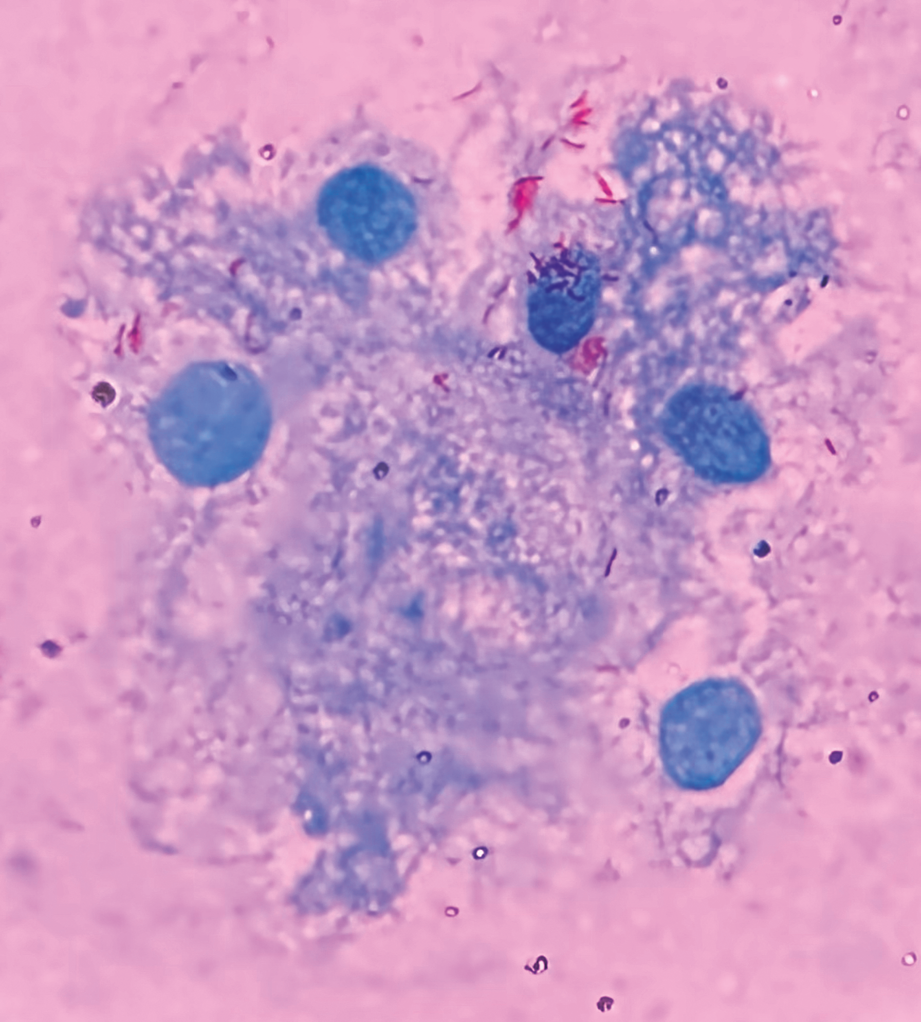
During all the infestations with A. dubitatum ticks, no guinea pig had fever or any other clinical manifestation. Further, no guinea pig seroconverted to R. bellii after any of the three consecutive exposures to R. bellii-infected A. dubitatum ticks (larvae, nymphs and adults). Despite the absence of seroconversion, R. bellii DNA was detected in salivary glands of A. dubitatum adult ticks that had fed on all guinea pigs from group GB and it was possible to visualise bacteria morphologically compatible with R. bellii in their salivary glands (Fig. 2). In addition, it was possible to visualise bacteria morphologically compatible with R. bellii (Fig. 3) and to detect R. bellii DNA in saliva samples from all tested females. These coupled results suggest that A. dubitatum are able to transmit R. bellii to hosts and that animals from the GB group were in fact exposed to R. bellii in spite of the absence of a humoral host response.
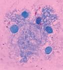
Optical microscopy analysis of the salivary glands of Amblyomma dubitatum ticks infected with Rickettsia bellii.
Giménez staining, 1000x original magnification.
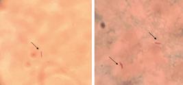
Optical microscopy analysis of saliva samples of Amblyomma dubitatum ticks infected with Rickettsia bellii.
Giménez staining, 1000x original magnification.
After exposure to a single R. rickettsii-infected tick, all the guinea pigs [except for one guinea pig (C31) from group GC] presented fever and other clinical manifestations compatible with R. rickettsii infection, which started between five and 12 DAI. One guinea pig from group GB, three from group GD and two from group GC died 12 to 21 DAI and five to nine days after fever onset (Table I).
Clinical and serological data of guinea pigs infested with Rickettsia rickettsii-infected Amblyomma sculptum females
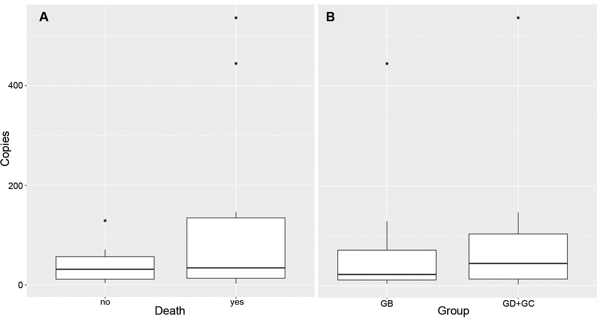
It was possible to detect R. rickettsii DNA in at least one blood sample from all animals, except for guinea pig C31. Noteworthy, R. rickettsii was detected only in one to four timepoints during the whole febrile period (Table I). Proportion of febrile days in which it was possible to detect R. rickettsii in blood samples was not different among experimental groups. Bacterial loads were slightly lower among animals from group GB and among animals that survived to infection, but no statistical significance was detected (Fig. 4).

Rickettsia rickettsii load in blood samples from guinea pigs.
(A) R. rickettsii load in blood samples (No. of R. rickettsii genome copies/ng of extracted DNA) from guinea pigs that died after infection and guinea pigs that survived infection with R. rickettsii. (B) R. rickettsii load in blood samples (No. of R. rickettsii genome copies/ng of extracted DNA) from guinea pigs infested with R. rickettsii-infected Amblyomma sculptum females. GB: guinea pigs previously exposed to Rickettsia bellii-infected Amblyomma dubitatum ticks; GD: guinea pigs previously exposed to R. bellii-uninfected A. dubitatum ticks; and GC: guinea pigs not exposed to A. dubitatum ticks.
All guinea pigs that had blood samples collected at 21 DAI seroconverted to R. rickettsii and R. bellii, generally with higher titres for R. rickettsii (Table I). Guinea pig C31 (group GC), which was the only one to remain afebrile, was also the only one that did not seroconvert, indicating that this animal was in fact not exposed to the pathogen. For this reason, it was not considered in further analyses.
Rickettsia rickettsii infection rates among Amblyomma sculptum unfed nymphs that had fed as larvae on guinea pigs previously exposed to R. rickettsii-infected A. sculptum females
From the A. sculptum nymphs that fed as larvae on guinea pigs during their febrile period, 191 nymphs underwent molecular analyses, which showed that 77 (40%) of them were infected by R. rickettsii. When evaluating the different experimental groups separately, ticks fed on GB guinea pigs showed 21% (17/80) infection rate, while ticks from groups GD and GC had infection rates of 50% (49/97) and 79% (11/14), respectively. When analysing ticks from GD and GC together, an infection rate of 54% (60/111) was observed. R. rickettsii infection rate in GB ticks was significantly lower than the infection rates recorded on the GD and GC groups (both when analysed separately or together) (Table II).
DISCUSSION
In the present study, the reservoir competence of guinea pigs for R. rickettsii was evaluated by comparing the infection rates of A. sculptum nymphs after feeding as larvae on three groups of R. rickettsii-infected guinea pigs: one group previously exposed to R. bellii-infected A. dubitatum ticks (GB), one group previously exposed to uninfected A. dubitatum ticks (GD), and one group never previously exposed to A. dubitatum (GC). As the GB A. sculptum nymphs had a significantly lower R. rickettsii-infection rate than the GD and GC nymphs, it was demonstrated under experimental conditions that exposure to R. bellii-infected A. dubitatum ticks reduced the reservoir competence of guinea pigs for R. rickettsii.
After being exposed to R. rickettsii-infected females, all guinea pigs (except for C31) presented clinical illness and seroconverted to R. rickettsii or died during acute infection. Although it was possible to detect R. rickettsii DNA in blood samples from all animals, it was not possible to detect the pathogen in all blood samples collected daily during the febrile period. R. rickettsii infects endothelial cells and has a very low concentration in the bloodstream, which makes blood qPCR an unsuitable assay for R. rickettsii diagnosis, as reported by several other studies, in which blood samples from infected animals yielded negative results in blood qPCR assays.10115960
Santos et al.61 showed that among BSF human cases, R. rickettsii DNA was detected in serum samples from all patients that had fatal outcomes, while in nonfatal cases, qPCR yielded positive results only for one third of the samples. Fatal outcomes are probably related to a vaster endothelial injury leading to severe vascular alterations and therefore higher R. rickettsii concentration in the blood. Interestingly, herein no statistical difference was observed in R. rickettsii load between animals that died or survived infection, even though a slightly higher load was recorded in animals that had fatal outcomes.
Even though guinea pigs from group GB showed a slightly lower R. rickettsii load, no statistical difference was observed among different experimental groups, precluding association between qPCR results and reservoir competence, assessed by the number of ticks becoming infected after blood feeding. This could be due to our small sample of guinea pigs or to an unsuitability of blood qPCR to assess hosts infectivity to ticks. In this regard it is important to keep in mind that molecular assays reflect one single moment in time when the respective blood sample was collected, while ticks remain attached ingesting blood for days. Further, it is not possible to affirm that R. rickettsii DNA detected by qPCR corresponds to viable bacteria, precluding inferences on infectivity.
Guinea pig C31 was not exposed to R. rickettsii as verified by the absence of seroconversion. This was unexpected, since the A. sculptum female fed on this guinea pig was shown to be infected. One possible reason for the absence of R. rickettsii transmission is the lack of time for the female to transmit the agent. Magalhães62 demonstrated that A. sculptum ticks may need 36 h to efficiently transmit R. rickettsii to a susceptible host. In our study, feeding chambers were inspected two times a day until the female was seen to be attached to the host. Forty-eight hours after attachment, females were removed from the chambers. Nevertheless, during this 48 h period, feeding chambers were not opened, being not possible to state that ticks were continuously attached to the host during this entire period.
Despite being repeatedly exposed to R. bellii, which was confirmed by detection of this bacterium in salivary glands of ticks fed on all animals, no guinea pig from group GB seroconverted to R. bellii prior to exposure to R. rickettsii. This was also noticed by other studies2942 and suggest that R. bellii exposure through tick bite is not able to elicit a humoral response in guinea pigs. These results are quite surprising, since many serosurveys have detected different wild and domestic animals with IgG titers for R. bellii higher than for other Rickettsia species, suggesting a homologous reaction to R. bellii.186364 One hypothesis is that these seropositive animals were exposed to R. bellii through a different infection route (e.g., ingestion of infected ticks), which enabled a humoral response due to exposure to a higher bacterial load. Another hypothesis is that antibodies detected in these studies do not derive from a previous exposure to R. bellii, but to another agent that cross-reacts with this bacterium.
In the study by Pacheco et al.,16 guinea pigs were inoculated intraperitoneally with different species of Rickettsia (R. bellii, Rickettsia canadensis, Rickettsia monteiroi and R. rickettsii). While guinea pigs inoculated with R. bellii had titres to the homologous antigen ranging from 256 to 1024, animals inoculated with other Rickettsia showed much higher homologous titres, ranging from 2048 to 32768. This low humoral response to R. bellii, even after exposure to a high number of bacteria via intraperitoneal inoculation, may indicate a low ability of R. bellii to elicit antibodies production.
Further studies should be performed to elucidate these findings and to ascertain whether IFA is a suitable assay for determining a previous exposure to R. bellii via tick bite, or not.
Since Rickettsia spp. are obligate intracellular bacteria, cell-mediated immunity, instead of humoral immunity, is expected to be the main immune response involved in their clearance, which is corroborated by studies that evaluated passive transfer of T cells in infections by different species of Rickettsia.65666768 Further, it was observed that mice lacking CD8+ T cells showed reduced survival and increased bacterial load after infection with Rickettsia conorii and enhanced susceptibility to fatal outcomes after infection with Rickettsia australis.6768 Thus, absence of detectable IgG against R. bellii does not necessarily mean that no effective adaptive immunity was induced. On the other hand, there is evidences that humoral response, specially antibodies to epitopes of OmpB, also play a relevant role in reinfections by Rickettsia, reducing the severity of the disease.697071 This could suggest that the adaptive immunity elicited by R. bellii may not be as effective as immune responses induced by SFG Rickettsia in protecting hosts to a second infection with a more pathogenic Rickettsia.
Indeed, exposure to R. bellii did not prevent clinical illness or fatal outcomes after infection by R. rickettsii, however, the lethality rate was lower in group GB (1/3) than in the other experimental groups (5/6). These results are different from other studies in which exposure to different Rickettsia fully prevented death due to R. rickettsii infection.37383941 These studies, however, used Rickettsia species phylogenetically closer to R. rickettsii than R. bellii, possibly inducing a stronger cross-reaction to R. rickettsii than the one observed in this study and, therefore, being more effective in protecting the hosts from severe illness and death.
Even though exposure to R. bellii-infected ticks did not fully protect animals from illness nor prevented A. sculptum larvae from being infected by R. rickettsii, guinea pigs from group GB (previously exposed to R. bellii-infected ticks) were less efficient in infecting A. sculptum larvae, when compared to guinea pigs from the other experimental groups (not previously exposed to R. bellii). Considering that A. sculptum is not very efficient in maintaining R. rickettsii across successive generations, reduction in host reservoir competence would probably have a great impact on the perpetuation of this pathogen. Nevertheless, since A. sculptum larvae were fed on a few animals, it is not possible to completely rule out the hypothesis that the difference in reservoir competence between experimental groups is, in part, due to natural variability among individuals rather than to previous exposure to R. bellii.
Binder et al.42 showed that A. dubitatum ticks were competent vectors for R. rickettsii and also able to transmit the pathogen transovarially, suggesting that these ticks may be important in the enzootic cycle of R. rickettsii. On the other hand, the same study noticed that A. dubitatum females coinfected by R. rickettsii and R. bellii generated a low number of viable larvae. Taken together, these results suggest that R. bellii could negatively impact the perpetuation of R. rickettsii among A. sculptum populations by two mechanisms: first, by reducing capybaras’ reservoir competence for R. rickettsii, and second, by minimising the role of A. dubitatum in R. rickettsii enzootic cycle due to the negative impact of superinfection on tick viability with a consequent reduction in the population of R. rickettsii-infected A. dubitatum ticks. These mechanisms could explain the association between a higher A. dubitatum/A. sculptum abundance ratio and the absence of R. rickettsii noticed by Luz et al.,18 being important drivers associated to spatial heterogeneity of R. rickettsii-infected A. sculptum populations within southeastern Brazil.
In conclusion, our results demonstrated that prior exposure of guinea pigs to R. bellii reduced their reservoir competence for R. rickettsii. Under natural conditions, this kind of interference among capybaras may significantly influence the long-term persistence of R. rickettsii in A. sculptum populations, a condition yet to be confirmed in the capybara model. Therefore, R. bellii may represent a promising strategy for disrupting the amplifying role of capybaras in the R. rickettsii transmission cycle.
-
Financial support: FAPESP (Grant 2021/06185-9).
-
How to cite
Binder LC, Fanchini CEC, Soares HS, dos Santos TBS, Taniwaki SA, Nassar MB, et al. Effects of exposure to Rickettsia bellii on reservoir competence of guinea pigs for Rickettsia rickettsii. Mem Inst Oswaldo Cruz. 2026; 121: e250195.
DATA AVAILABILITY
The contents underlying the research text are included in the manuscript.
REFERENCES
- 1. Pinter A, França AC, Souza CE, Sabbo C, Nascimento EMM, Santos FCP, et al. Febre maculosa brasileira. Bepa 2011; 8(Suppl. 1): 1-31.
- 2. Pinter A, Sabbo C, Leite R, Spinola R, Angerami R. Informe técnico sobre febre maculosa brasileira. Bepa 2021; 18: 54-78.
- 3. Krawczak FS, Nieri-Bastos FA, Nunes FP, Soares JF, Moraes-Filho J, Labruna MB. Rickettsial infection in Amblyomma cajennense ticks and capybaras (Hydrochoerus hydrochaeris) in a Brazilian spotted fever-endemic area. Parasit Vectors 2014; 7: 1-7.
- 4. Labruna MB, Krawczak FS, Gerardi M, Binder LC, Barbieri ARM, Paz GF, et al. Isolation of Rickettsia rickettsii from the tick Amblyomma sculptum from a Brazilian spotted fever-endemic area in the Pampulha Lake region, southeastern Brazil. Vet Parasitol Reg Stud Reports 2017; 8: 82-5.
- 5. Labruna MB, Ogrzewalska M, Martins TF, Pinter A, Horta MC. Comparative susceptibility of larval stages of Amblyomma aureolatum, Amblyomma cajennense, and Rhipicephalus sanguineus to infection by Rickettsia rickettsii J Med Entomol 2008; 45: 1156-9.
- 6. Soares JF, Soares HS, Barbieri AM, Labruna MB. Experimental infection of the tick Amblyomma cajennense, Cayenne tick, with Rickettsia rickettsii, the agent of Rocky Mountain spotted fever. Med Vet Entomol 2012; 26: 139-51.
- 7. Gerardi M, Ramírez-Hernández A, Binder LC, Krawczak FS, Gregori F, Labruna MB. Comparative susceptibility of different populations of Amblyomma sculptum to Rickettsia rickettsii Front Physiol 2019; 10: 653.
- 8. Labruna MB. Brazilian spotted fever: the role of capybaras. In: Moreira J, Ferraz K, Herrera E, Macdonald D, editors. Capybara New York: Springer; 2013. p. 371-83.
- 9. Polo G, Acosta CM, Labruna MB, Ferreira F. Transmission dynamics and control of Rickettsia rickettsii in populations of Hydrochoerus hydrochaeris and Amblyomma sculptum PLoS Negl Trop Dis 2017; 11: e0005613.
- 10. Souza CE, Moraes-Filho J, Ogrzewalska M, Uchoa FC, Horta MC, Souza SSL, et al. Experimental infection of capybaras Hydrochoerus hydrochaeris by Rickettsia rickettsii and evaluation of the transmission of the infection to ticks Amblyomma cajennense Vet Parasitol 2009; 161: 116-21.
- 11. Ramírez-Hernández A, Uchoa F, Serpa MC, Binder LC, Souza CE, Labruna MB. Capybaras (Hydrochoerus hydrochaeris) as amplifying hosts of Rickettsia rickettsii to Amblyomma sculptum ticks: evaluation during primary and subsequent exposures to R. rickettsii infection. Ticks Tick Borne Dis 2020; 11: 101463.
- 12. Ferraz KMPMB, Ferraz SFB, Moreira JR, Couto HTZ, Verdade LM. Capybara (Hydrochoerus hydrochaeris) distribution in agroecosystems: a cross‐scale habitat analysis. J Biogeog 2007; 34: 223-30.
- 13. Ferraz KMP, Peterson AT, Scachetti-Pereira R, Vettorazzi CA, Verdade LM. Distribution of capybaras in an agroecosystem, southeastern Brazil, based on ecological niche modeling. J Mammal 2009; 90: 189-94.
- 14. Vargas FC, Vargas SC, Moro MEG, Silva V, Carrer CRO. Monitoramento populacional de capivaras (Hydrochaeris hydrochaeris Linnaeus, 1766) em Pirassununga, SP, Brasil. Cienc Rural 2007; 37: 1104-8.
- 15. Souza CE, Pinter A, Donalisio MR. Risk factors associated with the transmission of Brazilian spotted fever in the Piracicaba river basin, State of São Paulo, Brazil. Rev Soc Bras Med Trop 2015; 48: 11-7.
- 16. Pacheco RC, Horta MC, Moraes-Filho J, Ataliba AC, Pinter A, Labruna MB. Rickettsial infection in capybaras (Hydrochoerus hydrochaeris) from São Paulo, Brazil: serological evidence for infection by Rickettsia bellii and Rickettsia parkeri Biomedica 2007; 27: 364-71.
- 17. Souza CE, Souza SSL, Lima VLC, Calic SB, Camargo MCGO, Savani ESMM, et al. Serological identification of Rickettsia spp from the spotted fever group in capybaras in the region of Campinas-SP-Brazil. Cienc Rural 2008; 38(6): 1694-99.
- 18. Luz HR, Costa FB, Benatti HR, Ramos VN, Serpa MCA, Martins TF, et al. Epidemiology of capybara-associated Brazilian spotted fever. Plos Negl Trop Dis 2019; 13: e0007734.
- 19. Nava S, Venzal JM, Labruna MB, Mastropaolo M, González EM, Mangold AJ, et al. Hosts, distribution and genetic divergence (16S rDNA) of Amblyomma dubitatum (Acari: Ixodidae). Exp Appl Acarol 2010; 51: 335-51.
- 20. Perez CA, Almeida ÁFD, Almeida A, Carvalho VHBD, Balestrin DDC, Guimarães MS, et al. Carrapatos do gênero Amblyomma (Acari: Ixodidae) e suas relações com os hospedeiros em área endêmica para febre maculosa no estado de São Paulo. Rev Bras Parasitol Vet 2008; 17: 210-17.
- 21. Brites-Neto J, Nieri-Bastos FA, Brasil J, Duarte KMR, Martins TF, Veríssimo CJ, et al. Environmental infestation and rickettsial infection in ticks in an area endemic for Brazilian spotted fever. Rev Bras Parasitol Vet 2013; 22: 367-72.
- 22. Brites-Neto J, Brasil J, Takeda GACG, Guillen AC, Labruna MB, Pinter A. Diferenciação morfométrica entre larvas de Amblyomma sculptum Berlese, 1888 e Amblyomma dubitatum Neumann, 1899. Arq Bras Med Vet Zoo 2018; 70: 1521-8.
- 23. Costa FB, Gerardi M, Binder LC, Benatti HR, Serpa MCDA, Lopes B, et al. Rickettsia rickettsii (Rickettsiales: Rickettsiaceae) infecting Amblyomma sculptum (Acari: Ixodidae) ticks and capybaras in a Brazilian spotted fever-endemic area of Brazil. J Med Entomol 2020; 57: 308-11.
- 24. Labruna MB, Whitworth T, Horta MC, Bouyer DH, McBride JW, Pinter A, et al. Rickettsia species infecting Amblyomma cooperi ticks from an area in the state of São Paulo, Brazil, where Brazilian spotted fever is endemic. J Clin Microbiol 2004; 42: 90-8.
- 25. Estrada DA, Schumaker TTS, Souza CED, Rodrigues Neto EJ, Linhares AX. Detecção de riquétsias em carrapatos do gênero Amblyomma (Acari: Ixodidae) coletados em parque urbano do município de Campinas, SP. Rev Soc Bras Med Trop 2006; 39: 68-71.
- 26. Horta MC, Labruna MB, Pinter A, Linardi PM, Schumaker TTS. Rickettsia infection in five areas of the state of São Paulo, Brazil. Mem Inst Oswaldo Cruz 2007; 102(7): 793-801.
- 27. Pacheco RC, Horta MC, Pinter A, Moraes-Filho J, Martins TF, Nardi MS, et al. Pesquisa de Rickettsia spp em carrapatos Amblyomma cajennense e Amblyomma dubitatum no Estado de São Paulo. Rev Soc Bras Med Trop 2009; 42: 351-3.
- 28. Brites-Neto J, Brasil J, Duarte KMR. Epidemiological surveillance of capybaras and ticks on warning area for Brazilian spotted fever. Vet World 2015; 8: 1143.
- 29. Sakai RK, Costa FB, Ueno TE, Ramirez DG, Soares JF, Fonseca AH, et al. Experimental infection with Rickettsia rickettsii in an Amblyomma dubitatum tick colony, naturally infected by Rickettsia bellii Ticks Tick Borne Dis 2014; 5(6): 917-23.
- 30. Costa FB, Barbieri AR, Ramos VN, Piovezan U, Labruna MB. New records of Rickettsia bellii-infected ticks in Brazil. Braz J Vet Res Anim Sci 2017; 54: 92-5.
- 31. Pajuaba-Neto AA, Ramos VDN, Martins MM, Osava CF, Pascoal JDO, Suzin A, et al. Influence of microhabitat use and behavior of Amblyomma sculptum and Amblyomma dubitatum nymphs (Acari: Ixodidae) on human risk for tick exposure, with notes on Rickettsia infection. Ticks Tick Borne Dis 2018; 9: 67-71.
- 32. Sousa ACP, Suzin A, Rodrigues V, Rezende LM, Maia R, Vieira RBK, et al. Ticks (Acari: Ixodidae) and rickettsiae associated with wild boars in a rural area of Minas Gerais, Brazil. Vet Parasitol Reg Stud Reports 2024; 50: 101016.
- 33. Szabó MPJ, Martins MM, de Castro MB, Pacheco RC, Tolesano-Pascoli GV, Dos Santos KT, et al. Ticks (Acari: Ixodidae) in the Serra da Canastra National Park in Minas Gerais, Brazil: species, abundance, ecological and seasonal aspects with notes on rickettsial infection. Exp Appl Acarol 2018; 76: 381-97.
- 34. Szabó MP, Queiroz CL, Suzin A, Rodrigues V, Vieira RBK, Martins MM, et al. Density and behavior of capybara (Hydrochoerus hydrochaeris) ticks (Acari: Ixodidae) Amblyomma sculptum and Amblyomma dubitatum with notes on Rickettsia bellii infection: Assessing human exposure risk. Ticks Tick Borne Dis 2024; 15: 102330.
- 35. Caixeta BT, Tolesano-Pascoli GV, Mundim FL, Pascoal JO, Rodrigues VS, Martins MM, et al. Survey of Rickettsia spp. in ticks (Acari: Ixodidae) infesting opossums (Didelphis albiventris) and capybaras (Hydrochoerus hydrochaeris) from an urban park in southeastern Brazil. Exp Appl Acarol 2024; 92: 463-77.
- 36. Pacheco RC, Moraes-Filho J, Marcili A, Richtzenhain LJ, Szabó MP, Catroxo MH, et al. Rickettsia monteiroi sp. nov., infecting the tick Amblyomma incisum in Brazil. Appl Environ Microbiol 2011; 77: 5207-11.
- 37. Feng WC, Waner JL. Serological cross-reaction and cross-protection in guinea pigs infected with Rickettsia rickettsii and Rickettsia montana Infect Immun 1980; 282: 627-9.
- 38. Gage KL, Jerrells TR. Demonstration and partial characterization of antigens of Rickettsia rhipicephali that induce cross-reactive cellular and humoral immune responses to Rickettsia rickettsii Infect Immun 1992; 60: 5099-5106.
- 39. Blanton LS, Mendell NL, Walker DH, Bouyer DH. Rickettsia amblyommii induces cross protection against lethal Rocky Mountain spotted fever in a guinea pig model. Vector Borne Zoonotic Dis 2014; 14: 557-62.
- 40. Levin ML, Zemtsova GE, Montgomery M, Killmaster LF. Effects of homologous and heterologous immunization on the reservoir competence of domestic dogs for Rickettsia conorii (israelensis). Ticks Tick Borne Dis 2014; 5(1): 33-40.
- 41. Rivas JJ, Moreira-Soto A, Alvarado G, Taylor L, Calderón-Arguedas O, Hun L, et al. Pathogenic potential of a Costa Rican strain of ‘Candidatus Rickettsia amblyommii’ in guinea pigs (Cavia porcellus) and protective immunity against Rickettsia rickettsii Ticks Tick Borne Dis 2015; 6: 805-11.
- 42. Binder LC, Fanchini CEC, Soares HS, Martins MP, Silito IS, Labruna MB. Vector competence and transovarial transmission of Rickettsia rickettsii in Rickettsia bellii-infected and -uninfected Amblyomma dubitatum ticks. Exp Appl Acarol 2025; 95: 3.
- 43. Sangioni LA, Horta MC, Vianna MC, Gennari SM, Soares RM, Galvão MA, et al. Rickettsial infection in animals and Brazilian spotted fever endemicity. Emerg Infect Dis 2005; 11: 265.
- 44. Regnery RL, Spruill CL, Plikaytis BD. Genotypic identification of rickettsiae and estimation of intraspecies sequence divergence for portions of two rickettsial genes. J Bacteriol 1991; 173: 1576-89.
- 45. Roux V, Fournier PE, Raoult D. Differentiation of spotted fever group rickettsiae by sequencing and analysis of restriction fragment length polymorphism of PCR-amplified DNA of the gene encoding the protein rOmpA. J Clin Microbiol 1996; 34: 2058-65.
- 46. Szabó MPJ, Nieri-Bastos FA, Spolidorio MG, Martins TF, Barbieri AM, Labruna MB. In vitro isolation from Amblyomma ovale (Acari: Ixodidae) and ecological aspects of the Atlantic rainforest Rickettsia, the causative agent of a novel spotted fever rickettsiosis in Brazil. Parasitology 2013; 140(6): 719-28.
- 47. Saraiva DG, Soares HS, Soares JF, Labruna MB. Feeding period required by Amblyomma aureolatum ticks for transmission of Rickettsia rickettsii to vertebrate hosts. Emerg Infect Dis 2014; 20: 1504-10.
- 48. Birck MM, Tveden-Nyborg P, Lindblad MM, Lykkesfeldt J. Non-terminal blood sampling techniques in guinea pigs. J Vis Exp 2014; 92: e51982.
- 49. Pinter A, Labruna MB. Isolation of Rickettsia rickettsii and Rickettsia bellii in cell culture from the tick Amblyomma aureolatum in Brazil. Ann NY Acad Sci 2006; 1078: 523-9.
- 50. Benatti HR, Binder LC, Costa FB, Soares HS, Luz HR, Labruna MB. Maintenance of the infection by Rickettsia amblyommatis in Amblyomma cajennense sensu stricto ticks and evaluation of vector competence. Exp Appl Acarol 2020; 82: 151-9.
- 51. Giménez DF. Staining Rickettsiae in Yolk-Sac cultures. Stain Technol 1964; 39: 135-40.
- 52. Guedes E, Leite RC, Prata MCA, Pacheco RC, Walker DH, Labruna MB. Detection of Rickettsia rickettsii in the tick Amblyomma cajennense in a new Brazilian spotted fever-endemic area in the state of Minas Gerais. Mem Inst Oswaldo Cruz 2005; 100(8): 841-5.
- 53. Kato CY, Chung IH, Robinson LK, Austin AL, Dasch GA, Massung RF. Assessment of real-time PCR assay for detection of Rickettsia spp. and Rickettsia rickettsii in banked clinical samples. J Clin Microbiol 2013; 51: 314-7.
- 54. Mangold A, Bargues M, Mas-Coma S. Mitochondrial 16S rDNA sequences and phylogenetic relationships of species of Rhipicephalus and other tick genera among Metastriata (Acari: Ixodidae). Parasitol Res 1998; 84: 478-84.
- 55. Steuber S, Abdel-Rady A, Clausen PH. PCR-RFLP analysis: a promising technique for host species identification of blood meals from tsetse flies (Diptera: Glossinidae). Parasitol Res 2005; 97: 247-54.
- 56. Hecht JA, Allerdice ME, Krawczak FS, Labruna MB, Paddock CD, Karpathy SE. Development of a Rickettsia bellii-specific TaqMan assay targeting the citrate synthase gene. J Med Entomol 2016; 53: 1492-5.
- 57. Esteves E, Bizzarro B, Costa FB, Ramírez-Hernández A, Peti APF, Cataneo AHD, et al. Amblyomma sculptum salivary PGE2 modulates the dendritic cell-Rickettsia rickettsii interactions in vitro and in vivo. Front Immunol 2019; 10: 118.
- 58. R Core Team R: A Language and Environment for Statistical Computing. R Foundation for Statistical Computing. Vienna: 2019 [cited 2025 Sept 25]. Available from: https://www.r-project.org/.
- 59. Horta MC, Moraes-Filho J, Casagrande RA, Saito TB, Rosa SC, Ogrzewalska M, et al. Experimental infection of opossums Didelphis aurita by Rickettsia rickettsii and evaluation of the transmission of the infection to ticks Amblyomma cajennense Vector Borne Zoonotic Dis 2009; 9: 109-18.
- 60. Binder LC, Ramírez-Hernández A, de Azevedo Serpa MC, Moraes-Filho J, Pinter A, Scinachi CA, et al. Domestic dogs as amplifying hosts of Rickettsia rickettsii for Amblyomma aureolatum ticks. Ticks Tick Borne Dis 2021; 12: 101824.
- 61. Santos FCP, Brasil RA, Nascimento EMM, Angerami RN, Colombo S, Pinter A, et al. Evaluation of real-time PCR for the diagnosis of Brazilian Spotted Fever. IV Congreso latinoamericano de enfermedades rickettsiales. Acta Médica Costarricense 2013; 55: 77.
- 62. Magalhães O. Contribuição ao conhecimento das doenças do grupo tifo exantemático no Brasil. Mem Inst Oswaldo Cruz 1956; 54(1): 279-306.
- 63. Neves LC, Barreto ALG, Souza MX, Martins DB, Barbieri ARM, Serpa MCA, et al. Serosurvey on rickettsiae of the spotted fever group and Rickettsia bellii among dogs in the state of Goiás, Brazil. Braz J Vet Parasitol 2020; 29: e021419.
- 64. Serpa MCDA, Luz HR, Costa FB, Weck BC, Benatti HR, Martins TF, et al. Small mammals, ticks and rickettsiae in natural and human-modified landscapes: Diversity and occurrence of Brazilian spotted fever in Brazil. Ticks Tick Borne Dis 2021; 12: 101805.
- 65. Shirai A, Catanzaro PJ, Phillips SM, Osterman JV. Host defenses in experimental scrub typhus: role of cellular immunity in heterologous protection. Infect Immun 1976; 14: 39-46.
- 66. Crist AE, Wisseman CL, Murphy JR. Characteristics of lymphoid cells that adoptively transfer immunity to Rickettsia mooseri infection in mice. Infect Immun 1984; 44: 55-60.
- 67. Feng H, Popov VL, Yuoh G, Walker DH. Role of T lymphocyte subsets in immunity to spotted fever group Rickettsiae. J Immunol 1997; 158: 5314-20.
- 68. Walker DH, Olano JP, Feng HM. Critical role of cytotoxic T lymphocytes in immune clearance of rickettsial infection. Infect Immun 2001; 69: 1841-6.
- 69. Lange JV, Walker DH. Production and characterization of monoclonal antibodies to Rickettsia rickettsii Infect Immun 1984; 46: 289-94.
- 70. Feng HM, Whitworth T, Olano JP, Popov VL, Walker DH. Fc-dependent polyclonal antibodies and antibodies to outer membrane proteins A and B, but not to lipopolysaccharide, protect SCID mice against fatal Rickettsia conorii infection. Infect Immun 2004; 72: 2222-28.
- 71. Chan YG, Riley SP, Chen E, Martinez JJ. Molecular basis of immunity to rickettsial infection conferred through outer membrane protein B. Infect Immun 2011; 79: 2303-13.
Edited by
-
Handling editor:
Ana Carolina Paulo Vicente | https://orcid.org/0000-0001-7086-2042
FIRST REVIEW ROUND - REVIEWERS COMMENTS
About the reviewerREVIEWER #1
The manuscript titled “Effects of exposure to Rickettsia bellii on reservoir competence of guinea pigs for Rickettsia rickettsii” contains relevant information, and I recommend it for publication. However, I suggest that the authors make some minor changes, if they deem it appropriate.
After reading about the experimental design, I felt that an experimental group exposed to R. bellii through intraperitoneal inoculation was missing, so that we would have a group with anti-R. bellii antibodies, since in non-endemic areas of BSF, the final titres in capybaras were significantly higher for R. bellii, according to Luz et al. (2019). My interest would be in the response regarding the behaviour of both the host reservoir and the Amblyomma sculptum tick population in this group “R. bellii-infected guinea pig via intraperitoneal route”, which was not carried out in this experiment. Is there any reason not to include this group in the experiment, even if it does not mimic what happens in the natural environment, besides the clarification related to the work of Pacheco et al. (2007)? As also discussed in the manuscript, capybaras could ingest R. bellii-infected ticks while feeding and for this reason would have anti-R. bellii antibodies. I apologise if I was not clear in these questions: I just wanted to understand the absence of this group in the experiment.
Another issue that I consider significant is that the authors reported that the guinea pigs were exposed to R. bellii because found this rickettsia in the salivary gland A. dubitatum tick, ok? The question is: were the animals really exposed to R. bellii, since they did not develop anti-R. bellii antibodies using the IFA technique? And if there is any “unknown factors inherent to A. dubitatum tick” that prevents the entry of this bacterium into guinea pigs during the tick’s blood meal? In this case, it is likely that the guinea pigs were not exposed to R. bellii, right?
Materials and Methods
Pag. 5; line 100: If possible, change “Ticks” to “Animals and Ticks”
Results
Pag. 11; lines 257-258: “These coupled results suggest that animals from the GB group were in fact exposed to R. bellii in spite of the absence of a humoral host response”. Please, review the work of Sakay et al. (2014) which states that “These results suggest that R. bellii was not tick-transmitted to CG guinea pigs. Guinea pigs exposed to ticks infected by both R. rickettsii and R. bellii generally elicited endpoint titers much higher to R. rickettsii than to R. bellii, indicating that reactions to the later agent were a result of cross-reactions with anti-R. rickettsii antibodies”. The question is: Does R. bellii-infected A. dubitatum tick transmit the bacterium to guinea pigs or not?
Pag. 11; lines 273-275: In this way, we can consider that all animals in the GB group were not exposed to rickettsia, correct?
Discussion
Pag. 11; lines 331-332: Or, for some unknown reason, the tick failed to transmit the bacteria during the blood meal, even though it was detected by direct methods in the salivary glands.
REVIEWER #2
The article by Binder and colleagues describes a study aimed at exploring an interesting phenomenon regarding the ecology of Rickettsia rickettsii in Brazil, where Rocky Mountain spotted fever (also known as Brazilian spotted fever) is endemic. In general, the manuscript is well written.
The overall experimental design is good, but this design is greatly limited by the very few animals. Although the total number of ticks is great, the number of animals for which the experiment was conducted was small (10 guinea pigs separated into 3 experimental groups) with lack of justification for these small numbers via power analysis. As an outbred animal model, one can expect more variation limiting validity of results with the few animals used. In addition, one animal (a control) was not analyzed due to the assumption that it was not infected (it should be noted that even ~ 12-15% of Hartley strain guinea pigs are completely resistant to infection with Rickettsia rickettsii). Although there is a statistical difference between the infection rate of nymphs, these nymphs were collected from very few animals, which questions if the comparison of these results to the situation in nature is valid.
The length of the manuscript is not commensurate for the amount and type of data presented. Both the introduction, discussion, and reference lists should be consolidated.
Lines 175 – 176: Please provide the dilution of the secondary antibody.
Line 180: Comment. A dilution of 1:64 is quite low for a positive control. Would usually expect a known positive to require a higher dilution for optimized viewing. If indeed this low dilution was required, it brings to question the quality of antigen used for the IFA analysis.
Llines 338 – 339: Seropositivity through ingestion of Rickettsia-infected ticks rather than tick bites? Seems unlikely.
AUTHORS’ RESPONSE TO THE REVIEWERS
We thank the reviewers for their valuable suggestions and important criticisms. Below are the responses to each of the comments.
We would also like reviewers to be aware that we conducted an additional experiment to address some of the questions raised, which is described in the new version of the manuscript (L. 237 – 262; L. 285 - 289).
Reviewer:1
The manuscript titled “Effects of exposure to Rickettsia bellii on reservoir competence of guinea pigs for Rickettsia rickettsii” contains relevant information, and I recommend it for publication. However, I suggest that the authors make some minor changes, if they deem it appropriate.
After reading about the experimental design, I felt that an experimental group exposed to R. bellii through intraperitoneal inoculation was missing, so that we would have a group with anti-R. bellii antibodies, since in non-endemic areas of BSF, the final titres in capybaras were significantly higher for R. bellii, according to Luz et al. (2019). My interest would be in the response regarding the behaviour of both the host reservoir and the Amblyomma sculptum tick population in this group “R. bellii-infected guinea pig via intraperitoneal route”, which was not carried out in this experiment. Is there any reason not to include this group in the experiment, even if it does not mimic what happens in the natural environment, besides the clarification related to the work of Pacheco et al. (2007)? As also discussed in the manuscript, capybaras could ingest R. bellii-infected ticks while feeding and for this reason would have anti-R. bellii antibodies. I apologise if I was not clear in these questions: I just wanted to understand the absence of this group in the experiment.
RESPONSE: Thanks for this interesting and relevant comment. Indeed, if we were to repeat this experiment today, we would definitely include a group with intraperitoneal inoculation of R. bellii. However, at the time when we planned this study, we were interested in mimicking a natural exposure to R. bellii via tick bite, because we thought that this would be the most common natural infection route. At that time, we did not imagine that guinea pigs would not develop a humoral response against R. bellii. Although it was published earlier, the other study in which our team observed this behavior (Binder et al., 2025) was carried out several months after this one. That is why we did not include other experimental groups in the study (e.g. a group exposed to R. bellii via tick ingestion).
Another issue that I consider significant is that the authors reported that the guinea pigs were exposed to R. bellii because found this rickettsia in the salivary gland A. dubitatum tick, ok? The question is: were the animals really exposed to R. bellii, since they did not develop anti-R. bellii antibodies using the IFA technique? And if there is any “unknown factors inherent to A. dubitatum tick” that prevents the entry of this bacterium into guinea pigs during the tick’s blood meal? In this case, it is likely that the guinea pigs were not exposed to R. bellii, right?
RESPONSE: We thought that detecting R. bellii in salivary glands was enough to state that this bacterium was inoculated in guinea pigs, but after this valuable questioning, we decided to verify whether R. bellii is in fact secreted in the saliva. Thus, we conducted an additional experiment, which is described in the new version of this manuscript, in which we were able to detect R. bellii in the saliva of infected A. dubitatum, corroborating the hypothesis that guinea pigs were indeed exposed to R. bellii, despite of the absence of detectable humoral response (L. 237 – 262; L. 285 - 289).
Materials and Methods
Pag. 5; line 100: If possible, change “Ticks” to “Animals and Ticks”
RESPONSE: The word “animals” was added.
Results
Pag. 11; lines 257-258: “These coupled results suggest that animals from the GB group were in fact exposed to R. bellii in spite of the absence of a humoral host response”. Please, review the work of Sakay et al. (2014) which states that “These results suggest that R. bellii was not tick-transmitted to CG guinea pigs. Guinea pigs exposed to ticks infected by both R. rickettsii and R. bellii generally elicited endpoint titers much higher to R. rickettsii than to R. bellii, indicating that reactions to the later agent were a result of cross-reactions with anti-R. rickettsii antibodies”. The question is: Does R. bellii-infected A. dubitatum tick transmit the bacterium to guinea pigs or not?
RESPONSE: Sakai et al (2014) did not attempt to detect R. bellli neither in the salivary glands or in the saliva of A. dubitatum, so, as there was no detectable humoral response, they assumed that R. bellii was not transmitted to guinea pigs. In this study we were able to better investigate the behavior of R. bellii in A. dubitatum ticks and it seems very unlikely that R. bellii is not transmitted to guinea pigs, since the bacterium was detected in salivary glands and saliva in 100% of the ticks that were tested.
Pag. 11; lines 273-275: In this way, we can consider that all animals in the GB group were not exposed to rickettsia, correct?
RESPONSE: In this paragraph we are referring to the absence of humoral response and clinical manifestations in animal C31 after exposure to R. rickettsii. All animals of the other groups had clinical signs and seroconverted after exposure to R. rickettsii. The absence of humoral response after exposure to R. bellii is discussed along the manuscript.
Discussion
Pag. 11; lines 331-332: Or, for some unknown reason, the tick failed to transmit the bacteria during the blood meal, even though it was detected by direct methods in the salivary glands.
RESPONSE: Since we were able to show that A. dubitatum secretes R. bellii in the saliva, we do not see a plausible reason for this bacterium not being transmitted to guinea pigs. Additionally, our findings suggest a different immunological behavior among guinea pigs previously exposed to R. bellii, which reinforces that these animals were indeed infected by this bacterium.
Reviewer 2:
The article by Binder and colleagues describes a study aimed at exploring an interesting phenomenon regarding the ecology of Rickettsia rickettsii in Brazil, where Rocky Mountain spotted fever (also known as Brazilian spotted fever) is endemic. In general, the manuscript is well written.
The overall experimental design is good, but this design is greatly limited by the very few animals. Although the total number of ticks is great, the number of animals for which the experiment was conducted was small (10 guinea pigs separated into 3 experimental groups) with lack of justification for these small numbers via power analysis. As an outbred animal model, one can expect more variation limiting validity of results with the few animals used. In addition, one animal (a control) was not analyzed due to the assumption that it was not infected (it should be noted that even ~ 12-15% of Hartley strain guinea pigs are completely resistant to infection with Rickettsia rickettsii). Although there is a statistical difference between the infection rate of nymphs, these nymphs were collected from very few animals, which questions if the comparison of these results to the situation in nature is valid.
RESPONSE: The small number of animals is unfortunately a limitation of the study. It is difficult nowadays to justify using a larger number of animals for this type of experiment. Rivas et al. (2015), for example, investigated protective immunity against R. rickettsii elicited by R. amblyommatis using only eight guinea pigs, while Levin et al. (2014) assessed effects of heterologous immunization in reservoir competence of dogs using only six animals. Furthermore, infection by R. rickettsii causes substantial distress in guinea pigs and this was a fairly long experiment, with animals being kept in individual cages without social interaction for several months, what makes even more difficult to advocate for the use of larger numbers of animals.
The length of the manuscript is not commensurate for the amount and type of data presented. Both the introduction, discussion, and reference lists should be consolidated.
RESPONSE: Reference list was consolidated. On the other hand, the Introduction was formulated to contextualize the application of the experiments used in this study, while the Discussion section discusses precisely the application of these results in the ecology of Brazilian spotted fever. Unfortunately, we were unable to determine what could be omitted as being of lesser importance. If this reviewer can suggest this in a more objective manner, we would be grateful.
Lines 175 – 176: Please provide the dilution of the secondary antibody.
RESPONSE: Dilution of the secondary antibody is now described.
Line 180: Comment. A dilution of 1:64 is quite low for a positive control. Would usually expect a known positive to require a higher dilution for optimized viewing. If indeed this low dilution was required, it brings to question the quality of antigen used for the IFA analysis.
RESPONSE: Although we used a dilution of 1:64 for the positive control serum, this serum had an endpoint titer higher than 1024, as obtained from guinea pigs previously inoculated with R. rickettsii in our laboratory. We have added the reference of Gerardi et al. (2019) for this positive control serum in the text. We used a dilution of 1:64 just because it was the cut-off dilution for the test serum samples. But indeed, this same positive control serum could be used at higher dilutions (e.g., 1:512 or 1:1024) without any interference in our serological analyses.
Lines 338 – 339: Seropositivity through ingestion of Rickettsia-infected ticks rather than tick bites? Seems unlikely
RESPONSE: It is just a hypothesis, not a conclusion or a statement.
- peer review recommendation: accept
REVIEWERS COMMENTS
About the reviewerREVIEWER #1
The authors included a microscopy image of tick saliva to demonstrate the presence of bacteria (R. belli) and, consequently, transmission, but they do not mention the bacterial load and viability in the saliva (I found it quite low—only two bacteria in the image). Were the bacteria viable and actually capable of establishing an infection and, “somehow,” preventing infection by R. rickettsii? Or is referee 2 correct in mentioning that resistance to infection can occur in these animals?
REVIEWER #2
The manuscript addresses this reviewer’s minor comments, but it is unresponsive to comments regarding animal numbers. Their justification based on other small studies with different experimental designs is not an appropriate approach to study design. Distress is here used as a convenient rationale, rather than a scientifically based one. No additional discussion about these weaknesses of low animal numbers is provided in the discussion. At the very least, the latter is required.
AUTHORS’ RESPONSE TO THE REVIEWERS
We thank the reviewers for their valuable suggestions and important criticisms. Below are the responses to each of the comments.
Reviewer 1:
The authors included a microscopy image of tick saliva to demonstrate the presence of bacteria (R. belli) and, consequently, transmission, but they do not mention the bacterial load and viability in the saliva (I found it quite low—only two bacteria in the image). Were the bacteria viable and actually capable of establishing an infection and, “somehow,” preventing infection by R. rickettsii? Or is referee 2 correct in mentioning that resistance to infection can occur in these animals?
Response:
In the previous version of the manuscript, this reviewer had made the following question: “In this case, it is likely that the guinea pigs were not exposed to R. bellii, right?” With our new procedure to demonstrate R. bellii organisms in the saliva of A. dubitatum, we can confirm that guinea pigs were exposed to R. bellii after been infested with ticks naturally infected with this agent. Since these A. dubitatum ticks were naturally infected with R. bellii, and were shown to maintain this infection through successive life stages and generations in our laboratory, we see no reasonable evidence that the bacteria were not viable.
Regarding our new figure of the manuscript, we see two bacteria at a magnification of 1000x in a single field. In 1µl of saliva (volume of saliva observed under the microscope), we observed more than 20 bacteria in all samples. Considering that ticks salivate for days on the host, we do not believe this to be a negligible amount of bacteria. We are unaware of any studies that have quantified Rickettsia in tick saliva, so any results we obtained would not be comparable to other studies or other Rickettsia species. Regardless, we recall that the procedure for demonstrating the presence of rickettsia in saliva was to answer this reviewer’s final question: “In this case, it is likely that the guinea pigs were not exposed to R. bellii, right?”. Obviously, quantification of rickettsia (bacteria load) in tick saliva is far beyond of the scope of this manuscript.
Please note that in our Discussion, we do not argue that R. bellii was able to establish an infection in the host, but only that it could have induced a cellular immune response, based on the general lower mortality of guinea pigs after being exposed to R. bellii-infected ticks. We would like to reinforce that guinea pigs are the animal model of choice for R. rickettsii infection, and we are unaware of any studies showing that any of these animals are resistant to highly virulent strains of R. rickettsii. Just to point out, we used a strain of R. rickettsii that is very virulent for R. rickettsii, as demonstrated in previous studies of experimental infection with this strain in guinea pigs (Soares et al. 2012, Gerardi et al. 2019).
Reviewer 2:
The manuscript addresses this reviewer’s minor comments, but it is unresponsive to comments regarding animal numbers. Their justification based on other small studies with different experimental designs is not an appropriate approach to study design. Distress is here used as a convenient rationale, rather than a scientifically based one. No additional discussion about these weaknesses of low animal numbers is provided in the discussion. At the very least, the latter is required.
Response: A discussion about limitations of using a small sample of animals was included in the manuscript (L 411 - 414).
- peer review recommendation: accept
REVIEWERS COMMENTS
About the reviewerREVIEWER #1
No comments.
REVIEWER #2
No comments.
- peer review recommendation: accept