 Uptake and survival of Leishmania amazonensis in Acanthamoeba: an adequate model organism?
Abstract
BACKGROUND Research on Leishmania pathogenesis, as well as drug and vaccine discovery, primarily relies on complex mammalian models that raise ethical concerns. To overcome these limitations, we tested a simpler and more accessible experimental system. In this study, we examined the interaction between Leishmania amazonensis and Acanthamoeba, a widespread free-living protozoan that interacts with various microorganisms.
OBJECTIVES To provide a deeper morphological and kinetic characterisation of the interaction between L. amazonensis and Acanthamoeba.
METHODS The parasite interaction was characterised using light microscopy, fluorescence microscopy, scanning electron microscopy (SEM), confocal microscopy, and live-cell imaging.
FINDINGS Co-culture was most optimal in RPMI medium at 26ºC. L. amazonensis promastigotes invaded Acanthamoeba trophozoites via their flagellum; trophozoites phagocytosed parasites through acanthopodia. Inside the amoeba, L. amazonensis became rounded and shortened, with no visible flagellum. These forms, isolated after 3 h of co-culture, differentiated back into promastigotes and were viable. The percentage of amoebas with L. amazonensis decreased over time.
MAIN CONCLUSIONS Acanthamoeba trophozoites can interact and clear out L. amazonensis. This model is unsuited for sustained infection studies necessary for drug screening. However, it could be an effective model for exploring cellular leishmanicidal mechanisms.
Key words:
Acanthamoeba
;
Leishmania
; models; protozoa
Leishmaniasis is classified by the World Health Organisation (WHO) as a neglected tropical disease.1 This parasitic disease is a global public health concern associated with impoverished populations.2 It is estimated that approximately 350 million people are at risk of infection, with about 1 million new cases reported annually.1 Epidemiological data indicate that the incidence of this disease has increased significantly in urban areas.1
Leishmania spp. is transmitted through the blood meal of infected female sandflies from the genera Phlebotomus (Africa, Europe, and Asia) and Lutzomyia (mainly in South and Central America).1 After replicating in the sandfly's digestive tract, promastigotes differentiate into the infective form, the metacyclic promastigotes. These are regurgitated during the blood meal, along with the insect's saliva, into the dermis of the mammalian host.3 The inoculated saliva attracts phagocytic cells to the bite site; once phagocytosed by the host's cells of the mononuclear phagocyte system, the promastigotes transform into amastigotes and start to proliferate.3
Among the various clinical manifestations of the disease, cutaneous leishmaniasis is the most common. This form typically occurs on exposed skin areas, with an erythematous base and well-defined edges.4 Mucocutaneous leishmaniasis damages mucosal tissue, mainly in the nasopharyngeal region, while visceral leishmaniasis is the most severe form and can be fatal if not treated.1,2 There are a few drugs available, and they often cause serious side effects and high treatment costs.2 Additionally, there are currently no vaccines to prevent leishmaniasis in humans. To develop more effective treatments, it is crucial to establish suitable models that can replicate the cellular environment and the parasite's life cycle.
Model organisms must necessarily represent the desired system and be easier to study than the target being modelled.5 Leishmania spp. strategies of infection, microbicidal functions, immunomodulation, and drug effectiveness have been inferred mainly from studies conducted in murine models, primary macrophages, and immortalised cell lines, and, more recently, from studies using three-dimensional cell culture models.6,7,8 However, all these models have limitations, and no single model mimics all aspects of infection and pathogenesis. It is expected that we will have different models that yield different answers, given that parasite development in humans is highly uncertain and depends on several factors, including virulence, immunological factors, the host microbiota, and Leishmania species.9 Therefore, developing additional models can help in understanding the diversity of the Leishmania-host relationship.
There is a wide diversity of model systems in protozoa, including Acanthamoeba, which are characterised by short generation times and easy storage, handling, and identification (morphological, genetic, and biochemical data), as well as phenotypic stability.10 Acanthamoeba is a ubiquitous, free-living protozoan that reproduces by mitosis, and its trophozoites are associated with biofilms.11,12 Cysts can be transmitted through the air (wind and dust storms) and via long-distance ventilation ducts.13
Acanthamoeba is known to interact with various microorganisms, including bacteria (e.g., Legionella pneumophila), fungi (e.g., Cryptococcus spp.), viruses (e.g., Yaravirus), and protozoa (e.g., Toxoplasma gondii and Cryptosporidium parvum), making it a potential model organism for studying Leishmania spp. Infection.14,15,16,17,18,19,20,21 These amoebas may act as a transmission vehicle when the microorganism lives within the amoeba without multiplying, or as a reservoir when the microorganism proliferates within the host.16 In addition, Acanthamoeba and mammalian macrophages share similarities in structure, cellular physiology, the presence of digestive vacuoles, phagotrophic capacity, and the ability to eliminate microorganisms.16,17,22 For example, oxidative attack in amoeba is similar to that observed in macrophages in the presence of reactive oxygen species (ROS) and nitric oxide (NO).16,17 The hypothesis that Acanthamoeba, at some point in its evolutionary history, may have been a host for Leishmania spp. and "trained" them to evade phagocytes and, consequently, the innate immune system, has previously been suggested, given that the two protists have been present on Earth for millions of years.23
There are only a few reports on the interaction between Leishmania spp. and amoeba, and the details of their relationship remain poorly understood.18,24,25 Based on electron microscopy image analysis, it has been suggested that the interaction between Acanthamoeba and Leishmania spp. results in the destruction of the amoeba,24 while another study analysing optical microscopy images concluded that Leishmania spp. subvert amoeba functions.25 Therefore, the main goal of this study was to provide a detailed morphological and kinetic characterisation of the interaction between these two protozoans.
MATERIALS AND METHODS
Parasite culture - Promastigotes of L. amazonensis (MHOM/BR/73/M2269), specie easily cultivated in vitro and highly infective in macrophages and murine models,26 were maintained in RPMI-1640 medium (Sinergia, Campinas, Brazil) at pH 7.4 supplemented with 10% foetal bovine serum (FBS) (Vitrocell, Freiburg, Germany) and 50 µg/mL gentamicin (Sigma-Aldrich. St. Louis, MO, USA) and grown in 25 cm2 cell culture flasks at 26ºC. The promastigote forms of L. amazonensis expressing green fluorescent protein [L. amazonensis-green fluorescent protein (GFP)] (strain MHOM/BR/75/Josefa) were maintained in RPMI culture medium + 10% FBS at pH 7.4, periodically selected with 200 µg/mL geneticin (G418) (Sigma-Aldrich), and grown in 25 cm2 cell culture flasks at 26ºC.26,27
Acanthamoeba castellanii trophozoites (ATCC 30010) were kindly provided by Dr. Cristina Elisa Alvez Martinez from the Department of Genetics, Evolution, Microbiology, and Immunology at the Institute of Biology, Universidade Estadual de Campinas (UNICAMP). The strain ATCC 30010 has been historically classified as A. castellanii. However, a recent taxonomic revision has reclassified it as A. terricola.28 To ensure taxonomic accuracy while maintaining clarity with the historical literature throughout the manuscript, we used only the genus name Acanthamoeba. Trophozoites were maintained in axenic peptone-yeast extract medium + 0.1 M glucose (PYG) at pH 6.5 supplemented with 300 µg/mL streptomycin and 100 µg/mL ampicillin, and grown in 25 cm2 cell culture flasks at 26ºC.18
Parasite proliferation curves - Promastigotes or trophozoites cultures were quantified, and 5 × 10⁵ parasites/mL were cultured in 6-well plates at a final volume of 5 mL in the indicated medium (RPMI, PYG, and PYG + 10% FBS) at 26ºC, 34ºC, or 37ºC. At 24 h intervals, parasites were quantified using a Neubauer chamber for 10 days. The assay was performed in biological triplicate for each proliferation curve produced.
Uptake assays - Assays were performed in 24-well plates with 13-mm-diameter coverslips. The interaction between Acanthamoeba trophozoites and L. amazonensis promastigotes or fluorescein isothiocyanate (FITC) labelled microbeads (diameter 2 μm) (Thermo Fisher, MA, USA) was carried out in PYG medium + 10% FBS or in RPMI medium + 10% FBS. The plates were incubated at 26ºC or 34ºC. 3 × 10⁵ trophozoites were added to the wells at ratios of 1:10 and 1:20 (trophozoite: promastigotes). To achieve the stated ratios: 3 × 10⁶ promastigotes were added for the 1:10 ratio (trophozoite: promastigotes); For the 1:20 ratio, 6 × 10⁶ promastigotes were added. Microbeads were added to the wells at a ratio of 1:10 (trophozoite: microbead). The coverslips were removed from the wells at 3, 24, 48, and 72 h of co-cultivation, washed in PBS, fixed in absolute methanol, and stained with Giemsa (Merck, Darmstadt, Germany) or rapid Panoptic stain (Laborclin, Pinhais, SP, Brazil).18,29 The slides were analysed using a standard optical microscope (Primo Star Zeiss), and images were captured using AxioVision 4.8. Image adjustment (brightness, contrast, and scale) and analysis were performed on ImageJ.
To determine the percentages of trophozoites with L. amazonensis, trophozoites containing microbeads, and the average number of parasites or microbeads per trophozoite, 200 trophozoites were counted per coverslip. All counts were made at 1,000× magnification using optical and fluorescence microscopes.29
Viability of L. amazonensis and Acanthamoeba in co-cultures - For L. amazonensis viability assessment, parasites were co-cultured for 3 h in RPMI or PYG medium at ratios of 1:10 or 1:20 (trophozoite: promastigotes). Then, the cultures were washed three times with 1× PBS, and trophozoites were lysed with 0.04% sodium dodecyl sulphate in PBS. The cell suspension was passed through seven passages using a 30-G needle in sterile 1-mL syringes.14 After the suspension was centrifuged (100 g) for 10 min, the supernatant was collected and centrifuged (800 g) for 10 min. The pellet containing the L. amazonensis intracellular forms was resuspended in RPMI medium + 10% FBS and seeded into 24-well plates with RPMI medium + 10% FBS for differentiation into promastigotes at 26ºC. The cultures were quantified daily using a Neubauer chamber over 10 days.14,30,31
For amoeba viability assessment, 3 × 10⁵ trophozoites suspended in either fresh RPMI or PYG were plated in 24-well plates containing 13-mm diameter coverslips. These were incubated in several time periods (3, 24, 48, and 72 h), and either at 26ºC or 34ºC. Adherent amoeba were counted in 20 random fields per coverslip.14,29 The percentage of viable amoeba co-cultured with promastigotes is depicted as a percentage of the control (trophozoites cultured without promastigotes).
Fluorescence and confocal microscopy - Acanthamoeba with L. amazonensis-GFP or microbeads were co-cultured at 26ºC at a 1:10 ratio (trophozoite: promastigotes or microbeads) in 24-well plates with 13-mm diameter coverslips in either PYG medium + 10% FBS, or in RPMI medium + 10% FBS. Parasites were fixed in 4% paraformaldehyde after the coverslips were removed, then washed with 1× PBS and permeabilised with Triton-X (0.3%) for 1 min. The coverslips were then stained with Celltracker (diluted 1:1,000 in 1× PBS) for 30 min to label the cytoplasm (Thermo Fisher Waltham, MA, USA), followed by 4',6-diamino-2-phenylindole (DAPI) (diluted 1:1000 in 1× PBS) for 40 min to label the nucleus (Thermo Fisher), and washed with 1× PBS. Lastly, the coverslips were placed onto 4 µL of VectaShield mounting medium.32 The slides were then analysed under a fluorescence microscope and a confocal microscope with Airyscan mode (Zeiss LSM880 Airyscan AG, Germany) was used for DAPI excitation (emission filter 450/40 nm), green (FITC) excitation (emission filter 510/20 nm), and Celltracker Red excitation (emission filter 620/30 nm), at 63× oil immersion objective, with zoom 1x. Images were captured using ZEN (Carl Zeiss Microscopy GmbH) software. Image adjustment (brightness, contrast, and scale) was performed using Fiji 3D Script/Vaa3D.
Live microscopy - The analysis of the real-time interaction of Acanthamoeba trophozoites with L. amazonensis-GFP promastigotes was carried out in a 16-well chamber slide system (LabTek, São Paulo, SP, Brazil), in RPMI medium + 10% FBS, at ratio of 1:10 (trophozoite: promastigotes) and incubated at 26ºC for 24 h. The recordings were performed under an inverted confocal microscope with Airyscan mode (Zeiss LSM880 Airyscan AG, Germany), at 20× oil immersion objective. Images were captured using ZEN (Carl Zeiss Microscopy GmbH) software. Image adjustment (brightness, contrast, and scale) was performed using Fiji.
Scanning electron microscopy (SEM) - Topological changes were examined. To this end, 1 × 10⁵ A. castellanii trophozoites and 1 × 10⁶ L. amazonensis promastigotes (ratio 1:10 trophozoite: promastigotes) were plated on 13-mm coverslips and, after 2 h, fixed in 2.5% glutaraldehyde (Electron Microscopy Sciences, Hatfield, PA, USA) in 0.1M PBS. After washing with PBS, the samples were post-fixed in 1% osmium tetroxide (1 h) (Electron Microscopy Sciences, Hatfield, PA), sequentially dehydrated in ethanol, washed, brought to the critical point dryer (Balzers CPD-030), and covered with Au, using Sputter Coater (Balzers SCD-050).33 Lastly, the samples were analysed using a SEM (JEOL JSM-5800LV), operating at a standard accelerating voltage of 10 kV. Image adjustment (brightness, contrast, and scale) was performed using Fiji.
Statistical analysis - The experiments were repeated at least three times independently. The mean and standard deviation (SD) were calculated for all assays, and the analysis was performed using GraphPad Prism version 8.1. Statistical comparisons between the two ratios (1:10 and 1:20) were performed at each time point (3, 24, and 48 hours) in the respective medium using Student's t-test. In addition, the percentage of viable amoeba at ratios (1:10 and 1:20) was compared with the control group (only trophozoites). Differences were considered statistically significant when p ≤ 0.05.
RESULTS
Uptake assays: interaction between Acanthamoeba trophozoites and L. amazonensis promastigotes - Firstly, we have confirmed that both microorganisms proliferate effectively in the culture medium commonly used for their cultivation; RPMI medium for cultivating L. amazonensis promastigotes, and PYG medium for culturing Acanthamoeba trophozoites, both at 26ºC [Supplementary data (Fig. 1)]. However, the conditions for L. amazonensis infection in macrophages in vitro require incubation at 34ºC. Consequently, although Acanthamoeba does not proliferate at 34ºC [Supplementary data (Fig. 1)], we also chose to evaluate the co-culture of the protozoans at this temperature. The amoeba remains viable for up to 72 h at 34ºC, predominantly in the trophozoite stage, before entering early encystment. We reasoned that this transitional physiological state could still allow for interaction and potential infection by L. amazonensis promastigotes.
The co-culture between trophozoites and promastigotes was performed at ratios of 1:10 and 1:20 (trophozoite: promastigotes), using either RPMI (Fig. 1) or PYG (Fig. 2). Regarding incubations in RPMI, L. amazonensis were only viable up to 24 h at 26ºC, with trophozoites (Fig. 1A). Interestingly, at 34ºC, it only survived up to a 3 h incubation period (Fig. 1D). The trophozoite: promastigote ratio did not affect the amoeba viability or the total number of amoeba with internalised promastigotes (Fig. 1A, C). However, this ratio affected the number of promastigotes internalised per trophozoite at both temperatures, with a slight increase observed at a 1:20 (trophozoite: promastigotes) ratio (Fig. 1B).

interaction between Acanthamoeba trophozoites and Leishmania amazonensis promastigotes. Trophozoites and promastigotes co-cultured at 26ºC (A, B, and C) and 34ºC (D, E, and F) in RPMI medium, at 3, 24, and 48 h, at ratios of 1:10 and 1:20 (trophozoite: promastigotes). The percentage of viable amoebas is relative to the control group culture without promastigotes (dotted line) (C, F). Data are representative of one of three experiments, performed in triplicate, and values are expressed as mean ± standard deviation (SD). Statistical significance for the two ratios (1:10 and 1:20) investigated at each time point (3, 24, and 48 h) is indicated by *, p ≤ 0.05.

interaction between Acanthamoeba trophozoites and Leishmania amazonensis promastigotes. Trophozoites and promastigotes were co-cultured at 26ºC (A, B, and C) and 34ºC (D, E, and F) in PYG medium + 10% foetal bovine serum (FBS), at 3, 24, and 48 h and ratios of 1:10 and 1:20 (trophozoite: promastigotes). The percentage of viable amoebas is relative to the control group culture without promastigotes (dotted line) (C, F). Data are representative of one of three experiments, performed in triplicate, and values are expressed as mean ± standard deviation (SD). Statistical significance for the two ratios (1:10 and 1:20) investigated at each time point (3, 24, and 48 h) is indicated by *, p ≤ 0.05.
The co-culture was also performed in PYG medium (Fig. 2), in which we found that L. amazonensis could survive only up to 24 h within amoeba at 26ºC (Fig. 2A, D). Amoeba viability was only affected at 34ºC (Fig. 2C), and the trophozoite: promastigotes ratio impacted only the total amoeba with internalised L. amazonensis at a 3 h incubation period at 26ºC (Fig. 2A).
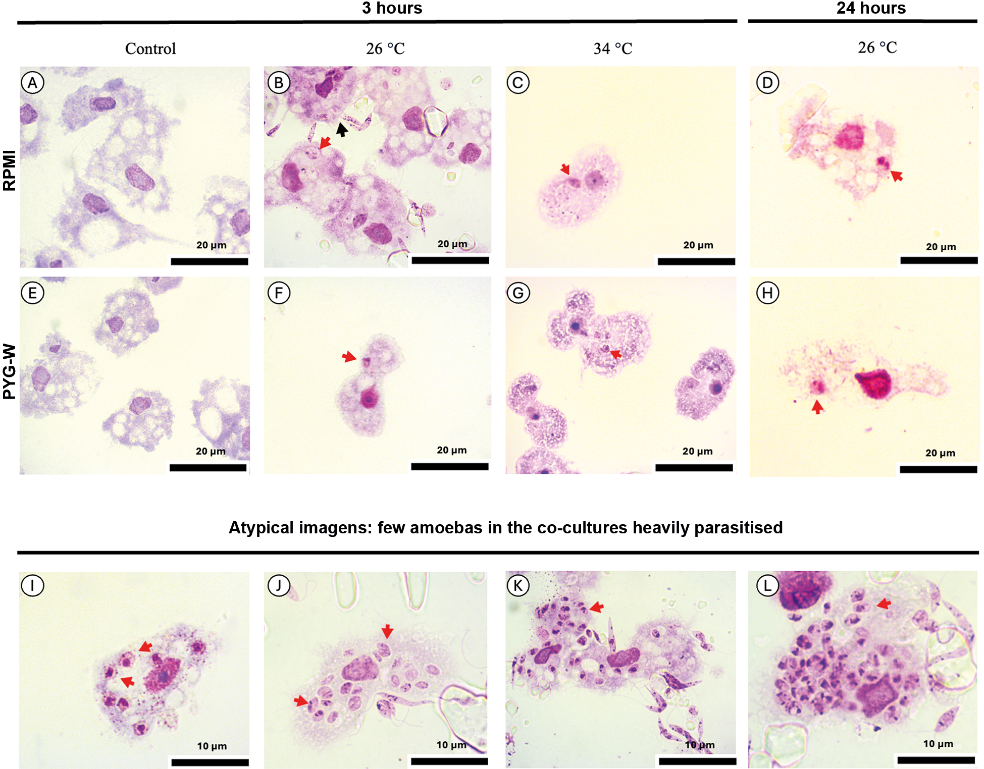
This interaction is represented in the photomicrographs of the kinetics of co-cultures in Fig. 3, which indicates the intracellular forms of L. amazonensis over time (Fig. 2A-H). Most of the internalised forms of L. amazonensis became rounded and shortened after 3 h of co-culture, and apparently lost their flagellum (Fig. 3), as it occurs with intracellular amastigotes. In addition, a very limited number of trophozoites were heavily parasitised with L. amazonensis (Fig. 3I-L).

Acanthamoeba trophozoites and Leishmania amazonensis were co-cultured in RPMI or PYG medium. Trophozoites cultured without promastigotes (A, E); trophozoites co-cultured with promastigotes at 26ºC (B, F) and at 34ºC (C, G) for 3 h; trophozoites co-cultured with promastigotes at 26ºC for 24 h (D, H); atypical images: a few amoebas in the co-cultures were heavily parasitised, as indicated by the presence of a large number of intracellular L. amazonensis (I, J, K, L). Cells were stained with Giemsa. Red arrows: trophozoites containing intracellular L. amazonensis and black arrows: promastigote interacting with trophozoites through the flagellum.
Altogether, based on the rates of L. amazonensis internalisation by the amoeba and amoeba viability, the most optimal condition for the co-culture of promastigotes and trophozoites is in RPMI medium at 26ºC at the ratio of 1:10.
Viability of internalised forms of L. amazonensis - Intracellular forms of L. amazonensis were successfully isolated from trophozoites after 3 h co-cultivation in RPMI medium, and differentiated into proliferating promastigotes, confirming L. amazonensis viability (Fig. 4). However, after 24 h of co-cultivation, no L. amazonensis were recovered from within the trophozoites. The trophozoites co-cultured in PYG medium also harboured viable L. amazonensis, but their ability to differentiate into promastigotes was reduced (Fig. 4).

detection of Leishmania amazonensis viability by proliferation curves of promastigotes recovered from co-cultures. Intracellular L. amazonensis forms were recovered from trophozoites co-cultured in RPMI or PYG medium, at ratios of 1:10 and 1:20 (trophozoite: promastigotes), after 3 h. The recovered intracellular forms were seeded in 24-well plates in RPMI medium for differentiation into promastigotes at 26ºC and counted using a Neubauer chamber over 10 days.
Optical, fluorescence, confocal microscopy, and live microscopy - Initially, to evaluate our staining methods for Acanthamoeba cultures, commercially available FITC microbeads were used as a reference34 [Supplementary data (Figs 2-3)]. The uptake, incorporation, and retention of microbeads were visualised in fluorescence microscopy. As expected, trophozoites were able to uptake microbeads, and after 3 h, up to 50% of amoebas contained microbeads (2-5 microbeads per amoeba) at both temperatures (26ºC and 34ºC) and culture medium (RPMI and PYG) [Supplementary data (Fig. 2)].
Subsequent assays using L. amazonensis-GFP revealed that approximately 27% of amoebas were found to contain the parasite, with an L. amazonensis/trophozoite rate of 0.8-2.8/amoeba after 3 h, which was similar to what was obtained with the L. amazonensis wild-type strain (Fig. 1). Using fluorescence microscopy, it was possible to observe acanthopodia in the trophozoites as well as the interaction of L. amazonensis-GFP promastigotes with trophozoites through their flagellum (Fig. 5). Through orthogonal visualisation of the XYZ axes using a confocal microscope (Fig. 5I-J, K), it was confirmed that L. amazonensis-GFP was located inside the amoeba instead of overlapping with it. This was corroborated by the 3D processing of 30 interaction planes [Supplementary data (Figs 4-5, Movie 1)].

confocal microscopy of co-cultures of Acanthamoeba trophozoites and Leishmania amazonensis-green fluorescent protein (GFP) promastigotes. Confocal microscopy of co-cultures of Acanthamoeba trophozoites and L. amazonensis-GFP promastigotes stained with Celltracker (cytoplasm) (A and E), and DAPI (nucleus) (B and F), L. amazonensis-GFP (C and G), and Merge (D and H). Orthogonal cutting confocal microscopy of Acanthamoeba trophozoites and L. amazonensis-GFP promastigotes co-cultures, stained with Celltracker and DAPI (L, I, and J). White arrow: promastigote interacting with trophozoite through the flagellum.
In addition, time-lapse microscopy demonstrated that the number of L. amazonensis-GFP decreased after 6 and 24 h of interaction compared with 3 h. In addition, this confirmed that the amoeba phagocytosed L. amazonensis-GFP promastigotes via its acanthopodia [Supplementary data (Movies 2-3)].

scanning electron microscopy (SEM) of Acanthamoeba trophozoites and Leishmania amazonensis promastigotes. Trophozoite cultured without promastigotes (control) (A), promastigote cultured without trophozoites (control); promastigote (B); interaction between trophozoites and promastigotes (C and D). green arrows: trophozoites, red arrows: promastigotes attached to trophozoite, and yellow arrow: trophozoite surface damage.
Topological analysis and parasite morphological changes in co-cultures - To examine the surface interactions between L. amazonensis and Acanthamoeba, we performed SEM on trophozoites (control) and promastigotes (control), and compared them with parasites co-cultured for 2 h (Fig. 6A-D). We confirmed that promastigotes interacted with trophozoites, primarily via their flagella. Interestingly, the surface of the trophozoites appeared damaged after the interaction with promastigotes, compared with the control (Fig. 6D).
DISCUSSION
Our results demonstrated the interaction between promastigotes and trophozoites and the presence of L. amazonensis within the amoeba visually. The amoeba phagocytosed the promastigotes via acanthopodia, which are spine-like structures on the surface used to capture particles.35 These results corroborate previous findings in which the interaction between L. braziliensis amastigotes isolated from a skin lesion and Acanthamoeba trophozoites was evaluated by transmission electron microscopy, demonstrating that the amoeba extended acanthopodia within 3 h of co-cultivation.24 Similar conclusions were obtained in a recent study using optical microscopy.25
Although our results confirm what was reported in the only two articles in the literature so far, our study provides a more detailed morphological and kinetic characterisation of the interaction between these protozoans, combining optical, fluorescence, electron, confocal, and live-video microscopy. Our three-dimensional approach overcomes the limitation of two-dimensional microscopy by conclusively confirming the intracellular localisation of Leishmania within amoeba. As a novel contribution, our group has captured protozoan interactions using SEM and observed membrane damage on the trophozoite during interaction with L. amazonensis. Furthermore, combining parasite counting with optical microscopy and real-time microscopy provides a better overview of what happens during the interaction. Interestingly, our results confirm that Acanthamoeba trophozoites can eliminate L. amazonensis rather quickly.
It should be noted that the population of L. amazonensis promastigotes used in our work exhibited persistent infection in RAW 264.7-derived macrophages after 72 h (data not shown). This indicates that these parasites were able to persist within macrophages, unlike what occurred in co-cultures with trophozoites.
Interestingly, most L. amazonensis promastigotes interact with trophozoites via their flagellum, a process that also occurs when promastigotes infect cells of the mammalian mononuclear phagocyte system.3,36 The presence of conserved immune receptors explains this interaction. For instance, amoeba express TLR-5, a pattern recognition receptor that binds flagellin, analogous to its function in mammalian phagocytic cells.37,38 Taken together, the results indicate that Leishmania can penetrate the amoeba both actively and passively via phagocytosis, in a manner comparable to infection of mammalian macrophages.36,39 The phagocytosed forms of L. amazonensis become rounded and shortened or lose the flagellum, and resemble amastigotes, the intracellular parasite form observed within the parasitophorous vacuoles of mammalian macrophages. Although it appears that these amastigote-like forms are located within amoeba vacuoles, as previously suggested,25 it will only be possible to confirm the localisation of Leishmania spp. with future analysis of vacuole markers inside an amoeba.
Regarding the presence of L. amazonensis within the amoeba, the data indicated that, during the first 3 h of co-culture, intracellular amastigote-like forms remained viable. At this time point, 20% of amoeba contained L. amazonensis, with an infection rate of 1.6 parasites per amoeba. The parasites were successfully isolated from trophozoites and differentiated back into axenic promastigotes, demonstrating their viability inside amoeba. However, no proliferation of L. amazonensis inside amoeba was detected over longer periods. Indeed, over time, the number of amastigote-like forms decreased, and no parasites were found inside the trophozoites after 48 h. Indeed, this result corroborates previous findings in which researchers, incidentally, observed a similar pattern in a control experiment with L. tropica. In their study, internalised Leishmania decreased and were completely cleared from the amoeba after 48 h of co-culture.18
It is worthwhile to compare the results with those described for mammalian macrophages. The kinetic profile, i.e., a decrease in L. amazonensis infection rate over time in co-cultures of trophozoites and promastigotes, was not observed in most commonly used cell systems, such as murine peritoneal macrophages, bone marrow cultures, and RAW 264.7-derived macrophages, in which the number of amastigotes increased over time.6,25 In any case, the infection process and the fate of intracellular parasites may differ in vivo.
The mechanism by which Acanthamoeba inactivates and kills Leishmania spp. remains unexplored at present. It is recognised that Acanthamoeba acts as a phagotrophic predator, processing a well-developed phagosome-associated feeding pathway, and producing oxidative stress through ROS and NO.16,17 Additional antimicrobial strategies have been described, including the production of pore-forming toxins, soft metal poisoning, and the exploitation of bacterial motility.40,41,42,43 Preliminary data from our laboratory (data not shown), measuring nitrite by the Griess method,44 indicated no elevated levels of nitrite in the co-cultures of Acanthamoeba trophozoites and L. amazonensis promastigotes.
The main objective of this study was to characterise in depth the fundamental interaction between Acanthamoeba and L. amazonensis. As a novel contribution to the literature, we determined that the optimal experimental condition was RPMI medium at 26ºC. Live microscopy revealed that L. amazonensis is briefly internalised by Acanthamoeba; however, its survival rate decreased after 24 h. Thus, considering that the amoeba can clear Leishmania spp. Regarding infection, we suggest that this amoeba model is not relevant for in vitro studies, such as drug screening. However, this interaction could serve as a model for studying cellular leishmanicidal mechanisms, i.e., strategies for killing microorganisms. Studies involving Acanthamoeba cytolytic mechanisms during the period of interaction with Leishmania spp. could reveal valuable information about molecules with potent microbicidal functions.
We do not know whether there is any ecological significance to the Acanthamoeba-Leishmania spp. interaction, nor whether this occurs in its natural environment. However, Acanthamoeba can act both as a predator and as an environmental host for several pathogens. There is a diversity of medically relevant microorganisms that the amoeba can interact with and influence their virulence in the environment. While amoebae can eliminate some microorganisms, they also internalize pathogens like Legionella and Cryptococcus into their vacuoles, thereby protecting these pathogens and enhancing their virulence.21,35 When challenged, the amoeba transforms into a remarkably resilient, double-walled cyst. This resistance ensures the environmental persistence of both the protozoan and any intracellular pathogens, allowing Acanthamoeba to thrive in virtually any environment.12,13,16
Thus, considering that the Acanthamoeba has been isolated from wild mosquitoes Aedes aegypti, it is possible that the amoeba interacts with the larval stage of the sandfly vector and, later, within the adult insect stage, interacts with Leishmania spp. promastigotes. The hypothesis is that larval stages of sandflies ingest the amoeba found in aquatic environments and biofilms and acquire them in the digestive tract. When adult sandflies take a blood meal from mammals infected by Leishmania spp., they ingest infected cells, and the amastigotes transform into procyclic promastigotes that interact with amoebas within the digestive tract. Likewise, adult sandflies can acquire amoeba, creating conditions for rapid interactions once they become infected with Leishmania promastigotes. Although the viability of trophozoites in this microenvironment may be limited, a rapid interaction with Leishmania spp. could exert some influence on the parasites. Future studies are required to assess Acanthamoeba's role as a natural host for various pathogens.
In conclusion, this study confirms that Acanthamoeba can interact and internalise L. amazonensis. Once internalised, the parasites shift into an amastigote-like form and can be cleared by the amoeba host. The application of reverse-engineering approaches, such as omics-based analyses, may help to elucidate the molecular pathways within the amoeba that lead to its leishmanicidal activity. Understanding these mechanisms could open new avenues for exploring host-pathogen interactions and for identifying innovative strategies with potential therapeutic relevance.
SUPPLEMENTARY MATERIALS
Supplementary data
ACKNOWLEDGEMENTS
To the Electron Microscope Laboratory (LME/UNICAMP) for access to equipment and assistance. We thank the National Institute of Science and Technology on Photonics Applied to Cell Biology (INFABIC) at the State University of Campinas for access to equipment and assistance. We acknowledge Editage (www.editage.com) for their support in improving the English language quality of this manuscript. We thank Victor Agostino for his careful review of the manuscript.
-
Financial support: CNPq (30439/2021-4), FAPESP (2018/23302-6). LFG was a recipient of CAPES (88887.713017/2022-00) and SG is a CNPq research productivity fellow.
DATA AVAILABILITY
The contents underlying the research text are included in the manuscript.
References
-
1 WHO - World Health Organization. Leishmaniasis 2025 [cited 2024 Nov 12]. Available from: https://www.who.int/news-room/fact-sheets/detail/leishmaniasis
» https://www.who.int/news-room/fact-sheets/detail/leishmaniasis -
2 Mann S, Frasca K, Scherrer S, Henao-Martínez AF, Newman S, Ramanan P, et al. A review of leishmaniasis: current knowledge and future directions. Curr Trop Med Rep. 2021; 8(2): 121-32. doi:10.1007/s40475-021-00232-7.
» https://doi.org/10.1007/s40475-021-00232-7 -
3 Serafim TD, Coutinho-Abreu IV, Dey R, Kissinger R, Valenzuela JG, Oliveira F, et al. Leishmaniasis: the act of transmission. Trends Parasitol. 2021; 37(11): 976-87. doi:10.1016/j.pt.2021.07.003.
» https://doi.org/10.1016/j.pt.2021.07.003 -
4 Reithinger R, Dujardin JC, Louzir H, Pirmez C, Alexander B, Brooker S. Cutaneous leishmaniasis. Lancet Infect Dis. 2007; 7(9): 581-96. doi:10.1016/S1473-3099(07)70209-8.
» https://doi.org/10.1016/S1473-3099(07)70209-8 -
5 Ankeny RA, Leonelli S. What's so special about model organisms? Stud Hist Philos Sci A. 2011; 42(2): 313-23. doi:10.1016/j.shpsa.2010.11.039.
» https://doi.org/10.1016/j.shpsa.2010.11.039 -
6 Bogdan C. Macrophages as host, effector and immunoregulatory cells in leishmaniasis. Cytokine X. 2020; 2(4): 100041. doi:10.1016/j.cytox.2020.100041.
» https://doi.org/10.1016/j.cytox.2020.100041 -
7 Terreros MJS, de Luna LAV, Giorgio S. Evaluation of antileishmanial drugs activities in an ex vivo model. Parasitol Int. 2019; 71: 163-6. doi:10.1016/j.parint.2019.04.011.
» https://doi.org/10.1016/j.parint.2019.04.011 -
8 O'Keeffe A, Hale C, Cotton JA, Yardley V, Gupta K, Ananthanarayanan A, et al. Novel 2D and 3D assays for anti-leishmanial drugs. Microorganisms. 2020; 8(6): 6. doi:10.3390/microorganisms8060831.
» https://doi.org/10.3390/microorganisms8060831 -
9 Gupta AK, Das S, Kamran M, Ejazi SA, Ali N. Pathogenicity and virulence of Leishmania - interplay of virulence factors with host defenses. Virulence. 2022; 13(1): 903-35. doi:10.1080/21505594.2022.2074130.
» https://doi.org/10.1080/21505594.2022.2074130 -
10 Montagnes D, Roberts E, Lukeš J, Lowe C. The rise of model protozoa. Trends Microbiol. 2012; 20(4): 184-91. doi:10.1016/j.tim.2012.01.007.
» https://doi.org/10.1016/j.tim.2012.01.007 -
11 Geres LF, Sartori E, Neves JMS, Miguel DC, Giorgio S. Amebicides against Acanthamoeba castellanii: the impact of organism models used in amebicide assays. Parasitologia. 2024; 4(1): 1. doi:10.3390/parasitologia4010002.
» https://doi.org/10.3390/parasitologia4010002 -
12 Zhang H, Cheng X. Various brain-eating amoebae: the protozoa, the pathogenesis, and the disease. Front Med. 2021; 15(6): 842-66. doi:10.1007/s11684-021-0865-2.
» https://doi.org/10.1007/s11684-021-0865-2 -
13 Lacerda AG, Lira M. Acanthamoeba keratitis: a review of biology, pathophysiology and epidemiology. Ophthalmic Physiol Opt. 2021; 41(1): 116-35. doi:10.1111/opo.12752.
» https://doi.org/10.1111/opo.12752 -
14 de Faria LV, do Carmo PHF, da Costa MC, Peres NTA, Chagas IAR, Furst C, et al. Acanthamoeba castellanii as an alternative interaction model for the dermatophyte Trichophyton rubrum Mycoses. 2020; 63(12): 1331-40. doi:10.1111/myc.13173.
» https://doi.org/10.1111/myc.13173 -
15 Fukaya S, Masuda L, Takemura M. Analysis of morphological changes in the nucleus and vacuoles of Acanthamoeba castellanii following Giant virus infection. Microbiol Spectr. 2023; 11(2): e04182-22. doi:10.1128/spectrum.04182-22.
» https://doi.org/10.1128/spectrum.04182-22 -
16 Mungroo MR, Siddiqui R, Khan NA. War of the microbial world: Acanthamoeba spp. interactions with microorganisms. Folia Microbiol. 2021; 66(5): 689-99. doi:10.1007/s12223-021-00889-7.
» https://doi.org/10.1007/s12223-021-00889-7 -
17 Rayamajhee B, Subedi D, Peguda HK, Willcox MD, Henriquez FL, Carnt N. A systematic review of intracellular microorganisms within Acanthamoeba to understand potential impact for infection. Pathogens. 2021; 10(2): 2. doi:10.3390/pathogens10020225.
» https://doi.org/10.3390/pathogens10020225 -
18 Winiecka-Krusnell J, Dellacasa-Lindberg I, Dubey JP, Barragan A. Toxoplasma gondii: uptake and survival of oocysts in free-living amoebae. Exp Parasitol. 2009; 121(2): 124-31. doi:10.1016/j.exppara.2008.09.022.
» https://doi.org/10.1016/j.exppara.2008.09.022 -
19 Boratto PVM, Oliveira GP, Machado TB, Andrade ACSP, Baudoin JP, Klose T, et al. Yaravirus: a novel 80-nm virus infecting Acanthamoeba castellanii Proc Natl Acad Sci USA. 2020; 117(28): 16579-86. doi:10.1073/pnas.2001637117.
» https://doi.org/10.1073/pnas.2001637117 -
20 Boratto PVM, Oliveira GP, Abrahão JS. "Yaraviridae": a proposed new family of viruses infecting Acanthamoeba castellanii Arch Virol. 2022; 167(2): 711-5. doi:10.1007/s00705-021-05326-1.
» https://doi.org/10.1007/s00705-021-05326-1 -
21 Carvalho JHS, Nascimento JKC, Silva KGV, Silveira Neto S, Macedo AT, França HL, et al. Yeast-amoeba interaction influences murine cryptococcosis. Microbes Infect. 2023; 25(7): 105153. doi:10.1016/j.micinf.2023.105153.
» https://doi.org/10.1016/j.micinf.2023.105153 -
22 Ferreira MS, Mendoza SR, Gonçalves DS, Rodríguez-de la Noval C, Honorato L, Nimrichter L, et al. Recognition of cell wall mannosylated components as a conserved feature for fungal entrance, adaptation and survival within trophozoites of Acanthamoeba castellanii and murine macrophages. Front Cell Infect Microbiol. 2022; 12: 858979. doi:10.3389/fcimb.2022.858979.
» https://doi.org/10.3389/fcimb.2022.858979 -
23 Ahmed K. Could Acanthamoeba have hosted and trained Leishmania to evade innate immune response?. Med Hypotheses. 2014; 83(3): 418-9. doi:10.1016/j.mehy.2014.05.015.
» https://doi.org/10.1016/j.mehy.2014.05.015 - 24 Campo-Aasen I, Convit J, Perez-Suarez E, Gallinotto ME. In vitro interaction of Acanthamoeba castellanii with Leishmania braziliensis Cell Mol Biol. 1988; 34(3): 247-54.
-
25 Santos HLC, Pereira GL, Reis RB, Rodrigues IC, d'Avila CM, Vidal VE. Using Acanthamoeba spp. as a cell model to evaluate Leishmania infections. PLoS Negl Trop Dis. 2024; 18(10): e0012517. doi:10.1371/journal.pntd.0012517.
» https://doi.org/10.1371/journal.pntd.0012517 -
26 Chang KP, Reed SG, McGwire BS, Soong L. Leishmania model for microbial virulence: the relevance of parasite multiplication and Patho antigenicity. Acta Trop. 2003; 85(3): 375-90. doi:10.1016/S0001-706X(02)00238-3.
» https://doi.org/10.1016/S0001-706X(02)00238-3 -
27 Costa SS, Golim MA, Rossi-Bergmann B, Costa FTM, Giorgio S. Use of in vivo and in vitro systems to select Leishmania amazonensis expressing green fluorescent protein. Korean J Parasitol. 2011; 49(4): 357-64. doi:10.3347/kjp.2011.49.4.357.
» https://doi.org/10.3347/kjp.2011.49.4.357 -
28 Corsaro D, Mrva M, Colson P, Walochnik J. Validation and redescription of Acanthamoeba terricola Pussard, 1964 (Amoebozoa: Acanthamoebidae). Eur J Protistol. 2024; 94: 126091. doi:10.1016/j.ejop.2024.126091.
» https://doi.org/10.1016/j.ejop.2024.126091 -
29 Barbosa AM, Costa SS, da Rocha JR, Montanari CA, Giorgio S. Evaluation of the leishmanicidal and cytotoxic effects of inhibitors for microorganism metabolic pathway enzymes. Biomed Pharmacother. 2015; 74: 95-100. doi:10.1016/j.biopha.2015.07.040.
» https://doi.org/10.1016/j.biopha.2015.07.040 -
30 Mendes B, Minori K, Consonni SR, Andrews NW, Miguel DC. Causative agents of American Tegumentary Leishmaniasis are able to infect 3T3-L1 adipocytes in vitro Front Cell Infect Microbiol. 2022; 12. doi:10.3389/fcimb.2022.824494.
» https://doi.org/10.3389/fcimb.2022.824494 -
31 Rayamajhee B, Willcox M, Henriquez FL, Vijay AK, Petsoglou C, Shrestha GS, et al. The role of naturally acquired intracellular Pseudomonas aeruginosa in the development of Acanthamoeba keratitis in an animal model. PLoS Negl Trop Dis. 2024; 18(1): e0011878. doi:10.1371/journal.pntd.0011878.
» https://doi.org/10.1371/journal.pntd.0011878 -
32 Garajová M, Mrva M, Vaškovicová N, Martinka M, Melicherová J, Valigurová A. Cellulose fibrils formation and organisation of cytoskeleton during encystment are essential for Acanthamoeba cyst wall architecture. Sci Rep. 2019; 9(1): 1. doi:10.1038/s41598-019-41084-6.
» https://doi.org/10.1038/s41598-019-41084-6 -
33 Albuquerque P, Nicola AM, Magnabosco DAG, Derengowski LS, Crisóstomo LS, Xavier LCG, et al. A hidden battle in the dirt: soil amoebae interactions with Paracoccidioides spp. PLoS Negl Trop Dis. 2019; 13(10): e0007742. doi:10.1371/journal.pntd.0007742.
» https://doi.org/10.1371/journal.pntd.0007742 -
34 Elloway EAG, Bird RA, Hewitt CJ, Kelly SL, Smith SN. Characterization of Acanthamoeba-microsphere association by multiparameter flow cytometry and confocal microscopy. Cytometry A. 2006; 69(4): 266-72. doi:10.1002/cyto.a.20210.
» https://doi.org/10.1002/cyto.a.20210 -
35 Siddiqui R, Khan NA. Biology and pathogenesis of Acanthamoeba Parasit Vectors. 2012; 5(1): 6. doi:10.1186/1756-3305-5-6.
» https://doi.org/10.1186/1756-3305-5-6 -
36 Vannier-Santos MA, Martiny A, Souza W. Cell Biology of Leishmania spp.: invading and evading. Curr Pharm Des. 2002; 8(4): 297-318. doi:10.2174/1381612023396230.
» https://doi.org/10.2174/1381612023396230 -
37 Gonçalves DS, Ferreira MS, Gomes KX, Rodríguez-de La Noval C, Liedke SC, da Costa GCV, et al. Unravelling the interactions of the environmental host Acanthamoeba castellanii with fungi through the recognition by mannose-binding proteins. Cell Microbiol. 2019; 21(10): e13066. doi:10.1111/cmi.13066.
» https://doi.org/10.1111/cmi.13066 -
38 Nasher F, Wren BW. Flagellin O-linked glycans are required for the interactions between Campylobacter jejuni and Acanthamoeba castellanii Microbiology. 2023; 169(8): 001386. doi:10.1099/mic.0.001386.
» https://doi.org/10.1099/mic.0.001386 -
39 Martínez-López M, Soto M, Iborra S, Sancho D. Leishmania hijacks myeloid cells for immune escape. Front Microbiol. 2018; 9: 883. doi:10.3389/fmicb.2018.00883.
» https://doi.org/10.3389/fmicb.2018.00883 -
40 Michalek M, Sönnichsen FD, Wechselberger R, Dingley AJ, Hung CW, Kopp A, et al. Structure and function of a unique pore-forming protein from a pathogenic Acanthamoeba Nat Chem Biol. 2013; 9(1): 37-42. doi:10.1038/nchembio.1116.
» https://doi.org/10.1038/nchembio.1116 -
41 Yabrag A, Ullah N, Baryalai P, Ahmad I, Zlatkov N, Toh E, et al. A new understanding of Acanthamoeba castellanii: dispelling the role of bacterial pore-forming toxins in cyst formation and amoebicidal actions. Cell Death Discov. 2025; 11(1): 1-12. doi:10.1038/s41420-025-02345-8.
» https://doi.org/10.1038/s41420-025-02345-8 -
42 German N, Doyscher D, Rensing C. Bacterial killing in macrophages and amoeba: do they all use a brass dagger? Future Microbiol. 2013; 8(10): 1257-64. doi:10.2217/fmb.13.100.
» https://doi.org/10.2217/fmb.13.100 -
43 de Schaetzen F, Fan M, Alcolombri U, Peaudecerf FJ, Drissner D, Loessner MJ, et al. Random encounters and amoeba locomotion drive the predation of Listeria monocytogenes by Acanthamoeba castellanii Proc Natl Acad Sci USA. 2022; 119(32): e2122659119. doi:10.1073/pnas.2122659119.
» https://doi.org/10.1073/pnas.2122659119 -
44 Karaś MA, Turska-Szewczuk A, Marczak M, Jaszek M, Janczarek M, Dworaczek K, et al. A mutation in the Mesorhizobium loti oatB gene alters the physicochemical properties of the bacterial cell wall and reduces survival inside Acanthamoeba castellanii Int J Mol Sci. 2018; 19(11): 11. doi:10.3390/ijms19113510.
» https://doi.org/10.3390/ijms19113510
Edited by
-
Handling editor:
Geovane Dias-Lopes | https://orcid.org/0000-0003-4448-1398
FIRST REVIEW ROUND - REVIEWERS' COMMENTS
About the reviewerREVIEWER #1
This manuscript investigates the use of Acanthamoeba as a model to study Leishmania amazonensis infection. The premise is interesting given the cellular and functional similarities between these free-living phagocytic amoebae and mammalian macrophages, the primary host cells for Leishmania. In specific contexts, it could serve as an alternative to complex or ethically demanding animal procedures. The topic has limited precedent, with only two major previous experimental studies directly focusing on the interaction of Acanthamoeba and Leishmania: Campos-Aisen in 1988 (PMID: 3208254), and a recent paper by Santos et al., 2024 (doi:10.1371/journal.pntd.0012517), who analyzed Acanthamoeba infectivity by four Leishmania species (including L. amazonensis) using light and fluorescence microscopy.
Considering this established context, I have three overall considerations of the manuscript: 1) It is crucial to define its novel contribution to avoid the perception of merely confirming or extending prior findings. For instance, the quantitative analysis of infection rates, combined with confocal and live microscopy, offers a deeper, more detailed examination of the interaction than previously reported. 2) The stated objective of evaluating A. castellanii as a model organism seems somewhat broad, considering the work did not apply the model to a mechanism investigation. In my view, the presented assays are descriptive, aimed at first understanding the biology of the interaction itself. 3) The manuscript needs a clearer logical story. The reasons for key experimental choices (like the temperatures used or why PCR was needed) are not well explained. The manuscript also requires revisions to English grammar, syntax, and style to improve sentence flow and ensure precise scientific expression.
Specific points to be addressed to strengthen the manuscript are detailed below:
Abstract:
1. Lines 28-31: Abstract opening sentence is factually correct, but it is a general statement that is dispensable. I recommend that the authors begin by directly stating the research challenge (e.g., "Research on Leishmania pathogenesis, drug, and vaccine discovery remains dependent mainly on mammalian models, which are complex and ethically challenging"), then introduce their proposed solution using a simpler, accessible system.
2. Line 33: It is known that the interaction between Acanthamoeba and L. amazonensis is not fully understood; thus, the study provided a more detailed morphological and kinetic characterization of the infection of this protozoan. To establish a more logical link between the knowledge gap and the methodology employed, I suggest that the authors refine their objective to better align with their experimental approach.
3. Line 45: The term "not relevant" carries an undervaluing tone. I suggest reframe the conclusion avoiding this term, focusing instead on the model's specific suitability. The idea can be preserved by stating that it is less suited for sustained infection studies, such as drug screening, while highlighting its value for investigating leishmanicidal mechanisms.
4. Lines 31,33,35,44: Revise the binomial designation A. castelllanii, considering the comment #5 below.
Introduction
5. The manuscript refers to the model organism as Acanthamoeba castellanii. However, not all cited studies or general statements about the genus are specific to this species. To improve taxonomic accuracy and readability, I recommend using Acanthamoeba for general statements (e.g., lines 89, 93, 97, 103, 108), reserving the full binomial nomenclature Acanthamoeba castellanii (or A. castellanii after first use) specifically when citing work that indeed used this species. This must also be applied to the article title.
6. Line 101: The mention of Yaravirus should be supported by citations of its foundational descriptions. I recommend including the seminal work by Boratto et al. 2020 (doi: 10.1073/pnas.2001637117) or a subsequent detailed analysis such as Boratto et al. 2022 (doi: 10.1007/s00705-021-05326-1)
7. Lines 118-124: The Objectives section requires reformulation. It currently mixes a broad objective with methodological details (e.g., the justification for using L. amazonensis) that are better placed in the Methods section. Please revise this section to focus on the study's core aim (see Comment #2).
Material and Methods
8. Line 135: The strain ATCC 30010 has been historically classified as Acanthamoeba castellanii. A recent taxonomic revision, however, has reclassified it as A. terricola (Corsaro et al., 2024; doi:10.1016/j.ejop.2024.126091). To ensure taxonomic accuracy while maintaining clarity with the historical literature, I recommend that the authors note the reclassification in this section. Throughout the rest of the manuscript, they could primarily use the genus name (Acanthamoeba) or the strain designation (ATCC 30010) to avoid confusion.
9. The standard medium used for the axenic culture of Acanthamoeba is designated as PYG (Peptone-Yeast Extract-Glucose). Please remove the 'W' throughout the manuscript to align with the conventional nomenclature in the field.
10. Line 179-182: The Mat & Met section 2.4 requires clarification. The text abruptly shifts from describing the protocol for lysing amoebae to recover Leishmania (lines 169-178) to a sentence about counting 'adherent and intact amoebas' (lines 179-180) without specifying that this was a separate, parallel experiment. I suggest inserting a transitional phrase such as 'In a separate set of wells,' or 'For amoeba viability assessment,' at the beginning of line 179. Furthermore, the reference [25] (line 180) does not refer to the Acanthamoeba analysis of viability.
Results
11. Line 250-255 and Fig S1: The opening statement of the results presents a known fact without providing novel insight. Furthermore, the citations (#30, #31) are dispensable here, as this section should focus on presenting new findings rather than referencing established methods. The primary concern, however, is the rationale for the temperature choices in the interaction assays. The data in Figure S1F clearly show that the amoeba (the host cell in this model) does not proliferate at 34°C in PYG medium. This choice of 34°C for subsequent co-culture experiments is unsuitable for studying a productive interaction, as the host is not totally viable. The authors must clarify this point.
12. Lines 255-301: This part is overly descriptive, detailing observational trends that are already visible in the figures. To improve clarity and impact, this section should be more synthetic, focusing on concisely stating the key findings and directing readers to the supporting data. A more effective strategy would be to structure the narrative around the key experimental variables. For instance, the 26°C condition led to a more sustained infection and higher host amoeba viability. The infection pattern in RPMI and PYG was similar.
13. Lines 302-306: I recommend moving this paragraph to the Discussion, since it provides an interpretation of the results
14. Lines 307-309: The interaction between L. amazonensis promastigotes and Acanthamoeba cysts is conceptually flawed and should be removed from the manuscript. Cysts are dormant, metabolically inactive stages characterized by a thick, protective cell wall. Its primary biological function is to resist environmental conditions, and no phagocytosis is expected.
15. Lines 310-314: Here, findings from a co-infection assay with amastigotes are mentioned, but this methodology is not described in the Materials and Methods section. Provide a complete description of the methodology for co-infection with amastigotes. Alternatively, as a preliminary, not a central, focus of the work, the authors should remove this mention and instead briefly comment on it in the Discussion as an informal observation.
16. Lines 321 – 325 (and corresponding methodology in Mat & Met), Figure 4: What is the rationale for the PCR assay? The authors present a more direct and functionally informative viability assay as definitive proof of parasite viability. In this context, the purpose of the conventional PCR is unclear and appears redundant. Also, PCR detection of Leishmania DNA does not confirm the presence of viable, replicating parasites, as the signal may originate from DNA released by degraded organisms.
17. Fig 6: The authors attribute membrane damage seen in a single SEM image to the interaction with Leishmania. However, a single micrography is not representative of a general pattern of interaction. Furthermore, the possibility that these structures are preparation artifacts cannot be ruled out.
Discussion
18. The Discussion begins with a summary of the methods and a repetition of basic findings. Consider removing this paragraph.
19. Line 374: This paragraph effectively cites previous studies with similar conclusions. This is an ideal point to highlight the novel contribution of the present work.
20. Winiecka-Krusnell et al. (2009) paper has a primary focus and title concern Toxoplasma, and the data on L. tropica appears to be a minor, comparative control. Mentioning that a similar pattern of clearance was incidentally observed by Winiecka-Krusnell et al. (2009) in a control experiment with L. tropica would prevent the reader from overestimating its role in the existing literature.
21. Line 417. Why would the lack of a three-dimensional structure be a limitation of the study?
22. Line 422: The statement that the objective was 'to evaluate whether Acanthamoeba could be used as a model organism sets an inappropriate expectation for the reader. A more precise aim would be: "to deeply characterize the fundamental interaction between Acanthamoeba and L. amazonensis." Authors should consider reframing it throughout the manuscript.
23. Lines 451-464: The Conclusion currently repeats specific methodological details, results, and discussion points that have already been presented. It should be reframed to synthesize the main achievement.
REVIEWER #2
The manuscript " Uptake and survival of Leishmania amazonensis in Acanthamoeba castellanii: An adequate model organism?" by Geres et al. describes the Acanthamoeba-Leishmania interaction as a model to study how Leishmania is able to survive within a phagocytic cell and with other related approaches.
I have several comments as below:
- Throughout the manuscript the language is sometimes not specific, based on scientific "slang," or not completely accurate. It should be proofread to improve accuracy.
- Authors should focus on the virulence factors of Leishmania, such as the tolerance to the oxidative stress within macrophages or how Leishmania is able to survive within them. Does Leishmania disturb the phagosome maturation? What are the mechanisms behind this?
- The optimal temperature range for Acanthamoeba cultivation is 25-31ºC. At 34 and 37 ºC, were the trophozoites growing flattened onto coverslip surface, or were tending to become rounded? Assays were performed for 3, 24, 48 and 72h. This conditions stimulate the trophozoites-to-cysts transition, and rounded cells are going to gradually leak the ability of phagocytosis. Acanthamoeba is very sensitive to changes in nutrients depletion and cellular density. How many Acanthamoeba and Leishmania cells composed the inocula based on 1:10 and 1:20 ratios? I think that the first 12h of interaction were important to evaluate phagocytosis dynamics.
- How was cell viability calculated after mechanical lysis with needle passages? Were Leishmania and Acanthamoeba separately subjected to the same lysis protocol to compare cell viability? How can you infer that Leishmania was killed by Acanthamoeba and not by the mechanical lysis procedure?
- Why was the co-culture timepoint of 2h chosen for scanning electron microscopy assay? What were the criteria?
- How many trophozoites were counted per slide in relation to the percentage shown in Fig 1? Were only internalized cells considered, or were other events, such as interaction with surface cells, also taken into account? The events of surface interaction and internalized cells should be compared to assess the phagocytosis efficiency over time. This article may help you (https://doi.org/10.1016/j.micinf.2023.105153)
- Please, explain the reason for the trophozoites augmentation (virtually no cysts) at 37 ºC (Fig. 1S), considering that the optimal temperature range for A. castellanii (ATCC 30010) cultivation is 25-31 ºC.
- Increase the size of Figs 1, 2, 3, 5, 1S, 2S
- The meaning of the term "intact trophozoites" is unclear, especially in the context of line 268. Please clarify its meaning or replace it throughout the manuscript.
- Line 278, "In addition, a very limited number of trophozoites were heavily parasitized with L. amazonensis (Figure 2J-L)." This may have occurred due to the prolonged co-culture period (> 6 h). Note the presence of a rounded trophozoite in Fig. 2L, probably in the process of becoming a cyst. Therefore, this cell is no longer expected to have phagocytic ability. Please provide the number of trophozoites interacting with Leishmania per slide and associate this information with the percentage for each time point of the co-culture. For example, one hundred or more trophozoites per slide can provide valuable information about the interaction between Leishmania and Acanthamoeba (cell surface interaction versus internalized cells).
- Lines 307-312, It is a long-established that cysts are a non-phagocytic, protective, and dormant stage in the parasite's life cycle. It was expected that this would happen.
- Lines 376-380, there is evidence that Acanthamoeba possesses a surface receptor similar to the TLR-5 of mammalian phagocytic cells, which recognizes flagellin. This article may be useful (https://doi.org/10.1099/mic.0.001386).
- Line 400, write "over time" instead of "over tine". I recommend that the author submit the manuscript to an English editing service.
- Lines 404-406, "only cytokine-activated macrophages were able to inhibit Leishmania growth (Bogdan 2020, Santos et al. 2024). Could it be write this way?
- Line 409, please, write "oxidative stress" instead of "oxidative attack".
- Lines 414-416, please, provide details about the method for NO measurement.
- Line 427, should delve deeper into the discussion "different strategies to kill microorganisms". It appears that a similar apoptosis pathway exists for protozoan parasites. If Acanthamoeba dies, should Leishmania also die (inside the amoeba)?. This article (https://doi.org/10.1186/1756-3305-4-44) may help you find recent studies based on this topic.
- Lines 440-448, you should explore the hypothesis that Acanthamoeba and Leishmania can interact within mosquitoes.
- Lines 431-438, please, clarify the discussion about Acanthamoeba in relation to the "trojan horse of the microbial world", "cystic resistance" and "microbial biofilm" terms.
Please, present a conclusion without rewriting your results. What gaps were filled? What was your contribution to the scientific knowledge about the Acanthamoeba-Leishmania interaction? What is possible to do and what should future studies focus on?
AUTHORS' RESPONSE TO THE REVIEWERS
REVIEWER COMMENTS: Reviewer:
1. This manuscript investigates the use of Acanthamoeba as a model to study Leishmania amazonensis infection. [...] Considering this established context, I have three overall considerations of the manuscript: 1) It is crucial to define its novel contribution to avoid the perception of merely confirming or extending prior findings. [...] We agree with the reviewer and appreciate this important point. We have revised the manuscript to more explicitly define its novel contributions. 2) The stated objective of evaluating A. castellanii as a model organism seems somewhat broad [...] We agree with the reviewer's assessment. Accordingly, we have revised the main objective of the study to focus on understanding the biology of the interaction between Acanthamoeba and Leishmania [...]. 3) The manuscript needs a clearer logical story. [...] We thank the reviewer for this important feedback. We have revised the manuscript to improve its overall logical flow [...]. In doing that, we decided to remove the PCR assay which did not directly contribute to the central objective of the study.
Abstract: 1. Lines 28-31: [...] We thank the reviewer for this valuable suggestion. The abstract was revised, and the opening sentences were restructured to be more direct and focused on the research challenge, as recommended (Line 27-31).
2. Line 33: [...] We thank the reviewer for this comment. The objective was revised to better align with the experimental approach employed in this study (Line 32-34).
3. Line 45: [...] We thank the reviewer for this valuable suggestion. The conclusion was revised. The term "not relevant" was removed and the text was reframed to emphasize the model's specific suitability (Line 44-45).
4. Lines 31,33,35,44: Revise the binomial designation A. castelllanii, considering the comment #5 below. We thank the reviewer for this important taxonomic recommendation. We revised the manuscript to improve taxonomic accuracy and readability as suggested. In response, we have adopted the genus-level nomenclature (Acanthamoeba) throughout the manuscript to avoid taxonomic ambiguity. [...] We also adapted the article title to reflect the new chosen nomenclature.
5. Introduction. [...] We thank the reviewer for this important taxonomic recommendation. We revised the manuscript to improve taxonomic accuracy and readability, as recommended by the point 4 made above (e.g., lines 38, 43, 88, 92, 96).
6. Line 101: [...] We thank the reviewer for this important suggestion. The mention of Yaravirus has now been supported by the suggested citations (Lines 99-100).
7. Lines 118-124: [...] We thank the reviewer for this important suggestion. We have reformulated this section to more clearly emphasize the core objective of the study, and the methodological details have been relocated to the Materials and Methods section (Lines 117-118).
8. Material and Methods. Line 135: [...] We thank the reviewer for this important taxonomic recommendation. We revised the manuscript to improve taxonomic accuracy and readability, as answered in the point 4 (Lines 134 – 137).
9. The standard medium [...] Please remove the 'W' [...]. We thank the reviewer for pointing this out. The manuscript was thoroughly revised to ensure consistent use of the conventional nomenclature PYG (Peptone–Yeast Extract–Glucose), and the letter "W" was removed throughout the text accordingly.
10. Line 179-182: [...] We thank the reviewer for this helpful comment. The text was revised to improve clarity and flow (Line 181). In addition, we included a specific reference related to amoeba viability assessment (https://doi.org/10.1371/journal.pntd.0011878) (Line 182).
11. Results. Line 250-255 and Fig S1: [...] We thank the reviewer for this detailed and constructive comment. [...] Regarding the choice of temperature, we agree that Acanthamoeba does not proliferate at 34 °C in PYG medium, as shown in Figure S1F. However, although proliferation is impaired, the amoebae remain viable for up to 72h, predominantly in the trophozoite stage, and subsequently enter an early encystment process. [...] We have modified the text to include this explanation (Lines 243-249).
12. Lines 255-301: [...] We agree with the reviewer that this section was overly descriptive. Accordingly, we have revised the text to make it more concise and synthetic [...] (Lines 250-272).
13. Lines 302-306: [...] We thank the reviewer for pointing this out. We have replaced this paragraph to the Discussion section (Lines 339-342).
14. Lines 307-309: [...] We thank the reviewer for this comment and agree with the assessment. This paragraph was removed from the manuscript.
15. Lines 310-314: [...] We thank the reviewer for this comment and agree with the assessment. We have removed this mention.
16. Lines 321 – 325 [...] Figure 4: What is the rationale for the PCR assay? [...] We agree with the reviewer's assessment. Therefore, we have removed this assay and its corresponding methodology from the manuscript.
17. Fig 6: [...] We thank the reviewer for this comment; however, the membrane alterations were consistently observed across multiple SEM fields, indicating a recurrent pattern rather than an isolated event. Importantly, a control sample consisting of only amoebae processed in parallel under identical preparation conditions, but without Leishmania interaction, did not display these alterations. This strongly suggests that the observed structures are not preparation artifacts.
18. Discussion. The Discussion begins with a summary of the methods [...] We agree with the reviewer's assessment. Therefore, we have removed this paragraph.
19. Line 374: [...] We thank the reviewer for this comment. We have reformulated this paragraph highlighting the novel contribution of our work (Lines 325 – 338).
20. Winiecka-Krusnell et al. (2009) [...] We fully agree with the reviewer's observation and have revised the text to explicitly indicate that this finding represents an incidental observation within the cited study (Lines 365 – 367).
21. Line 417. Why would the lack of a three-dimensional structure be a limitation of the study? We thank the reviewer for raising this point. This paragraph was misplaced in the Discussion. Accordingly, we have relocated it to the beginning of the Discussion section [...] (Lines 325 – 338).
22. Line 422: [...] We thank the reviewer for this insightful comment and agree [...]. Accordingly, we have revised the manuscript to clarify that the central objective of the study is to provide an in-depth characterization of this host–parasite interaction.
23. Lines 451-464: [...] We thank the reviewer for this comment. We have revised the Conclusion section to improve its clarity and focus [...] (Lines 423-430).
Reviewer: 2
- Throughout the manuscript the language is sometimes not specific [...] Thank you for your valuable feedback regarding the language in our manuscript. We have carefully revised the entire text. Authors should focus on the virulence factors of Leishmania [...] Thank you for these excellent and insightful suggestions. [...] Therefore, the primary scope and contribution of our current work was to establish a more accurate morphological analysis and a detailed quantitative kinetic profile of this host-parasite interaction [...].
- The optimal temperature range for Acanthamoeba cultivation is 25-31ºC [...] We thank the reviewer for these insightful questions [...] We acknowledge that the optimal proliferation range for Acanthamoeba is 25-31°C. The use of 34°C was an experimental choice, despite it not being a standard growth condition. Our rationale was based on the biological relevance of 34°C as the skin temperature of mammalian hosts [...]. Regarding the inoculum concentrations for the 1:10 and 1:20 ratios: For the co-culture assays, 3 × 10⁵ trophozoites were plated. To achieve the stated ratios: For a 1:10 ratio (amoeba:promastigote), 3 × 10⁶ promastigotes were added. For a 1:20 ratio, 6 × 10⁶ promastigotes were added. [...] This information were added in the material and Methods section [...] (Lines 154-157).
- How was cell viability calculated after mechanical lysis with needle passages? [...] Thank you for raising these important methodological points [...] Cell viability of the recovered Leishmania promastigotes was assessed based on the capacity for subsequent proliferation in culture. [...] To specifically rule out that the mechanical lysis procedure itself was responsible for reduced parasite viability, we performed a critical control experiment. Free-living Leishmania promastigotes (not exposed to amoebae) were subjected to the exact same mechanical lysis protocol [...]. Our conclusion that Leishmania was killed by Acanthamoeba and not by the experimental procedure is supported by a convergence of evidence [...].
- Why was the co-culture timepoint of 2h chosen for scanning electron microscopy assay? [...] Thank you for the opportunity to clarify this methodological point. The 2-hour co-culture timepoint for scanning electron microscopy (SEM) was selected based on direct evidence from our preliminary kinetic analyses. [...] This specific timepoint represents a critical phase in the interaction where a significant number of Leishmania promastigotes are adhered to the surface of Acanthamoeba trophozoites but have not yet been fully internalized.
- How many trophozoites were counted per slide in relation to the percentage shown in Fig 1? [...] Thank you for these pertinent questions regarding the quantitative analysis. To determine the percentages of trophozoites with L. amazonensis, 200 amoebas were counted per coverslip [...] (Line 166). [...] We acknowledge that distinguishing between adhesion and true internalization is crucial for precise phagocytosis efficiency metrics [...].
- Please, explain the reason for the trophozoites augmentation (virtually no cysts) at 37 ºC (Fig. 1S) [...] Thank you for this insightful question [...] We acknowledge that the optimal proliferation range for Acanthamoeba is 25-31°C. The use of 34°C was an experimental choice [...] We have revised the text to make this rationale clearer and to avoid any misinterpretation (Lines 243-249).
- Increase the size of Figs 1, 2, 3, 5, 1S, 2S. Thank you for your comment, as suggested, we have increased the size and resolution of figures 1, 2, 3, 5, 1S, and 2S [...].
- The meaning of the term "intact trophozoites" is unclear [...] We thank the reviewer for raising this important point. We have revised the manuscript and replaced the term "intact trophozoites" by viable amoeba throughout the manuscript.
- Line 278, "In addition, a very limited number of trophozoites were heavily parasitized with L. amazonensis (Figure 2J-L)." [...] We thank the reviewer for the observation. [...] The highly parasitized trophozoites shown in Figures 2J-L were captured at the 3-hour time point, not after a prolonged co-culture (>6h). This indicates that intense parasite association can occur even during the early phases of interaction. [...]
- Lines 307-312, It is a long-established that cysts are a non-phagocytic, protective, and dormant stage [...] We thank the reviewer for this comment and agree with the assessment. As the observations regarding the interaction between L. amazonensis promastigotes and Acanthamoeba cysts do not provide novel insight to the current literature, this paragraph was removed from the manuscript.
- Lines 376-380, there is evidence that Acanthamoeba possesses a surface receptor similar to the TLR-5 of mammalian phagocytic cells [...] We thank the reviewer for this valuable suggestion. The information highlighted in the cited article has now been incorporated into the Discussion section [...] (Lines 345-348).
- Line 400, write "over time" instead of "over tine" [...] We thank the reviewer for pointing out this typographical error. The term "over tine" has been corrected to "over time" (Line 369).
- Lines 404-406 [...] This information was misplaced in the Discussion, and could compromise clarity and understanding of the text, so it was removed from the discussion.
- Line 409, please, write "oxidative stress" instead of "oxidative attack". We thank the reviewer for this suggestion. The term "oxidative attack" has been replaced with "oxidative stress" (Line 378).
- Lines 414-416, please, provide details about the method for NO measurement. We thank the reviewer for this comment. [...] we have added a brief and informal methodological description of the NO measurement using the Griess method in this same section, and referenced (Line 381).
- Line 427, should delve deeper into the discussion "different strategies to kill microorganisms" [...] We appreciate this insightful comment and the suggested reference. In our experimental conditions, while Leishmania was all cleared, the Acanthamoeba trophozoites largely remained intact, suggesting that parasite death may not be linked to a classical apoptosis-like pathway shared by both organisms. [...]
- Lines 440-448, you should explore the hypothesis that Acanthamoeba and Leishmania can interact within mosquitoes. We thank the reviewer for this valuable suggestion. We have revised and reformulated this section.
- Lines 431-438, please, clarify the discussion about Acanthamoeba in relation to the "trojan horse of the microbial world", "cystic resistance" and "microbial biofilm" terms. We thank the reviewer for pointing out this issue. We have clarified and refined the discussion [...] (Line 397 – 404).
Please, present a conclusion without rewriting your results [...] We thank the reviewer for this important comment. We have substantially revised the Conclusion (Lines 423 – 430).
- peer review recommendation: accept
REVIEWERS' COMMENTS
About the reviewerREVIEWER #1
The authors have been responsive to reviewer comments and modifications have greatly improved rigor and readability of the manuscript.
My two suggestions for the manuscript are:
Lines 96-100. I think the amoeba-fungus interaction has been explored more thoroughly with Cryptococcus, at least in the last two decades, than with other fungi, such as dermatophytes. Nothing against mentioning new findings, but I think more established approaches to amoeba-fungus interaction should also be cited in this context (https://doi.org/10.1016/j.micinf.2023.105153).
Lines 389-401. I suggest mentioning, in addition to the bacteria in this paragraph, that Acanthamoeba is capable of modulating (increasing) the virulence of Cryptococcus (https://doi.org/10.1016/j.micinf.2023.105153), an important human pathogenic fungus that causes more than 600,000 deaths per year worldwide. This is important to show the diversity of microorganisms of medical interest that the amoeba can interact with and influence their virulence from the environment, as well as to reinforce the importance of using Acanthamoeba as a tool for cell interaction studies.
REVIEWER #2
The revisions are satisfactory and address all recommended points. Nevertheless, the added text would benefit from a final language polish. I would recommend a language edit to improve flow and conciseness in the following specific parts of the text:
1) Lines 365-367 (Original: "…who incidentally observed a similar pattern in a control experiment with L. tropica, which showed a decrease in Leishmania internalized by the amoeba, which were completely cleared after 48h of co-culture.). Suggestion: "…who incidentally observed a similar pattern in a control experiment with L. tropica. In their study, internalized Leishmania decreased and were completely cleared from the amoeba after 48h of co-culture."
2) Lines 385-388 (Original: "As a new contribution to literature, we have determined that the optimal experimental condition was RPMI medium at 26°C, by live microscopy we have seen that L. amazonensis is briefly internalized by Acanthamoeba, however, its survival rate decreased after 24 h."). Suggestion: "As a new contribution to the literature, we determined that the optimal experimental condition was RPMI medium at 26°C. Live microscopy revealed that L. amazonensis is briefly internalized by Acanthamoeba; however, its survival rate decreased after 24 h."
3) Lines 398-401 (Original: "While amoebae are able to eliminate some microorganisms from the environment, they can also internalize pathogens such as Legionella and Mycobacterium into their vacuoles, protecting them and enhancing their virulence [36].") Suggestion: "While amoebae can eliminate some microorganisms, they also internalize pathogens like Legionella and Mycobacterium into their vacuoles, thereby protecting these bacteria and enhancing their virulence [36]."
4) Lines 401-404: (Original: "Amoeba when challenged, transforms into double-walled cysts, structures remarkably resilient. This cystic resistance ensures the environmental persistence of both the protozoan and intracellular pathogens. It can be widespread in practically any environment [12,13,16]."). Suggestion: "When challenged, the amoeba transforms into a remarkably resilient, double-walled cyst. This resistance ensures the environmental persistence of both the protozoan and any intracellular pathogens, allowing Acanthamoeba to thrive in virtually any environment."
EDITOR COMMENTS:
I kindly ask the authors to incorporate the minor adjustments suggested by the reviewers and to take this opportunity to perform a comprehensive revision of the English language and grammar with an expert.
AUTHORS' RESPONSE TO THE REVIEWERS
REVIEWER COMMENTS:
Reviewer: 1
Lines 96-100. I think the amoeba-fungus interaction has been explored more thoroughly with Cryptococcus [...] Thank you for the valuable suggestion. The recommended article has now been cited and appropriately incorporated into the revised manuscript (Lines 107, 110). We also took this opportunity to carefully revise the references and improve the English language.
Lines 389-401. I suggest mentioning, in addition to the bacteria in this paragraph, that Acanthamoeba is capable of modulating (increasing) the virulence of Cryptococcus [...] Thank you for the suggestion. The recommended reference has been incorporated into the revised manuscript (Lines 384-388) to highlight the interaction between Acanthamoeba and Cryptococcus.
Reviewer: 2
The revisions are satisfactory and address all recommended points. Nevertheless, the added text would benefit from a final language polish [...] Thank you for your careful evaluation and constructive recommendation. We have performed a comprehensive revision of the entire manuscript to further improve the English language, grammar, clarity, and overall readability. The manuscript has also undergone professional English language editing by Editage prior to its submission to MIOC. A certificate confirming this linguistic review is available and has been attached.
1) Lines 365-367 [...] Thank you for the suggestion. The text has been revised accordingly to improve clarity and readability (Lines 351 – 353).
2) Lines 385-388 [...] Thank you for the suggestion. The text has been revised accordingly to improve clarity and readability (Lines 371 – 374).
3) Lines 398-401 [...] Thank you for the suggestion. The text has been revised accordingly to improve clarity and readability (Lines 385-388).
4) Lines 401-404 [...] Thank you for the suggestion. The text has been revised accordingly to improve clarity and readability (Lines 388-391).
EDITOR COMMENTS:
I kindly ask the authors to incorporate the minor adjustments suggested by the reviewers and to take this opportunity to perform a comprehensive revision of the English language and grammar with an expert.
Thank you for your careful evaluation and constructive recommendation. We have performed a comprehensive revision of the entire manuscript to further improve the English language, grammar, clarity, and overall readability. The manuscript has also undergone professional English language editing by Editage prior to its submission to MIOC. A certificate confirming this linguistic review is available and has been attached.
- peer review recommendation: accept
REVIEWERS' COMMENTS
About the reviewerREVIEWER #1
The revisions are satisfactory and address the recommended points.
REVIEWER #2
English editing and formatting are required, as recommended by the editor. The manuscript has been improved and now meets the requirements for acceptance by MIOC.
- peer review recommendation: accept