The role of the giacomini vein in preoperative mapping of lower limb varicose veins
Abstract
Background The Giacomini vein (GV) can transfer reflux from perineal veins, tributary veins, and perforators of the thigh to the small saphenous vein (SSV). Vascular ultrasound with Doppler (VUD) is the preferred method for detecting reflux in specific veins such as the GV.
Objective To identify GV depth and diameter, reflux in the GV, and presence of reflux in the SSV caused by the GV.
Methods A cross-sectional, retrospective study was conducted in women undergoing lower limb venous mapping for varicose vein surgery. The following parameters were analyzed in GVs in which reflux was detected: segmental or diffuse reflux pattern; GV diameter and depth; and reflux in the SSV caused by the GV.
Results 340 of the 2368 women evaluated were included in the study because they had a GV, totaling 511 veins analyzed, 150 (29.4%) of which had reflux. The diameters of the 150 GVs with reflux ranged from 1.5 to 7.8 mm and their depth varied from 4 to 25 mm. Most GVs with reflux (91.3%) had a segmental reflux pattern. The majority (66%) of refluxing GVs drained reflux into the popliteal vein through the saphenopopliteal junction, while reflux was transferred to the SSV in 34 veins (22.7%), and was drained by a tributary vein in the thigh in 15 veins (11.3%).
Conclusions Approximately one-third of the studied GVs had reflux, mostly segmental, mean caliber was 2.7 mm, and mean depth was 11 mm. Reflux in the SSV originating from the GV was detected in 22% of the evaluated veins.
Keywords:
ultrasonography; Doppler; venous insufficiency; varicose veins; preoperative period
Resumo
Contexto A veia de Giacomini (VG) pode transferir refluxo de veias perineais, veias tributárias e veias perfurantes da coxa para a veia safena parva (VSP). A ultrassonografia vascular com Doppler (USVD) é o exame de escolha para detecção do refluxo em veias específicas como a VG.
Objetivos Identificar a profundidade, o diâmetro e o refluxo na VG e a presença de refluxo na VSP causado pela VG.
Métodos Estudo transversal e retrospectivo, em mulheres que realizaram mapeamento venoso para cirurgia de varizes de membros inferiores. Nas VGs com refluxo, foram considerados os seguintes parâmetros: refluxo segmentar ou difuso; diâmetro, profundidade; e refluxo na VSP causado pela VG.
Resultados Das 2.368 mulheres avaliadas, 340 foram incluídas no estudo por apresentarem VG, totalizando 511 veias analisadas, sendo 150 (29,4%) veias com refluxo. Nas 150 VGs com refluxo, o diâmetro variou entre 1,5 e 7,8 mm e a profundidade, entre 4 e 25 mm. O padrão de refluxo na maioria das VGs (91,3%) foi do tipo segmentar. Em relação à drenagem do refluxo das VGs, a maioria (66%) drenou o refluxo para a veia poplítea através da junção safenopoplítea; em 34 veias (22,7%), o refluxo foi transferido para a VSP e, em 15 veias (11,3%), foi escoado por veia tributária na coxa.
Conclusões Aproximadamente um terço das VGs estudadas apresentou refluxo, majoritariamente segmentar, com calibre médio de 2,7 mm e profundidade média de 11 mm. Refluxo na VSP originado da VG foi detectado em 22% das veias avaliadas.
Palavras-chave:
ultrassonografia Doppler; insuficiência venosa; varizes; período pré-operatório
INTRODUCTION
The Giacomini vein (GV) is a tributary vein of the great saphenous vein (GSV) or the posterior accessory saphenous vein that ascends obliquely in the posterior thigh, with both subfascial and subcutaneous segments, and is, therefore, an intersaphenous vein. The GV may originate from the small saphenous vein (SSV) or from its cranial extension.1
This intersaphenous vein originates superficially in the popliteal fossa, arising from the segment between the pre-terminal and terminal valves of the SSV and initially ascends between the semimembranosus and biceps femoral muscles and then within the sulcus between the biceps femoral muscle and the semitendinosus muscle, alongside the posterior femoral cutaneous nerve. Its valves are configured to direct blood from the SSV to the GSV, preventing distal reflux, and can be found in both the subfascial portion and the subcutaneous segment, close to the junction with the GSV.2
Valve incompetence in the GV can be responsible for emergence of varicose veins in the posterior thigh or in the topography of the SSV and must be identified in preoperative assessments to ensure better results of surgical treatment.
Vascular ultrasonography with Doppler (VUSD) has been used for more than twenty years for detecting and assessing reflux of blood in veins of the lower limbs (LL), primarily using color flow mapping (to identify retrograde flow) and spectral Doppler (reflux time). It is thus possible to precisely identify the distribution and extent of venous reflux. This examination has become the method of choice for assessment of the peripheral venous system.3
In order to detect sources of reflux and their repercussions for the superficial vein system and chronic venous disease, it is necessary to identify incompetent venous segments that cause varicose veins, such as a GV with reflux.
The GV can be a source of reflux into the SSV, giving rise to varicose veins in the posterior leg, and may constitute indications for surgery, which should also include the GV and can be performed with endovenous procedures. It is therefore important to detect reflux and identify the specific reflux pattern present in the GV.
The objectives of this study were to identify presence of GVs; their depth and diameter, reflux in the GV, and any possibility of reflux in the SSV caused by the GV.
METHODS
A cross-sectional, retrospective, observational study was conducted of data from 2,368 women referred to the Angiolab vascular laboratory (Curitiba, Paraná, Brazil), for venous mapping. Inclusion criteria were age greater than 18 years and primary varicose veins in the LL. Patients with a prior history of surgery for lower limb varicose veins were excluded.
A sample size calculation was conducted for a 0.05 margin of error and 0.95 confidence level and considering a mean prevalence in the literature of 29%, indicating a minimum sample size of 317 GVs.
Ultrasonographic assessment
The ultrasonographic assessment was conducted using Siemens-Acuson Antares® and X 700® ultrasound machines (Issaquah®, United States), with 5 MHz transducers to assess the deep vein system and rule out deep venous thrombosis, with the patient lying down, and 7 MHz transducers to assess the superficial vein system, with the patient standing upright.
The GSV and SSV were examined and presence of GVs identified with the patient standing upright, acquiring anatomic images of the veins in transverse and longitudinal views with the ultrasound equipament in B-mode.
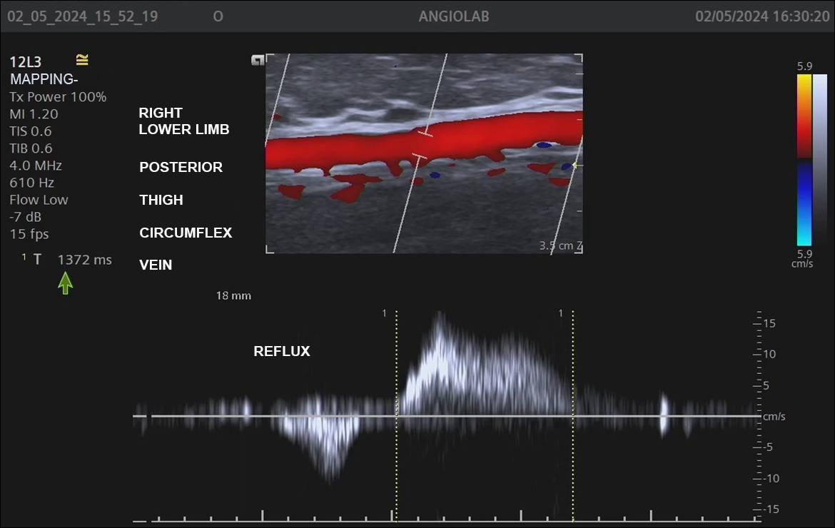
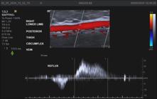
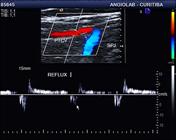
With the help of color flow mapping and spectral Doppler, valve competence was assessed by applying manual muscle compression, distal of the transducer, in order to provoke and detect reflux in the saphenous veins and GV, when present. Reflux in these veins was defined as present if there was retrograde flow with a duration exceeding 0.5s4 (Figure 1).
The GV-specific assessment considered the following parameters: reflux absent, segmental, or diffuse; presence of reflux in the SSV caused by the GV; and diameter, depth, and height (distance above the sole of the foot) of communication with the GSV and the SSV (for GVs with reflux only) (Figure 2).
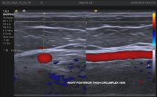
Measurement of the depth (13 mm) of the Giacomini vein, in the muscular fascia in relation to the skin.
For statistical analysis, quantitative variables were expressed as mean, standard deviation, median, and range. For categorical variables, frequency and percentage were calculated. Data were organized in an Excel® spreadsheet and analyzed with IBM SPSS Statistics v.28.0 (Armonk: IBM Corp).
The study was approved by the Research Ethics Committee at the Pontifícia Universidade Católica do Paraná (PUCPR), under decision number 3.987.576.
RESULTS
A total of 340 out of the 2,368 women assessed (14.3%) had GVs and were included in the study, totaling 511 veins analyzed, with a similar distribution between limbs. Half of the sample had bilateral GVs. Of the 511 GVs analyzed, 150 (29.4%) had reflux. The diameter of these 150 GVs with reflux ranged from 1.5 to 7.8 mm (mean of 2.7 mm); mean depth was 11.5 mm, varying from 4 to 25 mm; and the level of the connections with the GSV and with the SSV ranged from 60 to 82 cm (mean of 71 cm) and from 40 to 60.5 cm (mean of 50.5 cm), respectively (Table 1).
Among GVs with reflux (29.4% of the sample), the great majority (91.3%) had segmental reflux and just 8.7% of the veins had diffuse reflux. The origin of reflux in 98% (147) of the veins with segmental reflux was a tributary vein on the posterior aspect of the thigh (Table 2).
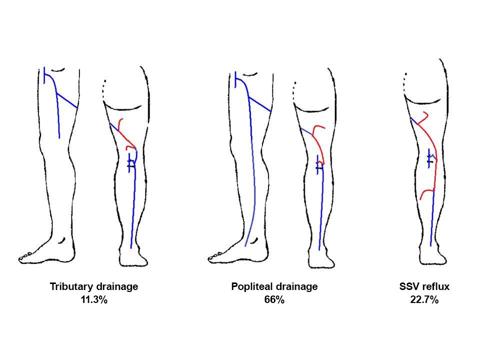
With relation to drainage of the reflux in the 150 incompetent veins, the majority (66%) drained reflux to the popliteal vein via the saphenopopliteal junction (SPJ); in 34 veins (22.7%), reflux was transferred to the SSV, and in 15 veins (11.3%) it was drained by a tributary vein in the thigh (Figure 3).
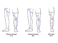
Diagram illustrating the drainage possibilities of reflux from the Giacomini vein. SSV = small saphenous vein.
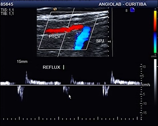
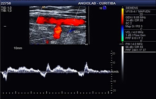
Figures 4 to 7 illustrate examples of reflux in the GV draining into the SPJ without compromising the SSV and of reflux in the SSV caused by an incompetent GV.
Ultrasound image of Giacomini vein with reflux. PTCV = Posterior thigh circumflex vein (Giacomimni Vein); SPJ = saphenopopliteal junction.
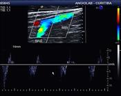
Ultrasound image of the small saphenous vein (same case as in Figure 4) without reflux, demonstrating that reflux in the Giacomini vein is drained via the saphenopopliteal junction. SSV = Small saphenous vein; SPJ = saphenopopliteal junction.
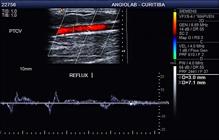
Ultrasound image of the Giacomini vein with reflux, with caliber of 3 mm and depth of 7.1 mm. PTCV = Posterior thigh circumflex vein (Giacomini Vein).
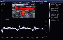
Ultrasound image of the small saphenous vein (same case as in Figure 6) with reflux, demonstrating that reflux in the Giacomini vein is transferred to the small saphenous vein. PTCV = Posterior thigh circumflex vein; SSV = small saphenous vein; SPJ = saphenopopliteal junction.
DISCUSSION
The definition of a GV is a little controversial in the literature. Carlo Giacomini’s original description, from 1873, lists eight different types. Type 1, the definition adopted in our study, is the most common (52.9%) and is described as an anastomotic branch between the SSV (terminating at the popliteal vein) and the GSV.5
In 2001, the International Union of Phlebology, with the support of the Federative International Committee on Anatomical Terminology, established new terminology for the superficial veins of the lower limbs, in which the intersaphenous vein corresponds to the Giacomini vein. Along the same lines, in 2002, the International Interdisciplinary Consensus Committee on Venous Anatomical Terminology stated that when the cranial continuation of the SSV communicates with the GSV (via the posterior thigh circumflex vein), it should be called the GV.6,7
The prevalence of GVs varies from 2.5% in a phlebographic study,8 through 2 to 86% in ultrasound assessments,9,10 and even as high as 95% in cadaveric studies.11-13
In our study, the prevalence observed with ultrasonography was 14.5% of a specific population of women referred for venous mapping for varicose veins surgery.
With relation to the caliber of the 150 GVs with reflux assessed in our study, diameters ranged from 1.5 to 7.8 mm (mean of 2.7 mm) and mean depth was 11.5 mm, varying from 4 to 25 mm. Delis et al. reported a similar mean caliber (2.68 mm) to our study, with a range of 0.2 to 7.7 mm.14
Considering that the GV has both subfascial and subcutaneous segments, we believe that the depth of a GV with reflux is relevant information in the context of possible endovascular treatment. This information is not available elsewhere in the literature.
Six different SSV reflux patterns are described, including perijunctional, proximal, segmental, multisegmental (with and without SPJ involvement), and distal reflux.15,16
Among these patterns, the perijunctional type (reflux in the SSV below the SPJ) is directly related to the GV. In this pattern, the incompetent GV transfers reflux to the SSV below the SPJ, making it incompetent and potentially originating varicose veins in the leg. In these cases, failure to treat the GV may lead to relapse in the future.
In the literature, rates of reflux in the GV detected by VUSD vary from 2 to 19%.17,18 Our study identified reflux in 29.4% of GVs, which was segmental in the great majority (91.3%) and was diffuse in just 8.7%. In the majority of cases (66%), reflux is drained by the SPJ, without causing reflux in the SSV. The higher incidence of reflux in our study may be related to the fact that GV assessment was performed routinely in our vascular laboratory.
Among the incompetent GVs, 22.7% transferred reflux to the SSV, constituting the perijunctional reflux pattern. Veltman et al. observed 10% perijunctional reflux with VUSD in 1,142 lower limbs, caused by Giacomini veins or cranial continuation of the SSV.19
Considering the 29% incidence of reflux in the GV vein and the possibility of a perijunctional reflux pattern in 23% of SSVs, we believe that preoperative mapping of lower limbs varicose veins should routinely include proactive screening for the GV and assessment of GVs when found.
In cases with GV reflux, the caliber and height of the connections with the GSV and SSV should be measured. In our study they had mean height (above the sole of the foot) of 71 cm for the GSV and 50.5 cm for the SSV.
In conclusion, the incidence of GV presence in this study was 14%. One third of these veins had reflux, which was segmental in the majority of cases. In 23% of GV, reflux was transferred to the SSV, which makes assessment relevant during preoperative mapping of varicose veins and for planning surgery.
-
How to cite: Engelhorn CA, Engelhorn ALDV, Oliveira ES, Macedo JM, Anizelli LB, Mendonça MLO. The role of the giacomini vein in preoperative mapping of lower limb varicose veins. J Vasc Bras. 2024;23:e20240058. https://doi.org/10.1590/1677-5449.202400582
-
Financial support: None.
-
The study was carried out at Angiolab Laboratório Vascular, Curitiba, PR, Brazil.
References
-
1 Labropoulos N, Delis K, Nicolaides NA, Leon M, Ramaswami G, Volteas N. The role of distribution and anatomic extent of reflux in the development of signs and symptoms in chronic venous insufficiency. J Vasc Surg. 1996;23(3):504-10. http://doi.org/10.1016/S0741-5214(96)80018-8 PMid:8601895.
» http://doi.org/10.1016/S0741-5214(96)80018-8 -
2 Romualdo AP, Bastos RM, Fatio M, et al. Extensão cranial da veia safena parva: quando o fluxo caudal é normal. J Vasc Bras. 2009;8(2):166-1703. http://doi.org/10.1590/S1677-54492009000200011
» http://doi.org/10.1590/S1677-54492009000200011 -
3 Gloviczki P, Lawrence PF, Wasan SM, et al. The 2022 Society for Vascular Surgery, American Venous Forum, and American Vein and Lymphatic Society clinical practice guidelines for the management of varicose veins of the lower extremities. Part I. Duplex Scanning and Treatment of Superficial Truncal Reflux: Endorsed by the Society for Vascular Medicine and the International Union of Phlebology. J Vasc Surg Venous Lymphat Disord. 2023;11(2):231-261.e6. http://doi.org/10.1016/j.jvsv.2022.09.004 PMid:36326210.
» http://doi.org/10.1016/j.jvsv.2022.09.004 -
4 Labropoulos N, Tiongson J, Pryor L, et al. Definition of venous reflux in lower-extremity veins. J Vasc Surg. 2003;38(4):793-8. http://doi.org/10.1016/S0741-5214(03)00424-5 PMid:14560232.
» http://doi.org/10.1016/S0741-5214(03)00424-5 - 5 Natsis K, Paraskevas G, Lazaridis N, Sofidis G, Piagkou M. Giacomini vein: thigh extension of the small saphenous vein - report of two cases and review of the literature. Hippokratia. 2015;19(3):263-5. PMid:27418788.
- 6 Whitmore I, editor. Terminologia Anatomica: International Anatomical Terminology. Stuttgart: Thieme, Federative Committee on Anatomical Terminology; 1998.
-
7 Caggiati A, Bergan JJ, Gloviczki P, Jantet G, Wendell-Smith CP, Partsch H. Nomenclature of the veins of the lower limbs: an international interdisciplinary consensus statement. J Vasc Surg. 2002;36(2):416-22. http://doi.org/10.1067/mva.2002.125847 PMid:12170230.
» http://doi.org/10.1067/mva.2002.125847 - 8 Zierau UT, Kullmer A, Kunkel HP. Stripping the Giacomini vein--pathophysiologic necessity or phlebosurgical games? Vasa. 1996;25(2):142-7. PMid:8659216.
-
9 Farrah J, Saharay M, Georgiannos SN, Scurr JH, Smith PD. Variable venous anatomy of the popliteal fossa demonstrated by duplex scanning. Dermatol Surg. 1998;24(8):901-3. http://doi.org/10.1111/j.1524-4725.1998.tb04271.x PMid:9723058.
» http://doi.org/10.1111/j.1524-4725.1998.tb04271.x - 10 Georgiev M, Myers KA, Belcaro G, St May’s Society of Vascular Fellows. Giacomini’s observations on the superficial veins of the abdominal limb and principally the external saphenous. Int Angiol. 2001;20(3):225-33. PMid:11573057.
- 11 Giacomini C. Osservazioni anatomiche per servire allo Studio della circolazione venosa delle estremita inferiori. Parte I: Delle vene superficiali dell’arto addominale e principalmente Dell asaphena esterna. Giornale della Reale Accademia di Medicina di Torino. 1873;14:109-36.
- 12 Prakash JM, Nishanth R, Kalyani P, Ramya PT, Singh G. A review of literature along with a cadaveric study of the prevalence of the Giacomini vein (the thigh extension of the small saphenous vein) in the Indian population. Rom J Morphol Embryol. 2008;49(4):537-9. PMid:19050803.
- 13 Stolic E. Posterior subaponeurotic vein of the thigh. C R Assoc Anat. 1970;149:1016-26. PMid:5525227.
-
14 Delis KT, Knaggs ALI, Khodabakhsh P. Prevalence, anatomic patterns, valvular competence, and clinical significance of the Giacomini vein. J Vasc Surg. 2004;40(6):1174-83. http://doi.org/10.1016/j.jvs.2004.09.019 PMid:15622372.
» http://doi.org/10.1016/j.jvs.2004.09.019 -
15 Engelhorn CA, Engelhorn AL, Cassou MF, Salles-Cunha SX. Patterns of saphenous reflux in women with primary varicose veins. J Vasc Surg. 2005;41(4):645-51. http://doi.org/10.1016/j.jvs.2004.12.051 PMid:15874929.
» http://doi.org/10.1016/j.jvs.2004.12.051 - 16 Engelhorn AL, Engelhorn CA, de Morais D Fo, Barros FS, Coelho NA. Ultrassonografia vascular na avaliação de varizes dos membros inferiores. In: Engelhorn CA, Engelhorn ALDV, editores. Guia Prático de Ultrassonografia Vascular. 4. ed. Rio de Janeiro: DiLivros Editora; 2019. p. 555-79.
-
17 Labropoulos N, Leon M, Nicolaides AN, Giannoukas AD, Volteas N, Chan P. Superficial venous insufficiency: correlation of anatomic extent of reflux with clinical symptoms and signs. J Vasc Surg. 1994;20(6):953-8. http://doi.org/10.1016/0741-5214(94)90233-X PMid:7990191.
» http://doi.org/10.1016/0741-5214(94)90233-X -
18 Labropoulos N, Giannoukas AD, Delis K, et al. The impact of isolated saphenous vein incompetence on clinical signs and symptoms of chronic venous disease. J Vasc Surg. 2000;32(5):954-60. http://doi.org/10.1067/mva.2000.110349 PMid:11054227.
» http://doi.org/10.1067/mva.2000.110349 -
19 Veltman HJ, Zollmann P, Zollmann M, et al. Reflux origin of the insufficient small saphenous vein by duplex ultrasound determination and consequences for therapy considering the saphenopopliteal junction type. J Vasc Surg Venous Lymphat Disord. 2023;11(6):1114-21. http://doi.org/10.1016/j.jvsv.2023.07.004 PMid:37442273.
» http://doi.org/10.1016/j.jvsv.2023.07.004