 Phenolic compounds involved in the inhibition of root and seedling regeneration in seeds of Eugenia pyriformis Cambess.
ABSTRACT:
Eugenia pyriformis produces recalcitrant and monoembryonic seeds with high regenerative capacity, although the simultaneous formation of multiple roots is inhibited by chemical self-control. This study analyzed the phytochemical profile at different stages of development to investigate the regulatory role of phenols and tannins. Ethanolic extracts were purified by Solid Phase Extraction (SPE-C18) and analyzed by High-Performance Liquid Chromatography with Photodiode Array Detection (HPLC-PDA). The profiles revealed the predominance of hydrolyzable tannins and phenolic acids, notably gallic and ellagic acids. There was a progressive increase of gallic acid in the cotyledons during germination, with a peak at the seedling stage. Gallic acid acts as a chemical marker in the suppression of new meristems, preventing the protrusion of secondary roots while the primary structure is established, preserving the energy reserves of the seed.
Index terms:
gallic acid; germination self-control; recalcitrant seeds
RESUMO:
Eugenia pyriformis produz sementes recalcitrantes e monoembriônicas com alta capacidade regenerativa, embora a formação simultânea de múltiplas raízes seja inibida por autocontrole químico. Este estudo analisou o perfil fitoquímico em diferentes estádios de desenvolvimento para investigar o papel regulador de fenóis e taninos. Extratos etanólicos foram purificados por Extração em Fase Sólida (EFS-C18) e analisados por Cromatografia Líquida de Alta Eficiência com Detector de Arranjo de Fotodiodos (HPLC-PDA). Os perfis revelaram o predomínio de taninos hidrolisáveis e ácidos fenólicos, notadamente os ácidos gálico e elágico. Ocorreu um incremento progressivo de ácido gálico nos cotilédones durante a germinação, com pico no estádio de plântula. O ácido gálico atua como marcador químico na supressão de novos meristemas, impedindo a protrusão de raízes secundárias enquanto a estrutura primária se estabelece, preservando as reservas energéticas da semente.
Termos para indexação:
ácido gálico; autocontrole da germinação; sementes recalcitrantes
INTRODUCTION
Seeds of Eugenia species (Myrtaceae) have been the subject of attention for their capacity to regenerate roots and seedlings from cotyledons when the first germination is lost (Alonso and Barbedo, 2020; Delgado et al., 2022; Amorim et al., 2023; Alonso et al., 2024). Such capacity is considered an essential survival strategy for these seeds, which have recalcitrant behavior (sensitive to desiccation and with active metabolism), which limits their longevity in the seed bank (Barbedo, 2018).
In addition, these seeds can initiate the formation of several roots or seedlings from a single seed, despite being monoembryonic, but this rarely occurs simultaneously (Amador and Barbedo, 2015; Prataviera et al., 2015; Alonso et al., 2019). Eugenia seeds show a unique regeneration behavior after damage to the embryonic axis, allowing multiple germination attempts (Barbedo, 2018). However, it is observed that, once the first root is established, the development of new structures is suppressed, configuring a process of germination self-control. This phenomenon, physiologically characterized as a chemical suppression of the development of new meristems (Amador and Barbedo, 2015), is evidenced by the formation of new roots only in fractionated seeds, where perivascular cells of the cotyledons are stimulated (Delgado et al., 2022). However, such regeneration does not occur in intact seeds (Amador and Barbedo, 2011), reinforcing the existence of an endogenous system that inhibits new protrusions during the development of primary structures.
Phenolic compounds, such as phenolic acids and tannins, are widely recognized as endogenous inhibitors of germination and seed vigor in several botanical families (Bewley et al., 2013). This metabolic interference occurs in a multifactorial way, involving everything from the modulation of phytohormonal homeostasis to direct enzymatic restriction in root development (Nurjannah et al., 2015; Nakabayashi et al., 2022). Such evidence suggests that the phenolic constituents of E. pyriformis can act as central chemical markers, regulating the processes of self-control and regeneration observed in the species.
The genus Eugenia (Myrtaceae) has high phytochemical relevance, with seeds rich in flavonoids and tannins, such as gallic and ellagic acids, which act in oxidative protection and metabolic modulation (Nicácio et al., 2017; Saber et al., 2023). Although species such as E. uniflora and E. stipitata are well characterized, seeds of E. pyriformis (E. pyriformis) remain chemically underexploited (Nogueira et al., 2022). The choice of E. pyriformis is based on the robust volume of physiological and anatomical information already consolidated for the species, allowing an accurate interpretation of the data without the need for complementary biological studies. Thus, investigating its phenolic profile is essential to elucidate the chemical mechanisms that regulate germination in recalcitrant seeds (Silva and Kempka, 2023; Honaiser et al., 2025).
The complexity of the Eugenia seed matrix, rich in sugars and polymers, requires purification via Solid Phase Extraction (SPE) to ensure the selectivity of the analytes (Oh and Lee, 2014). Integrated with SPE, high-performance liquid chromatography with photodiode array detection (HPLC-PDA) allows fingerprints to be obtained and the unambiguous identification of compounds such as gallic and ellagic acids via UV absorption spectra (Lyu et al., 2019; Pathania, 2023). This approach ensures the resolution needed to monitor phenolic markers at the physiological stages of E. pyriformis (Feenstra et al., 2017).
Despite the regenerative potential of E. pyriformis seeds, there remains a gap in knowledge about the dynamics of secondary metabolites during their maturation and how the fluctuation of these compounds modulates the germination process. In light of this context, the present study aimed to characterize the phytochemical profile of hydroethanolic extracts of E. pyriformis seeds at different stages of development, using SPE and HPLC-PDA approaches. This investigation seeks to provide the basis to understand the role of phenolic compounds as endogenous regulators of germination, contributing to the advancement of knowledge about the chemical mechanisms of self-control of seeds of this genus and the basis for understanding their high capacity for embryo regeneration.
MATERIAL AND METHODS
Ripe E. pyriformis fruits were collected at the Agronomic Institute of Campinas (22°52’S, 47°04’W, 645 m; Cwa), and their seeds were manually extracted in the laboratory. The seeds were placed to germinate in polyethylene trays, containing vermiculite as substrate, kept in a germination chamber at 25 °C under a 12-hour photoperiod (Amador and Barbedo, 2011).
To obtain the extracts, the samples were standardized into three distinct stages: (i) ungerminated seeds, called intact seeds (IS); (ii) seeds germinated until the formation of the primary root, fractionated into cotyledons (CSR) and primary roots (RSR); and (iii) established seedlings, subdivided into cotyledons (CSS), roots (RSS) and shoots (SHSS). The morphological aspect of these stages and the delimitation of the fractions collected are illustrated in Figure 1. After collection, the materials were dried in an oven with forced air circulation at 44 °C for 48 hours, ground in a knife mill, homogenized, weighed, packed and stored in a dry place protected from light, thus maintaining the stabilization of the constituents, a procedure that ensures enzymatic inactivation and stabilization of the phenolic constituents (Khoddami et al., 2013).
Phenolic compounds were extracted by percolation using a solution of ethanol and water in the proportion of 7:3 (EtOH:H2O) (v/v), following the methodology of Prista et al. (1995). Cold percolation was chosen to preserve the integrity of the thermolabile tannins, avoiding the degradation that could occur in methods such as Soxhlet (Azwanida, 2015). Leachates were concentrated in a rotary evaporator under reduced pressure (50 °C), lyophilized and stored in amber flasks protected from light (Khoddami et al., 2013).
In order to mitigate matrix interference and optimize chromatographic resolution, the extracts were subjected to solid-phase extraction (SPE) using C18 reversed-phase cartridges (500 mg, 6 mL, Supelco®), following purification protocols for phenolic analytes (Luthria et al., 2006). Conditioning consisted of the sequential elution of 15 mL of methanol (MeOH) and 10 mL of water (H2O). Previously solubilized aliquots of 10 mg of extract were applied to the cartridges for selective fractionation of the analytes. The higher-polarity fraction was obtained by elution with MeOH:H2O (2:8, v/v), while the lower-polarity fraction was eluted with 100% MeOH. The resulting eluates were concentrated, filtered through a 0.45 μm membrane and subjected to HPLC-PDA analysis (Luthria et al., 2006).
The biochemical complexity of E. pyriformis required a High-Performance Liquid Chromatography (HPLC) system with quaternary gradient (PU-2089S Plus, Jasco®), ideal for polyphenol separation in plant matrices (Stalikas, 2007). The detection used a photodiode array detector (PDA; MD-2015 Plus, Jasco®; 200-900 nm scan) for spectral authentication in ultraviolet. The system featured a column oven (CO-2060 Plus, Jasco®) for thermal stability and an automatic injector (AS-2055, Jasco®) for accuracy in 50 μL injections, operated via ChromPass software (v. 1.8.1.6).
The analytical strategy involved combining different stationary phases and chromatographic scales to optimize selectivity and isolate markers. Selectivity was evaluated comparatively between the analytical columns Luna C18(2) (250 × 4.6 mm i.d., 5 μm, 100 Å, Phenomenex®) and Synergi Hydro-RP (250 × 4.6 mm i.d., 4 μm, Phenomenex®), the latter being selected for the development of calibration curves due to the higher retention of polar analytes. For preparative purposes, the method was transposed to the semi-preparative (Luna C18(2), 250 × 10 mm i.d., 5 μm) and preparative (Dynamax RP18, 250 × 41.4 mm i.d., 8 μm, Varian®) scales, aiming at the fractionation of major constituents. Separations were conducted under a flow rate of 1.0 mL.min⁻¹ and a temperature of 40 °C, using a linear gradient elution system composed of MeOH + 0.1% formic acid (v/v) (Phase A) and H2O + 0.1% formic acid (v/v) (Phase B). The elution profile started at 5% A (0 min), reaching 100% A at 60 min, with isocratic maintenance for 5 min and rebalancing of the column for 10 min under the initial condition. The 20 μL injections were monitored at λ = 254 nm, with the aid of a corresponding guard column (4 × 3 mm i.d.) (Snyder et al., 2012).
For chromatographic characterization, 10-mg aliquots of extract were solubilized in methanol:water (1:1, v/v) and filtered through PTFE membrane (0.45 μm). Based on previous screenings, gallic and ellagic acids (Sigma-Aldrich®) were the standards for identification and quantification (Ignat et al., 2011). The analyses and curves were recorded in a PDA detector at 254 nm (1 nm step), with injections via the Rheodyne® 7125 valve. The volumes were organized by loops of 20 μL (analytical), 100 μL (semi-preparative) and 1 mL (preparative). Methanol and ethanol A.R. (Sigma-Aldrich®), HPLC-grade methanol and formic acid (Tedia® and Synth®), and ultrapure water (Milli-Q®, Millipore®) were used.
RESULTS AND DISCUSSION
The analysis of E. pyriformis seed extracts at the initial stage (IS, Figure 1) revealed matrices of high complexity, with low resolution of peaks and presence of extended bands (humps), characteristic of the occurrence of condensed tannins and phenolic polymers (Figure 2). Purification via SPE-C18 eliminated these interferents, selectively increasing the chromatographic resolution: elution with 100% MeOH (Figure 2D) concentrated constituents of lower polarity, while the MeOH:H2O system (2:8, v/v) (Figure 2B) isolated the polar fraction. In both eluates, there was suppression of broad bands, resulting in profiles with discrete peaks (Figure 2).

Stages of germination development and fractionation of tissues of Eugenia pyriformis for phytochemical analysis. IS: intact seeds, without germination; CSR: cotyledons, after germination and removal of the primary root; RSR: primary roots produced by intact seeds, after removal of cotyledon; SHSS: shoot of seeds that produced seedlings, after removal of cotyledons and roots; CSS: cotyledons of seeds that produced seedlings, after removal of roots and shoots; RSS: roots of seeds that produced seedlings, after removal of cotyledons and shoots.

Analytical chromatograms (HPLC-PDA, 210 nm) of the fractions obtained by SPE-C18 from the hydroethanolic extract (70% EtOH) of the IS fraction of Eugenia pyriformis seeds. Elution eluents: A: water (100%); B: methanol:water (2:8, v/v); C: methanol:water (1:1, v/v); and D: methanol (100%). x-axis: Time (min); y-axis: Absorbance (mAU).
In the 100% MeOH eluate (Figure 2D), a major peak was identified at retention time (RT) = 28.53 min, corresponding to 81.49% of the total area, confirmed as ellagic acid by the overlapping of the UV spectrum (λ max = 254 nm) with the standard (Figures 3 and 4). In the MeOH:H2O eluate (2:8, v/v) (Figure 2B), the higher polarity of the fraction resulted in the predominance of early signals, especially gallic acid (RT = 5.56 min), identified via coelution and spectral profiles (Figures 5 and 6). In both eluates, the other detected signals showed low relative contributions, not allowing identification.

Analytical chromatogram obtained by HPLC-PDA (254 nm) of the methanol fraction (100% MeOH) obtained by SPE-C18 from the extract of intact seeds (IS) of Eugenia pyriformis (injection: 500 μg mL⁻¹). Major peak at RT = 28.53 min (81.49% of the area), identified as ellagic acid by comparison of retention time and UV spectrum with the standard. x-axis: RT (min); y-axis: absorbance (mAU).

UV absorption spectra (200-700 nm) obtained by HPLC-PDA: standard of ellagic acid (black line) and methanol eluate (SPE-C18, 100% MeOH) from the extract of intact seeds (IS) of Eugenia pyriformis (red line). Maximum values are observed at 254 and 362 nm, compatible with ellagic acid. X-axis: Wavelength (nm); y-axis: Absorbance (mAU).

Analytical chromatogram obtained by HPLC-PDA of the MeOH:H₂O eluate (2:8, v/v) (SPE-C18) from the extract of intact seeds (IS) of Eugenia pyriformis. Traces extracted at λ = 254 nm (red) and λ = 596 nm (blue); the signal at 596 nm is shown as a reference and does not represent relevant absorption for phenolic acids. x-axis: time (min); Y-axis: absorbance (mAU).
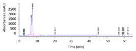
Confirmation of gallic acid by HPLC-PDA. Analytical chromatogram of the standard (magenta line) and the MeOH:H₂O eluate (2:8, v/v) of the extract of intact seeds (IS) of Eugenia pyriformis (blue line), both monitored at 254 nm, showing coincidence of retention time (RT ≈ 5.56 min) and the characteristic UV spectrum. x-axis: time (min); Y-axis: absorbance (mAU).
The chromatographic profiles revealed ellagic acid as the predominant constituent in the lower-polarity fractions, while gallic acid was consolidated as a recurrent marker in polar eluates. In the literature, both compounds are associated with the negative modulation of germination through the potential inhibition of hydrolytic enzymes, such as α-amylase and α-glucosidase (Kam et al., 2013; Muscolo et al., 2014). The presence of these phenolic acids in E. pyriformis suggests, therefore, a biochemical basis for the control of reserve mobilization, an inhibition mechanism already reported in other ellagitannin-rich matrices (Kam et al., 2013).
Relative quantification of phenolic markers was performed via area normalization in intact seeds (IS) and in developing tissues (Figure 7). In seeds at early stage (IS), ellagic acid (100% MeOH eluate, Figure 2D) represented 81.49% of the total chromatographic area, while gallic acid (MeOH:H2O 2:8 eluate, Figure 2B) had a minority, but reproducible contribution among the biological replications. With the progression of germination (root emergence stage), there was a reduction in the relative levels of ellagic acid in the cotyledons (CSR samples), concomitantly with a significant increase in the levels of gallic acid (Figure 7). This profile suggests an intense mobilization of phenolic derivatives during the differentiation of E. pyriformis germination tissues.

HPLC-PDA estimation of ellagic acid (A) and gallic acid (B) contents of non-germinating and germinating seeds of Eugenia pyriformis at different stages of development. IS: Intact seeds, without germination; CSR: cotyledons, after germination and removal of the primary root; RSR: primary roots produced by intact seeds, after removal of cotyledon; SHSS: shoot of seeds that produced seedlings, after removal of cotyledons and roots; CSS: cotyledons of seeds that produced seedlings, after removal of roots and shoots; RSS: roots of seeds that produced seedlings, after removal of cotyledons and shoots.
The detection and quantification of gallic and ellagic acids in Eugenia seeds are corroborated by studies in related species, such as E. punicifolia and E. involucrata (Nicácio et al., 2017; Silva et al., 2023). The effectiveness in isolating these acids is attributed to the use of polar solvents, a consolidated method for the extraction of phenolics in complex plant matrices (Zhang et al., 2014; Mohamed et al., 2020). Thus, the expected chromatographic profile for E. pyriformis is in line with the standards reported for the genus, ratifying the use of HPLC-PDA techniques for the characterization of these constituents.
With the advancement of seedling development, gallic acid tended to accumulate in the cotyledons, a profile not observed for ellagic acid. Qualitatively, a higher intensity of gallic acid signals is observed in the root axes (RSS) and in the shoots (SHSS) of the seedlings compared to seeds in the initial stage of root emergence. In contrast, ellagic acid showed signals of low magnitude in roots and shoots in both stages of development (Figure 7). These profiles suggest that germination progression and seedling establishment coincide with increased detection of gallic acid. This phenomenon may be associated with the regulation of subsequent germination in seeds of Eugenia species, as discussed in studies on inhibition by endogenous allelochemicals (Amador and Barbedo, 2015; Alonso et al., 2019; Alonso and Barbedo, 2020).
The predominance of gallic and ellagic acid derivatives in Eugenia (Shakeri et al., 2018; Evtyugin et al., 2020) corroborates the profiles observed in E. pyriformis eluates. The low aqueous solubility of ellagic acid (García-Niño and Zazueta, 2015) justifies its majority detection in cotyledons and the low magnitude of its signals in the root axes and shoots (Figure 7), suggesting a restricted mobility that can act as a local chemical barrier. On the other hand, the greater water solubility of gallic acid favors its diffusion, explaining the increase in its detection in developing tissues (RSS and SHSS) as germination progresses.
The relative quantification data (Figure 7 B) reveal an aspect of E. pyriformis metabolism: the transition from the intact seed (IS) to the root emergence (CSR) stage is marked by a robust increment in gallic acid levels in the cotyledons. This immediate accumulation after protrusion of the primary root suggests that the seed intensifies the production of chemical markers to establish the suppression of the development of new meristems, ensuring that the energy investment is directed exclusively to the already active meristem.
The distribution dynamics of these phenolics seems to influence the morphological development following root protrusion. The higher water solubility and diffusibility of gallic acid favor its detection in growing tissues, where it can modulate auxin signaling and root elongation (Xu et al., 2024), while the low solubility of ellagic acid (García-Niño and Zazueta, 2015) reinforces the existence of a chemical barrier located in the cotyledons. In short, these acids act as central regulators of germination self-control in E. pyriformis, combining the suppression of reserve mobilization (Kam et al., 2013; Muscolo et al., 2014) to the modulation of hormonal and redox signaling pathways (Xu et al., 2024), essential for the staggering of germination processes and for the regeneration strategy of the species (Shakeri et al., 2018).
The observed profiles suggest that the germination progression and seedling establishment coincide with the increase of inhibitory compounds, such as gallic acid. This explains why, although the E. pyriformis embryo has plasticity to regenerate meristems (Barbedo, 2018), this ability is suppressed as soon as the first root is established. Therefore, the seed maintains its regeneration potential only as long as the chemical dominance system exerted by the growing axis is not consolidated, confirming that the self-control of germination has a specific phytochemical basis (Amador and Barbedo, 2015).
Eugenia pyriformis seeds are sensitive to desiccation (recalcitrant), as are those of other species of the genus. Therefore, the propagation strategies of these seeds, unlike species with desiccation-tolerant seeds (orthodox), are not based on the formation of seed banks in the soil for long periods (Amorim and Barbedo, 2020, and contained references). Among the propagation strategies used by species with recalcitrant seeds, the most frequent are germination immediately after their dispersal and the continuous and regular production and dispersal of seeds in the field (Barbedo, 2018). As demonstrated in previous studies, seeds of Eugenia species, despite being recalcitrant, have characteristics that allow their germination and colonization of areas for relatively long periods (Amorim and Barbedo, 2020). To this end, they have characteristics favorable to this strategy, such as the large amount of reserves in the cotyledons, the ability to regenerate seedlings successively from the same seed (Alonso and Barbedo, 2020; Delgado et al., 2022), resistance to water loss, capacity to germinate even under water deficit (Inocente and Barbedo, 2019; 2021), and germination self-control, preventing simultaneous germination and unnecessary expenditure of reserves (Amador and Barbedo, 2015). Among these characteristics, the system that avoids these simultaneous germination events had not yet been demonstrated. In the present study, it was demonstrated that this system is based on the concentration of two important phenolic compounds, gallic and ellagic acids, especially the former, whose concentration increase in the cotyledons when germination and the formation of new seedlings begin, inhibiting the formation of new roots or seedlings.
CONCLUSIONS
Phytochemical characterization identified gallic and ellagic acids as the major markers in the regulation of E. pyriformis germination. While ellagic acid acts as a stable and major chemical barrier in reserve tissues, the significant increase of gallic acid in cotyledons after germination (CSR) characterizes it as dynamically responsible for the self-control. Together, the stability of ellagic acid and the mobilization of gallic acid underlie the processes of germination and regeneration inhibition in E. pyriformis seeds, allowing the seed to temporarily suppress new meristems in favor of the established seedling. This phenolic dynamic explains, at the molecular level, the survival strategy and the sequential regeneration typical of this recalcitrant species.
ACKNOWLEDGMENTS
In memoriam
This article is dedicated to the memory of Dr. Luce Maria Brandão Torres († December/2024), whose supervision and intellectual generosity were fundamental to carrying out this study.
We thank the Agronomic Institute of Campinas (collections), the LQPN-FC/Bauru (HPLC-PDA) and the Seed Laboratory-IPA (germination tests). This study was funded by the Coordination for the Improvement of Higher Education Personnel - Brazil (CAPES), through a doctoral scholarship (PPG-Botany, IB-Botucatu, UNESP).
REFERENCES
-
ALONSO, C.R.; ASPERTI, L.M.; GUARDIA, M.C.; BARBEDO, C.J. Cutting and regeneration of roots and seedlings from seeds of Eugenia candolleana DC. at different maturity stages. Journal of Seed Science, v.41, n.2, p.160-167, 2019. https://doi.org/10.1590/2317-1545v41n2200571
» https://doi.org/https://doi.org/10.1590/2317-1545v41n2200571 -
ALONSO, C.R.; BARBEDO, C.J. Germinações sucessivas em sementes de Eugenia spp. Hoehnea, v.47, e412019, 2020. https://doi.org/10.1590/2236-8906-41/2019
» https://doi.org/https://doi.org/10.1590/2236-8906-41/2019 -
ALONSO, C.R.; RIBEIRO, M.I.; GUARDIA, M.C.; BARBEDO, C.J. Regeneration of roots and shoots as a propagation strategy in Eugenia candolleana DC. (Myrtaceae) seeds. Journal of Seed Science , v.46, e202446003, 2024. https://doi.org/10.1590/2317-1545v46275827
» https://doi.org/https://doi.org/10.1590/2317-1545v46275827 -
AMADOR, T.S.; BARBEDO, C.J. Germination inhibits the growth of new roots and seedlings in Eugenia uniflora and Eugenia brasiliensis Journal of Seed Science , v.37, n.3, p.241-247, 2015. https://doi.org/10.1590/2317-1545v37n3150595
» https://doi.org/https://doi.org/10.1590/2317-1545v37n3150595 -
AMADOR, T.S.; BARBEDO, C.J. Potencial de inibição da regeneração de raízes e plântulas em sementes germinantes de Eugenia pyriformis Pesquisa Agropecuária Brasileira, v.46, n.8, p.814-821, 2011. https://doi.org/10.1590/S0100-204X2011000800005
» https://doi.org/https://doi.org/10.1590/S0100-204X2011000800005 -
AMORIM, I.P.; SILVA, J.P.N.; BARBEDO, C.J. Germination and regeneration of Eugenia involucrata (Myrtaceae) seeds correlated with reactive oxygen species. Journal of Seed Science , v.45, e202345021, 2023. https://doi.org/10.1590/2317-1545v45262916
» https://doi.org/https://doi.org/10.1590/2317-1545v45262916 -
AMORIM, I.P.; SILVA, J.P.N.; BARBEDO, C.J. As sementes de Eugenia spp. (Myrtaceae) e seus novos conceitos sobre propagação. Hoehnea , v.47, e292020, 2020. https://doi.org/10.1590/2236-8906-29/2020
» https://doi.org/https://doi.org/10.1590/2236-8906-29/2020 -
AZWANIDA, N.N. A review on the extraction methods use in medicinal plants, principle, strength and limitation. Medicinal & Aromatic Plants, v.4, n.3, p.1-6, 2015. https://www.longdom.org/open-access/a-review-on-the-extraction-methods-use-in-medicinal-plants-principle-strength-and-limitation-2167-0412-1000196.pdf
» https://www.longdom.org/open-access/a-review-on-the-extraction-methods-use-in-medicinal-plants-principle-strength-and-limitation-2167-0412-1000196.pdf -
BARBEDO, C.J. A new approach towards the so-called recalcitrant seeds. Journal of Seed Science , v.40, n.3, p.221-236, 2018. http://dx.doi.org/10.1590/2317-1545v40n3207201
» https://doi.org/http://dx.doi.org/10.1590/2317-1545v40n3207201 -
BEWLEY, J.D.; BRADFORD, K.J.; HILLHORST, H.W.M.; NONOGAKI, H. Seeds: Physiology of development, germination and dormancy 3rd ed. New York: Springer. 392 p. 2013. https://doi.org/10.1007/978-1-4614-4693-4
» https://doi.org/https://doi.org/10.1007/978-1-4614-4693-4 -
DELGADO, L.F.; TEIXEIRA, S.P.; CARMELLO-GUERREIRO, S.M.; BARBEDO, C.J. The origin of new roots from cut seeds of Eugenia species. Journal of Seed Science , v.44, e202244260855, 2022. https://doi.org/10.1590/2317-1545v44260855
» https://doi.org/https://doi.org/10.1590/2317-1545v44260855 -
EVTYUGIN, D.D.; MAGINA, S.; EVTUGUIN, D.V. Recent advances in the production and applications of ellagic acid and its derivatives: a review. Molecules, v.25, n.12, 2745, 2020. https://doi.org/10.3390/molecules25122745
» https://doi.org/https://doi.org/10.3390/molecules25122745 -
FEENSTRA, A.D.; ALEXANDER, L.E.; SONG, Z.; KORTE, A.R.; YANDEAU-NELSON, M.D.; NIKOLAU, B.J.; LEE, Y.J. Spatial mapping and profiling of metabolite distributions during germination. Plant Physiology, v.174, n.4, p.2532-2548, 2017. https://doi.org/10.1104/PP.17.00652
» https://doi.org/https://doi.org/10.1104/PP.17.00652 -
GARCÍA-NIÑO, W.R.; ZAZUETA, C. Ellagic acid: pharmacological activities and molecular mechanisms involved in liver protection. Pharmacological Research, v.97, p.84-103, 2015. https://doi.org/10.1016/j.phrs.2015.04.005
» https://doi.org/https://doi.org/10.1016/j.phrs.2015.04.005 -
HONAISER, T.C.; SEHN, A.L.; MOMBACH, H.C.; SEHN, K.F.; ARNOLD, L.; FABIANE, K.C.; ARCARI, S.G. Effects of in vitro digestion on the phenolic profile and antioxidant capacity of the brazilian native fruit Eugenia involucrata DC. ACS Food Science & Technology, 2025. https://doi.org/10.1021/acsfoodscitech.5c00540
» https://doi.org/https://doi.org/10.1021/acsfoodscitech.5c00540 -
IGNAT, I.; VOLF, I.; POPA, V.I. A critical review of methods for characterisation of polyphenols in fruits and vegetables. Food Chemistry, v.126, n.4, p.1821-1835, 2011. https://doi.org/10.1016/j.foodchem.2010.12.026
» https://doi.org/https://doi.org/10.1016/j.foodchem.2010.12.026 -
INOCENTE, M.C.; BARBEDO, C.J. Germination of Eugenia brasiliensis, E. involucrata, E. pyriformis, and E. uniflora (Myrtaceae) under water-deficit conditions. Journal of Seed Science , v.41, p.76-85, 2019. https://doi.org/10.1590/2317-1545v41n1212109
» https://doi.org/https://doi.org/10.1590/2317-1545v41n1212109 -
INOCENTE, M.C.; BARBEDO, C.J. Regeneration of roots and seedlings from Eugenia involucrata seeds under water deficit conditions. Journal of Seed Science , v.43, e202143015, 2021. https://doi.org/10.1590/2317-1545v43248394
» https://doi.org/https://doi.org/10.1590/2317-1545v43248394 -
KAM, A.; LI, K.M.; RAZMOVSKI-NAUMOVSKI, V.; NAMMI, S.; SHI, J.; CHAN, K.; LI, G.-Q. A comparative study on the inhibitory effects of different parts and chemical constituents of pomegranate on α-amylase and α-glucosidase. Phytotherapy Research, v.27, n.11, p.1614-1620, 2013. https://doi.org/10.1002/ptr.4913
» https://doi.org/https://doi.org/10.1002/ptr.4913 -
KHODDAMI, A.; WILKES, M.A.; ROBERTS, T.H. Techniques for analysis of plant phenolic compounds. Molecules, v.18, p.2328-2375, 2013. https://doi.org/10.3390/molecules18022328
» https://doi.org/https://doi.org/10.3390/molecules18022328 - LYU, H.; XU, S.; CHEN, Y.; PAETZ, C. Secondary metabolites in seed development of Musella lasiocarpa Berichte aus dem Julius Kühn-Institut, v.204, p.121, 2019.
-
LUTHRIA, D.L.; MUKHOPADHYAY, P.N.; KRIZEK, D.T. Content of total phenolics and phenolic acids in tomato (Solanum lycopersicum L.) fruits as influenced by cultivar and solar UV radiation. Journal of Food Composition and Analysis, v.19, n.8, p.771-777, 2006. https://doi.org/10.1016/j.jfca.2006.04.005
» https://doi.org/https://doi.org/10.1016/j.jfca.2006.04.005 -
MOHAMED, I.T.A.; KHAN, W.; CHESTER, K.; MOHAMED, A.H.; AHMAD, S.; AYOUB, S.M.H. Simultaneous quantitative estimation of ellagic acid and gallic acid in Sudanese Solanum dubium seed by high performance thin layer chromatography (HPTLC). GSC Biological and Pharmaceutical Sciences, v.13, n.1, p.054-061, 2020. https://doi.org/10.30574/GSCBPS.2020.13.1.0311
» https://doi.org/https://doi.org/10.30574/GSCBPS.2020.13.1.0311 -
MENCIN, M.; MIKULIC-PETKOVSEK, M.; VEBERIC, R.; TERPINC, P. Development and optimisation of solid-phase extraction of extractable and bound phenolic acids in spelt (Triticum spelta L.) seeds. Antioxidants, v.10, n.7, p;1085, 2021. https://doi.org/10.3390/antiox10071085
» https://doi.org/https://doi.org/10.3390/antiox10071085 -
MUSCOLO, A.; SIDARI, M.; ANASTASI, U.; SANTONOCETO, C.; MAGGIO, A. Effect of PEG-induced drought stress on seed germination of four lentil genotypes. Journal of Plant Interactions, v.9, n.1, p.354-363, 2014. https://doi.org/10.1080/17429145.2013.835880
» https://doi.org/https://doi.org/10.1080/17429145.2013.835880 -
NAKABAYASHI, K.; WALKER, M.; IRWIN, D.; COHN, J.; GUIDA-ENGLISH, S.; GARCÍA, L.; PAVLOVIC, I.; NOVÁK, O.; TARKOWSKÁ, D.; STRNAD, M.; PÉREZ, M.; SEVILLE, A.; STOCK, D.; LEUBNER-METZGER, G. The phytotoxin Myrigalone A triggers a phased detoxification programme and inhibits Lepidium sativum seed germination via multiple mechanisms including interference with auxin homeostasis. International Journal of Molecular Sciences, v.23, n.9, p.4618, 2022. https://doi.org/10.3390/ijms23094618
» https://doi.org/https://doi.org/10.3390/ijms23094618 -
NICÁCIO, A.E.; ROTTA, E.M.; BOEING, J.S.; BARIZÃO, É.O.; KIMURA, E.; VISENTAINER, J.V.; MALDANER, L. Antioxidant activity and determination of phenolic compounds from Eugenia involucrata DC. fruits by UHPLC-MS/MS. Food Analytical Methods, v.10, n.8, p.2718-2728, 2017. https://doi.org/10.1007/S12161-017-0840-3
» https://doi.org/https://doi.org/10.1007/S12161-017-0840-3 -
NOGUEIRA, L.A.; FIGUEIREDO, Y.G.; RAMOS, A.L.C.C.; CORREIA, V.T.D.V.; NUNES, B.V.; RIBEIRO, L.V.; FRANCO, A.O.; FERREIRA, R.B.; SOUSA, I.; MOTA, J.; BATISTA-SANTOS, P.; ARAÚJO, R.L.B.; MELO, J.O.F. The presence of flavonoids in some products and fruits of the genus Eugenia: an integrative review. Frontiers in Food Science and Technology, v.2, 899492, 2022. https://doi.org/10.3389/frfst.2022.899492
» https://doi.org/https://doi.org/10.3389/frfst.2022.899492 - NURJANNAH, U.; YERNISA, P.; S., A.T.; S., D. Allelochemistry of jengkol (Pithecolobium jiringa (Jack) Prain ex King) coat aqueous-extract on agronomical processes of rice seed germination Bengkulu: Badan Penerbitan Faculdade de Agronomia, Universidade de Bengkulu, 2015.
-
OH, J.H.; LEE, Y.J. Sample preparation for liquid chromatographic analysis of phytochemicals in biological fluids. Phytochemical Analysis, v.25, n.4, p.314-330, 2014. https://doi.org/10.1002/pca.2484
» https://doi.org/https://doi.org/10.1002/pca.2484 -
PATHANIA, A. An overview-advances in chromatographic techniques in phytochemistry. E3S Web of Conferences, v.391, 01038, 2023. https://doi.org/10.1051/e3sconf/202339101038
» https://doi.org/https://doi.org/10.1051/e3sconf/202339101038 -
PRATAVIERA, J.S.; LAMARCA, E.V.; TEIXEIRA, C.C.; BARBEDO, C.J. The germination success of the cut seeds of Eugenia pyriformis depends on their size and origin. Journal of Seed Science , v.37, n.1, p.47-54, 2015. https://doi.org/10.1590/2317-1545v37n1141425
» https://doi.org/https://doi.org/10.1590/2317-1545v37n1141425 - PRISTA, L.N.; ALVES, A.C.; MORGADO, R. Tecnologia farmacêutica 5.ed. Lisboa: Fundação Calouste Gulbenkian, 1995.
-
SABER, F.R.; MUNEKATA, P.E.S.; RIZWAN, K.; EL-NASHAR, H.A.S.; FAHMY, N.M.; ALY, S.H.; EL-SHAZLY, M.; BOUYAHYA, A.; LORENZO, J.M. Family Myrtaceae: The treasure hidden in the complex/diverse composition. Critical Reviews in Food Science and Nutrition, p.1-19, 2023. https://doi.org/10.1080/10408398.2023.2173720
» https://doi.org/https://doi.org/10.1080/10408398.2023.2173720 -
SHAKERI, A.; ZIRAK, M.R.; SAHEBKAR, A. Ellagic acid: a logical lead for drug development? Current Pharmaceutical Design, v.24, p.106-122, 2018. https://doi.org/10.2174/1381612823666171115094557
» https://doi.org/https://doi.org/10.2174/1381612823666171115094557 -
SILVA, L.S.; OLIVEIRA, M.G.; MARTINS, C.F.; BORGES, L.L.; FIUZA, T.S.; CONCEIÇÃO, E.C.; PAULA, J.R. Validation HPLC-DAD method for quantification of gallic and ellagic acid from Eugenia punicifolia leaves, extracts and fractions. Journal of the Brazilian Chemical Society, 2023. https://doi.org/10.21577/0103-5053.20220117
» https://doi.org/https://doi.org/10.21577/0103-5053.20220117 -
SILVA, V.R.F.; KEMPKA, A.P. Campomanesia xanthocarpa (Mart.) O. Berg: Therapeutic potential through a comprehensive review of biological activities and phenolic compound interactions. Biocatalysis and Agricultural Biotechnology, 2023. https://doi.org/10.1016/j.bcab.2023.102927
» https://doi.org/https://doi.org/10.1016/j.bcab.2023.102927 -
SNYDER, L.R.; KIRKLAND, J.J.; DOLAN, J.W. Introduction to modern liquid chromatography 3.ed. Hoboken: John Wiley & Sons, 2012. https://doi.org/10.1002/9780470508183
» https://doi.org/https://doi.org/10.1002/9780470508183 -
STALIKAS, C.D. Extraction, separation, and detection methods of phenolic acids and flavonoids. Journal of Separation Science, v.30, n.18, p.3268-3295, 2007. https://doi.org/10.1002/jssc.200700261
» https://doi.org/https://doi.org/10.1002/jssc.200700261 -
XU, Z.; YANG, B.; FAN, J.; YUAN, Q.; HA, F.; LIANG, H.; CHEN, F.; LIU, W. Gallic acid regulates primary root elongation via modulating auxin transport and signal transduction. Frontiers in Plant Science, v.15, 1464053, 2024. https://doi.org/10.3389/fpls.2024.1464053
» https://doi.org/https://doi.org/10.3389/fpls.2024.1464053 -
ZHANG, L.; WANG, Y.; XU, M.; WU, D.; JIAHONG, C. Quantification of gallic acid and ellagic acid from the seed of Cornus officinalis by uhplc method and their antioxidant activity. Chemical Engineering Communications, v.201, n.4, p.545-556, 2014. https://doi.org/10.1080/00986445.2013.780165
» https://doi.org/https://doi.org/10.1080/00986445.2013.780165
Additional data will be made available by the authors upon reasonable request.


