
 Dermatological Characteristics in a Cohort of Patients with Mucopolysaccharidosis from Southwestern Colombia
Abstract
Background: Mucopolysaccharidoses (MPS) are rare lysosomal storage disorders characterized by glycosaminoglycan accumulation and multisystem involvement, including underreported dermatological features.
Objective: To describe the dermatological characteristics of a cohort of patients with mucopolysaccharidosis from southwestern Colombia.
Methods: We conducted a prospective cross-sectional study of 16 patients with a clinical, enzymatic, and/or molecular diagnosis of MPS, evaluated in Cali, Colombia (January-June 2024). Sociodemographic variables were collected, and cutaneous manifestations were categorized as facial, body, adnexal, or other.
Results: The median age was 14.5 years (IQR 10-29); MPS IV-A was the most frequent subtype (n=11). Dermatologically, all patients presented with a flat nasal bridge, broad nose, and brachyonychia. Hypertrichophrydia, telangiectasias, and dermal melanocytosis were frequently observed in MPS IV-A.
Conclusions: This cohort confirms consistent dermatological markers in MPS, particularly coarse facial features and universal brachyonychia, which may represent an underrecognized phenotypic sign. Early dermatologic evaluation can contribute to clinical suspicion and multidisciplinary management in vulnerable populations.
Keywords
Mucopolysaccharidosis; lysosomal storage disorder; dermatological manifestations
Introduction
Mucopolysaccharidoses (MPS) are a group of rare inherited lysosomal storage disorders caused by deficiencies in enzymes required for glycosaminoglycan (GAG) degradation [1]. Except for MPS II, which is X-linked, all subtypes are inherited in an autosomal recessive manner [2].
Eight types of MPS have been identified, each with distinct clinical features, onset, diagnostic strategies, therapeutic options, and complications. Their prevalence varies among populations, influenced by ethnic and sociodemographic factors, though epidemiological data remain incomplete [3].
The estimated overall incidence of all MPS types is ~1 in 20,000 live births, with MPS II representing ~55% of cases [3]. Prevalence ranges from <1 to >3 per 100,000 live births [4]. Although typically diagnosed in childhood, attenuated forms may present in adulthood [2,5].
MPS affects multiple organ systems [6], with early involvement of the integumentary system [7]. Dermatological signs play a critical role in early recognition, improving quality of life and prognosis by reducing morbidity and mortality [8].
Despite their relevance, dermatological manifestations of MPS remain underreported. This study aimed to describe the demographic and clinical profile of a Colombian cohort, focusing on skin, hair, and nail findings to enrich the literature and guide future research.
Materials and Methods
This was an observational, descriptive, cross-sectional study with prospective data collection. Patients had a clinical diagnosis of MPS confirmed by enzymatic and/or molecular testing. Recruitment occurred between January and June 2024 at a medical genetics referral canter in Cali, Colombia. Patients of all ages and sexes were included after informed consent was obtained from patients or their legal guardians.
Data collection involved structured interviews and dermatological examinations by two dermatologists. Sociodemographic variables (age, sex, origin, socioeconomic status, occupation, health insurance) and clinical variables (MPS type/subtype, affected enzyme and gene, enzyme activity levels) were recorded. Dermatological findings were categorized into coarse facial features, body manifestations, adnexal findings, and “other” dermatological features not fitting prior categories.
Quantitative variables were summarized using medians and interquartile ranges, and qualitative variables were reported as absolute frequencies. The study was approved by the ethics committee of Universidad Libre (Cali) and classified as minimal risk according to Colombian Ministry of Health Resolution 8430 of 1993.
Written informed consent for publication of clinical images was obtained from patients or their legal guardians.
Results
Sociodemographic Characteristics
A total of 16 patients with MPS were included. The median age was 14.5 years (IQR 10-29), with 9 males and 7 females.
Regarding socioeconomic level, most patients were from stratum 2 (n=8), followed by stratum 1 (n=5) and stratum 3 (n=3). In terms of education, 7 had completed or were attending primary school, 6 had secondary education, 2 had no formal education, and 1 had technical training.
Most participants were from Valle del Cauca (n=13), mainly from Cali (n=7), Palmira (n=3), Tuluá (n=2), and El Dovio (n=2). Other cases came from Cauca (n=2) and Nariño (n=1). Fourteen lived in urban areas, and 2 in rural zones.
Nine participants were students, 4 had no occupation, and 3 performed various trades. Twelve patients were under the subsidized health system, while 4 had contributory insurance. (Table 1)
Classification of MPS and Skin Phototype.
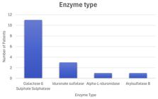
Identified MPS types included type IV (Morquio syndrome) in 11 patients, type II (Hunter syndrome) in 3, type VI (Maroteaux-Lamy syndrome) in 1, and type I (Hurler syndrome) in 1 (Figure 1). Subtype IV-A was the most frequent (n=11), followed by II-B (n=3) and I-IS (n=1). The most commonly deficient enzyme was galactose-6-sulfate sulfatase (n=11), and the most affected gene was GALNS (n=11).

Enzyme activity varied widely: 4 patients had null enzyme activity (0.00), while others had extremely low levels (≤ 0.10). The median enzymatic value was 0.10 (IQR 0.01-0.65). Most patients had skin phototype III (n=9), followed by IV (n=4), VI (n=2), and II (n=1). (Figure 2)
Dermatological Findings.
Coarse facial features were frequent (Figure 3). Flat nasal bridge (n=16) and broad nose (n=15) were the most common, followed by prominent forehead (n=8) and thick lips (n=6). Macroglossia (n=2) was less common. Body skin findings included multiple telangiectasias (n=6), thickened skin over hands (n=5), dermal melanocytosis (n=5), and ivory-colored papules or nodules (n=2) (Figure 4).
Among adnexal findings, hypertrichophrydia (n=10) and hypertrichosis (n=7) were most frequent, followed by synophrys (n=6). Lanugo hair (n=1) and hirsutism (n=1) were less common (Figure 5). All patients had brachyonychia. Other notable findings included seborrheic dermatitis (n=5), xerosis (n=3), acne (n=2), sparse hair (n=2), and isolated cases of tinea capitis, eyelid eczema, and melasma (Figure 6). (Table 2) (Table 3).
Individual clinical and dermatological characteristics of patients with mucopolysaccharidosis (n = 16).


Discussion
This study describes a cohort of patients diagnosed with MPS, with a predominance of type IV-A (Morquio-A), aligning with reports highlighting its relative frequency in certain populations across the Americas. For example, a study by Çelik B. et al. reported a prevalence of 2.23 per 100,000 births in Mexico, with MPS IV accounting for 49% of all cases [4]. Similarly, Gómez et al. (2012) in Colombia found MPS type IV to be the most frequent subtype [9].
In our cohort, enzymatic activity did not correlate linearly with the number of dermatological findings, suggesting that other genetic or environmental modulators may influence cutaneous manifestations independently of systemic severity. While literature associates enzyme deficiency with systemic disease severity [10], this pattern was not reflected in our patients’ dermatologic profiles.
Our study confirmed that most patients were in the pediatric age group, consistent with previous findings [2]. Lin HY et al. described a cohort with a mean age of 8.2 years (range 2.7-26.5) [11]. In our study, although the majority were children, the median age was 14.5 years, with a wide range (3 to 66 years) and similar distribution between sexes.
Sociodemographically, most patients came from urban areas in Valle del Cauca, likely due to the availability of specialized centers in these locations. This reflects the centralization of diagnostic and therapeutic resources in urban settings, as also stated by the EUCERD (2013) [12].
Educational attainment was low, with nearly half only completing primary school. This may reflect both physical/neurological limitations and socioeconomic barriers. Our findings mirror those from Yekedüz MK et al., who described compromised quality of life in patients with MPS [13]. Similarly, the high representation of patients from low socioeconomic strata and the subsidized health system highlights their vulnerability. The BURQOL-RD review and studies like Chung et al. in Hong Kong also support the heavy financial burden rare diseases place on low-income families [14,15].
Coarse facial features such as flat nasal bridge and broad nose were consistently observed. These facial alterations are described in the literature as part of the typical phenotype of the disease, together with the prominent forehead and thickened lips, related to the accumulation of glycosaminoglycans in soft tissues [16]. These align with features described by Swetha et al. in their analysis of 46 patients, where coarse facies and thick lips were common early signs [17].
Other findings like dermal melanocytosis and telangiectasias have been reported in the context of lysosomal storage diseases [18,19], though in our cohort, melanocytosis in MPS IV-A is a novel observation.
Thickened skin on the hands, a sign of connective tissue infiltration in MPS [20], was not prevalent in this cohort, though it is known to contribute to joint stiffness.
Adnexal findings such as hypertrichophrydia, hypertrichosis, and synophrys were frequently seen, consistent with altered follicular regulation in MPS [7,21]. These traits, especially synophrys and hirsutism, are well documented in MPS III. Escolar M et al. emphasized that their early detection should prompt metabolic or developmental evaluation [21].
brachyonychia (shortening of the nail plate, with a width greater than its length) was identified in all patients evaluated, a finding that has not been described in classical studies on MPS to date. Previous studies have consistently documented dysostosis and other bone abnormalities characteristic of MPS [22, 23], but have not described this dermatological manifestation. The presence of brachyonychia could be related to underlying skeletal abnormalities, opening the possibility of using it as a complementary clinical marker.
Other non-specific dermatoses such as seborrheic dermatitis, xerosis, and acne may be influenced by altered mucopolysaccharide metabolism or immune dysfunction [2]. Fungal and inflammatory conditions (e.g., tinea capitis, eyelid eczema) further reinforce the importance of regular dermatologic monitoring in MPS.
Conclusions
MPS IV-A was the most frequent subtype in this cohort. Most patients were pediatric and equally distributed by sex. Coarse facial features were consistent markers across patients, and brachyonychia was universally observed, suggesting it may represent an underrecognized phenotypic sign. Dermal melanocytosis and adnexal involvement were particularly notable in MPS IV-A. Recognition of these dermatological findings may function as clinical red flags that prompt metabolic evaluation and facilitate earlier multidisciplinary management, particularly in resource-limited settings. Strengthening referral systems for timely management remains essential.
Abbreviations
FNB: flat nasal bridge; BN: broad nose; PF: prominent forehead; TL: thick lips; MG: macroglossia; TS: thickened skin (hands); IPN: ivory papules/nodules; DM: dermal melanocytosis; Tel: telangiectasias; HT: hypertrichosis; Lan: lanugo hair; HTF: hypertrichophrydia; Syn: synophrys;
Hir: hirsutism; Br: brachyonychia; Xer: xerosis; SD: seborrheic dermatitis; Tinea: tinea capitis; Acne: acne vulgaris; EE: eyelid eczema; Hg: sparse hair; Mel: facial melasma.
REFERENCES
-
1. Moreno LJ, Satizábal JM, Sanchéz A. Genomic variability of the mucopolysaccharidosis complex in southwestern Colombia. Genet Mol Res 2020;19(2):GMR18502. doi: 10.4238/gmr18502
» https://doi.org/10.4238/gmr18502 -
2. Zhou J, Lin J, Leung WT, Wang L. A basic understanding of mucopolysaccharidosis: Incidence, clinical features, diagnosis, and management. Intractable Rare Dis Res 2020 Feb;9(1):1-9. doi: 10.5582/irdr.2020.01011
» https://doi.org/10.5582/irdr.2020.01011 -
3. Khan SA, Peracha H, Ballhausen D, et al Epidemiology of mucopolysaccharidoses. Mol Genet Metab 2017 Jul;121(3):227-240. doi: 10.1016/j.ymgme.2017.05.016
» https://doi.org/10.1016/j.ymgme.2017.05.016 -
4. Çelik B, Tomatsu SC, Tomatsu S, Khan SA. Epidemiology of Mucopolysaccharidoses Update. Diagnostics (Basel) 2021 Feb 10;11(2):273. doi: 10.3390/diagnostics11020273
» https://doi.org/10.3390/diagnostics11020273 - 5. Suárez-Guerrero JL, Bello AK, Vargas MC, Contreras GA. Caracterización clínica, estudios genéticos, y manejo de la Mucopolisacaridosis tipo IV A. Medicas UIS 2013;26(2):43-50.
-
6. Suarez-Guerrero JL, Gómez Higuera PJ, Arias Flórez JS, Contreras-García GA. Mucopolisacaridosis: características clínicas, diagnóstico y de manejo. Rev Chil Pediatr 2016;87(4):295-304. doi: 10.1016/j.rchipe.2015.10.004
» https://doi.org/10.1016/j.rchipe.2015.10.004 -
7. Tran MC, Lam JM. Cutaneous Manifestations of Mucopolysaccharidoses. Pediatr Dermatol 2016 Nov;33(6):594-601. doi: 10.1111/pde.12971
» https://doi.org/10.1111/pde.12971 -
8. Huertas JC, Giraldo LM. Manifestaciones dermatológicas de las mucopolisacaridosis. Piel 2025;41(1):1-82. doi:10.1016/j.piel.2025.04.001
» https://doi.org/10.1016/j.piel.2025.04.001 -
9. Gómez AM, García-Robles R, Suárez-Obando F. Estimación de las frecuencias de las mucopolisacaridosis y análisis de agrupamiento espacial en los departamentos de Cundinamarca y Boyacá. Biomedica 2012;32(4):602-609. doi: 10.1590/S0120-41572012000400015
» https://doi.org/10.1590/S0120-41572012000400015 -
10. Piotrowska E, Jakóbkiewicz-Banecka J, Tylki-Szymańska A, Czartoryska B, Wegrzyn A, Wegrzyn G. Correlation between severity of mucopolysaccharidoses and combination of the residual enzyme activity and efficiency of glycosaminoglycan synthesis. Acta Paediatr 2009 Apr;98(4):743-749. doi: 10.1111/j.1651-2227.2008.01153.x
» https://doi.org/10.1111/j.1651-2227.2008.01153.x -
11. Lin HY, Chuang CK, Lee CL, Tu RY, Lo YT, Chiu PC, Niu DM, Fang YY, Chen TL, Tsai FJ, Hwu WL, Lin SJ, Chang TM, Lin SP. Mucopolysaccharidosis III in Taiwan: Natural history, clinical and molecular characteristics of 28 patients diagnosed during a 21-year period. Am J Med Genet A 2018;176(9):1799-1809. doi: 10.1002/ajmg.a.40351
» https://doi.org/10.1002/ajmg.a.40351 -
12. EUCERD - European Union Committee of Experts on Rare Diseases. EUCERD Recommendations on Quality Criteria for Centres of Expertise for Rare Diseases in Member States Brussels: EUCERD; 2011. https://www.rd-action.eu/eucerd/EUCERD_Recommendations/eucerd-recommendations(1).pdf Published October 24, 2016.
» https://www.rd-action.eu/eucerd/EUCERD_Recommendations/eucerd-recommendations(1).pdf -
13. Yekedüz MK, Cilesiz K, Kara İS, İnci A, Kose E, Tümer L, Eminoğlu FT. Quality of Life and Related Factors in Patients Diagnosed with Mucopolysaccharidosis and Their Caregivers. Klin Padiatr 2026 May;238(3):133-146. doi: 10.1055/a-2451-6562
» https://doi.org/10.1055/a-2451-6562 -
14. Angelis A, Tordrup D, Kanavos P. Socio-economic burden of rare diseases: A systematic review of cost of illness evidence. Health Policy 2015 Jul;119(7):964-979. doi: 10.1016/j.healthpol.2014.12.016
» https://doi.org/10.1016/j.healthpol.2014.12.016 -
15. Chung CC, Ng NY, Ng YN, Lui AC, et al Socio-economic costs of rare diseases and the risk of financial hardship: A cross-sectional study. Lancet Reg Health West Pac 2023;34:100711. doi:10.1016/j.lanwpc.2023.100711
» https://doi.org/10.1016/j.lanwpc.2023.100711 -
16. Nagpal R, Goyal RB, Priyadarshini K, Kashyap S, Sharma M, Sinha R, Sharma N. Mucopolysaccharidosis: A broad review. Indian J Ophthalmol 2022 Jul;70(7):2249-2261. doi: 10.4103/ijo.IJO_425_22
» https://doi.org/10.4103/ijo.IJO_425_22 -
17. Swetha P, Panigrahi I, Saxena A, Kaur A, Thakur R. Clinical and Radiological Characterisation of Patients with Mucopolysaccharidosis in a Genetic Clinic. J Inborn Errors Metab Screen 2021;9:e20200008. doi:10.1590/2326-4594-JIEMS-2020-0008
» https://doi.org/10.1590/2326-4594-JIEMS-2020-0008 -
18. Alimi Y, Iwanaga J, Loukas M, Oskouian RJ, Rizk E, Oakes WJ, Tubbs RS. A comprehensive review of Mongolian spots with an update on atypical presentations. Childs Nerv Syst 2018 Dec;34(12):2371-2376. doi: 10.1007/s00381-018-3929-0
» https://doi.org/10.1007/s00381-018-3929-0 -
19. Romagnuolo M, Moltrasio C, Gasperini S, Marzano AV, Cambiaghi S. Extensive and Persistent Dermal Melanocytosis in a Male Carrier of Mucopolysaccharidosis Type IIIC (Sanfilippo Syndrome): A Case Report. Children (Basel) 2023 Dec 13;10(12):1920. doi: 10.3390/children10121920
» https://doi.org/10.3390/children10121920 -
20. Michaud M, Belmatoug N, Catros F, Ancellin S, Touati G, Levade T, Gaches F. Mucopolysaccharidoses: Quand y penser? Rev Med Interne 2020 Mar;41(3):180-188. doi: 10.1016/j.revmed.2019.11.010
» https://doi.org/10.1016/j.revmed.2019.11.010 -
21. Escolar M, Bradshaw J, Byers VT, et al. Development of a clinical algorithm for the early diagnosis of Mucopolysaccharidosis III. J Inborn Errors Metab Screen . 2020;8:e20200002. doi:10.1590/2326-4594-JIEMS-2020-0002
» https://doi.org/10.1590/2326-4594-JIEMS-2020-0002 -
22. Borgo A, Cossio A, Gallone D, Vittoria F, Carbone M. Orthopaedic challenges for mucopolysaccharidoses. Ital J Pediatr 2018 Nov 16;44(Suppl 2):123. doi: 10.1186/s13052-018-0557-y
» https://doi.org/10.1186/s13052-018-0557-y -
23. Morishita K, Petty RE. Musculoskeletal manifestations of mucopolysaccharidoses. Rheumatology (Oxford) 2011;50 (Suppl 5):v19-25. doi: 10.1093/rheumatology/ker397
» https://doi.org/10.1093/rheumatology/ker397
-
Funding
This research received no specific grants from public, commercial, or not-for-profit funding agencies.
-
Ethics Approval and Consent to Participate
This study was conducted in accordance with international research ethics standards and was approved by the institutional ethics committee.
-
Data Availability
The dataset supporting the results of this study is not publicly available.
The dataset supporting the results of this study is not publicly available.


 Photo A: 13-year-old male with MPS IV-A, presenting prominent forehead, flat nasal bridge and thick lips. Photo B: 38-year-old female with MPS IV-A, showing broad nose, thick lips, associated with typical coarse facial features.
 Photos A and B: Multiple telangiectasias, clinically and dermoscopically observed in a 3-year-old male with MPS IV-A. Photo C: 13-year-old male with MPS IV-A, showing persistent beyond early childhood dermal melanocytosis on the anterior thorax. Photo D: 13-year-old male with MPS II- B, presenting thickened skin over the dorsal hands with reduced skin elasticity and underlying joint stiffness, consistent with connective tissue infiltration.
 Photo A: 3-year-old male with MPS IV- A, exhibiting marked hypertrichophrydia and synophrys, associated with coarse facial features including broad nasal bridge, possibly related to progressive dermal thickening due to glycosaminoglycan accumulation, contributing to a pseudo-aging or sagging appearance. Photo B: 18-year-old male with MPS IV- A, demonstrating generalized hypertrichosis involving the lower limbs and dorsum of the feet, along with evident progressive contractures of the feet, consistent with underlying skeletal involvement due to glycosaminoglycan accumulation. Photo C and D: Clinical and dermoscopy examination of the frontotemporal scalp in a 3-year-old patient with MPS IV-A, revealing reduced hair density, variation in hair shaft diameter, and areas of follicular miniaturization without evidence of inflammatory scarring.
 Photo A and B: 13-year-old male with MPS II-B, presenting pseudoalopecic plaques on the scalp and dystrophic hairs observed via trichoscopy. Photo C: 34-year-old male with MPS II- B and Fitzpatrick skin phototype IV, demonstrating centrofacial hyperpigmented macules consistent with melasma, with symmetric distribution over the malar regions and forehead. Photo D: Close-up image of fingernails in a 34-year-old patient with MPS II- B, revealing brachyonychia characterized by nail plates wider than their longitudinal length, with preserved nail surface and absence of pitting or dystrophy. Photo E: Toenails from a 16-year-old female patient with MPS IV-A, also demonstrating brachyonychia, reinforcing the consistent presence of this finding across the cohort. Additionally, progressive contractures of the toes are evident, consistent with skeletal involvement secondary to glycosaminoglycan accumulation.