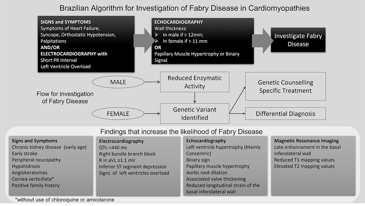
 Algorithm for Investigation of Fabry Disease in Cardiomyopathies
Abstract
Fabry disease (FD) is a rare, x-linked lysosomal storage disease caused by mutations in the GLA gene that leads to total or partial alfa galactosidase A deficiency. Its prevalence ranges between 1:117,000 and 1:8,454. Mutations in the GLA gene result in alpha galactosidase A deficiency leading to the progressive accumulation of globotriaosylceramide (Gb3) in lysosomes of different types of cells of the heart, kidneys, skin, eyes, central nervous system, and gastrointestinal system, and may lead to different clinical scenarios. The cardiac manifestation most frequently found in FD is the presence of left ventricular hypertrophy (LVH) in a concentric pattern, although asymmetric forms are also reported. In clinical practice, it is often difficult to differentiate between FD and other diseases associated with LVH. In adults with LVH, a prevalence of FD of 3-5% has been reported. Therefore, diagnosing these patients is of fundamental importance, as specific treatment for FD has the potential to change the prognosis, especially if instituted early. The purpose of this article is to describe an algorithm developed through a thorough literature review to assist in the identification of FD in patients with cardiomyopathies.
Keywords.
Hypertrophic; cardiomyopathy; Fabry; disease; left; ventricular; hypertrophy
Introduction
Fabry disease (FD) is a rare, X-linked lysosomal storage disorder caused by mutations in the GLA gene leading to total or partial deficiency of alfa-galactosidase A and the progressive accumulation of globotriaosylceramide (Gb3) deposits in several tissues and organs [1,2]. The classical phenotype is more commonly seen in males due to complete enzyme inactivity leading to multisystemic symptoms such as neuropathic pain, cornea verticillata, angiokeratomas, sweating abnormalities, gastrointestinal impairment, stroke at young ages, and cardiac and kidney disfunction [3,4]. Manifestations of the later-onset phenotype appear later in life usually during adulthood due to higher levels of residual α-Gal A may be seening in both males and females. Although hemizygous males and heterozygous females are affected by Fabry disease, heterozygous females can present a heterogeneous range of signals and symptoms, from milder to severe clinical manifestations [5,6]. Heterozygous females can present a heterogeneous range of signals and symptoms, disease severity and organ involvement, that are related to the proportion of skewed X-chromosome inactivation of normal or mutant alleles [5,6,7]. As a result of the residual enzyme activity often found in their lysosomes, the disease may clinically manifest with single-organ involvement of the heart or kidney [8-11]. Long-term complications of the disease include progressive renal failure, stroke, and cardiomyopathy. All tissues in the heart are affected, leading to left ventricular hypertrophy (LVH), disturbances in the conduction system such as short PR interval and bradycardia [12-14], valvar regurgitation and ischemic myocardial disease [15-17].
The prevalence of FD among patients with LVH is 3 to 5% [2,18,19]. Therefore, diagnosing these patients is of crucial importance, as specific treatment for FD can potentially change the prognosis, especially if instituted early [20-23]. However, clinically, it can be challenging for physicians to differentiate Fabry cardiomyopathy from other diseases associated with LVH, such as sarcomeric disease, amyloidosis, hemochromatosis, Noonan Syndrome, and neuromuscular disorders [24-26]. Currently, there are many algorithms for the investigation of FD [25-30], but little have a focus on cardiac involvement [31,32]. In this review, a group of cardiologists with expertise in Fabry cardiomyopathy propose an algorithm to enable an easy, accurate, timely and cost-effective screening of the Fabry cardiomyopathy.
Methods
From March to June 2019, we searched PubMed and the Cochrane Library in the attempt to retrieve articles that defined criteria and methods for investigation of FD in patients with unexplained LVH. The key words used in the search were: “Fabry disease”, “left ventricular hypertrophy”, “hypertrophic cardiomyopathy”, and “screening criteria”. We found eight review articles and two clinical trials that were included in our review [16,24,33-40]. We read and discussed these articles with the aim of building the algorithm. By discussing the literature in a group of FD experts, we reached a consensus among authors based on a common understanding of the available evidence that lends support to the algorithm, shown below.
Rationale for the Presentation of Cardiology Algorithm in Fabry Disease
As proposed in the algorithm displayed in Figure 1, the investigation of Fabry cardiomyopathy should be initiated with the follow timelines: 1) anamnesis with the understanding of cardiac signs and symptoms and/or electrocardiographic changes related to Fabry and; 2) the next step is echocardiographic analysis of changes followed by 3) FD specific investigation.

Proposed algorithm for screening and diagnosis of Fabry disease. The algorithm starts with the analyses of signs and symptoms and/or electrocardiography alterations follow by echocardiography investigation. As the patient reaches those criteria’s, the suspicion of FD is substantial, a specific investigation is indicated. aVL: augmented vector left; QTc: corrected QT interval.
In that way, once the clinical history reported refers to an individual presenting clinical complaints of heart failure that might start during the third decade of life associated with chest pain, palpitations resulting from supraventricular or ventricular arrhythmias, symptoms related to autonomic dysfunction such as unexplained syncope, orthostatic hypotension, recurrent dizziness, or chronotropic incompetence that are not explained by other conditions such as hypertension, diabetes mellitus or atherosclerotic coronary disease [16,18,38,41,42]. It is important to note that cardiac changes can be present at different ages even during childhood [21]. Moreover, the suspicion of FD may be enhanced by a previous history of chronic kidney disease with unknown etiology, history of stroke before 40 years of age, peripheral neuropathy at young ages, hypohidrosis or anhidrosis, the presence of angiokeratomas, or the presence of another subject with FD in the family. These criteria are not mandatory for full investigation of FD and are more likely to be seen in patients with classical mutation than in those with late-onset disease.
Most adult patients with FD show abnormalities in the electrocardiogram, a low-cost exam routinely performed on the first cardiological evaluation. Variable brady or tachycardic rhythms associated with a short PR interval (<120 milliseconds) and changes in the corrected QT interval are observed [43,44]. Namdar et al. reported a significant reduction in the P wave and PQ/PR interval in patients with FD when compared with healthy individuals, demonstrating a high sensitivity and specificity for diagnosis [43,45]. Hoigné et al. also described a tendency to reduce the PR interval in these patients when compared with other heart diseases presenting with LVH [24]. Another finding is the reduction of the corrected QT interval at the early stage of the disease. The corrected QT <440 ms, combined with the PQ interval minus the P-wave width in lead II <40 ms, showed 100% sensitivity and 99% specificity for FD diagnosis [44]. Arrhythmias, such as bradycardia and atrioventricular blocks, can be found in later stages [45]. Recently, Vitale et al. suggested the following independently predictive signs for FD diagnosis: short PR interval, prolonged QRS duration, right bundle branch block (RBBB), R in aVL ≥1.1 mV, and inferior ST depression [46]. Contrasting with other storage diseases such as amyloidosis, the deposition of Gb3 in the conduction system manifests as signs of left-ventricular overload by variable criteria (Sokolov, Cornel) [47,48].
Following patient evaluation, there is the need to perform imaging methods that allow an in-depth assessment of cardiac involvement. In this regard, the echocardiogram should be highlighted, as it is an accessible, low-cost exam capable of providing a morpho-functional substrate for the complaints previously presented, as well as providing data that influence patient prognosis [49].
Data derived from Fabry registries show that LVH is the chief manifestation [24,37,40]. Cardiac hypertrophy affects more than two-thirds of patients with FD, according to the literature [50]. Aspects related to the type of pathogenic genetic variant, age group, associated diseases and clinical management can influence the severity of LVH [24,49,51]. Concentric involvement without obstruction of the left ventricular outflow tract is most frequently seen; however, concentric remodeling, eccentric hypertrophy or even isolated apical forms have already been described, as well as hypertrophy of the papillary muscles [37,49,52]. The cut-off values for left ventricular wall thickness proposed in this algorithm are >11 mm in females and >12 mm in males. These are similar to more recent therapeutic guidelines focused on FD, such as the Canadian FD treatment guidelines [53].
Other echocardiographic findings typically seen are papillary hypertrophy, enlightenment of ventricular endocardium (Binary sign), and thickening of mitral and aortic valves leading to mild regurgitations and aortic-root dilatation. The binary sign was defined as suggested by Pieroni et al. as a hyperechogenic and bright endocardium and a hypoechogenic space between endo- and myocardium, especially visible in the interventricular septal and the apical region of the left ventricle, respectively, thus forming a binary appearance [54]. Systolic function remains preserved until the late stages of the disease unless there is the coexistence of other cardiac diseases such as uncontrolled hypertension, renal failure, diabetes mellitus, or coronary disease [41,55].
Novel echocardiographic techniques, such as the analysis of myocardial deformity or cardiac strain using the speckle-tracking technique, detects early cardiac changes in FD. These include subclinical forms of systolic dysfunction in contractile function that are sometimes imperceptible using conventional methods and result in ejection fraction disturbances. In early stages of ventricular involvement, substrate deposits and the underlying inflammatory process determine a slight reduction in the left-ventricular global longitudinal strain (LV GLS) at the basal portion of the infero-lateral wall even before the development of left ventricular hypertrophy. The development of hypertrophy generates more marked reductions in the LV GLS as well as in the radial strain (RS) of the inferolateral wall. There is also a post-systolic contraction described as a double-peak sign in the graphical representation of the deformity. In advanced stages of the disease, and with the development of fibrosis, there is substantial worsening of the values of LV GLS and RS [56,57]. Similar phenomena are observed in the free-wall strain of the right ventricle and the left atrium, even before the appearance of clinical symptoms [58]. Moreover, several studies have demonstrated the value of this novel echocardiographic method for the differential diagnosis of cardiomyopathies.
Laboratory findings of low cost and easy access may help FD cardiomyopathy staging before going to disease-specific diagnosis. Hypertrophic cardiomyopathy biomarkers such as highly sensitive troponin and N-terminal pro-B-type natriuretic peptide (NT-pro-BNP) are associated with inflammatory myocardial injury and left-ventricle volume and pressure overload as extensively discussed in the cardiomyopathies guidelines [59]. The augment of those markers may anticipate the onset of heart-failure symptoms and assist the management of disease progression [60-62].
The combined analysis of clinical information, electrocardiographic and echocardiographic changes and bio-markers seems to increase the likelihood of FD cardiomyophaty [63-65]. Moreover, this information is crucial for initial diagnosis of Fabry Disease, for disease-specific therapy indication and monitoring, as well as to manage cardiovascular symptoms and prevent undesired cardiovascular events in order to maximize therapeutic benefits [26,66].
Fabry Disease-Specific Diagnosis
Genetic confirmatory testing is mandatory in both suspected males and females, as GLA gene mutations may range from benign polymorphisms to very severe classical phenotypes [21]. However, in case of males clinical suspicion to have FD, α-Gal A activity should be measured before genotyping, as the absence of low α-Gal activity can exclude FD and reduce diagnostic costs. Is important to note that, patients with higher enzyme activity tend to have more attenuated disease [67,68]. In females, because of genetic mosaicism, the α-Gal A activity is variable and can range from low to within normal levels [69]. In this reason, the genetic testing is indicated in suspected females without the need of enzymatic testing [21]. In that way, the timeline for Fabry disease specific diagnostic in males and females is described in Figure 1.
The assessment of specific biomarkers of FD contributes not only to diagnosis but it is also used as a tool to assess therapeutic efficacy [70,71]. In this sense, the serum levels of Lyso-Gb3 were shown to be superior to the assessment of Gb3, especially for female patients [72].
Other Useful Elements for FD Diagnosis
Magnetic resonance imaging
Cardiac magnetic resonance imaging (MRI) is considered the gold standard for the diagnosis and deeper characterization of cardiac involvement in hypertrophic diseases. Notwithstanding many favorable features, cardiac MRI is not yet widely accessible, especially in less economically developed regions. The equipment is usually installed in large urban centers, and tests are often considered expensive. Thus, in places with scarce resources, the methods presented above in the algorithm are sufficient for the initial diagnosis. Furthermore, more detailed assessments of cardiac involvement, such as the presence of fibrosis, should not delay the diagnosis or the initiation of specific treatment. In addition, some peculiarities associated with the technique, such as the need for a regular heart rhythm and the cooperation on the part of the patient when asked to perform respiratory maneuvers, may be challenging in the attempt to obtain reasonable results.
Cardiac MRI provides not only diagnostic and staging information in FD but also allows differential diagnosis with other disorders and provides prognostic information. Information regarding cardiac mass, wall thickness, cavity volumes and systolic function are accurately measured even in the presence of a difficult echocardiographic window [73,74]. The use of the MRI delayed enhancement technique with gadolinium makes it possible to locate and quantify the degree of myocardial fibrosis, typically located in the mesocardial portion of the lower basal region. The fibrosis is a marker of chronic heart disease and may lead to systolic disfunction and heart failure. Prognosis in these cases is worse due to the low response to enzyme replacement therapy [75] and increased occurrence of malignant arrhythmias, heart failure, and sudden cardiac death [76-78]. It is important to highlight that this technique is not recommended in individuals with glomerular filtration rate <30 mL/min due to the risk of nephrogenic systemic fibrosis and death [79].
The development of T1 and T2 mapping techniques expanded the diagnostic horizon for MRI by enabling the early detection of edema, inflammation and fibrosis even in individuals with end-stage renal disease [80]. The cardiomyopathy in FD is characterized by a decrease in T1 values on the parametric map, associated with an increase in extracellular volume. Besides Fabry cardiomyopathy, only hemosiderosis cardiomyopathy has low T1 mapping values and the finding of iron deposits in other tissues helps to differentiate between the two diseases. Other disorders that present with hypertrophy are characterized by a T1 map of higher intensity, helping in their differentiation from FD [80,81].
Differential Diagnosis
The heart involvement in FD may be confirmed by Gb3 deposits in the heart and can be evaluate by biopsy [31]. Moreover, it is important to highlight that biopsy brings more information concerning patients with low Lyso-Gb3 levels and variants of unknown significance [31].
In addition, the finding of low-voltage QRS complexes on the ECG may suggest other diagnoses, such as amyloidosis while high voltages QRS makes differential diagnosis with sarcomeric hypertrophyc cardiomiophaty [59]. Echocardiographic findings of severe hypertrophy, systolic dysfunction or valve disorders in the first years of life may be indicative of other diagnoses, such as Danon or Pompe disease [26,82]. Therefore, a confirmed diagnosis of another disorder with cardiac involvement may rule out the diagnosis of FD. Such disorders include amyloidosis, mitochondrial cardiomyopathies, Friedreich ataxia or other muscular dystrophies, valve diseases, other deposit diseases (Pompe, mucopolysaccharidosis), and genetic syndromes (Noonan, LEOPARD, and Costello) [26,82].
Discussion
In FD, early diagnosis is essential for the relief of cardiac symptoms that are highly disabling and therefore cause of intense psychological suffering. The systematization of a therapeutic strategy helps the attending physician to establish a highly effective and cost-effective investigative strategy by requesting specific diagnostic tests not only through conventional techniques but also new technologies. In this sense, it is known that the journey of a patient with a diagnosis of a rare disease is challenging. In general, patients are evaluated by physicians from different specialties with no control of symptoms or the interruption of disease progression as there is a lack of effective action on the primary cause of the disease, which is in this case the glycosphingolipid deposits. In addition, the multisystemic manifestations of FD make it necessary to perform multiple tests, some of which are highly complex and not always available in the vicinity of the patient’s home. As a result, literature data indicate that there may be a delay of 14 years for male patients and of 19 years for female patients between the onset of symptoms and the establishment of the diagnosis of FD [83].
Is a matter of fact, that in locations where the resources for diagnosis are scarce, the proposed algorithm together with the low-complexity exams may be useful to indicate the possibility of heart disease related to FD. Based on this, the patient can receive adjuvant drug treatment for symptom relief while waiting for care in reference centers where exams of higher complexity are available.
Conclusion
The proposed algorithm aims at optimizing the clinical management of FD to prevent patients from developing cardiac fibrosis or high-grade blocks in the conduction system that require high-cost procedures such as the implantation of pacemaker/cardiac defibrillators or even heart transplantation. At this stage of disease progression, it is known that specific therapies for FD have low effectiveness and may not prevent the occurrence of unfavorable outcomes such as malignant arrhythmias or sudden death [84].
Acknowledgments
Editorial support in the preparation of this publication was provided by Ana Elisa B. Bueno da Silva and Everardo D. Saad (Dendrix Research, Sao Paulo) and paid for by Sanofi. The authors, individually and collectively are responsible for all content and editorial decisions and received no payment from Sanofi directly or indirectly (through a third party) related to the development/presentation of this publication.
References
-
Zarate YA, Hopkin RJ. Fabry’s disease. Lancet 2008;372(9647):1427-1435.doi:10.1016/S0140-6736(08)61589-5.
» https://doi.org/10.1016/S0140-6736(08)61589-5. -
Nakao S, Takenaka T, Maeda M, et al. An atypical variant of Fabry’s disease in men with left ventricular hypertrophy. N Engl J Med 1995;333(5):288-293. doi:10.1056/NEJM199508033330504.
» https://doi.org/10.1056/NEJM199508033330504 -
Desnick RJ, Brady R, Barranger J, et al. Fabry disease, an under-recognized multisystemic disorder: Expert recommendations for diagnosis, management, and enzyme replacement therapy. Ann Intern Med 2003;138(4):338-346. doi:10.7326/0003-4819-138-4-200302180-00014.
» https://doi.org/10.7326/0003-4819-138-4-200302180-00014. - Desnick RJ, Ioannou YA, Eng CM. α-Galactosidase A deficiency: Fabry disease. In: Scriver CR, Beaudet AL, Sly WS, Valle D, eds. The metabolic and molecular bases of inherited disease 7th ed. Vol. 2. New York, NY: McGraw-Hill; 1995:2741-2784.
-
Elstein D, Schachamorov E, Beeri R, Altarescu G. X-inactivation in Fabry disease. Gene 2012;505(2):266-268. doi:10.1016/j.gene.2012.06.013.
» https://doi.org/10.1016/j.gene.2012.06.013. -
Boggio P, Luna PC, Abad ME, Larralde M. Doença de Fabry. An Bras Dermatol 2009;84(4):367-376. doi:10.1590/s0365-05962009000400008.
» https://doi.org/10.1590/s0365-05962009000400008. -
Echevarria L, Benistan K, Toussaint A, et al. X-chromosome inactivation in female patients with Fabry disease. Clin Genet. 2016;89(1):44-54. doi:10.1111/cge.12613.
» https://doi.org/10.1111/cge.12613 -
Arends M, Wanner C, Hughes D, et al. Characterization of classical and nonclassical Fabry disease: A multicenter study. J Am Soc Nephrol 2017;28(5):1631-1641. doi:10.1681/ASN.2016090964.
» https://doi.org/10.1681/ASN.2016090964. -
Nagao Y, Nakashima H, Fukuhara Y, et al. Hypertrophic cardiomyopathy in late-onset variant of Fabry disease with high residual activity of alpha-galactosidase A. Clin Genet 1991;39(3):233-237. doi:10.1111/j.1399-0004.1991.tb03018.x.
» https://doi.org/10.1111/j.1399-0004.1991.tb03018.x. -
Ogawa K, Sugamata K, Funamoto N, et al. Restricted accumulation of globotriaosylceramide in the hearts of atypical cases of Fabry’s disease. Hum Pathol 1990;21(10):1067-1073. doi:10.1016/0046-8177(90)90258-7.
» https://doi.org/10.1016/0046-8177(90)90258-7. -
von Scheidt W, Eng CM, Fitzmaurice TF, et al. An atypical variant of Fabry’s disease with manifestations confined to the myocardium. N Engl J Med 1991;324(6):395-399. doi:10.1056/NEJM199102073240607.
» https://doi.org/10.1056/NEJM199102073240607. -
Aryana A, Fifer MA, Ruskin JN, Mela T. Short PR interval in the absence of preexcitation: A characteristic finding in a patient with Fabry disease. Pacing Clin Electrophysiol 2008;31(6):782-783. doi:10.1111/j.1540-8159.2008.01088.x.
» https://doi.org/10.1111/j.1540-8159.2008.01088.x. -
Jastrzebski M, Bacior B, Dimitrow PP, Kawecka-Jaszcz K. Electrophysiological study in a patient with Fabry disease and a short PQ interval. Europace 2006;8(12):1045-1047. doi:10.1093/europace/eul121.
» https://doi.org/10.1093/europace/eul121. -
Kubo T. Fabry disease and its cardiac involvement. J Gen Fam Med 2017;18(5):225-229. doi:10.1002/jgf2.76.
» https://doi.org/10.1002/jgf2.76. -
Barbey F, Brakch N, Linhart A, et al. Cardiac and vascular hypertrophy in Fabry disease: evidence for a new mechanism independent of blood pressure and glycosphingolipid deposition. Arterioscler Thromb Vasc Biol 2006;26(4):839-844. doi:10.1161/01.ATV.0000209649.60409.38.
» https://doi.org/10.1161/01.ATV.0000209649.60409.38. -
Linhart A, Kampmann C, Zamorano JL, et al. Cardiac manifestations of Anderson-Fabry disease: results from the international Fabry outcome survey. Eur Heart J 2007;28(10):1228-1235. doi:10.1093/eurheartj/ehm153.
» https://doi.org/10.1093/eurheartj/ehm153. -
Weidemann F, Breunig F, Beer M, et al. The variation of morphological and functional cardiac manifestation in Fabry disease: Potential implications for the time course of the disease. Eur Heart J 2005;26(12):1221-1227. doi:10.1093/eurheartj/ehi143.
» https://doi.org/10.1093/eurheartj/ehi143. -
Fernandes F, Antunes MO, Hotta VT, Rochitte CE, Mady C. Deposit Diseases as Differential Diagnosis of Left Ventricular Hypertrophy in Patients with Heart Failure and Preserved Systolic Function. Arq Bras Cardiol 2019;113(5):979-987. doi:10.36660/abc.20180370.
» https://doi.org/10.36660/abc.20180370. -
Sachdev B, Takenaka T, Teraguchi H, et al. Prevalence of Anderson-Fabry disease in male patients with late onset hypertrophic cardiomyopathy. Circulation 2002;105(12):1407-1411. doi:10.1161/01.cir.0000012626.81324.38.
» https://doi.org/10.1161/01.cir.0000012626.81324.38. -
Kampmann C, Perrin A, Beck M. Effectiveness of agalsidase alfa enzyme replacement in Fabry disease: Cardiac outcomes after 10 years’ treatment. Orphanet J Rare Dis 2015;10:125. doi:10.1186/s13023-015-0338-2.
» https://doi.org/10.1186/s13023-015-0338-2. -
Ortiz A, Germain DP, Desnick RJ, et al. Fabry disease revisited: Management and treatment recommendations for adult patients. Mol Genet Metab 2018;123(4):416-427. doi:10.1016/j.ymgme.2018.02.014.
» https://doi.org/10.1016/j.ymgme.2018.02.014. -
Spinelli L, Pisani A, Sabbatini M, et al. Enzyme replacement therapy with agalsidase beta improves cardiac involvement in Fabry’s disease. Clin Genet 2004;66(2):158-165. doi:10.1111/j.1399-0004.2004.00284.x.
» https://doi.org/10.1111/j.1399-0004.2004.00284.x. -
Weidemann F, Breunig F, Beer M, et al. Improvement of cardiac function during enzyme replacement therapy in patients with Fabry disease: A prospective strain rate imaging study. Circulation 2003;108(11):1299-1301. doi:10.1161/01.CIR.0000091253.71282.04.
» https://doi.org/10.1161/01.CIR.0000091253.71282.04. -
Hoigné P, Attenhofer Jost CH, Duru F, et al. Simple criteria for differentiation of Fabry disease from amyloid heart disease and other causes of left ventricular hypertrophy. Int J Cardiol 2006;111(3):413-422. doi:10.1016/j.ijcard.2005.08.023.
» https://doi.org/10.1016/j.ijcard.2005.08.023. -
Monserrat L, Gimeno-Blanes JR, Marín F, et al. Prevalence of fabry disease in a cohort of 508 unrelated patients with hypertrophic cardiomyopathy. J Am Coll Cardiol 2007;50(25):2399-2403. doi:10.1016/j.jacc.2007.06.062.
» https://doi.org/10.1016/j.jacc.2007.06.062. -
Task Force members; Elliott PM, Anastasakis A, et al. 2014 ESC Guidelines on diagnosis and management of hypertrophic cardiomyopathy: The Task Force for the Diagnosis and Management of Hypertrophic Cardiomyopathy of the European Society of Cardiology (ESC). Eur Heart J 2014;35(39):2733-2779. doi:10.1093/eurheartj/ehu284.
» https://doi.org/10.1093/eurheartj/ehu284. -
Geske JB, Ommen SR, Gersh BJ. Hypertrophic Cardiomyopathy: Clinical Update. JACC Heart Fail 2018;6(5):364-375. doi:10.1016/j.jchf.2018.02.010.
» https://doi.org/10.1016/j.jchf.2018.02.010. -
Maron BJ. Clinical course and management of hypertrophic cardiomyopathy. N Engl J Med 2018;379(7):655-668. doi:10.1056/NEJMra1710575.
» https://doi.org/10.1056/NEJMra1710575. -
Chimenti C, Pieroni M, Morgante E, et al. Prevalence of Fabry disease in female patients with late-onset hypertrophic cardiomyopathy. Circulation 2004;110(9):1047-1053. doi:10.1161/01.CIR.0000139847.74101.03.
» https://doi.org/10.1161/01.CIR.0000139847.74101.03. -
Mawatari K, Yasukawa H, Oba T, et al. Screening for Fabry disease in patients with left ventricular hypertrophy. Int J Cardiol 2013;167(3):1059-1061. doi:10.1016/j.ijcard.2012.10.076.
» https://doi.org/10.1016/j.ijcard.2012.10.076. -
Pieroni M, Moon JC, Arbustini E, et al. Cardiac Involvement in Fabry Disease: JACC Review Topic of the Week. J Am Coll Cardiol 2021;77(7):922-936. doi:10.1016/j.jacc.2020.12.024.
» https://doi.org/10.1016/j.jacc.2020.12.024. -
Linhart A, Germain DP, Olivotto I, et al. An expert consensus document on the management of cardiovascular manifestations of Fabry disease. Eur J Heart Fail 2020;22(7):1076-1096. doi:10.1002/ejhf.1960.
» https://doi.org/10.1002/ejhf.1960. -
Hagège AA, Caudron E, Damy T, et al. Screening patients with hypertrophic cardiomyopathy for Fabry disease using a filter-paper test: The FOCUS study. Heart 2011;97(2):131-136. doi:10.1136/hrt.2010.200188.
» https://doi.org/10.1136/hrt.2010.200188. -
Wu JC, Ho CY, Skali H, et al. Cardiovascular manifestations of Fabry disease: Relationships between left ventricular hypertrophy, disease severity, and alpha-galactosidase A activity. Eur Heart J 2010;31(9):1088-1097. doi:10.1093/eurheartj/ehp588.
» https://doi.org/10.1093/eurheartj/ehp588. -
Patel MR, Cecchi F, Cizmarik M, et al. Cardiovascular events in patients with Fabry disease natural history data from the fabry registry. J Am Coll Cardiol 2011;57(9):1093-1099. doi:10.1016/j.jacc.2010.11.018.
» https://doi.org/10.1016/j.jacc.2010.11.018. - Anastasakis A, Sevdalis E, Papatheodorou E, Stefanadis C. Anderson-Fabry disease: A cardiomyopathy that can be cured. Hellenic J Cardiol 2011;52(4):316-326.
-
Seydelmann N, Wanner C, Störk S, Ertl G, Weidemann F. Fabry disease and the heart. Best Pract Res Clin Endocrinol Metab 2015;29(2):195-204. doi:10.1016/j.beem.2014.10.003.
» https://doi.org/10.1016/j.beem.2014.10.003. -
Seo J, Kim M, Hong GR, et al. Fabry disease in patients with hypertrophic cardiomyopathy: A practical approach to diagnosis. J Hum Genet 2016;61(9):775-780. doi:10.1038/jhg.2016.52.
» https://doi.org/10.1038/jhg.2016.52. -
Hsu TR, Niu DM. Fabry disease: Review and experience during newborn screening. Trends Cardiovasc Med 2018;28(4):274-281. doi:10.1016/j.tcm.2017.10.001.
» https://doi.org/10.1016/j.tcm.2017.10.001. -
Hagège A, Réant P, Habib G, et al. Fabry disease in cardiology practice: Literature review and expert point of view. Arch Cardiovasc Dis 2019;112(4):278-287. doi:10.1016/j.acvd.2019.01.002.
» https://doi.org/10.1016/j.acvd.2019.01.002 -
Linhart A, Lubanda JC, Palecek T, et al. Cardiac manifestations in Fabry disease. J Inherit Metab Dis 2001;24 Suppl 2:75-65. doi:10.1023/a:1012428009627.
» https://doi.org/10.1023/a:1012428009627. -
Shah JS, Hughes DA, Sachdev B, et al. Prevalence and clinical significance of cardiac arrhythmia in Anderson-Fabry disease. Am J Cardiol 2005;96(6):842-846. doi:10.1016/j.amjcard.2005.05.033.
» https://doi.org/10.1016/j.amjcard.2005.05.033. -
Namdar M, Kampmann C, Steffel J, et al. PQ interval in patients with Fabry disease. Am J Cardiol 2010;105(5):753-756. doi:10.1016/j.amjcard.2009.10.056.
» https://doi.org/10.1016/j.amjcard.2009.10.056. -
Namdar M, Steffel J, Jetzer S, et al. Value of electrocardiogram in the differentiation of hypertensive heart disease, hypertrophic cardiomyopathy, aortic stenosis, amyloidosis, and Fabry disease. Am J Cardiol 2012;109(4):587-593. doi:10.1016/j.amjcard.2011.09.052.
» https://doi.org/10.1016/j.amjcard.2011.09.052. -
Namdar M. Electrocardiographic Changes and Arrhythmia in Fabry Disease. Front Cardiovasc Med 2016;3:7. doi:10.3389/fcvm.2016.00007.
» https://doi.org/10.3389/fcvm.2016.00007. -
Vitale G, Ditaranto R, Graziani F, et al. Standard ECG for differential diagnosis between Anderson-Fabry disease and hypertrophic cardiomyopathy. Heart 2022;108(1):54-60. doi:10.1136/heartjnl-2020-318271.
» https://doi.org/10.1136/heartjnl-2020-318271. -
Sheth KJ, Thomas JP Jr. Electrocardiograms in Fabry’s disease. J Electrocardiol 1982;15(2):153-156. doi:10.1016/s0022-0736(82)80010-1.
» https://doi.org/10.1016/s0022-0736(82)80010-1. -
Kampmann C, Wiethoff CM, Martin C, et al. Electrocardiographic signs of hypertrophy in fabry disease-associated hypertrophic cardiomyopathy. Acta Paediatr 2002;91(439):21-27. doi:10.1111/j.1651-2227.2002.tb03105.x.
» https://doi.org/10.1111/j.1651-2227.2002.tb03105.x. -
Chaves-Markman ÂV, Markman M, Calado EB, et al. GLA Gene Mutation in Hypertrophic Cardiomyopathy with a New Variant Description: Is it Fabry’s Disease? Arq Bras Cardiol 2019;113(1):77-84. doi:10.5935/abc.20190112.
» https://doi.org/10.5935/abc.20190112. -
Yousef Z, Elliott PM, Cecchi F, et al. Left ventricular hypertrophy in Fabry disease: A practical approach to diagnosis. Eur Heart J 2013;34(11):802-808. doi:10.1093/eurheartj/ehs166.
» https://doi.org/10.1093/eurheartj/ehs166. -
Beer G, Reinecke P, Gabbert HE, Hort W, Kuhn H. Fabry disease in patients with hypertrophic cardiomyopathy (HCM). Z Kardiol 2002;91(12):992-1002. doi:10.1007/s00392-002-0870-7.
» https://doi.org/10.1007/s00392-002-0870-7. -
Maron MS, Xin W, Sims KB, et al. Identification of fabry disease in a tertiary referral cohort of patients with hypertrophic cardiomyopathy. Am J Med 2018;131(2):200.e1-200.e8. doi:10.1016/j.amjmed.2017.09.010.
» https://doi.org/10.1016/j.amjmed.2017.09.010. - Sirrs S, Bichet D, Iwanochko M, et al. Canadian Fabry disease treatment guidelines 2016 Toronto; 2016.
-
Pieroni M, Chimenti C, De Cobelli F, et al. Fabry’s disease cardiomyopathy: Echocardiographic detection of endomyocardial glycosphingolipid compartmentalization. J Am Coll Cardiol 2006;47(8):1663-1671. doi:10.1016/j.jacc.2005.11.070.
» https://doi.org/10.1016/j.jacc.2005.11.070. -
Niemann M, Weidemann F. Echocardiography in Fabry disease. Cardiogenetics 2013;3:17-21. doi:10.4081/cardiogenetics.2013.e3.
» https://doi.org/10.4081/cardiogenetics.2013.e3. -
Cikes M, Sutherland GR, Anderson LJ, Bijnens BH. The role of echocardiographic deformation imaging in hypertrophic myopathies. Nat Rev Cardiol 2010;7(7):384-396. doi:10.1038/nrcardio.2010.56.
» https://doi.org/10.1038/nrcardio.2010.56. -
Yeung DF, Sirrs S, Tsang MYC, et al. Echocardiographic Assessment of Patients with Fabry Disease. J Am Soc Echocardiogr 2018;31(6):639-649.e2. doi:10.1016/j.echo.2018.01.016.
» https://doi.org/10.1016/j.echo.2018.01.016. -
Perry R, Shah R, Saiedi M, et al. The Role of Cardiac Imaging in the Diagnosis and Management of Anderson-Fabry Disease. JACC Cardiovasc Imaging 2019;12(9):1230-1242. doi:10.1016/j.jcmg.2018.11.039.
» https://doi.org/10.1016/j.jcmg.2018.11.039. -
Arbelo E, Protonotarios A, Gimeno JR, et al. 2023 ESC Guidelines for the management of cardiomyopathies. Eur Heart J. 2023;44(37):3503-3626. doi:10.1093/eurheartj/ehad194
» https://doi.org/10.1093/eurheartj/ehad194 -
Augusto JB, Nordin S, Vijapurapu R, et al. Myocardial Edema, Myocyte Injury, and Disease Severity in Fabry Disease. Circ Cardiovasc Imaging 2020;13(3):e010171. doi:10.1161/CIRCIMAGING.119.010171.
» https://doi.org/10.1161/CIRCIMAGING.119.010171. -
Seydelmann N, Liu D, Krämer J, et al. High-Sensitivity Troponin: A Clinical Blood Biomarker for Staging Cardiomyopathy in Fabry Disease. J Am Heart Assoc 2016;5(6):e002839. doi:10.1161/JAHA.115.002839.
» https://doi.org/10.1161/JAHA.115.002839. -
Yogasundaram H, Nikhanj A, Putko BN, et al. Elevated inflammatory plasma biomarkers in patients with Fabry Disease: A critical link to heart failure with preserved ejection fraction. J Am Heart Assoc 2018;7(21):e009098. doi:10.1161/JAHA.118.009098.
» https://doi.org/10.1161/JAHA.118.009098. -
Germain DP. Fabry disease. Orphanet J Rare Dis 2010;5:30. doi:10.1186/1750-1172-5-30.
» https://doi.org/10.1186/1750-1172-5-30. -
Hagège AA, Germain DP. Adult patients with Fabry disease: what does the cardiologist need to know? Heart 2015;101(12):916-918. doi:10.1136/heartjnl-2015-307472.
» https://doi.org/10.1136/heartjnl-2015-307472 -
Lang RM, Badano LP, Mor-Avi V, et al. Recommendations for cardiac chamber quantification by echocardiography in adults: An update from the American Society of Echocardiography and the European Association of Cardiovascular Imaging. J Am Soc Echocardiogr 2015;28(1):1-39.e14. doi:10.1016/j.echo.2014.10.003.
» https://doi.org/10.1016/j.echo.2014.10.003. -
Gersh BJ, Maron BJ, Bonow RO, et al. 2011 ACCF/AHA Guideline for the Diagnosis and Treatment of Hypertrophic Cardiomyopathy: A report of the American College of Cardiology Foundation/American Heart Association Task Force on Practice Guidelines. Developed in collaboration with the American Association for Thoracic Surgery, American Society of Echocardiography, American Society of Nuclear Cardiology, Heart Failure Society of America, Heart Rhythm Society, Society for Cardiovascular Angiography and Interventions, and Society of Thoracic Surgeons. J Am Coll Cardiol 2011;58(25):e212-e260. doi:10.1016/j.jacc.2011.06.011.
» https://doi.org/10.1016/j.jacc.2011.06.011. -
Branton MH, Schiffmann R, Sabnis SG, et al. Natural history of Fabry renal disease: Influence of alpha-galactosidase A activity and genetic mutations on clinical course. Medicine (Baltimore). 2002;81(2):122-138. doi:10.1097/00005792-200203000-00003.
» https://doi.org/10.1097/00005792-200203000-00003 -
Desnick RJ. Alpha-galactosidase A deficiency: Fabry disease. In: The online metabolic and molecular bases of inherited disease. New York, NY: McGraw-Hill; 2014. doi:10.1036/ommbid.181.
» https://doi.org/10.1036/ommbid.181 -
Schiffmann R, Fuller M, Clarke LA, Aerts JM. Is it Fabry disease? Genet Med 2016;18(12):1181-1185. doi:10.1038/gim.2016.55.
» https://doi.org/10.1038/gim.2016.55. -
Sakuraba H, Togawa T, Tsukimura T, Kato H. Plasma lyso-Gb3: A biomarker for monitoring fabry patients during enzyme replacement therapy. Clin Exp Nephrol 2018;22(4):843-849. doi:10.1007/s10157-017-1525-3.
» https://doi.org/10.1007/s10157-017-1525-3. -
Smid BE, van der Tol L, Biegstraaten M, Linthorst GE, Hollak CE, Poorthuis BJ. Plasma globotriaosylsphingosine in relation to phenotypes of Fabry disease. J Med Genet 2015;52(4):262-268. doi:10.1136/jmedgenet-2014-102872.
» https://doi.org/10.1136/jmedgenet-2014-102872. -
Nowak A, Mechtler TP, Desnick RJ, Kasper DC. Plasma LysoGb3: A useful biomarker for the diagnosis and treatment of Fabry disease heterozygotes. Mol Genet Metab 2017;120(1-2):57-61. doi:10.1016/j.ymgme.2016.10.006.
» https://doi.org/10.1016/j.ymgme.2016.10.006. -
Cuocolo A, Nappi C, Gaudieri V, Pisani A, Imbriaco M. New insights for early assessment of cardiac involvement in Anderson-Fabry disease. J Nucl Cardiol 2021;28(6):2500-2502. doi:10.1007/s12350-019-01635-w.
» https://doi.org/10.1007/s12350-019-01635-w. -
Serra W, Marziliano N. Role of cardiac imaging in Anderson-Fabry cardiomyopathy. Cardiovasc Ultrasound 2019;17(1):1. doi:10.1186/s12947-019-0151-5.
» https://doi.org/10.1186/s12947-019-0151-5. -
Weidemann F, Beer M, Kralewski M, Siwy J, Kampmann C. Early detection of organ involvement in Fabry disease by biomarker assessment in conjunction with LGE cardiac MRI: Results from the SOPHIA study. Mol Genet Metab 2019;126(2):169-182. doi:10.1016/j.ymgme.2018.11.005.
» https://doi.org/10.1016/j.ymgme.2018.11.005. -
Hanneman K, Karur GR, Wasim S, Wald RM, Iwanochko RM, Morel CF. Left ventricular hypertrophy and late gadolinium enhancement at cardiac MRI are associated with adverse cardiac events in fabry disease. Radiology 2020;294(1):42-49. doi:10.1148/radiol.2019191385.
» https://doi.org/10.1148/radiol.2019191385. -
Krämer J, Niemann M, Störk S, et al. Relation of burden of myocardial fibrosis to malignant ventricular arrhythmias and outcomes in Fabry disease. Am J Cardiol 2014;114(6):895-900. doi:10.1016/j.amjcard.2014.06.019.
» https://doi.org/10.1016/j.amjcard.2014.06.019. -
Hanneman K, Karur GR, Wasim S, Morel CF, Iwanochko RM. Prognostic significance of cardiac magnetic resonance imaging late gadolinium enhancement in Fabry disease. Circulation 2018;138(22):2579-2581. doi:10.1161/circulationaha.118.037103.
» https://doi.org/10.1161/circulationaha.118.037103. -
Sado DM, White SK, Piechnik SK, et al. Identification and assessment of Anderson-Fabry disease by cardiovascular magnetic resonance noncontrast myocardial T1 mapping. Circ Cardiovasc Imaging 2013;6(3):392-398. doi:10.1161/circimaging.112.000070.
» https://doi.org/10.1161/circimaging.112.000070. -
Fernandes JL. Perspectivas em Ressonância Magnética Cardíaca-Mapa T1 e T2: Fundamentos e Utilidade Clínica. Arq Bras Cardiol 2015;28:175-184. doi:10.5935/2318-8219.20150021.
» https://doi.org/10.5935/2318-8219.20150021. -
Camporeale A, Pieroni M, Pieruzzi F, et al. Predictors of clinical evolution in prehypertrophic Fabry disease. Circ Cardiovasc Imaging 2019;12(4):e008424. doi:10.1161/circimaging.118.008424.
» https://doi.org/10.1161/circimaging.118.008424. -
Perry R, Selvanayagam JB. Echocardiography in infiltrative cardiomyopathy. Heart Lung Circ 2019;28(9):1365-1375. doi:10.1016/j.hlc.2019.04.017.
» https://doi.org/10.1016/j.hlc.2019.04.017. -
Eng CM, Fletcher J, Wilcox WR, et al. Fabry disease: baseline medical characteristics of a cohort of 1765 males and females in the Fabry Registry. J Inherit Metab Dis 2007;30(2):184-192. doi:10.1007/s10545-007-0521-2.
» https://doi.org/10.1007/s10545-007-0521-2. -
Ortiz A, Abiose A, Bichet DG, et al. Time to treatment benefit for adult patients with Fabry disease receiving agalsidase β: Data from the Fabry Registry. J Med Genet 2016;53(7):495-502. doi:10.1136/jmedgenet-2015-103486.
» https://doi.org/10.1136/jmedgenet-2015-103486.