
 Neurogenetics and Inborn Errors of Metabolism: Insights from Complex Cases
Abstract
This study presents a series of three pediatric clinical cases involving patients evaluated at the Hospital da Criança during 2024, with suspected inborn errors of metabolism (IEM) and neurogenetic conditions. The patients presented with complex clinical manifestations and underwent extensive diagnostic evaluation. All three patients exhibited metabolic disturbances and significant neurological findings on MRI assessment. This paper aims to highlight the challenges in diagnosing rare and complex conditions, the importance of a multidisciplinary approach, and the critical role of early intervention and continuous monitoring in patient management. Furthermore, this study contributes to the understanding of IEMs and neurogenetic disorders by emphasizing the necessity of a comprehensive clinical approach in pediatric care.
Keywords:
Neurogenetics; metabolic diseases; mitochondrial disease; urea cycle disorders; Sulfite Metabolism
Clinical Cases
Patient 1
The first case involved a first daughter of a non-consanguineous couple, 1 year and 6 months old, who presented with multiorgan deterioration after an environmental trigger. The condition began after an infectious insult (acute otitis media), where the patient developed refractory seizures, eventually progressing to a status epilepticus episode.
Initially, she was managed as a case of refractory sepsis, due to altered of the level of consciousness and lactic acute acidosis. After further investigation, persistent alterations were observed in serum lactate and cerebrospinal fluid, despite the control of the epileptic seizures.
Due to the complex clinical manifestation previously described, the patient was transferred to your specialized center. The patient underwent extensive metabolic evaluation (urinary organic acids, plasma quantitative aminoacids, acyl carnitines profile and plasmatic ammonia, were performed in a supportive clinical laboratory), only an increase in lactate in urinary organic acids was identified, with no other metabolic findings (the arterial gasometry yields lactic acid of 3-4 [upper reference of 2.1 mmol/L] and values floating around 45-45 mg/dL[upper reference of 40mg/dL] respectively for plasma and cerebrospinal fluid).
The patient presented with hypotonia, respiratory difficulty and the need for a gastrostomy. She was extensively investigated, with no apparent damage to organs other than the nervous system.
Neurological imaging through MRI revealed diffuse cerebral volumetric reduction, as illustrated in Figure 1. Consistent with the observations in Figure 1B, this finding was more pronounced in the hippocampus, which is considered pathological for the age group. Additionally, brain spectroscopy demonstrated the presence of an abnormal peak between 1 and 1.6 ppm, suggestive of the lactate metabolite (Figure 1C).
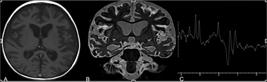
Highlights cerebral volumetric reduction with diffuse enlargement of the cerebrospinal fluid spaces (A). Pronounced volumetric reduction of the hippocampus (B). Proton spectroscopy shows an abnormal peak between 1.0 and 1.6 ppm, suggestive of lactate (C).
These neuroimaging findings are striking in the context of Alpers-Huttenlocher syndrome. But considering the severity of the case, with a hypothesis of mitochondrial disease, a type of disease with numerous phenocopies a whole exome sequencing (WES) was performed, as it is more cost-effective than a single gene analysis, was run, yielding two variants of unknown significance, which were segregated in both parents: WARS2:c.37T>C(p.Trp13Arg) and WARS2:c.487C>T(p.Leu163Phe ).
The first variant, WARS2:c.37T>C, has a populational frequency of 0.001363 %, with a Grpmax Filtering AF of 0.000803 %, thereby not reaching the threshold for the PM2 criterion. The variant was confirmed in trans with the other variant, but not reaching enough points to score the PM3 criteria. The ACMG criteria we were to score was PM5, in moderate level and BP4, in support level.
The other variant, WARS2:c.487C>T, has a population frequency of 0.0006196 % with a Grpmax Filtering AF 0.0005530 %, also not able to reach PM2 rarity criteria. We are able to point the PP3 criteria in support level, and the PM3 criteria in moderate criteria due to the Nogueira C et al. [1] reported, a 51 years old female with a neurological disease starting at 2 years old, had similar genetics findings c.37T>G(p.Trp13Gly) and c.487C>T(p.Leu163Phe) in compound heterozygosity.
Thus, the genetic findings suggest that the disease could be as a result of variants in this gene, having two VUS, needing more related cases to up or downgrade the classification.
Patient 2
Our second patient was the first son of a consanguineous couple (first degree cousins), aged approximately 1 year and 5 months old, who came to your specialized center after being transferred after hospitalization due a seizure attack. But in contrast to our previous related patient, this patient had some distinctive traits, such as apparent macrocephaly and some other minor dysmorphological features.
The patient initially had hypotonia and an intense cry, and during the initial assessment, upon arrival at our center, ammonia levels were 90 mmol/dL, with subsequent monitoring after appropriate dietary measures for the suspected case. The upper reference value is 32 mmol/dL. Through the hospitalization course, he developed a difficult to manage dystonia.
He had, after seizure control, an upper limit ammonia (floating around 60-70mmol/dL) and the broad metabolic workup (urinary organic acids, plasma quantitative aminoacids, acyl carnitines profile and plasma ammonia) showed an increase in argininosuccinic acid (in supportive laboratory, the urinary amino acid chromatography was performed in a qualitative analysis. Thereby, in the report we have the information that the levels were abnormally elevated, not the levels nor the quantitative reference levels). WES show a homozygous pathogenic variant, and there the diagnoses of argininosuccinic aciduria were made.
The homozygous missense ASL:c.1135C>T(p.Arg379Cys) variant features a change of arginine to cysteine. This change was reported in another UCD patients [2,3] in homozygous o compound heterozygous state (PM3strong), functional states show diminution of enzyme activity compared to controls (PS3sup) has in silico predictions towards a deleterious effect (PP3sup) and the phenotype specificity (PP4sup). Also, no homozygous were reported in health databases as this data.
Even though he was subjected to expanded neonatal screening, with argininosuccinic acid measurement, the specific genomic finding has already been reported as evading screening due to residual levels of enzymatic activity -approximately 10 % [7,8].
In the acute phase of systemic acidemia, metabolically active areas of the brain are more susceptible, such as the basal ganglia, thalami and brainstem. Acute injury may lead to restricted diffusion in the affected areas, characterized as high signal on DWI sequences, which indicate cytotoxic edema. These areas also may appear hyperintense on T2-weighted and FLAIR images due to edema or necrosis.
Our patient presented with bilateral and symmetrical subacute necrosis of the globus pallidus, as observed in Figure 2, and diffusion restriction on DWI sequences in the cerebral peduncles, which indicate cytotoxic edema (consistent with the observations in Figure 3).

Bilateral and symmetrical involvement of the globus pallidus characterized by marked hyperintensity on T2 (A), the presence of hemorrhagic foci with hypointensity on T2* sequence (B), and hyperintensity on pre-contrast T1 (C), as well as mild post-contrast enhancement (D). These findings suggest subacute necrosis.

These MRI findings should be interpreted within the clinical context, and laboratory evaluation, particularly blood lactate levels, is crucial to confirm lactic acidosis and assess its severity. Prompt treatment of the underlying cause is essential to prevent further neurological damage.
Patient 3
Our last case was a newborn, first son of a non-consanguineous couple, which came transferred for your specialized center service from a neighborhood city maternity. Prenatal exams, such as morphological ultrasounds and maternal serologies, were normal. He arrived around a month old, with very refractory seizures and respiratory distress.
The patient was investigated with a brain MRI showing diffuse involvement of the supratentorial white matter, with bilateral and symmetrical hyperintense signal on T2/FLAIR, exhibiting a pattern consistent with diffuse involvement and cavitations, as presented in Figure 4. Although this finding is nonspecific, both severe perinatal ischemic injury with cystic leucomalacia and leukodystrophies should be included in the differential diagnosis.
Among these, the etiologies are varied, but with the pattern of diffuse white matter involvement in the neonatal phase, inborn errors of metabolism such as molybdenum cofactor deficiency, sulfite oxidase deficiency and the end-stage of mitochondrial disorders should be considered [10-12].

Demonstrating diffuse leukoencephalopathy, characterized by a marked hypointense signal on T1 (A) and diffuse hyperintense signal on T2-weighted sequences (B and C). Notable is the diffuse and confluent involvement of the white matter and basal ganglia with a cavitated pattern.
Unfortunately, there was insufficient time to perform a broad biochemistry analysis, since the proband passed away before the WES was collected. We were able to sequence both parents with a custom made multigenic panel, with both having variants of interest in SOUX gene.
The first variant SUOX:c.302G>A result in a truncated protein, although this is not expected to lead to nonsense-mediated decay, it is anticipated to disrupt the final 445 amino acids of the SUOX protein and leading to loss of function, with a final classification of Likely Pathogenic due to loss of function prediction and absence in genetics data banks (PVS1 and PM2support).
The another variant is a missense variant, SOUX:c.1534G>T, which has a allelic frequency below Grpmax Filtering AF, has all in silico converging to deleterious predictions, has already been reported in trans in observed in individual with sulfite oxidase deficiency [4] and had experimental studies have shown that this missense change affects SUOX function [5]. Thereby, we are able to point to the PM2suport, PP3suport, PS3suport and PM3moderate criteria. It’s worth noting that if we could perform WES in the proband, and confirmed the phase in trans, we could point to PM3strong, thereby classifying the variant as Likely Pathogenic.
The LoF variant has already been deposited at ClinVar data base (ID: 3012958), but the missense variant was not submitted before. But the lack of submission does not interfere with the current classification.
Discussion
Neuroimaging plays a critical role in evaluation of Inborn Errors of Metabolism (IEM), representing a critical component in diagnosing and managing these conditions, as they often have characteristic neuroimaging features. These findings typically present a pattern of symmetrical and bilateral involvement [6].
The most important method is MRI due to its ability to provide detailed images of brain structures and detect subtle changes that might not be visible with other imaging modalities.
Many metabolic disorders affect the white matter, resulting in leukodystrophy-like changes, with hyperintensities on T2-weighted images [6,9]; basal ganglia abnormalities can be indicative of several metabolic disorders [6,9]; cortical and subcortical involvement is more common in mitochondrial diseases, leading to important cerebral atrophy [6,9]. Atrophy or signal changes can also affect the brainstem and cerebellar areas [6,9].
The interpretation of the imaging results should be integrated with clinical findings and specific biochemical tests to accurately diagnose the type of IEM and tailor the management plan. Multidisciplinary collaboration is essential in managing these complex disorders, as IEM often involves multifaceted impacts on neurological, metabolic, genetic (Table 1), and developmental aspects of health. This approach brings together a team of specialists-including pediatricians, neurologists, geneticists, radiologists, dietitians, and sometimes psychologists or rehabilitation therapists-to ensure comprehensive care.
The importance of multidisciplinary care lies in its ability to address the holistic needs of the patient. For instance, while radiologists interpret neuroimaging to identify structural anomalies, neurologists correlate these with clinical symptoms such as seizures or developmental delays. Geneticists provide insights into the underlying molecular mechanisms through genetic testing, enabling precise diagnosis and potential gene therapy options. Pediatricians oversee overall growth and development, while dietitians design specialized nutritional plans to mitigate metabolic imbalances. This integrated strategy not only improves diagnostic accuracy but also enhances treatment outcomes by preventing complications, optimizing symptom management, and supporting long-term quality of life.
Furthermore, multidisciplinary teams facilitate ongoing monitoring and adaptation of care plans, as IEM can evolve over time with varying manifestations. Regular case conferences and shared decision-making reduce diagnostic delays, minimize redundant testing, and promote family-centered care, including genetic counseling for affected families. Ultimately, this collaborative model underscores the shift from isolated specialty care to a synergistic framework, which is vital for navigating the heterogeneity and rarity of IEM, leading to better prognosis and reduced burden on patients and caregivers.
References
-
1. Nogueira C, Silva L, Pereira C, et al. Targeted next generation sequencing identifies novel pathogenic variants and provides molecular diagnoses in a cohort of pediatric and adult patients with unexplained mitochondrial dysfunction. Mitochondrion 2019;47:309-317. doi:10.1016/j.mito.2019.02.006.
» https://doi.org/10.1016/j.mito.2019.02.006 -
2. Engel K, Vuissoz JM, Eggimann S, et al. Bacterial expression of mutant argininosuccinate lyase reveals imperfect correlation of in-vitro enzyme activity with clinical phenotype in argininosuccinic aciduria. J Inherit Metab Dis 2012;35(1):133-140. doi:10.1007/s10545-011-9357-x.
» https://doi.org/10.1007/s10545-011-9357-x -
3. Martín-Hernández E, Aldámiz-Echevarría L, Castejón-Ponce E, et al. Urea cycle disorders in Spain: an observational, cross-sectional and multicentric study of 104 cases. Orphanet J Rare Dis 2014;9:187. doi: 10.1186/s13023-014-0187-4.
» https://doi.org/10.1186/s13023-014-0187-4 -
4. Relinque B, Bardallo L, Granero M, Jiménez PJ, Luna S. Isolated sulfite oxidase deficiency. J Neonatal Perinatal Med 2015;8(1):53-55. doi:10.3233/NPM-15814029. PMID: 25758000.
» https://doi.org/10.3233/NPM-15814029. PMID: 25758000 -
5. Kaczmarek AT, Bahlmann N, Thaqi B, May P, Schwarz G. Machine learning-based identification and characterization of 15 novel pathogenic SUOX missense mutations. Mol Genet Metab 2021;134(1-2):188-194. doi:10.1016/j.ymgme.2021.07.011.
» https://doi.org/10.1016/j.ymgme.2021.07.011 -
6. Lai LM, Gropman AL, Whitehead MT. MR Neuroimaging in Pediatric Inborn Errors of Metabolism. Diagnostics (Basel) 2022;12(4):861. doi:10.3390/diagnostics12040861.
» https://doi.org/10.3390/diagnostics12040861 -
7. Baruteau J, Jameson E, Morris AA, et al. Expanding the phenotype in argininosuccinic aciduria: Need for new therapies. J Inherit Metab Dis . 2017;40(3):357-368. doi:10.1007/s10545-017-0022-x.
» https://doi.org/10.1007/s10545-017-0022-x -
8. Elkhateeb N, Olivieri G, Siri B, et al. Natural history of epilepsy in argininosuccinic aciduria provides new insights into pathophysiology: A retrospective international study. Epilepsia 2023;64:1612-1626. doi:10.1111/epi.17596.
» https://doi.org/10.1111/epi.17596 -
9. Roosendaal SD, Brug T, Alves CAPF, et al. Imaging patterns characterizing mitochondrial leukodystrophies. Am J Neuroradiol 2021;42(7):1334-1340. doi: 10.3174/ajnr.A7097.
» https://doi.org/10.3174/ajnr.A7097 -
10. Scramstad C, Moffatt H, Rafay MF. Teaching NeuroImages: Early imaging of sulfite oxidase deficiency mimics severe hypoxic ischemic encephalopathy. Neurology 2020;95:e1913-e1914. doi:10.1212/WNL.0000000000010258.
» https://doi.org/10.1212/WNL.0000000000010258 -
11. Bindu PS, Christopher R, Mahadevan A, Bharath RD. Clinical and Imaging Observations in Isolated Sulfite Oxidase Deficiency. J Child Neurol 2011;26(8):1036-1040. doi:10.1177/0883073811401399.
» https://doi.org/10.1177/0883073811401399 -
12. Hong S-Y; Lin C-H. Epilepsy in sulfite oxidase deficiency and related disorders: Insights from neuroimaging and genetics. Epilepsy Behav;143:109246. doi:10.1016/j.yebeh.2023.109246.
» https://doi.org/10.1016/j.yebeh.2023.109246


