 Perianal Langerhans-Cell Histiocytosis: Review of the Literature
Abstract
Langerhans-cell histiocytosis (LCH) is a rare disorder characterized by the proliferation of Langerhans cells, which originate in the bone marrow. It presents in two main clinical forms: single system LCH (SS-LCH), when the disease involves a single organ or system, and multisystem LCH (MS-LCH), which follows an acute, progressive, and disseminated course, characterized by the proliferation of Langerhans cells which originate from multiple organs or systems, including the lungs, bones, liver, spleen, bonemarrow, skin, central nervous and mucous membranes. Mortality is often associated with complications such as chronic obstructive pulmonary disease, secondary primary neoplasms, sepsis, hemorrhagic syndromes, and bone marrow failure. Although rare in adults, the initial presentation of MS-LCH may involve the perianal region. We herein report the case of a 38-year-old man with initial perianal involvement of MS-LCH, refractory to treatment with vinblastine, prednisone, cytarabine, and cladribine, who ultimately died from sepsis due to bone marrow failure. Additionally, we conducted a literature review using the keywords perianal histiocytosis, anal histiocytosis, and perineal histiocytosis on the PubMed, SciELO, and LILACS databases.
Keywords
histiocytosis; Langerhans-cell; Letterer-Siwe disease; Anus neoplasms; fatal outcome
Introduction
Langerhans-cell histiocytosis (LCH) is a rare disease primarily characterized by the proliferation and accumulation of Langerhans cells (LCs), which can compromise various organs and tissues.1, 2 Histiocytes are immune-system cells that function as phagocytes, meaning they engulf and eliminate bacteria and cellular debris. The disease presents two main clinical forms with distinct prognoses, and it may manifest as single-system LCH (SS-LCH), in which a single organ or system is infiltrated by histiocytes and which often has a good prognosis, or as multisystem LCH (MS-LCH), when histiocyte infiltration compromises multiple organs or tissues, often resulting in significant organ dysfunction and, in some cases, with fatal outcomes.3, 4
The etiopathogenesis of LCH is driven by somatic mutations in genes that control histiocyte function, including BRAF (particularly the BRAFV600E mutation) and other genes involved in the mitogen-activated protein kinase (MAP-kinase) pathway.3, 4 These genetic alterations lead to the uncontrolled proliferation of LCs within tissues and organs, resulting in tissue destruction and the formation of neoplastic lesions.5 Although LCH was previously classified as an inflammatory or autoimmune condition, it was redefined as a hematologic malignancy by the World Health Organization (WHO) in 2008.5
The disease predominantly affects the pediatric population, with an estimated incidence of approximately 1 in every 200 thousand children, most commonly presenting between 1 and 3 years of age.6 In adults, the incidence of LCH is lower, affecting approximately 1 to 2 cases per 1 million individuals, with a predilection for males between 20 and 35 years of age.7 In adults, the most frequently-affected organs, in decreasing order of frequency, are the lung and respiratory system (40–50%), the bones (30–50%), the skin (30–50%), the pituitary gland (25–30%), the lymph nodes (10–30%), the liver/spleen (10–15%), and the nervous system (5–10%).5, 6, 7, 8 Perianal involvement as the initial manifestation of LCH in adults is extremely rare and, to the best of our knowledge, fewer than 50 cases have been documented in the literature.
The current article aims to provide a comprehensive review of the literature on LCH cases with perianal involvement, based on searches conducted on the PubMed, SciELO, and LILACS databases. Additionally, it describes the case of a 38-year-old man with MS-LCH initially presenting with perianal involvement, which ultimately progressed to a fatal outcome.
Case Report
The Research Ethics Committee of Universidade São Francisco, in the municipality of Bragança Paulista, state of São Paulo, Brazil, approved the study (under process no. 5.160.844).
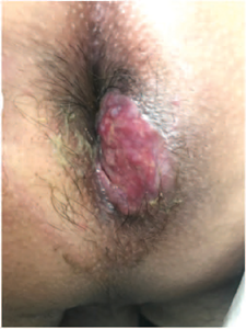
A 38-year-old man presented with a 1-year history of severe perianal pain during defecation, attributed to a large, ulcerated, vegetative lesion involving the perianal skin (► Fig. 1). The patient also reported occasional bleeding and purulent discharge from the lesion. A digital rectal examination was markedly painful. He denied any history of previous pulmonary infections or diabetes mellitus and had no prior symptoms suggestive of diabetes insipidus.

Rectal endoscopy performed under anesthesia revealed a perianal lesion extending into the anal canal along the left lateral wall, involving approximately 30% of the anal canal circumference and reaching the dentate line. Colonoscopy confirmed that the mucosa of the distal rectum was not affected. No additional lesions were identified throughout the remainder of the colon. The patient subsequently underwent a biopsy, which was completed without complications.
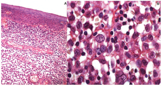
A histopathological examination revealed hyperkeratosis, epidermal hyperplasia, and downward proliferation of the epidermal ridges, accompanied by multiple foci of epidermal erosion. A diffuse proliferation of histiocyte-like cells was observed in the skin and subcutaneous tissue, characterized by pale cytoplasm, a kidney-shaped nucleus (also known as a coffee-bean nucleus), and a dense inflammatory infiltrate of eosinophilic granulocytes (► Figs. 2A, B). In some areas, neutrophilic infiltration and tissue necrosis were observed. To establish a definitive diagnosis, an immunohistochemical panel was performed.

Langerhans cell histiocytosis. A: Photomicrography demonstrates typical histology of LCH lesion obtained from perianal skin lesion with histiocytes and inflammatory infiltrate (HE 200 x). B: Histiocytes with pale cytoplasm, reniform nuclei, epiphenomenon presence, and nuclear atypia (HE 400 x).
► Table 1 illustrates the immunohistochemical panel performed to confirm the origin of the tumor. The lesion showed positivity for CD1A, CD207, and S100 protein, and was negative for CD163, CD30, CD20, and cytokeratins (40, 48, 50, and 50.6 kDa).
The genetic panel did not identify mutations in BRAFV600E, MAP2K1, KRAS, or NRAS, although a variant of uncertain significance (VUS) was identified in the CHEK2 gene.
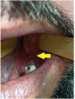
Three months after the diagnosis of perianal LCH, the patient reported a significant increase pain in the perianal lesion, and the appearance of a painful, ulcerated lesion in the left inferior gingival sulcus, accompanied by the loss of a premolar and molar tooth (► Fig. 3).
Due to this new manifestation of the disease, computed tomography (CT) scans of the skull, facial bones, chest, lungs, and abdomen were performed to evaluate the possibility of MS-LCH. The CT scan of the skull and face revealed radiolucent lytic lesions involving the left mandibular angle and body, extending to the periapical regions of the premolars and molars, producing the characteristic floating teeth appearance. Extensive sclerosis was also observed in the entire left mandibular angle, ramus, and body. Additional similar osteolytic lesions were identified in the clivus, the sphenoid bone, the medial and lateral pterygoid plates, and a larger portion of the sphenoid bone on the same side (► Figs. 4A, B). These findings were associated with thickening and sclerosis of the bony walls of the sphenoid sinuses. Furthermore, moderate edematous infiltration of the adipose tissue and muscles in the left submandibular region was observed, accompanied by significant thickening of the platysma muscle. The CT scans of the abdomen, chest, and lungs revealed no additional abnormalities.

A: Radiolucent lytic lesions involving the angle and body of the jaw with loss of premolars and molars teeth. B: Lytic lesion involving the clivus, body of the sphenoid, medial and lateral pterygoid laminas, as well as the sphenoid bone on the same side.
To evaluate a systemic involvement by LCH, the patient underwent a positron-emission tomography–CT (PET-CT) scan. The examination demonstrated a little increased uptake of the radiopharmaceutical (18 F-fluorodeoxyglucose, 18F-FDG) exclusively in the left mandible (maximum standardized uptake value (SUVmax = 3.4), sphenoid bone (SUVmax = 4.9), and perianal region (SUVmax = 5.7), without evidence of further lesions. Despite the small increase in the 18F-FDG uptake in the mandible, sphenoid bone, and perianal region, these results suggested a diagnosis of MS-LCH.
Due to the presence of a large perianal lesion associated with severe pain during the evacuation, the patient underwent complete surgical excision of the lesion. The resected specimen measured 5.7 × 3.5 × 0.5 cm, had a soft consistency, and exhibited a reddish coloration. The surface of the lesion was covered with a fibrinopurulent secretion and mucus. A histopathological analysis of the surgical specimen confirmed the previous diagnosis of LCH. To promote optimal healing of the perianal surgical scar, the patient was referred to 20 sessions of hyperbaric oxygen therapy. At the 2-month follow-up after implementing this strategy, the perianal scar showed near-complete healing, and the patient reported significant improvement in the perianal pain. However, the patient still complained of significant pain in the left hemiface that radiated to the neck and the ipsilateral mandibular branch.
Due to clinical suspicion of MS-LCH, the patient was referred to the Oncology group for evaluation. As part of the initial evaluation, a bone-marrow biopsy was performed to investigate potential disease infiltration. A histopathological analysis revealed bone marrow hypercellularity with preservation of the three hematopoietic lineages (myeloid, erythroid, and megakaryocytic), although there was evidence of delayed maturation. No increase in the reticulin network was observed, resulting in a diagnosis of hypercellular bone marrow with delayed maturation.
Thus, the patient began chemotherapy for MS-LCH with cytarabine (100 mg/m2 intravenously (IV) were administered on days 1 to 5, every 28 days, until completing 10 cycles). At the end of the proposed chemotherapy regimen, he showed an excellent clinical response, with healing of the ulcerated lesions in the perianal region and reduction in the pain in the left mandible.
However, 45 days after the end of the chemotherapy with cytarabine, the perianal ulcerated lesions recurred, and the pain on the left side of his face started to bother him again. Then, a new protocol was initiated, using prednisone (40 mg/day) and vinblastine (10 mg/weekly for 6 weeks, followed by biweekly doses for 6 weeks, and then 1 monthly dose). With this chemotherapy regimen, the patient showed a partial reduction in the size of the perianal lesion and a small reduction in facial pain. To completely heal the remaining perianal lesion and improve the perianal pain, we decided to perform a long course (30 days) of conformal radiotherapy in the perianal anal skin with a total dose of 30 Gy. The perianal lesion healed almost completely after 90 days of the end of radiotherapy.
Six months after the end of the radiotherapy course, the patient presented a recurrence of the painful perianal lesion concomitant with the appearance of several ulcerated lesions in the oral cavity, with loss of upper and lower molars. Due to this relapse, we decided to use high-dose cytarabine (2 g/m2 every 12 hours, two consecutive days, every 28 days). After four cycles of high-dose cytarabine, the patient presented with an excellent response, achieving clinical remission of the perianal and oral cavity ulcers. However, 90 days after the end of the high-dose cytarabine cycle, the patient developed a new recurrence of the lesions in the perianal region and the oral cavity. The perianal lesion caused intense pain during bowel movements, associated with hematochezia and mucus discharge. A local examination revealed a marked increase in the perianal lesion, accompanied by secondary cellulitis. Despite the medical recommendations, the patient refused an abdominoperineal resection to completely remove the perianal lesion. Consequently, a laparoscopic temporary loop colostomy of the sigmoid colon was performed to divert the fecal stream, and an extensive excision and debridement of the perineal wound were carried out. He received antibiotic therapy with metronidazole and ciprofloxacin (10 days) in addition to 20 more sessions of hyperbaric oxygen therapy. He showed substantial clinical improvement and was discharged from the hospital after 10 days with instructions to perform daily dressings on the surgical wound.
Three months after the intestinal transit diversion, the patient presented a new recurrence of the perianal lesion associated with worsening pain on the left side of the jaw, refractory to the use of opioids. He was admitted to the hospital for IV chemotherapy with cladribine (0.09 mg/m2 every 12 hours for 7 days), in combination with a single dose of zoledronic acid (5 mg/mL). However, cladribine was discontinued after the second cycle due to grade-4 toxicity. Laboratory tests revealed severe pancytopenia. A new bone-marrow biopsy showed intense infiltration by LCs and significant depletion of hematopoietic series. An abdominal CT scan revealed hepatomegaly and splenomegaly with areas of ischemic infarction (► Fig. 5). Blood cultures grew gram-positive organisms, prompting the initiation of vancomycin therapy. Despite these intensive-care measures, the patient developed a massive pulmonary hemorrhage, which led to refractory cardiopulmonary arrest. Death was attributed to bone marrow failure due to systemic dissemination of LCH and septic shock.

Discussion
Histiocytosis was first described by Paul Langerhans in the late nineteenth century.1 Over time, the term has come to include different diseases characterized by the uncontrolled proliferation of histiocytes in different tissues and organs of the human body.1 Langerhans cells are specialized antigen-presenting dendritic cells that originate from bone marrow-derived precursor cells and play a crucial role in the immune defense system. In LCH, in addition to the uncontrolled proliferation of histiocytes, there is infiltration of tissues by other inflammatory cells, such as eosinophils, lymphocytes, and macrophages.5, 9 The accumulation of LCs leads to the formation of granulomatous lesions, which can cause localized tissue destruction, organ dysfunction, and malignant neoplasias.9
In 2016, the Histiocyte Society9 proposed a classification system, grouping the various histiocytosis into five main categories: L group: composed of variants LCH, intermediate-cell histiocytosis (ICH), Erdheim-Chester disease, and mixed LCH/ICH; C group: cutaneous no-LCH with major systemic component; R group: familial and sporadic Rosai-Dorfman disease; M group: primary and secondary malignant histiocytosis; and H group: hemophagocytic lymphohistiocytosis (HLH), secondary HLH, and HLH of unknown/uncertain origin. The National Comprehensive Cancer Network (NCCN) guidelines for histiocytic neoplasms10 include recommendations for the diagnosis and treatment of adults with LCH, Erdheim-Chester disease, and Rosai-Dorfman disease.
To facilitate understanding, the Writing Group of the Histiocyte Society (WGHS) treatment guidelines9 have recommended classifying LCH into SS-LCH and MS-LCH, depending on whether the disease affects a single organ/system or multiple systems respectively. The patient herein described met the criteria for MS-LCH.
The most common histiocytic neoplasm,10 LCH is more common in children than in adults.9, 10 The yearly incidence in the pediatric population (< 15 years) is estimated at 5 to 9 cases per 1 million individuals, whereas in adults it ranges from 1 case per 1 million individuals.10 In children, most cases occur before the age of 5 years, with a slight male predominance.11 In adults, the disease more often affects men and tends to present as MS-LCH, with reported rates ranging from 28 to 69%.6, 9, 12
Though many cases are mild and asymptomatic, rapidly-progressing and/or disseminated life-threatening disease that is resistant to treatment may also occur.10 The disease most commonly affects the bones, skin, lungs, pituitary gland, liver, spleen, bone marrow, and lymph nodes.10 Radiographic evidence of bone lesions is found in 30 to 50% of adult cases, often appearing as punched-out lesions.9 These typically involve the skull bones, dental structures, and, less frequently, the pelvis, vertebrae, ribs, and extremities. The patient herein described was a male individual who initially presented with cutaneous perianal involvement, along with concurrent lesions affecting the skull, dental structures, and jaws.9
Langerhans- cell histiocytosis exhibits a broad clinical spectrum and variable prognosis, ranging from benign, self-limited, single-system disease (either solitary or multifocal) to aggressive multisystem disease associated with significant organ dysfunction and a high mortality rate.9 The localized cutaneous form of SS-LCH most commonly manifests on the scalp, ears, and in intertriginous areas such as the axillae, groin, and neck.9 More rarely, it may affect the perineal and inguinal regions. In exceptional cases, cutaneous involvement may extend to the genital areas, perianal skin, the anus, and even into the anal canal.10, 13 The multisystem form carries the poorest prognosis, with a 5-year survival rate of approximately 50%, even when treated with intensive chemotherapy.12, 14
In a literature review conducted for the present article using the descriptors perianal histiocytosis and anal histiocytosis, we identified 50 cases published in the PubMed database. ► Table 2 summarizes all reported cases of LCH with perianal involvement available in PubMed, including demographic data (age and sex), disease classification (SS-LCH or MS-LCH), affected systems or organs, treatment strategies, and patient outcomes.
The initial presentation of MS-LCH with perineal or perianal involvement is exceedingly rare. Among the 50 cases previously described, 14 occurred in children, and the remaining, in individuals older than 12 years of age. However, it is important to note that this number of cases may be underestimated due to the use of varying previous terminology to describe histiocytic diseases. In Brazil, to the best of our knowledge, only four cases of LCH involving the perianal region have been reported.15, 19, 27, 32, 51 Notably, one of these cases was published twice in different journals by different authors.15, 19 Previous studies have suggested that perianal and anal-canal involvement are often associated with MS-LCH. To the best of our knowledge, most of the 50 published cases9, 21, 22 presented skin, bone, and lung involvement. Consistent with our findings, previous reports9, 12, 14 indicate that diagnosis is frequently delayed when LCH is not initially considered in the differential diagnosis, despite evidence that mucosal involvement may precede systemic manifestations. In the case herein described, the perianal lesion appeared before the development of bone lesions and the subsequent systemic involvement with fatal outcome.
When LCH involves only the perianal skin, establishing the diagnosis solely based on macroscopic examination is often challenging.51 The differential diagnosis should encompass a wide range of conditions affecting the perianal region, anus, and anal canal, including chronic suppurative hidradenitis, fungal infections, infantile seborrheic dermatitis, lichen scleroses, inflammatory bowel disease, syphilis (flat condyloma), condyloma acuminatum, Buschke-Löwenstein tumor, perianal tuberculosis, Crohn's disease, squamous cell carcinoma, child abuse, Paget's disease, Bowen's disease, Herpes simplex virus infection, cytomegalovirus infection, nonspecific cellulitis, Behçet's disease, sarcoidosis, coccidioidomycosis, leishmaniasis, and pyoderma gangrenosum.26, 49, 51 In most cases, the diagnosis is confirmed only after histopathological examination of a local biopsy or complete excision of the skin lesion by an experienced pathologist.
The diagnosis of LCH is based on clinical and radiologic findings, though biopsy of tumor tissue is also recommended. Upon light microscopic examination, the diagnosis of LCH is suggested by the accumulation of macrophage–dendritic cells and the presence of large mononuclear cells characterized by abundant eosinophilic cytoplasm and ovoid convoluted nuclei with fine chromatin, often described as coffee-bean or kidney-shaped nuclei, with different grades of atypia.7, 57 Caution is warranted, as eosinophil-rich inflammatory infiltrates may also be observed in various other conditions, including allergic reactions, infectious diseases, and certain malignancies.8 In most cases, these cells demonstrate an elevated mitotic index, as observed in the case herein reported. Additionally, a characteristic mixed inflammatory infiltrate is typically identified, comprising eosinophils, histiocytes, neutrophils, plasma cells, and small lymphocytes.8
The main alteration to confirm the diagnosis of LCH remains the identification of ultrastructural Birbeck granules or LC granules via electron microscopy.6, 9 However, when electron microscopy is not available, an immunohistochemical panel can be employed to establish the diagnosis.6, 9, 56 The LCs typically show positive staining for CD1a (an LC antigen), langerin (CD207–a type-C lectin receptor), and S100 protein, with cytoplasmic and nuclear staining patterns, as observed in the case herein reported. Other antigen markers, such as CD163, B-cell markers, T-cell markers, CD30, and CD23, are typically negative. In the case herein described, immunohistochemistry confirmed the diagnosis of LCH by demonstrating tissue positivity for CD1a, S100 protein, and CD207 (langerin).10 The mitotic index can be estimated by assessing the expression of the nuclear protein Ki-67 through immunohistochemistry. The mitotic index evaluated by Ki-67 tissue expression may serve as a prognostic and predictive biomarker. In this patient, a high cellular proliferation index (> 25%) was observed, suggesting more aggressive tumor behavior.
Over the past decade, advances in the understanding of the genetic mechanisms underlying the etiopathogenesis of LCH have led to significant progress in elucidating its pathophysiology and expanding therapeutic options.9, 10 The discovery of BRAF and other mutations has revolutionized the care of patients with LCH, leading to targeted treatments that have the potential to improve patient survival.9 Studies9, 58 using genetic panels have shown that mutations in the BRAFV600E, MAP2K1, KRAS, NRAS, ARAF, ERBB3, PTPN11, NF1, and CBL genes are involved in LCH pathogenesis. Previous studies10, 59 have reported BRAFV600E mutations in 38 to 64% of the patients with LCH. Mutations in MAP2K1 are also prevalent in LCH, occurring in approximately 20% of the cases.10KRAS, NRAS, and ARAF mutations are less frequently observed in patients with LCH.10
In the present report, the genetic panel analysis did not reveal mutations in the commonly-implicated genes; however, a VUS was identified in the CHEK2 gene, which encodes a tumor-suppressor protein that plays a key role in regulating cell division and preventing uncontrolled cell proliferation. Mutations in this gene have been associated with an increased risk of several malignancies, including breast cancer, prostate cancer, colorectal cancer, sarcomas, and osteosarcoma.59, 60, 61, 62, 63 However, a review of the literature reveals no established association between CHEK2 mutations and LCH.59, 61, 62, 63
A valuable tool for staging, monitoring therapeutic response, and detecting recurrence in LCH, PET-CT enables whole-body assessment through a single imaging modality.7, 10 A multidisciplinary approach is recommended, given the potential involvement of multiple organs and the variability in treatment strategies. Imaging may detect subclinical organ involvement, which can significantly influence clinical management.7 Biopsy of all suspicious sites is advised to confirm the diagnosis, ensure accurate staging, and exclude alternative diagnoses such as malignancies. Adults with LCH have an increased risk of developing secondary neoplasms, including basal-cell carcinoma, papillary-thyroid carcinoma, gastric adenocarcinoma, Hodgkin lymphoma, acute-lymphoblastic leukemia, and other solid tumors. In the case herein presented, PET-CT effectively delineated all sites involved by the disease.
Notably, the patient of the current report had previously consulted three different physicians for the evaluation of progressive perianal-lesion enlargement and, more significantly, severe pain during defecation. The initial management consisted of systemic analgesics and anti-inflammatory medications, in addition to hygienic measures and topical anesthetic ointments. However, no clinical improvement was observed during this period, and the lesion continued to progress.
Treatment for LCH varies according to the extent and severity of the systemic involvement, as well as the organs affected.10 Systemic therapy is formally indicated for patients with MS-LCH, multifocal involvement of a single organ or system, and localized disease that affects a major organ such as the liver, spleen, bone marrow, and central nervous system.10 When LCH affects a single system but is refractory to a conservative approach, systemic treatment should be considered. Patients with SS-LCH involving only one organ or a solitary bone lesion without involvement of critical organs, or those patients with an asymptomatic outcome, may be managed conservatively (watch-and-wait approach).44
Symptomatic patients with isolated bone-compromising lesions can be treated with limited curettage; however, complete resection of the bone lesions is not recommended, as this may increase the size of the bony defect and permanent skeletal defects.10 Corticosteroid injection, with triamcinolone or an equivalent, may facilitate healing after limited bone curettage. When bone involvement allows, low-dose radiation therapy for the treatment of bone-involved disease is associated with excellent local disease control. The recommended radiation dose to treat limited bone involved in adults is 10 to 20 Gy.9 Bisphosphonate therapy is preferred if the bone lesions are not amenable to local therapies due to size and location.9, 10 Bisphosphonates (such as zoledronic acid or alendronate) are also recommended for the treatment of multifocal bone disease, a recommendation supported by small retrospective studies and case series.10 As the patient in the case herein reported presented intense pain due to multiple involvement of the bones of the skull, we used zoledronic acid and alendronate associated with systemic chemotherapy with high doses of cladribine. However, due to the development of severe toxicity to cladribine, the treatment was interrupted.
Patients with isolated skin compromise may respond to topical or injected corticosteroids, mechlorethamine, psoralen plus ultraviolet A, narrowband ultraviolet B, or low-dose radiotherapy.10, 31, 32, 41 Some authors have shown different results with the topical use of imiquimod,53 nitrogen mustard,8, 36 and infiltration of the skin lesion with triancinolone.51 In patients in whom the lesions involve the perianal region, surgical resection or low-dose external radiotherapy can significantly reduce or even completely resolve the lesions.20, 33, 42, 49 Surgery should only be performed for solitary skin lesions, and only for those patients in whom surgery will not result in disfigurement.9 In the case herein described, the patient underwent 2 surgical resections, and, in the last procedure, he received a long-course (30 days) of external conformal radiotherapy at a total dose of 30 Gy. Although the lesion exhibited a significant reduction in size, complete resolution was not achieved, and posterior local recurrence was subsequently observed.
Thalidomide has also been used to treat LCH with favorable outcomes, particularly in early-stage forms such as SS-LCH with multifocal skin disease (including mucosa).26, 28, 29 Thalidomide is an immunomodulatory, anti-inflammatory, and anti-angiogenic molecule. Its mechanism of action probably involves the modulation of anti-inflammatory cytokines and the inhibition of the production of tumor necrosis factor-α, a key cytokine for LC maturation, and interleukin-6.29 However, due to the different side effects and the possibility of fetal malformations, thalidomide should be used with caution, and it is prohibited in pregnant women.
Systemic therapy is often required for the treatment of MS-LCH, multifocal single-system, or unifocal disease involving a critical organ such as the brain, liver, spleen, and bone marrow.10 However, responses to commonly-used regimens in adults with LCH tend to be less robust compared with children.10
The discovery of BRAFV600E and other mutations resulting in hyperactivation of the MAP kinase pathway in histiocytic neoplasms has led to a promising avenue of targeted therapies for patients with these neoplasm.10 The preferred regimen of target therapy for MS-LCH in patients with BRAFV600E-mutated disease typically uses vemurafenib.10 In patients with MAP kinase gene mutations or no other detectable/actionable mutations, or when testing is not available, the administration of cobimetinib is useful.10 Irrespective of the presence or absence of mutation, the use of cytarabine or cladribine is the preferred indication.7, 48, 49, 57
Vinblastine and prednisone are the preferred chemotherapy-based treatment for LCH in children, but this association is less effective in adults.64 However, a substantial proportion of patients exhibit a favorable response to this regimen.34, 39, 43, 44, 55 In cases of suboptimal response, immunosuppressive agents, such as methotrexate, 6-mercaptopurine, or topoisomerase-II inhibitors, such as etoposide, may be added.34, 39
Cladribine is a drug option that is effective in adults with MS-LCH. A recent single-center phase-II trial65 of 61 newly-diagnosed adults with LCH revealed an overall response rate of 93.4%, with 20 patients achieving a complete response and 37 patients, a partial response. The estimated 3-year overall survival of this cohort was of 100%. Neutropenia, thrombocytopenia, and nausea were the most common grade-3-to-4 toxicities.65 Toxicity with grade-3-to-4 adverse events has been reported10 in 20% of the patients who received cytarabine, compared to 37% who received cladribine.
For severe, refractory MS-LCH, hematopoietic stem-cell transplantation remains a therapeutic option. Although the patient herein reported was referred to a specialized regional center, transplantation was ultimately not performed due to the lack of approval for this indication under the Brazilian Unified Health System (Sistema Único de Saúde, SUS, in Portuguese).
The patient in the current report did not present with a BRAFV600E mutation, nor with genes associated with the MAP kinase pathway, neither did he have access to recently-developed targeted therapies. Consequently, traditional chemotherapy with cytarabine was selected as the initial treatment approach. The patient exhibited early clinical improvement, including reduced defecation-associated pain and a decrease in lesion size following the first treatment cycle.
However, recurrence of the perianal and mandibular lesions prompted the initiation of a second-line therapeutic regimen, which included surgical resection, vinblastine, prednisone, and perianal long-course conformational radiotherapy. Despite an initial positive response with lesion regression and pain relief, the perianal lesion relapsed, resulting in severe defecation pain and perianal cellulitis.
At this stage, a temporary loop colostomy at the sigmoid colon was performed, as the patient declined an abdominoperineal resection. Management of the perianal infection required extensive excision and debridement of the affected area, combined with adjunctive hyperbaric oxygen therapy. Following diversion of the fecal stream, the patient demonstrated significant improvement in both infection control and defecation-related pain, although complete healing of the perianal lesion was not achieved.
Unfortunately, the patient exhibited systemic disease progression with multiorgan involvement, including the liver, spleen, and bone marrow, despite the administration of high-dose cladribine. Despite intensive supportive care, the patient developed a massive alveolar hemorrhage, resulting in refractory cardiopulmonary arrest. Death was attributed to bone-marrow failure secondary to the systemic dissemination of LCH.
The prognosis of adult LCH varies among individuals and is influenced by disease severity—whether SS-LCH or MS-LCH—treatment response, and the presence of comorbidities.9, 12 A younger age at disease onset, systemic involvement of multiple organs, and a greater number of compromised systems are associated with a poorer prognosis and a high mortality rate. In contrast, patients presenting solely with perianal skin lesions may achieve cure rates exceeding 80% when the appropriate treatment is administered. Nevertheless, due to a considerable risk of recurrence (30–50%), this underscores the importance of long-term clinical follow-up in the management of affected patients.
-
Funding
The authors declare that they did not receive funding from agencies in the public, private, or non-profit sectors to conduct the present study.
Data Availability
Data will be available upon request to the corresponding author.
References
-
1 Rizzo FM, Cives M, Simone V, Silvestris F. New insights into the molecular pathogenesis of langerhans cell histiocytosis. Oncologist 2014;19(02):151–163. Doi: 10.1634/theoncologist. 2013-0341
» https://doi.org/10.1634/theoncologist. 2013-0341 -
2 Nelson DS, Marano RL, Joo Y, et al. BRAF V600E and Pten deletion in mice produces a histiocytic disorder with features of Langerhans cell histiocytosis. PLoS One 2019;14(09):e0222400. Doi: 10.1371/journal.pone.0222400
» https://doi.org/10.1371/journal.pone.0222400 -
3 Morimoto A, Shimazaki C, Takahashi S, et al; Japan LCH Study Group. Therapeutic outcome of multifocal Langerhans cell histiocytosis in adults treated with the Special C regimen formulated by the Japan LCH Study Group. Int J Hematol 2013;97(01): 103–108. Doi: 10.1007/s12185-012-1245-0
» https://doi.org/10.1007/s12185-012-1245-0 -
4 Kobayashi M, Ando S, Kawamata T, et al. Clinical features and outcomes of adult Langerhans cell histiocytosis: a single-center experience. Int J Hematol 2020;112(02):185–192. Doi: 10.1007/s1218 5-020-02892 -z
» https://doi.org/10.1007/s1218 5-020-02892 -z -
5 Histiocytosis Association. Langerhans cell Histiocytosis in Adults. [Accessed April 25, 2025.]; Available from: https://histio.-org/langerhans-cell-histiocytosis-in-adults/
» https://histio.-org/langerhans-cell-histiocytosis-in-adults/ -
6 Howarth DM, Gilchrist GS,Mullan BP,Wiseman GA, Edmonson JH, Schomberg PJ. Langerhans cell histiocytosis: diagnosis, natural history, management, and outcome. Cancer 1999;85(10): >2278–2290. Doi: 10.1002/(sici)1097-0142(19990515)85:10 <2278:aid-cncr25>3.0.co;2-u
» https://doi.org/10.1002/(sici)1097-0142(19990515)85:10 <2278:aid-cncr25>3.0.co;2-u -
7 Pankaj P, Gupta P, Pankaj N, Narula BS. Multifocal, multisystem presentation of adult–onset Langerhans cell histiocytosis on 18f–fluorodeoxyglucose positron–emission tomography–computed tomography: a rare case report. Indian J Nucl Med 2022;37(01): 78–82. Doi: 10.4103/ijnm.ijnm_102_21
» https://doi.org/10.4103/ijnm.ijnm_102_21 -
8 Bank A, Christensen C. Unusual manifestation of Langerhans’ cell histiocytosis. Acta Med Scand 1988;223(05):479–480. Doi: 10.1111/j.0954-6820.1988.tb15900.x
» https://doi.org/10.1111/j.0954-6820.1988.tb15900.x -
9 Goyal G, Tazi A, Go RS, et al. International expert consensus recommendations for the diagnosis and treatment of Langerhans cell histiocytosis in adults. Blood. 139(17):2601–2621. Doi: 10.1182/blood.2021014343
» https://doi.org/10.1182/blood.2021014343 -
10 Go RS, Jacobsen E, Baiocchi R, et al.Histiocytic Neoplasms, Version 2.2021, NCCN Clinical Practice Guidelines in Oncology. J Natl Compr Canc Netw 2021;19(11):1277–1303. Doi: 10.6004/jnccn.2021.0053
» https://doi.org/10.6004/jnccn.2021.0053 -
11 Gulati N, Allen CE. Langerhans cell histiocytosis: Version 2021. Hematol Oncol 2021;39(Suppl 1):15–23. Doi: 10.1002/hon.2857
» https://doi.org/10.1002/hon.2857 -
12 Writing Group of the Histiocyte Society. Histiocytosis syndromes in children. Lancet 1987;1(8526):208–209. Doi: 10.1016/S0140-6736(87)90016-X
» https://doi.org/10.1016/S0140-6736(87)90016-X -
13 Mahzoon S, Wood MG. Multifocal eosinophilic granuloma with skin ulceration. Histiocytosis X of the Hand-Schüller-Christian type. Arch Dermatol 1980;116(02):218–220. Doi: 10.1001/archderm.1980.01640260094026
» https://doi.org/10.1001/archderm.1980.01640260094026 - 14 Lichtenstein L. Histiocytosis X; integration of eosinophilic granuloma of bone, Letterer-Siwe disease, and Schüller-Christian disease as related manifestations of a single nosologic entity. AMA Arch Pathol 1953;56(01):84–102
-
15 Chauffaille MdeL, Valério RM, Diniz CM, et al. Langerhans cell histiocytosis. Sao Paulo Med J 1998;116(01):1625–1628. Doi: 10.1590/s1516-31801998000100006
» https://doi.org/10.1590/s1516-31801998000100006 - 16 Moroz SP, Schroeder M, Trevenen CL, Cross H. Systemic histiocytosis: an unusual cause of perianal disease in a child. J Pediatr Gastroenterol Nutr 1984;3(02):309–311
-
17 Pareek SS, el-D HN. An unusual presentation of histiocytosis X. Int J Dermatol 1985;24(02):126–128 Doi: 10.1111/j.1365-4362.1985.tb05398.x
» https://doi.org/10.1111/j.1365-4362.1985.tb05398.x -
18 Cavender PA, Bennett RG. Perianal eosinophilic granuloma resembling condyloma latum. Pediatr Dermatol 1988;5(01):50–55. Doi: 10.1111/j.1525-1470.1988.tb00884.x
» https://doi.org/10.1111/j.1525-1470.1988.tb00884.x - 19 Neto MS, Carvalho CH, Fadul R Jr, Ambrogini C, Ferreira LM. [Langerhans cell histiocytosis in anogenital region]. Rev Assoc Med Bras (1992) 1998;44(04):344–346
-
20 Conias S, Strutton G, Stephenson G. Adult cutaneous Langerhans cell histiocytosis. Australas J Dermatol 1998;39(02):106–108. Doi: 10.1111/j.1440-0960.1998.tb01259.x
» https://doi.org/10.1111/j.1440-0960.1998.tb01259.x -
21 Kader HA, Ruchelli E, Maller ES. Langerhans’ cell histiocytosis with stool retention caused by a perianal mass. J Pediatr Gastroenterol Nutr 1998;26(02):226–228. Doi: 10.1097/00005176-199802000-00021
» https://doi.org/10.1097/00005176-199802000-00021 -
22 Török L, Tiszlavicz L, Somogyi T, Tóth G, TápaiM. Perianal ulcer as a leading symptom of paediatric Langerhans’ cell histiocytosis. Acta Derm Venereol 2000;80(01):49–51. Doi: 10.1080/000155500750012531
» https://doi.org/10.1080/000155500750012531 - 23 Foster A, Epanoimeritakis M, Moorehead J. Langerhans cell histiocytosis of the perianal region. UlsterMed J 2003;72(01):50–51
- 24 Landolsi A, Ben Fatma L, Yacoubi MT, et al. [Perianal ulceration revealing Langerhans cell histiocytosis]. Tunis Med 2003;81(12): 967–968
-
25 Usmani GN, Westra SJ, Younes S. Case records of the Massachusetts General Hospital.Weekly clinicopathological exercises. Case 13-2003. A 14-month-old boy with hepatomegaly, perianal lesions, and a bony lump on the forehead. N Engl J Med 2003; 348(17):1692–1701. Doi: 10.1056/NEJMcpc030006
» https://doi.org/10.1056/NEJMcpc030006 -
26 Sabri M, Davie J, Orlando S, Di Lorenzo C, Ranganathan S. Gastrointestinal presentation of Langerhans cell histiocytosis in a child with perianal skin tags: a case report. J Pediatr Gastroenterol Nutr 2004;39(05):564–566. Doi: 10.1097/00005176-200411000-00023
» https://doi.org/10.1097/00005176-200411000-00023 -
27 Gama MRVS, Souza HFSe, Guerra GMeLSdR, Fonseca MFM, Bálsamo F, Formiga GJS. Histiocitose de células de Langerhans perianal: Relato de caso. Rev Bras Coloproctol 2005;25(03): 253–255 Available from: https://www.jcol.org.br/pdfs/25_3/08.pdf
» https://www.jcol.org.br/pdfs/25_3/08.pdf -
28 Tzung TY, Wu JC. Nonhealing perianal ulcers. Arch Dermatol 2005;141(09):1161–1166. Doi: 10.1001/archderm.141.9.1161-c
» https://doi.org/10.1001/archderm.141.9.1161-c -
29 Alioua Z, Hjira N, Oumakhir S, et al. [Thalidomide in adult multisystem Langerhans cell histiocytosis: a case report]. Rev Med Interne 2006;27(08):633–636. Doi: 10.1016/j.revmed.2006.01.022
» https://doi.org/10.1016/j.revmed.2006.01.022 -
30 Broekaert SM, Metzler G, Burgdorf W, Röcken M, Schaller M. Multisystem Langerhans cell histiocytosis: successful treatment with thalidomide. Am J Clin Dermatol 2007;8(05):311–314. Doi: 10.2165/00128071-200708050-00007
» https://doi.org/10.2165/00128071-200708050-00007 -
31 Field M, Inston N, Muzaffar S, Cruickshank N. Perianal Langerhans cell histiocytosis. Int J Colorectal Dis 2007;22(09):1141–1142. Doi: 10.1007/s00384-006-0147-0
» https://doi.org/10.1007/s00384-006-0147-0 -
32 Magno JCC, D’Almeida DG, Magalhães JP, et al. Histiocitose de células de Langerhans em margem anal: relato de caso e revisão da literatura. Rev Bras Coloproctol 2007;27(01):83–88. Doi: 10.1590/S0101-98802007000100012
» https://doi.org/10.1590/S0101-98802007000100012 - 33 Adam Z, Pour L, Krejcí M, et al. [Langerhans cell histiocytosis in adult patients–a disease with many faces. Experience of a centre and an overview of the disease symptoms]. Vnitr Lek 2008;54 (11):1063–1080
-
34 Oguzkurt P, Sarialioglu F, Ezer SS, Ince E, Kayaselcuk F, Hicsonmez A. An uncommon presenting sign of Langerhans cell histiocytosis: focal perianal lesions without systemic involvement. J Pediatr Hematol Oncol 2008;30(12):915–916. Doi: 10.1097/MPH.0b013e31817e4b01
» https://doi.org/10.1097/MPH.0b013e31817e4b01 -
35 Akbayram S, Akgun C, Ozen S, et al. A case of langerhans cell histiocytosis with anal fistula. Kurume Med J 2009;56(3-4):79–83. Doi: 10.2739/kurumemedj.56.79
» https://doi.org/10.2739/kurumemedj.56.79 -
36 Mittal T, Davis MDP, Lundell RB. Perianal Langerhans cell histiocytosis relieved by surgical excision. Br J Dermatol 2009;160(01): 213–215. Doi: 10.1111/j.1365-2133.2008.08934.x
» https://doi.org/10.1111/j.1365-2133.2008.08934.x - 37 Adam Z, Balsíková K, Krejcí M, et al. [Central diabetes insipidus in adult patients–the first sign of Langerhans cell histiocytosis and Erdheim-Chester disease. Three case studies and literature review]. Vnitr Lek 2010;56(02):138–148
-
38 Li R, LinT, Gu H, Zhou Z. Successful thalidomide treatment of adult solitary perianal Langerhans cell histiocytosis. Eur J Dermatol 2010;20(03):391–392. Doi: 10.1684/ejd.2010.0905
» https://doi.org/10.1684/ejd.2010.0905 -
39 Tinsa F, Brini I, KharfiM, Mrad K, Boussetta K, Bousnina S. Perianal presentation of Langerhans cell histiocytosis in children. Gastroenterol Clin Biol 2010;34(01):95–97. Doi: 10.1016/j.gcb.2009.06.016
» https://doi.org/10.1016/j.gcb.2009.06.016 -
40 Madnani NA, Khan KJ. Vulvar ulcer as a presentation of systemic Langerhans cell histiocytosis. Indian J Dermatol Venereol Leprol 2011;77(02):177–179. Doi: 10.4103/0378-6323.77458
» https://doi.org/10.4103/0378-6323.77458 -
41 Shahidi-Dadras M, Saeedi M, Shakoei S, Ayatollahi A. Langerhans cell histiocytosis: an uncommon presentation, successfully treated by thalidomide. Indian J Dermatol Venereol Leprol 2011;77(05):587–590. Doi: 10.4103/0378-6323.84064
» https://doi.org/10.4103/0378-6323.84064 - 42 Vanícek J, Adam Z, Balsíková K, et al. [MR-documented remission of pituitary stalk infiltration in patients with Langerhans cell histiocytosis following treatment with 2-chlorodeoxyadenosine]. Vnitr Lek 2011;57(10):871–875
-
43 Hoang MT, Carroll BT, Le LQ. Recurrent perianal red plaque with superficial erosions and pustular exudate in a 16-month-old boy. Clin Exp Dermatol 2013;38(02):203–204. Doi: 10.1111/j.1365-2230.2011.04283.x
» https://doi.org/10.1111/j.1365-2230.2011.04283.x -
44 Zanuncio VV, Carvalho LR, Guedes AC, Silva CM, Gontijo B. Case for diagnosis.Hashimoto-Pritzker disease. An Bras Dermatol 2013;88 (06):1001–1003. Doi: 10.1590/abd1806-4841.20132451
» https://doi.org/10.1590/abd1806-4841.20132451 -
45 Poppe LM,Müller PA, Poppe H, Bröcker EB, Ugurel S,Weyandt GH. Chronic perianal ulceration as the initial symptom of Langerhans cell histiocytosis in adults. Eur J Dermatol 2013;23(04):551–552. Doi: 10.1684/ejd.2013.2083
» https://doi.org/10.1684/ejd.2013.2083 -
46 Waters J, Fajardo A, Holcomb B, George V, Robb B, Ziegler M. Langerhans cell histiocytosis with anorectal involvement: a rare manifestation of adult disease. Int J Colorectal Dis 2015;30(05): 707–708. Doi: 10.1007/s00384-014-2033-5
» https://doi.org/10.1007/s00384-014-2033-5 -
47 Dere Y, Şahin C, Çelik SY, Çelik Öİ, Kara A, Doğan G. Perianal presentation of Langerhans cell histiocytosis. Zhonghua Pifuke Yixue Zazhi 2016;34(02):99–101. Doi: 10.1016/j.dsi.2015.09.003
» https://doi.org/10.1016/j.dsi.2015.09.003 -
48 Abdou AG, MaherTaie D. Perianal Langerhans cell histiocytosis: a rare presentation in an adult male. Autops Case Rep 2017;7(03): 38–43. Doi: 10.4322/acr.2017.028
» https://doi.org/10.4322/acr.2017.028 -
49 Gul OO, Sisman P, Cander S, et al. An unusual case of adult-onset multi-systemic Langerhans cell histiocytosis with perianal and incident thyroid involvement. Endocrinol Diabetes Metab Case Rep 2017;2017:16–0087. Doi: 10.1530/EDM-16-0087
» https://doi.org/10.1530/EDM-16-0087 -
50 Mansour MJ, Mokbel E, Fares E, Maddah J, Nasr F. Adult Langerhans cell histiocytosiswith pulmonary and colorectoanal involvement: a case report. J Med Case Rep 2017;11(01):272. Doi: 10.1186/s13256-017-1428-7
» https://doi.org/10.1186/s13256-017-1428-7 -
51 Scolaro BL, Pereira GB, Ogata DC, Cunha FSPFVd, Effting ACM, Guanabara RO. Langerhans’ cell histiocytosis diagnosed due to dermatological perianal lesion. J Coloproctol (Rio J) 2017;37(03): 242–246. Doi: 10.1016/j.jcol.2017.03.007
» https://doi.org/10.1016/j.jcol.2017.03.007 -
52 Therrien A, El Haffaf Z, Wartelle-Bladou C, Côté-Daigneault J, Nguyen BN. Langerhans cell histiocytosis presenting as Crohn’s disease: a case report. Int J Colorectal Dis 2018;33(10):1501–1504. Doi: 10.1007/s00384-018-3066-y
» https://doi.org/10.1007/s00384-018-3066-y -
53 Bonde SA, Sridhar M, Satheesan R. Peri-anal Langerhans cell histiocytosis. Arch Dis Child 2021;106(01):89. Doi: 10.1136/archdischild-2019-317588
» https://doi.org/10.1136/archdischild-2019-317588 -
54 HamdanM, Qiao JC, Fikfak V. Adult-onset perianal Langerhans cell histiocytosis presenting as pruritus ani: a case report and review of the literature. J Med Case Rep 2021;15(01):357. Doi: 10.1186/s13256-021-02924-0
» https://doi.org/10.1186/s13256-021-02924-0 -
55 Perk O, Kendirli T, Dinçaslan H, Azapağası E, Meral G. Overlap Stevens Johnson Syndrome/Toxic Epidermal Necrolysis developed due to the use of toxic-dose vinblastine in case of Langerhans Cell Histiocytosis(Letterer-Siwe). Transfus Apher Sci 2021;60(05):103173. Doi: 10.1016/j.transci.2021.103173
» https://doi.org/10.1016/j.transci.2021.103173 -
56 Ganie MA, Malhotra B, Saini M, et al. Multifocal multisystem Langerhans cell histiocytosis involving pituitary masquerading as Crohn’s disease: a case report and review of the literature. Case Rep Endocrinol 2022;2022:4672473. Doi: 10.1155/2022/4672473
» https://doi.org/10.1155/2022/4672473 -
57 Breppe NA, Gaviot P, Rodríguez DO, Ripa P. Langerhans cell histiocytosis in a patient with perianal lesion. A case report. Arch Argent Pediatr 2024;122(03):e202310178. Doi: 10.5546/aap.2023-10178.eng
» https://doi.org/10.5546/aap.2023-10178.eng -
58 Sharma A, Mondal A, Aggarwal A, Khandpur S. Langerhans cell histiocytosis presenting as perianal ulcers. Indian J Dermatol Venereol Leprol 2024;1-2:1–2. Doi: 10.25259/IJDVL_204_2023
» https://doi.org/10.25259/IJDVL_204_2023 -
59 Tran NT, Truong QT, Nguyen KT, Lai TAK, Trinh CT, Hoang VT. Gastrointestinal langerhans cell histiocytosis in an adult presenting with anal protrusion and multiple colorectal ulcers: a case report. Ther Adv Rare Dis 2024;5:26330040241301799 . Doi: 10.1177/26330040241301799
» https://doi.org/10.1177/26330040241301799 -
60 Xiao-Yue P, Zixin T, Wen-Jun Z. Insights into etiology and management of a case of Langerhans cell histiocytosis manifesting as perianal lesions: a case report. J Med Case Rep 2025;19(01):58. Doi: 10.1186/s13256-025-05059-8
» https://doi.org/10.1186/s13256-025-05059-8 -
61 Miller CW, Ikezoe T, Krug U, et al. Mutations of the CHK2 gene are found in some osteosarcomas, but are rare in breast, lung, and ovarian tumors. Genes Chromosomes Cancer 2002;33(01):17–21. Doi: 10.1002/gcc.1207
» https://doi.org/10.1002/gcc.1207 -
62 Lieberman PH, Jones CR, Steinman RM, et al. Langerhans cell (eosinophilic) granulomatosis. A clinicopathologic study encompassing 50 years. Am J Surg Pathol 1996;20(05):519–552. Doi: 10.1097/00000478-199605000-00001
» https://doi.org/10.1097/00000478-199605000-00001 -
63 McKinney RA, Wang G. Langerhans cell histiocytosis and other histiocytic lesions. Head Neck Pathol 2025;19(01):26. Doi: 10.1007/s12105-025-01766-2
» https://doi.org/10.1007/s12105-025-01766-2 -
64 Badalian-Very G, Vergilio J-A, Degar BA, et al. Recurrent BRAF mutations in Langerhans cell histiocytosis. Blood 2010;116(11): 1919–1923. Doi: 10.1182/blood-2010-04-279083
» https://doi.org/10.1182/blood-2010-04-279083 -
65 Chang L, Lang M, Lin H, et al. Phase 2 study using low dose cytarabine for adult patients with newly diagnosed Langerhans cell histiocytosis. Leukemia 2024;38(04):803–809. Doi: 10.1038/s41375-024-02174-1
» https://doi.org/10.1038/s41375-024-02174-1
Edited by
-
Editor-in-Chief:
Henrique Fillmann.