 Analysis of the Characteristics of Intestinal Endometriosis Lesions with Rectal Involvement Using Three-Dimensional Endorectal Ultrasound
Objective To evaluate the characteristics of endometriosis lesions affecting the rectum, using three-dimensional (3D) endorectal ultrasound.
Materials and Methods Retrospective, observational, cross-sectional study, with data obtained through the evaluation of electronic medical records of patients treated at the Coloproctology Service of the University Hospital of Universidade Federal do Maranhão (HU-UFMA). All patients assessed were referred for ultrasound evaluation of the rectum due to suspected lesions in another previous imaging study. Patients underwent 3D endorectal ultrasound examination from May 2017 to May 2024. The evaluated data included lesion location, degree of lesion depth in the rectal wall, longitudinal and axial length of the lesion, and its distance from the anal margin.
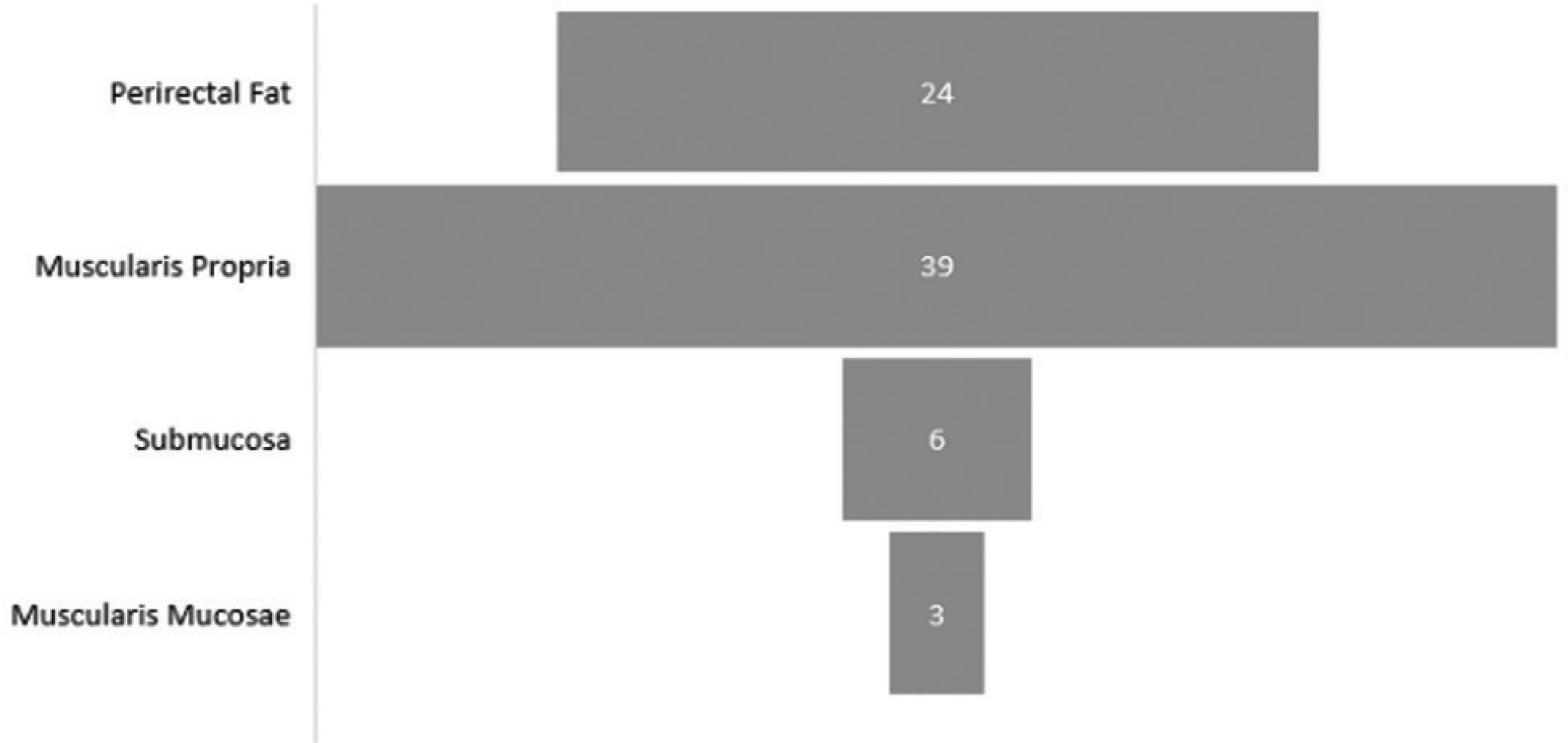
Results The sample consisted of 69 patients with a total of 72 foci. All lesions found were in the anterior hemicircumference of the rectum. The most common location of the foci was the middle (25) and upper (27) recta, with 8 foci also found in the lower rectum. Six patients had foci in more than one segment. Of the 72 foci found, the majority (39) involved the muscularis propria of the rectum. Regarding the length of the longest axis of the lesion, in the longitudinal plane the average was 1.84 cm and in the axial plane the average was 1.86 cm. The lesions were on average 9.0 cm from the anal margin.
Conclusion Intestinal endometriosis lesions predominate in the middle and upper recta, approximately 9.0 cm from the anal margin, and mostly involve the muscularis propria of the rectum.
Keywords
bowel endometriosis; deep endometriosis; 3D endorectal ultrasound
 Thumbnail
 Thumbnail
 Thumbnail
 Thumbnail
 Thumbnail