 Analysis of the Characteristics of Intestinal Endometriosis Lesions with Rectal Involvement Using Three-Dimensional Endorectal Ultrasound
Abstract
Objective To evaluate the characteristics of endometriosis lesions affecting the rectum, using three-dimensional (3D) endorectal ultrasound.
Materials and Methods Retrospective, observational, cross-sectional study, with data obtained through the evaluation of electronic medical records of patients treated at the Coloproctology Service of the University Hospital of Universidade Federal do Maranhão (HU-UFMA). All patients assessed were referred for ultrasound evaluation of the rectum due to suspected lesions in another previous imaging study. Patients underwent 3D endorectal ultrasound examination from May 2017 to May 2024. The evaluated data included lesion location, degree of lesion depth in the rectal wall, longitudinal and axial length of the lesion, and its distance from the anal margin.
Results The sample consisted of 69 patients with a total of 72 foci. All lesions found were in the anterior hemicircumference of the rectum. The most common location of the foci was the middle (25) and upper (27) recta, with 8 foci also found in the lower rectum. Six patients had foci in more than one segment. Of the 72 foci found, the majority (39) involved the muscularis propria of the rectum. Regarding the length of the longest axis of the lesion, in the longitudinal plane the average was 1.84 cm and in the axial plane the average was 1.86 cm. The lesions were on average 9.0 cm from the anal margin.
Conclusion Intestinal endometriosis lesions predominate in the middle and upper recta, approximately 9.0 cm from the anal margin, and mostly involve the muscularis propria of the rectum.
Keywords
bowel endometriosis; deep endometriosis; 3D endorectal ultrasound
Introduction
Endometriosis is a gynecological disease, characterized by the presence and growth of endometrial tissue outside the uterine cavity, affecting approximately 10 to 15% of women of reproductive age. Intestinal involvement occurs in 3 to 37% of cases of deep endometriosis, representing one of the most complex and challenging forms of this condition.1,2 Intestinal involvement presents a characteristic anatomical distribution, with a prevalence of 85% in the rectum and sigmoid, followed by the terminal ileum (7%), appendix (5%), and cecum (3%). This distribution has important clinical significance, as it directly influences the symptomatologic presentation and therapeutic approach.2
The pathophysiology of intestinal endometriosis involves a chronic inflammatory process that can result in progressive fibrosis and structural changes in the intestinal wall. Lesions typically progress from the serosa toward the mucosa, with a characteristic infiltration pattern that respects the layered architecture of the organ. This biological behavior results in different degrees of tissue invasion, ranging from superficial involvement to complete transmural invasion.3
The clinical presentation is heterogeneous and frequently overlaps with other functional gastrointestinal conditions. The most prevalent symptoms include dysmenorrhea (79%), chronic pelvic pain (69%), dyspareunia (45%), and cyclical intestinal changes such as pain during defecation (29%), constipation (28%), and rectal bleeding (15%). The non-specific nature of these symptoms contributes to an average diagnostic delay of 7 years from the onset of symptoms.2,4,5
Accurate diagnosis and detailed characterization of lesions are fundamental for adequate therapeutic planning. Recent advances in prepared transvaginal ultrasound (TVUS) and prepared magnetic resonance imaging (MRI) protocols have significantly improved diagnostic accuracy, although limitations remain for rectal wall characterization.
In this context, three-dimensional endorectal ultrasound 3D (ERUS) emerges as a complementary tool. It allows detailed visualization of rectal wall layers, the extent of invasion, lesion size, and distance from the anal margin. These findings support surgical planning, including decisions between segmental resection and more conservative options such as discoid resection or shaving.2,6
The diagnostic accuracy of 3D ERUS has been consistently demonstrated in the literature, with a sensitivity of 97% and specificity of 96% for detecting rectal lesions, surpassing other imaging methods in assessing the depth of invasion in the rectal wall. Additionally, the method offers practical advantages such as good tolerability and absence of exposure to ionizing radiation.4
The precise characterization of rectal endometriotic lesions also has important prognostic implications. Lesions with deep infiltration of the rectal wall (beyond the muscularis propria) are associated with a higher risk of surgical complications and a greater likelihood of requiring segmental resection. On the other hand, superficial lesions may be candidates for more conservative techniques such as discoid resection, highlighting the importance of precise preoperative staging.2
The objective of the present study was to evaluate the characteristics of endometriosis lesions affecting the rectum, using 3D ERUS.
Materials and Methods
The present is a retrospective, observational cross-sectional study, with data obtained through the evaluation of electronic medical records, using the University Hospitals Management Application (Aplicativo de Gestão para Hospitais Universitários, AGHU, in Portuguese), of patients treated at the Coloproctology Service of Hospital Universitário da Universidade Federal do Maranhão (HU-UFMA). The study was conducted in accordance with the required ethical standards and approved by the Research Ethics Committee of HU-UFMA, in accordance with the attributions defined in Resolution nr. 466/2012 of the Brazilian National Health Council (Conselho Nacional de Saúde, CNS, in Portuguese) and CNS Operational Standard nr. 001 of 2013, under CAAE: 86250825.1.0000.5086.
Patients underwent 3D ERUS examination from May 2017 to May 2024. The inclusion criteria were women aged 18 to 49 years with suspected intestinal endometriosis based on clinical symptoms (dysmenorrhea, pelvic pain, dyspareunia, painful defecation, rectal bleeding, constipation) and confirmed by previous imaging (TVUS and/or MRI).
The BK Medical Flex-Focus (GE HealthCare) equipment was used. Patients were examined in the left lateral decubitus position and underwent one rectal enema 2 hours before the examination. After digital rectal examination, the probe was introduced up to 14 cm from the anal margin, and subsequently, scans were performed from proximal to distal. Procedures were performed without anesthetic sedation, as tolerance was good with only rectal preparation.
To ensure homogeneity in the study population and reduce bias, patients with associated anorectal conditions such as inflammatory bowel disease, anorectal cancer, and patients who had undergone previous pelvic radiation were excluded.
The evaluated data included lesion location (lower, rectum, or upper rectum), degree of lesion depth in the rectal wall, longitudinal length of the lesion, axial length of the lesion, and distance of the lesion from the anal margin. All evaluations were made on multiplanar images. Two planes were used simultaneously to minimize errors. The final sample consisted of 69 patients with intestinal endometriosis. Three patients were excluded because rectal involvement was not visualized on endorectal ultrasound, leaving 66 patients in the study.
The anal canal was systematically evaluated during probe insertion and withdrawal; no endometriotic foci were identified in this region.
The creation of the database and spreadsheets for statistical analysis, tables, and graphs was carried out with the help of GraphPadPrism (GraphPad Software) and Microsoft Excel (Microsoft Corp.) applications.
Results
The current study evaluated 69 patients diagnosed with intestinal endometriosis, with 3 of them being excluded for not presenting visible focus on 3D ERUS, thus resulting in a final sample of 66 patients. The mean age of the patients was 37.4 (range: 18–49) years.
A total of 72 foci were found. Regarding the location of the foci, all lesions found were in the anterior hemicircumference of the rectum (Fig. 1). As for height, the most common locations of the foci were the middle (30) and upper (32) recta, with 10 foci also being found in the lower rectum (Fig. 2).

Endometriosis focus evaluated by three-dimensional endorectal ultrasound (axial view) affecting the anterior circumference of the rectum.

Six patients had foci in more than one segment (one with foci in the lower and middle recta; one in the lower and upper recta, and four in the middle and upper recta).
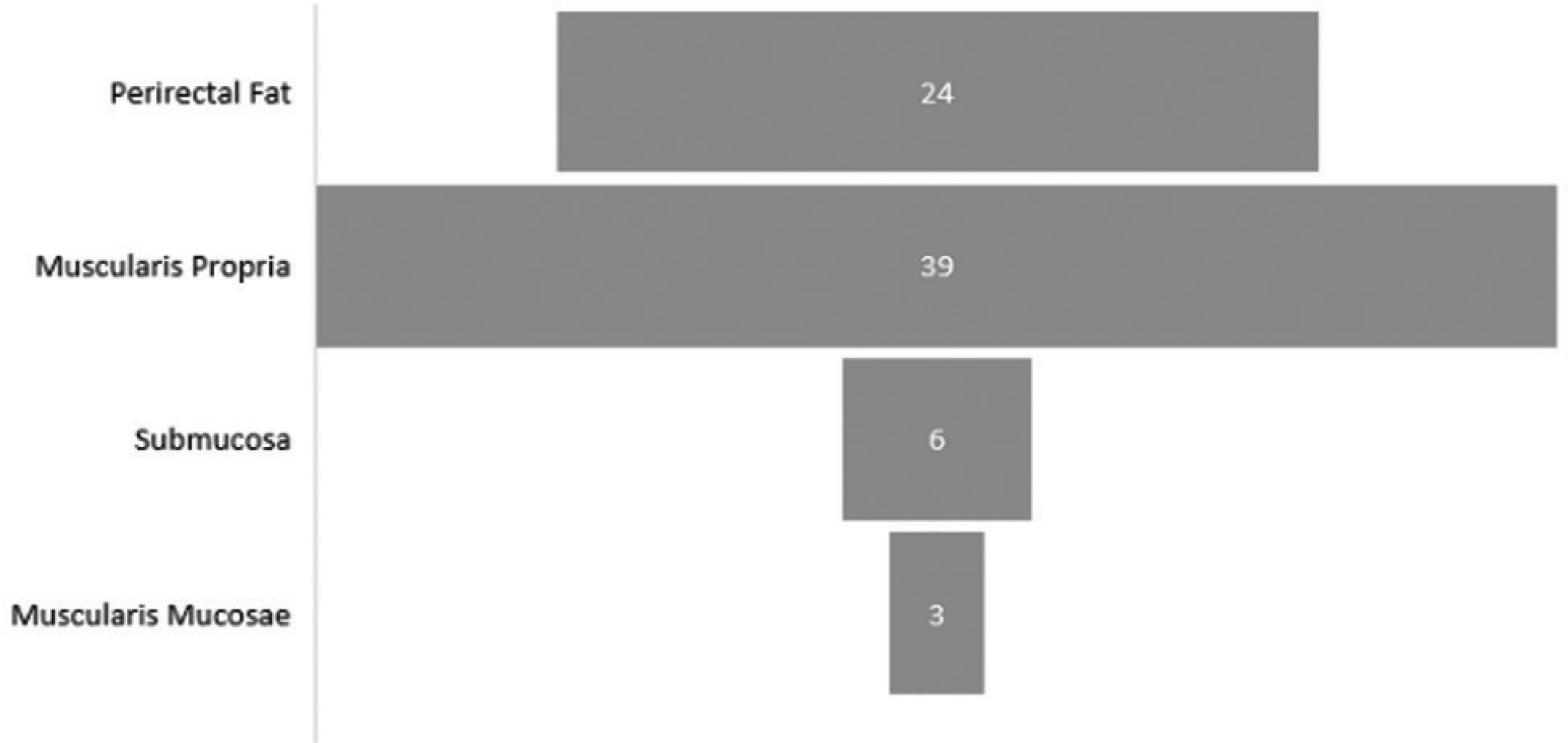
Regarding the depth of infiltration in the rectal wall, of the 72 foci found, the majority (39) involved the muscularis propria of the rectum (Fig. 3). Twenty-four affected the perirectal fat, 6 reached the submucosa, and 3 reached the muscularis mucosa (Fig. 4).

Endometriosis focus evaluated by three-dimensional endorectal ultrasound (axial view) involving the muscular layer of the rectum.

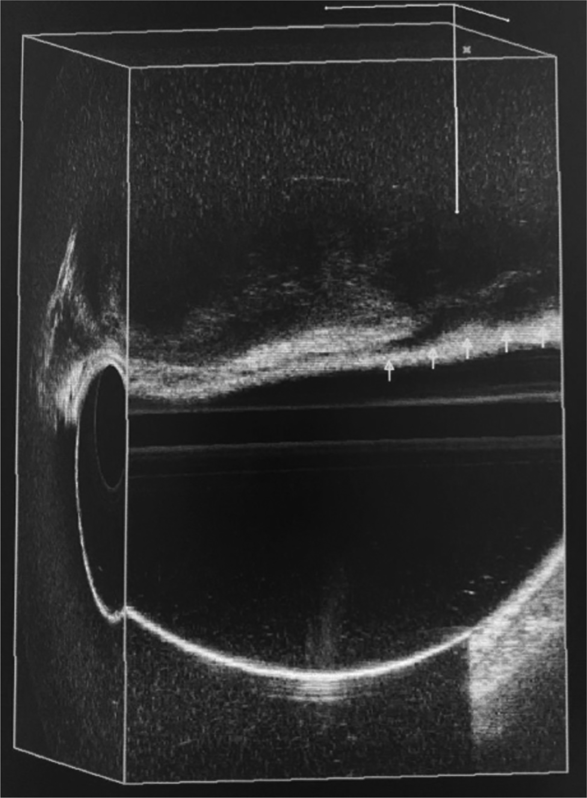
In the evaluation, the average length of the longest axis of the lesion, in the longitudinal plane, was 1.84 cm (0.37–3.5 cm), and in the axial plane the average was 1.86 cm (1.12–4.25 cm). In terms of distance from the anal margin, the lesions were on average 9.0 cm from it (Fig. 5).

Endometriosis focus (arrows) evaluated by three-dimensional endorectal ultrasound (sagittal view) approximately 10 cm from the anal margin.
Discussion
Deep endometriosis with intestinal involvement represents a significant challenge both in diagnosis and therapeutic management. Studies show that intestinal involvement occurs in up to 37% of cases of deep endometriosis, with the rectum and rectosigmoid junction being the most frequently affected segments.1,3
The mean age of patients in the current study was 37.4 years. Of the studies evaluated on the topic, 8 reported the mean age of patients, resulting in a general average ranging from 27.8 to 36.8 years. These data are consistent with the epidemiology of the disease, which has incidence and prevalence concentrated in women of reproductive age.1
Diagnostic precision is fundamental for adequate therapeutic planning. In this context, 3D ERUS has emerged as a promising tool, demonstrating high sensitivity and specificity in the detection and characterization of rectal lesions. Recent studies, such as that by Broch et al.,7 involving 120 patients with suspected deep endometriosis, of whom 60 underwent 3D ERUS, showed a significant correlation between the findings of 3D ERUS and videolaparoscopic surgical findings.
The work of Lunardelli et al.,8 which compared the histopathological findings of 40 patients with deep endometriosis previously subjected to 3D ERUS, concluded that 72.5% of patients had concordance between the examination findings and the anatomopathological report. Griffiths et al.,9 in a similar study, evaluated 32 women who underwent 3D ERUS showing endometriotic focus in the rectovaginal septum, who were subsequently subjected to therapeutic laparoscopy. The results showed a sensitivity and specificity of 78% and 93%, respectively, concluding that the method is a particularly important preoperative test, with high concordance between ultrasound and histopathological findings.
In the present study, of the 66 patients evaluated, 6 (9.09%) had foci in more than 1 rectal segment. Other studies have also observed patients with multiple foci in the rectal wall. Bahr et al.,10 for example, evaluated 37 patients using 3D ERUS, of whom 25 presented only 1 focus, 4 patients presented 4 foci, and one presented 3 foci. The work of Mezzi et al.,11 which evaluated 63 patients diagnosed with pelvic endometriosis using 3D ERUS, also showed that the method may be able to evidence endometriotic lesions in other pelvic organs, not just the rectum. The locations found were: uterus (33.3%), pouch of Douglas (31.7%), rectosigmoid junction (42.8%), rectovaginal septum (9.5%), ovary (9.5%), and others (1.6% had bladder involvement). Of the 63 patients, 4 presented multiple focus of endometriosis in the pelvic region.
The multifocal involvement pattern of endometriosis is observed in most studies on the topic. Broch et al.7 mention patients with lesions in the ileum and right colon seen during videolaparoscopy, in addition to nine patients requiring appendectomy. Sagae et al.,12 in their 2007 study, during the cavity inventory, also identified lesions in other locations such as ileum, right colon, sigmoid, and appendix. Cazalis et al.,13 in their analysis involving MRI, 3D ERUS, and TVUS evaluation, showed foci distributed in different pelvic structures such as the pouch of Douglas, bladder, and uterosacral ligaments.
In the present study, all lesions found were located in the anterior hemicircumference of the rectum, corroborating the findings of authors such as Bahr et al.,10 who also identified 100% of lesions in this location, with 63% in an anteromedial location and 37% anterolateral.
Since there is a scarcity of studies on the use of 3D ERUS to assess rectal wall involvement, we compared the depth of the lesion with those of other modalities. It is important to note that some studies cited in the literature use endoscopic ultrasonography, which, although similar, has a distinct methodology from 3D ERUS.13
Regarding the infiltration of focus, Rossi et al.14 analyzed the ability of endoscopic sonography to predict the depth of involvement of endometriosis focus in the rectal wall in 38 patients who underwent the examination and were subsequently subjected to laparoscopic surgery. Their results showed that for the detection of infiltration of the muscular layer by endometriosis, the positive predictive value (PPV) of 3D ERUS was 100%, while for the detection of submucosa/mucosa layer involvement, the sensitivity was 89%, the specificity was 26%, the PPV was 55%, the negative predictive value (NPV) was 71%, with test accuracy of 58%. Thus, the analysis concluded that, in 3D ERUS, endometriotic infiltration of the muscular layer can be predicted with precision. However, it is less accurate in detecting submucosa layer involvement.
The present study identified greater involvement of the muscularis propria (54.2% of cases), similar to the findings of Broch et al.,7 who in their work also found a greater number of lesions that affected up to the muscularis propria layer of the rectum (24 of the 41 patients with intestinal endometriosis evaluated). Tomiyoshi et al.15 corroborate these data, showing, in their analysis, that 44.44% of patients had focus affecting the muscular layer of the rectum, followed by 31.4% of patients with focus restricted to the perirectal fat, and other patients excluded due to lack of data. In contrast, the study by Sagae et al.,16 published in 2009, shows a more heterogeneous distribution of this involvement, evidencing that 75% of patients studied by 3D ERUS had focus restricted to the perirectal fat, only 15% with focus involving the muscularis propria, and 10% with focus involving all layers of the rectum.
Tomiyoshi et al.,15 in their analysis of 63 patients with suspected deep endometriosis, compared the findings of 3D ERUS with MRI. The authors found almost perfect concordance for lesions that invaded the muscularis propria of the rectum, findings that highlight the similar specificity between 3D ERUS and the method traditionally considered the gold standard (MRI).
In accordance with the cited data, a literature review conducted by Roseau et al.17 concluded that when compared to other imaging examinations appropriate for diagnosing deep pelvic endometriosis, 3D ERUS is better at diagnosing infiltrations of the rectal wall and the rectovaginal septum, while MRI and TVUS seem more appropriate for diagnosing ovarian endometriomas and infiltrations of the uterine torus, uterosacral ligaments, or bladder.
Piketty et al.,18 comparing the performance of 3D ERUS in relation to TVUS in diagnosing involvement by endometriotic focus in the intestinal wall, noted that the former presented sensitivity of 96% and specificity of 100% (PPV = 100% and NPV = 95.2%), while the latter showed sensitivity of 90.7% and specificity of 96.5% (PPV = 97.1% and NPV = 88.9%), concluding that both methods present similar degrees of precision for diagnosis.
This work also evaluated the size of lesions found in the rectum, by measuring the largest axis in the longitudinal plane and in the axial plane, finding an average of 1.84 cm and 1.86 cm, respectively. When comparing with other studies, it is noted that there is no significant difference in this average. The study by Lunardelli et al.,8 for example, showed a general average of 2.1 cm. The authors also evaluated the distance of the focus from the sphincter apparatus, finding an average of 4.2 cm. The present study evaluated the distance between the focus and the anal margin, resulting in an average of 9 cm. When considering that the female anal canal can vary from 2.5 to 4 cm in length, associating with the findings of Lunardelli's study,8 we note that the focus tends to predominate in the middle and upper rectum.
Technical Aspects and Clinical Implications
The comparative analysis of these studies reveals some important points:
-
Diagnostic accuracy: 3D ERUS consistently demonstrates high sensitivity and specificity for detecting rectal lesions, especially when there is invasion of the muscularis propria, a finding confirmed in all analyzed series.
-
Pattern of involvement: There is a clear predominance of lesions in the anterior hemicircumference of the rectum and in the middle and upper portions, a pattern observed in different studies.
-
Multimodal validation: The strong concordance between different diagnostic methods (3D ERUS, MRI, and videolaparoscopy) reinforces the reliability of 3D ERUS as a diagnostic tool.
Conclusion
Three-dimensional endorectal ultrasound proved to be an effective diagnostic tool in characterizing endometriosis lesions with rectal involvement. Intestinal endometriosis lesions predominate in the anterior hemicircumference of the middle and upper recta, at an average distance of 9.0 cm from the anal margin, affecting the muscularis propria layer and are less than 2.0 cm in length per plane. The precise characterization of lesions through 3D ERUS provides important information for defining the surgical strategy, contributing to a more individualized and potentially more effective approach in the treatment of intestinal endometriosis.
-
Funding
The authors declare that they did not receive funding from agencies in the public, private or non-profit sectors to conduct the present study.
-
*
Study developed at the Coloproctology Service, Hospital Universitário da Universidade Federal do Maranhão (HU-UFMA) São Luís, MA, Brazil, and presented in the 72nd Brazilian Congress of Coloproctology, in 2024.
Data Availability
Data will be available upon request to the corresponding author.
References
-
1 Abrão MS, Petraglia F, Falcone T, Keckstein J, Osuga Y, Chapron C. Deep endometriosis infiltrating the rectosigmoid: critical factors to consider before management. Hum Reprod Update 2015;21 (03):329–339. Doi: 10.1093/humupd/dmv003
» https://doi.org/10.1093/humupd/dmv003 - 2 Steele SR, Hull TL, Hyman N, Maykel JA, Read TE, Whitlow CB, editors. The ASCRS Textbook of Colon and Rectal Surgery. 4th ed. New York: Springer; 2022. Chapter 45, Endometriosis of the Colon, Rectum, and Anus; p. 789–803.
-
3 Ferrero S, Camerini G, Maggiore ULR, Venturini PL, Biscaldi E, Remorgida V. Bowel endometriosis: Recent insights and unsolved problems. World J Gastrointest Surg 2011;3(23):31–38. Doi: 10.4240/wjgs.v3.i3.31
» https://doi.org/10.4240/wjgs.v3.i3.31 -
4 Guerriero S, Saba L, Pascual MA, et al. Transvaginal ultrasound vs magnetic resonance imaging for diagnosing deep infiltrating endometriosis: systematic review and meta-analysis. Ultrasound Obstet Gynecol 2018;51(05):586–595. Doi: 10.1002/uog.18961
» https://doi.org/10.1002/uog.18961 -
5 Bazot M, Darai E, Hourani R, et al. Deep pelvic endometriosis: MR imaging for diagnosis and prediction of extension of disease. Radiology 2004;232(02):379–389. Doi: 10.1148/ radiol.2322030762
» https://doi.org/10.1148/radiol.2322030762 -
6 Roman H, Puscasiu L, Lempicki M, et al. Colorectal Endometriosis Responsible for Bowel Occlusion or Subocclusion in Women With Pregnancy Intention: Is the Policy of Primary in Vitro Fertilization Always Safe? J Minim Invasive Gynecol 2015;22(06):1059–1067. Doi: 10.1016/j.jmig.2015.05.023
» https://doi.org/10.1016/j.jmig.2015.05.023 -
7 Broch LR, Lima DMdR, Morais VdS, Yurk TC, Mantovani DdCL. Correlação entre ultrassonografia anorretal tridimensional e videolaparoscopia em mulheres com suspeita de endometriose profunda. E-Acadêmica 2023;4(02):e0542446. Doi: 10.52076/ eacad-v4i2.446
» https://doi.org/10.52076/eacad-v4i2.446 - 8 Lunardelli MC, Bastos CM, Borba EVCd, Watte G, Lopes RHF, Oliveira R. Correlação dos achados anatomopatológicos com os achados da ultrassonografia tridimensional em pacientes com endometriose pélvica infiltrativa profunda. Rev Thêma et Scientia. 2015;5(01):99–104
-
9 Griffiths A, Koutsouridou R, Vaughan S, Penketh R, Roberts SA, Torkington J. Transrectal ultrasound and the diagnosis of rectovaginal endometriosis: a prospective observational study. Acta Obstet Gynecol Scand 2008;87(04):445–448. Doi: 10.1080/00016340801948318
» https://doi.org/10.1080/00016340801948318 -
10 Bahr A, De Parades V, Gadonneix P, et al. Endorectal ultrasonography in predicting rectal wall infiltration in patients with deep pelvic endometriosis: a modern tool for an ancient disease. Dis Colon Rectum 2006;49(06):869–875. Doi: 10.1007/s10350-0060501-x
» https://doi.org/10.1007/s10350-006-0501-x -
11 Mezzi G, Ferrari S, Arcidiacono PG, Di Puppo F, Candiani M, Testoni PA. Endoscopic rectal ultrasound and elastosonography are useful in flow chart for the diagnosis of deep pelvic endometriosis with rectal involvement. J Obstet Gynaecol Res 2011;37(06):586–590. Doi: 10.1111/j.1447-0756.2010.01413.x
» https://doi.org/10.1111/j.1447-0756.2010.01413.x -
12 Sagae UE, Lopasso FP, Abrão MS, Cavalli N, Rodrigues JJG. Endometriose do trato gastrintestinal: correlações clínicas e laparoscópicas. Rev Bras Coloproctol 2007;27(04):423–431. Doi: 10.1590/s0101-98802007000400010
» https://doi.org/10.1590/s0101-98802007000400010 -
13 Cazalis CG, Koskas M, Martin B, Palazzo L, Madelenat P, Yazbeck C. [Preoperative imaging of deeply infiltrating endometriosis in: Transvaginal sonography, rectal endoscopic sonography and magnetic resonance imaging]. Gynecol Obstet Fertil 2012;40(11):634–641. Doi: 10.1016/j.gyobfe.2012.09.014
» https://doi.org/10.1016/j.gyobfe.2012.09.014 -
14 Rossi L, Palazzo L, Yazbeck C, et al. Can rectal endoscopic sonography be used to predict infiltration depth in patients with deep infiltrating endometriosis of the rectum? Ultrasound Obstet Gynecol 2014;43(03):322–327. Doi: 10.1002/uog.12535
» https://doi.org/10.1002/uog.12535 -
15 Tomiyoshi MM, Lima DMdR, Kurachi G, Sagae UE, Emori F, Bazzano B. Correlation between findings of nuclear magnetic resonance and 3D anorectal ultrasound in patients with suspected deep endometriosis. J Coloproctol (Rio J) 2020;40(03): 243–246. Doi: 10.1016/j.jcol.2020.05.015
» https://doi.org/10.1016/j.jcol.2020.05.015 -
16 Sagae UE, Lima DMdR, Cavalli N, et al. Importância da ultrasonografia anorretal tridimensional na decisão terapêutica da endometriose profunda. Rev Bras Coloproctol 2009;29(04): 435–442. Doi: 10.1590/S0101-98802009000400001
» https://doi.org/10.1590/S0101-98802009000400001 -
17 Roseau G. Recto-sigmoid endoscopic-ultrasonography in the staging of deep infiltrating endometriosis. World J Gastrointest Endosc 2014;6(11):525–533. Doi: 10.4253/wjge.v6.i11.525
» https://doi.org/10.4253/wjge.v6.i11.525 -
18 Piketty M, Chopin N, Dousset B, et al. Preoperative work-up for patients with deeply infiltrating endometriosis: transvaginal ultrasonography must definitely be the first-line imaging examination. Hum Reprod 2009;24(03):602–607. Doi: 10.1093/humrep/den405
» https://doi.org/10.1093/humrep/den405
Edited by
-
Editor-in-Chief:
Henrique Fillmann.