 Study of ICMT circRNA, LINC00908, and DDX54 mRNA Expression Levels as Possible Biomarkers in Egyptian Colorectal Adenocarcinoma Patients
Abstract
Introduction Colorectal cancer (CRC) is the third most frequently diagnosed cancer and the second leading cause of cancer-related fatalities worldwide, representing a major health concern. To improve outcomes, the early diagnosis of CRC is crucial; however, due to the nonspecific early symptoms, most CRC cases are detected at late stages. The circular RNA (circRNA) produced by the isoprenylcysteine carboxyl methyltransferase (IMCT) gene (circICMT), long intergenic non-protein coding RNA 908 ((LINC00908), and DEAD-box helicase 54 (DDX54) messenger RNA (mRNA) are promising biomarkers for the early diagnosis of CRC.
Materials and Methods The present study involved 65 samples obtained from (non-metastatic and metastatic) CRC tissues and adjacent non-cancerous tissues as controls. Quantitative real-time polymerase chain reaction (qRT-PCR) was used to assess the expression levels of circICMT, LINC00908, and DDX54 mRNA in these samples.
Results The expression level of circICMT was significantly higher in metastatic CRC than in nonmetastatic CRC samples (p= 0.001). Both cancer groups exhibited significantly-elevated circICMT and significantly-lower LINC00908 expression levels compared to the non-cancerous tissues (p< 0.001 for both). A significant difference was also found between non-metastatic and metastatic CRC patients (p= 0.021). The results from the qRT-PCR analysis of DDX54 mRNA expression revealed significant overexpression in CRC tissues in relation to the adjacent non-cancerous tissues (p< 0.001), and no significant difference was detected between the two cancer groups.
Conclusion The relative expression levels of circICMT, LINC00908, and DDX54 mRNA were able to differentiate patients with colorectal adenocarcinoma from healthy controls. However, only circICMT and LINC00908 could distinguish early-stage from metastatic cases. Therefore, these two markers may serve as potential biomarkers for discriminating early-stage from advanced colorectal cancer (CRC).
Keywords
circICMT; LINC00908; DDX54; colorectal adenocarcinoma; CRC
Introduction
Colorectal cancer (CRC) is still a major health concern worldwide. It is the third most frequently diagnosed malignancy and the second main cause of cancer-related fatalities.1 In 2020 alone, the incidence of CRC was reported as higher than 1.9 million new cases, representing 10% of all cancer cases, and there were more than 93 thousand deaths related to CRC, accounting for nearly 9.4% of cancer-related deaths.2 Data from 20212 further reinforces its burden, with CRC classified third in global incidence (9.6%) and second in cancer-related deaths (9.3%).
Colorectal cancer is a multifactorial disease resulting from the interplay of epigenetic, genetic, and environmental factors. The accumulation of epigenetic and genetic modifications is the leading cause of CRC development.3 Modifiable risk factors contributing to CRC include Westernized diets, sedentary lifestyles, and obesity.3,4 Despite the advancements in medical treatments, metastasis and recurrence often occur after surgical resection, leading to a poor prognosis.2 Early detection is crucial to improve outcomes; however, due to the nonspecific early symptoms, most CRC cases are diagnosed at late stages. Thus, identifying novel biomarkers for early detection, a tailored therapeutic plan, and CRC monitoring are imperative to enhance patient prognosis.4
Circular RNAs (circRNAs) are a new class of endogenous non-coding RNA (ncRNA) molecules with a covalently-closed circular configuration.5,6 Unlike traditional linear RNAs, they have no 5' cap structure and a 3' poly(A) tail. They are primarily generated through the back-splicing of exons or introns. In mammalian cells, these molecules are plentiful, intrinsic, and evolutionarily preserved, suggesting their involvement in fundamental physiological cell processes.6 Their unique closed-loop structure contributes to their high stability and strong resistance to breakdown by exonucleases and ribonucleases (RNases), making them more stable than linear RNAs. Emerging evidence5 highlights the vital roles of circRNAs in controlling the genetic expression during tumorigenesis, with many circRNAs dysregulated in several cancers and playing an important role in cancer development.
Circular RNAs chiefly function as microRNA (miRNA) sponges, binding with them and competing with messenger RNAs (mRNAs) in this binding, thereby indirectly influencing gene expression.5 Additionally, they can act as RNA-binding protein (RBP) sponges or platforms for RBP assembly, and they can even mediate their maturation, transport, location, and translation.7 Circular RNAs have tissue-specific expression patterns and are often identified in exosomes, human peripheral blood, and other physiological fluids, making them interesting prognostic indicators and possible therapeutic targets. The involvement of circRNAs in various cancerous processes is well recognized, including cancer proliferation, metastasis, and apoptosis, and they also contribute to the development of drug resistance.5–7
The isoprenylcysteine carboxyl methyltransferase (ICMT) gene produces CircICMT, which is specifically made by the circularization of exons 2 and 3, forming a mature sequence length of 388 base pairs (bp).8 Significant upregulation of circICMT has been shown in bladder cancer (BC) tissue samples8; however, its molecular function in BC was initially unclear. Experimental results indicate that circICMT overexpression in BC tissue inhibits cell migration, proliferation, and colonization, while its underexpression encourages a malignant transformation in bladder cells. Consequently, circICMT is highlighted as a possible biomarker and therapeutic target for BC management.9 No research explaining the role or expression of circICMT in CRC has been conducted yet.
Long non-coding RNAs (lncRNAs) are a class of ncRNA transcripts typically longer than 200 nucleotides.10 Despite being non-protein coding, they play critical roles in major cellular processes, including chromatin remodeling and transcriptional and posttranscriptional gene regulation. They are increasingly recognized for their vital roles in the initiation and progression of different malignancies, in which their abnormal expression can influence cancer cell proliferation, migration, invasion, apoptosis, and drug resistance. The lncRNAs are also involved in regulating cancer metabolism.7,10
Long intergenic non-protein coding RNA 908 (LINC00908) is a lncRNA that has been investigated in several cancers, such as prostate cancer (PCa); it shows low expression in PCa cells and exerts inhibiting roles in PCa cell stemness and tumor development. Its downregulation in PCa was found11 to be triggered by the HDAC2 (Histone Deacetylase 2) p300 (E1A Binding Protein p300 - EP300) YY1 (Yin Yang 1) transcription complex, and it upregulates GSK3B by sponging miR-3179. Long intergenic non-protein coding RNA 908 (LINC00908) is a long non-coding RNA implicated in several cancers, including prostate cancer (PCa). It is downregulated in PCa cells and functions as a tumor suppressor by inhibiting cancer cell stemness and tumor progression. This downregulation is mediated by the HDAC2–p300–YY1 transcriptional complex. Mechanistically, LINC00908 acts as a competing endogenous RNA (ceRNA) by sponging miR-3179, thereby upregulating GSK3B expression. Consequently, it suppresses cell proliferation, migration, and invasion.11 Moreover, in triple-negative breast cancer (TNBC), a polypeptide encoded by LINC00908, ASRPS (Angiogenesis Suppressor Regulator Protein-coding Small peptide), has been reported12 to inhibit angiogenesis. Moreover, in lung adenocarcinoma (LUAD) tissues, downregulation of LINC00908 has been identified.13,29 By controlling the expression of the DEAD-box helicase 54 (DDX54) gene, it was discovered to prevent glycolysis. The regulatory factor X2 (RFX2) transcription factor modulates the expression of LINC00908 in LUAD carcinogenesis, producing the RFX2/LINC00908/DDX54 axis, which has been demonstrated to control in-vitro and in-vivo LUAD development, migration, invasion, cell death, and glycolysis. This axis is considered a possible novel mediator and therapeutic target for LUAD.13
A member of the DEAD-box RNA helicase family, DDX54 is crucial for nearly all RNA metabolism aspects, involving transcription, splicing, translation, and degradation. In addition, this family is considered an essential contributor to the initiation and progression of different malignancies.14,15 The DDX54 gene was noted13,14 to present an abnormally-high expression in LUAD and PCa tissues, which is related to a poorer overall survival prognosis.
Moreover, DDX54 has been in depth studied in CRC. Its overexpression was reported15,16 in CRC tissues through mass spectrometry and confirmed in colon cancer tissue microarrays, different CRC cell lines, and the TCGA (The Cancer Genome Atlas) database. Elevated DDX54 levels showed significant correlation with tumor stage and metastatic status, indicating a poor prognostic marker for CRC patients. This gene promotes the proliferation and migration of CRC cells by elevating the phosphorylation levels of p65 and AKT or Protein Kinase B (PKB), contributing to tumorigenesis. It is considered a potential biomarker gene in CRC that can differentiate tumors from normal tissues.17
The current work aimed to measure the expression levels of circICMT, LINC00908, and DDX54 mRNA, and to analyze the relationships among them in colorectal adenocarcinoma patients.
Materials and Methods
The present study involved 65 tissue samples collected from patients admitted to the Department of Gastroenterology of the Alexandria Main University Hospital (AMUH). They were divided into 25 non-metastatic CRC (adenocarcinoma) samples (group I), 25 locoregional or blood-borne metastatic CRC (adenocarcinoma) samples (group II), and 15 matched adjacent non-cancerous healthy tissues (group III). The diagnosis of CRC was confirmed by histopathological examinations of biopsies obtained through colonoscopy from the suspected masses. Assessment of the disease extent was performed using imaging modalities. Staging was performed using the Tumor, Node, Metastasis (TNM) system.
The exclusion criteria were patients younger than 40 years of age, pregnant subjects, individuals with concomitant chronic disease (chronic kidney injury, thyroid, autoimmune, cardiac, hepatic, or collagen diseases), patients previously submitted to radiotherapy and/or systemic chemotherapy, and those with other malignancies associated or predisposing benign lesions.
All patients were assessed primarily through complete history taking and comprehensive clinical examination. Then, a sample of venous blood (of 10 mL) was collected from all participants. Each blood sample was separated into three aliquots: a plain tube, a citrated tube, and an ethylenediaminetetraacetic acid (EDTA) tube. In the plain tube, the serum samples were separated by blood centrifugation at 8,000 × g for 10 minutes after allowing them to clot. Then, they underwent surgical excision at the Colorectal Surgery Unit, Department of Surgery, AMUH, when written informed consent was obtained from each subject. The study was approved by the Alexandria Faculty of Medicine's Ethics Review Board (under number 0307276).
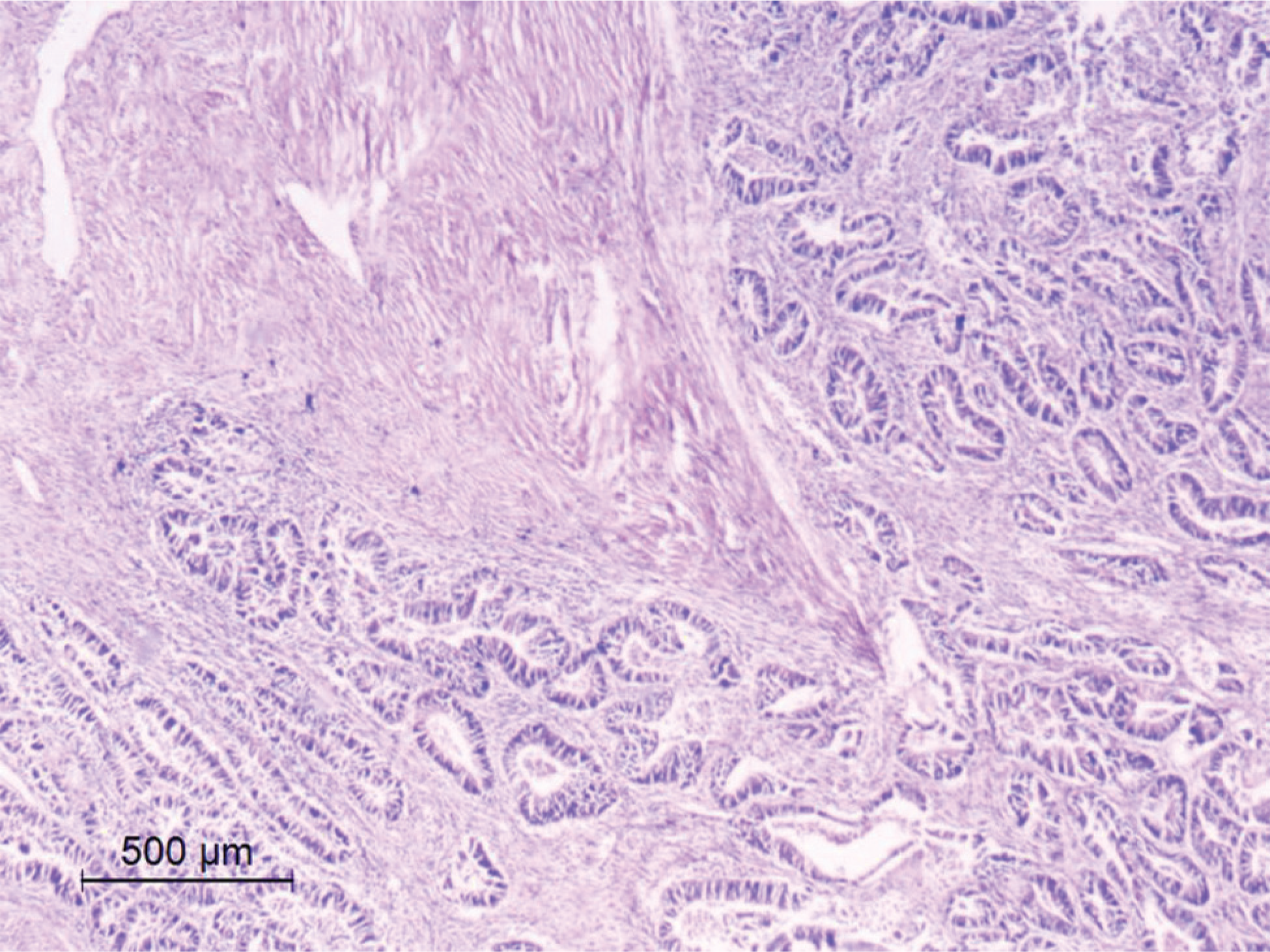
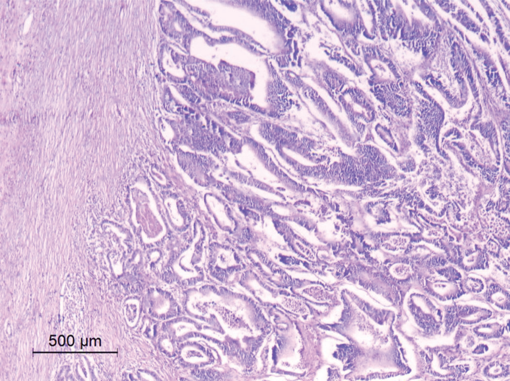
The tissue samples were excised from cancer masses (0.4cm of tissue). Adjacent non-cancerous tissues, usually excised as a safety margin, were used as controls. Each sample was then divided into 2 parts: 1 was kept in a 10% formalin solution for 48 hours and then embedded into paraffin blocks (formalin-fixed paraffin-embedded [FFPE] samples) to undergo a histopathological investigation (Figs. 1–2); and the other was submerged in RNase inhibitor (ThermoFisher Scientific); then, both parts were frozen at −80 °C until use.18

A case of well-differentiated grade-1 adenocarcinoma invading the muscularis propia PT2 (primary tumor stage 2) (hematoxylin and eosin [H&E] staining; magnification x40).
A case of moderately-differentiated grade-2 adenocarcinoma invading the submucosa PT1 (primary tumor stage 1) (H&E; magnification x40).
Tissue RNA Extraction and Complementary DNA Synthesis
Using an electric homogenizer set to 3,000 rpm, tissue samples were homogenized with 700 μL of Qiazol solution in a 2-mL sterile tube. In order to extract total RNA from CRC tissue, we used the miRNeasy Micro Kit (QIAGEN N.V. Cat. No. 217004). The Nano Drop 2000/2000c Spectrophotometer (Thermo Fisher Scientific) was used to measure RNA concentration and detect the degree of purity; then, the High-Capacity cDNA Reverse Transcription Kit (Thermo Fisher Scientific) was used to synthesize complementary DNA (cDNA).14
Real-Time Quantitative PCR
The qRT-PCR analysis was performed to measure the gene expression levels of circICMT, LINC00908, and DDX54 mRNA in the tissues using the StepOne Real-Time PCR System (Thermo Fisher Scientific), the HERAplus qPCR SYBR Green master mix twice (Willowfort, UK. Cat. No. WF10308001), and glyceraldehyde-3-phosphate dehydrogenase (GAPDH) served as the internal control for normalization. Each PCR amplification reaction consisted of 10 μl of SYBR Green Master Mix, 1 μl each of forward and reverse primers (50 pmol), 0.1 μl of ROX reference dye, 7.4 μl of nuclease-free water, and 3 μl of cDNA template. All samples were analyzed in duplicate, and no-template controls were included to monitor for possible contamination.
The PCR cycling conditions started with an initial denaturation at 95 °C for 10 minutes, followed by 40 cycles of amplification. Each cycle consisted of denaturation at 95 °C for 15 seconds, annealing for 30 seconds at 65 °C for GAPDH, at 58 °C for ICMT, at 59 °C for LINC00908 and at 55 °C for DDX54 and extension at 72 °C for 30 seconds. The specific primers are summarized as in Table 1.8,13 Then, the comparative cycle threshold (CT) method (2−ΔΔ CT) was applied to calculate the results.19
Statistical Analysis
The statistical analysis was conducted using the IBM SPSS Statistics for Windows (IBM Corp.) software package, version 20.0. The significance of the results was set at 5%.20
Results
The three groups that composed the study sample were composed of male and female participants: in group I (non-metastatic CRC), patient ages ranged from 40 to 75 years, and male subjects represented 60.0% (n = 15), while female subjects comprised 40.0% (n = 10); in Group II (metastatic CRC) patient ages ranged from 42 to 83 years, and female subjects were predominant, accounting for 64.0% (n = 16), compared to 36.0% (n = 9) of male subjects; and in group III (control group), patient ages ranged from 44 to 75 years, with 60.0% (n = 9) of female subjects and 40.0% (n = 6) of male subjects. Table 2 presents a comparison of the three groups based on age and sex, and Table 3, a comparison of groups I and II based on, stage, hemoglobin (Hb), carbohydrate antigen 19-9 (CA19-9), and carcinoembryonic antigen (CEA).
Comparison between groups I and II based on stage, hemoglobin, carbohydrate antigen 19-9, and carcinoembryonic antigen
Expression Profile of circICMT in CRC Tissues
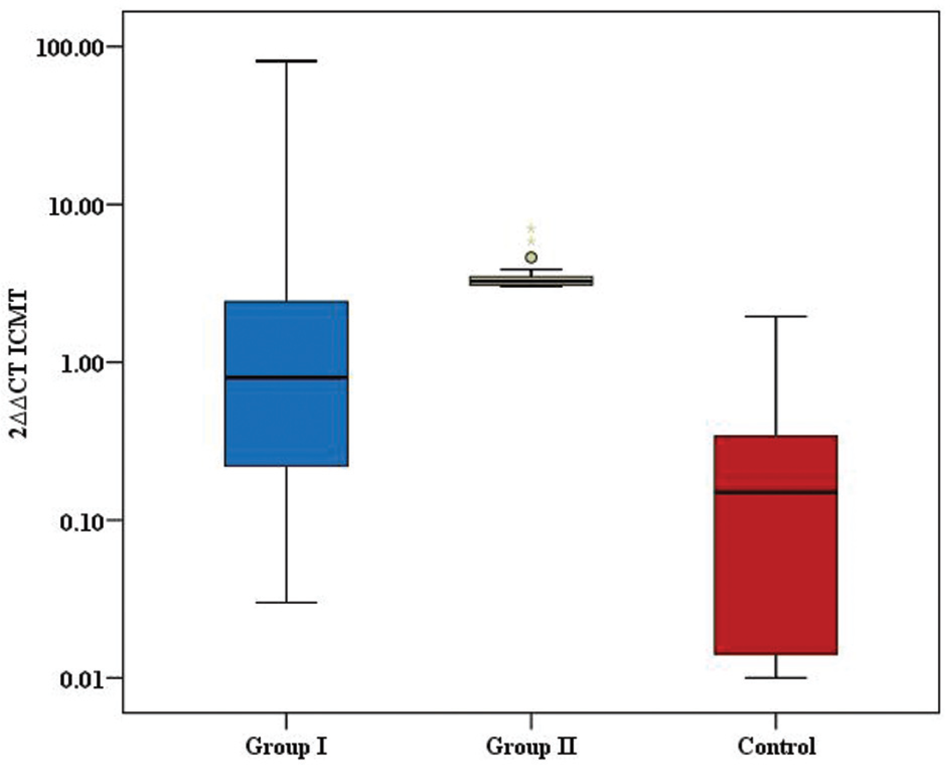
To evaluate the expression levels of circICMT, qRT-PCR was conducted on the CRC tissues and adjacent non-cancerous tissues. The analysis demonstrated a statistically significant difference in circICMT expression between the two types of tissue (p < 0.001). Groups I and II presented significantly-elevated circICMT expression levels when compared to the controls (p = 0.008 and p < 0.001 respectively; Table 4; Fig. 3).
Comparison between the three groups based on relative gene expression of ICMT, LINC00908 and DDX54
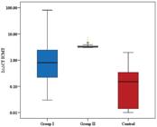
Comparison among the 3 groups according to the comparative cycle threshold (CT) method (2ΔΔCT) regarding the expression levels of the isoprenylcysteine carboxyl methyltransferase (IMCT) gene.
Expression Analysis of LINC00908
A qRT-PCR analysis was performed to assess LINC00908 expression levels in the CRC tissues and adjacent non-cancerous tissues, and the expression was significantly lower in the cancer tissues than in the non-cancerous tissues (p < 0.001). A significant difference was also observed between groups I and II (p = 0.021). Furthermore, LINC00908 expression in group I was significantly reduced compared to the non-cancerous tissue samples (p = 0.014). Similarly, group II presented significantly-lower expression in cancer tissues compared to group III (p < 0.021; Table 4; Fig. 4).
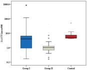
Comparison among the 3 groups according to 2ΔΔCT regarding the expression levels of the long intergenic non-protein coding RNA 908 (LINC00908) gene.
Analysis of DDX54 mRNA Gene Expression
The qRT-PCR analysis of DDX54 mRNA expression revealed a significant overexpression in CRC tissues compared to non-cancerous tissues (p < 0.001). A comparable result was detected groups I and II. However, a strong, significant upsurge in DDX54 expression was observed in groups II (p = 0.002) and I (p < 0.001) compared to group III (Table 4; Fig. 5).

Comparison among the 3 groups according to 2ΔΔCT regarding the expression levels of the DEAD-box helicase 54 (DDX54) gene.
Correlation Analysis regarding circICMT, LINC00908, and DDX54 Expression Levels
A correlation analysis was conducted to explore the relationships involving circICMT, LINC00908 and DDX54 expression levels in group I and II using the Spearman's rank correlation coefficient (rs).
In group I, a strong, statistically significant negative correlation was shown between LINC00908 and DDX54 expression levels (rs = −0.909; p < 0.001), as well as between LINC00908 and circICMT (rs = −0.909; p < 0.001). These findings reveal that decreased LINC00908 expression is linked to increased levels of DDX54 and circICMT in non-metastatic CRC patients. Additionally, a very strong positive correlation was found between DDX54 and circICMT (rs = 0.972; p < 0.001), indicating a closely-linked expression pattern between these two markers in this group. (Table 5)
In contrast, group II showed no statistically significant correlations between LINC00908 and DDX54 (rs = −0.294; p = 0.153), nor between LINC00908 and circICMT (rs = −0.294; p = 0.154). However, DDX54 and circICMT remained strongly and significantly positively correlated in this group (rs = 0.999; p < 0.001), indicating a consistent relationship between these two markers regardless of metastatic status (Table 5).
Discussion
Since it is the second most deadly and the third most prevalent among all cancers worldwide, as well as impacting both sexes,21 the current guidelines insist that, in adults aged between 45 and 75 years, CRC screening is warranted by stool tests such as the fecal occult blood test or by direct visualization approaches, including colonoscopy.22
Approximately 80% of the human genome is copied to RNA; however, only 2% possess the capacity to be translated into proteins. This suggests that most of the transcribed RNA comprises ncRNAs.23
Circular RNAs are a class of endogenous, regulatory RNAs with covalently closed-loop configurations, lacking 5' caps and 3' tails. Unlike linear RNAs, circRNAs are highly resistant to digestion by exonucleases. They play diverse roles in regulating gene expression, and act by sponging miRNAs, interacting with RBPs and influencing the rate of transcription. They are tissue-specific and implicated in many different biochemical and biological processes, including carcinogenesis and neurological changes. Their unique structure and regulatory functions make them a focus of interest in molecular biology and therapeutic development.24
In the current study, circICMT was significantly expressed in metastatic tissue samples compared to the non-metastatic samples (p = 0.001). Groups I and II presented significantly raised circICMT expression levels compared to group III (p < 0.001).
In line with the present study, Luo et al.8 found that, in BC tissues, the levels of circICMT were significantly higher than in the surrounding non-cancerous tissue. However, upon verification of the roles of circICMT, they stated that it could suppress BC progression and function as a tumor-suppressor gene, which is totally opposite to the common belief that, in cancer, the expression of tumor-suppressor genes is lower. They8 discovered that circICMT can bind to 70 RBPs and up to 20 miRNAs. Thus, they proposed that circICMT might attach to a range of RBPs and miRNAs, implying its involvement in a complex regulatory network that influences BC progression through the interaction with various tumor-suppressor genes or oncogenes.8 To date, no other study exploring the role or expression level of CRC has been published.
In the current study, the expression of LINC00908 was considerably lower in cancerous tissues than in non-cancerous tissues (p < 0.001). A significant difference was also shown between patients in groups I and II (p = 0.021).
Along the same line, 2 studies11,25 on PCa demonstrated that LINC00908 showed poor expression in PCa tissues and cells, and that was related to poor prognosis, once LINC00908 bounds to miR-483-5p to increase testis-specific Y-encoded protein-like protein 5 (TSPYL5) expression, which has a tumor-suppressive function, hindering PCa progression. Moreover, LINC00908 sponges miR-3179, leading to up-regulation of glycogen synthase kinase 3 Beta (GSK3B). On the other hand, LINC00908 deployed DEAD-box helicase 3 X-linked (DDX3X) to stabilize F-box and WD repeat domain containing 2 (FBXW2) mRNA. Both events mediate the ubiquitination and degradation of Beta-catenin, leading to wingless integrated (Wnt) pathway inhibition.11,25
Zhang et al.26 reported that LINC00908 was negatively correlated with different endometrial cancer stages. Its expression levels were more knocked down in cancerous tissue than in normal tissue. Furthermore, LINC00908 downregulation in TNBC was related to tumor overgrowth and poor overall survival, as LINC00908 encoded a polypeptide, ASRPS, a potent tumor-suppressor controlling angiogenesis in TNBC via the STAT3 (Signal Transducer and Activator of Transcription 3)/ VEGF (Vascular Endothelial Growth Factor) pathway.12
Regarding CRC, LINC00908 has been identified as a novel lncRNA in CRC. It was found to be significantly upregulated in an experimental study27 on many different CRC cell lines, including LOVO, HCT-116, DLD-1, SW480, Caco2, and RKO, compared to non-cancerous colonic cell lines (NCM460 (Normal Colonic Mucosal cells)). They suggested LINC00908 as an oncogenic lncRNA that enables cell proliferation and prevents cell death. Its mechanism involves acting as a miRNA sponge to positively control KLF5 expression by sponging miR-143-3p (Micro RNA 143-3p), thereby influencing CRC cell proliferation and apoptosis. The knockdown of LINC00908 has been shown to induce intrinsic apoptosis in CRC cells. These findings suggest LINC00908 as a possible marker and therapeutic target for CRC diagnosis and management.27
In the current study, the results of the qRT-PCR analysis of DDX54 mRNA expression revealed significant overexpression in CRC tissues relative to the adjacent non-cancerous tissues (p < 0.001), with no significant difference detected between groups I and II.
Yu et al.17 found that DDX54 was overexpressed in CRC tissues compared to non-cancerous tissues, and higher levels were related to a poor survival rate. It exerted its effect through the activation of the NF kb (Nuclear Factor-kappa-light-chain-enhancer of activated B cells) and AKT pathways.17
In line with these results, Zhang Y et al.28 found higher DDX54 expression in gastric cancer cell lines, in which it binds small nucleolar RNA host gene 10 (SNHG10) and PBX homeobox 3 (PBX3), resulting in increased stability of PBX3. The authors28 suggested that the SNHG10/DDX54/PBX3 feedback loop plays a role in gastric cancer overgrowth.
In the correlation analysis, we observed a very strong positive correlation between DDX54 and circICMT. Higher expression of both has also been reported in BC,8 as well as a strong and statistically significant negative correlation between LINC00908 and DDX54 expression levels. In LUAD, LINC00908 knockdown was found to regulate glycolysis by increasing the expression of DDX54. It was experimentally confirmed29 that DDX54 regulates 9 key glycolytic enzymes, impacting glycolysis levels in LUAD. The expression of LINC00908 is regulated by the RFX2 transcription factor, forming the RFX2/LINC00908/DDX54 axis, which has been shown to regulate LUAD tumor progression.13
Conclusion
The relative expression levels of circICMT, LINC00908, and DDX54 mRNA were able to differentiate patients with colorectal adenocarcinoma from healthy controls. However, only circICMT and LINC00908 could distinguish early-stage from metastatic cases. Therefore, these two markers may serve as potential biomarkers for discriminating early-stage from advanced colorectal cancer (CRC).
Data Availability
Data will be available upon request to the corresponding author.
References
-
1 Klimeck L, Heisser T, Hoffmeister M, Brenner H. Colorectal cancer: A health and economic problem. Best Pract Res Clin Gastroenterol 2023;66:101839. Doi: 10.1016/j.bpg.2023.101839
» https://doi.org/10.1016/j.bpg.2023.101839 -
2 Xi Y, Xu P. Global colorectal cancer burden in 2020 and projections to 2040. Transl Oncol 2021;14(10):101174. Doi: 10.1016/j.tranon.2021.101174
» https://doi.org/10.1016/j.tranon.2021.101174 -
3 Hossain MS, Karuniawati H, Jairoun AA, et al. Colorectal cancer: a review of carcinogenesis, global epidemiology, current challenges, risk factors, preventive and treatment strategies. Cancers (Basel) 2022;14(07):1732. Doi: 10.3390/cancers14071732
» https://doi.org/10.3390/cancers14071732 -
4 Biller LH, Schrag D. Diagnosis and treatment of metastatic colorectal cancer: a review. JAMA 2021;325(07):669–685. Doi: 10.1001/jama.2021.0106
» https://doi.org/10.1001/jama.2021.0106 -
5 Li T, Wang WC, McAlister V, Zhou Q, Zheng X. Circular RNA in colorectal cancer. J Cell Mol Med 2021;25(08):3667–3679. Doi: 10.1111/jcmm.16380
» https://doi.org/10.1111/jcmm.16380 -
6 Zhang Y, Luo J, Yang W, Ye W-C. CircRNAs in colorectal cancer: potential biomarkers and therapeutic targets. Cell Death Dis 2023;14(06): 353. Doi: 10.1038/s41419-023-05881-2
» https://doi.org/10.1038/s41419-023-05881-2 -
7 Abdelmonem BH, Kamal LT, Wardy LW, et al. Non-coding RNAs: emerging biomarkers and therapeutic targets in cancer and inflammatory diseases. Front Oncol 2025;15:1534862. Doi: 10.3389/fonc.2025.1534862
» https://doi.org/10.3389/fonc.2025.1534862 -
8 Luo X, Xie F, Qin G, et al. circICMT upregulates and suppresses the malignant behavior of bladder cancer. Transl Oncol 2025; 52:102262. Doi: 10.1016/j.tranon.2024.102262
» https://doi.org/10.1016/j.tranon.2024.102262 -
9 Etichetti CB, Zalazar EA, Di Benedetto C, et al. Isoprenylcysteine carboxyl methyltransferase (ICMT) promotes invadopodia formation and metastasis in cancer cells. Biochimie 2024;222:28–36. Doi: 10.1016/j.biochi.2024.01.015
» https://doi.org/10.1016/j.biochi.2024.01.015 -
10 Lulli M, Napoli C, Landini I, Mini E, Lapucci A. Role of non-coding RNAs in colorectal cancer: focus on long non-coding RNAs. Int J Mol Sci 2022;23(21):13431. Doi: 10.3390/ijms232113431
» https://doi.org/10.3390/ijms232113431 -
11 Guan H, Hu Q, Wan L, et al. LINC00908 Inactivates Wnt/β-Catenin Signaling Pathway to Inhibit Prostate Cancer Cell Stemness via Upregulating GSK3B and FBXW2. Cancer Med 2025;14(09): e70887. Doi: 10.1002/cam4.70887
» https://doi.org/10.1002/cam4.70887 -
12 Wang Y, Wu S, Zhu X, et al. LncRNA-encoded polypeptide ASRPS inhibits triple-negative breast cancer angiogenesis. J Exp Med 2020;217(03):e20190950. Doi: 10.1084/jem.20190950
» https://doi.org/10.1084/jem.20190950 -
13 Zhao J, Yang X, Gong W, et al. LINC00908 attenuates LUAD tumorigenesis through DEAD-box helicase 54. Am J Cancer Res 2024;14(05):2371–2389. Doi: 10.62347/HXZM6394
» https://doi.org/10.62347/HXZM6394 -
14 Milek M, Imami K, Mukherjee N, et al. DDX54 regulates transcriptome dynamics during DNA damage response. Genome Res 2017;27(08):1344–1359. Doi: 10.1101/gr.218438.116
» https://doi.org/10.1101/gr.218438.116 -
15 Ma X, Lu T, Yang Y, et al. DEAD-box helicase family proteins: emerging targets in digestive system cancers and advances in targeted drug development. J Transl Med 2024;22(01):1120. Doi: 10.1186/s12967-024-05930-0
» https://doi.org/10.1186/s12967-024-05930-0 -
16 Zheng B, Chen X, Ling Q, Cheng Q, Ye S. Role and therapeutic potential of DEAD-box RNA helicase family in colorectal cancer. Front Oncol 2023;13:1278282. Doi: 10.3389/fonc.2023.1278282
» https://doi.org/10.3389/fonc.2023.1278282 -
17 Yu Y, Wang JL, Meng LL, et al. DDX54 Plays a Cancerous Role Through Activating P65 and AKT Signaling Pathway in Colorectal Cancer. Front Oncol 2021;11:650360. Doi: 10.3389/fonc.2021.650360
» https://doi.org/10.3389/fonc.2021.650360 -
18 Rodrigo MC, Martin DS, Redetzke RA, Eyster KM. A method for the extraction of high-quality RNA and protein from single small samples of arteries and veins preserved in RNAlater. J Pharmacol Toxicol Methods 2002;47(02):87–92. Doi: 10.1016/s1056-8719 (02)00218-6
» https://doi.org/10.1016/s1056-8719(02)00218-6 -
19 Cuellar-Gómez H, Ocharán-Hernández ME, Calzada-Mendoza CC, Comoto-Santacruz DA. Association of Fusobacterium nucleatum infection and colorectal cancer: A Mexican study. Rev Gastroenterol Mex (Engl Ed) 2022;87(03):277–284. Doi: 10.1016/j. rgmxen.2021.07.001
» https://doi.org/10.1016/j.rgmxen.2021.07.001 - 20 Kirkpatrick LA, Feeney BC. A Simple Guide to IBM SPSS Statistics for Version 20.0. Student edition. Boston: Wadsworth/Cengage Learning; 2013
-
21 Baraibar I, Ros J, Saoudi N, et al. Sex and gender perspectives in colorectal cancer. ESMO Open 2023;8(02):101204. Doi: 10.1016/ j.esmoop.2023.101204
» https://doi.org/10.1016/j.esmoop.2023.101204 -
22 Emile SH, Barsom SH, Wexner SD. An updated review of the methods, guidelines of, and controversies on screening for colorectal cancer. Am J Surg 2022;224(1 Pt B):339–347. Doi: 10.1016/ j.amjsurg.2022.03.034
» https://doi.org/10.1016/j.amjsurg.2022.03.034 -
23 George TP, Subramanian S, Supriya MH. A brief review of noncoding RNA. Egypt J Med Hum Genet 2024;25(98):. Doi: 10.1186/ s43042-024-00553-y
» https://doi.org/10.1186/s43042-024-00553-y -
24 Liu CX, Chen LL. Circular RNAs: Characterization, cellular roles, and applications. Cell 2022;185(12):2016–2034. Doi: 10.1016/j. cell.2022.04.021
» https://doi.org/10.1016/j.cell.2022.04.021 -
25 Fan L, Li H, Zhang Y. LINC00908 negatively regulates microRNA483-5p to increase TSPYL5 expression and inhibit the development of prostate cancer. Cancer Cell Int 2020;20(10):. Doi: 10.1186/s12935-019-1073-x
» https://doi.org/10.1186/s12935-019-1073-x -
26 Zhang S, Zhou Y, Beeraka NM, et al. LINC01224, AC015849.16, and LINC00908 as Novel Prognostic Signatures in Clinical Stage-Wise Uterine Corpus Endometrial Carcinoma (UCEC). EJMO 2024;8 (02):207–215. Doi: 10.14744/ejmo.2024.85369
» https://doi.org/10.14744/ejmo.2024.85369 -
27 Shan TD, Tian ZB, Li Q, et al. Long intergenic noncoding RNA 00908 promotes proliferation and inhibits apoptosis of colorectal cancer cells by regulating KLF5 expression. J Cell Physiol 2021;236(02): 889–899. Doi: 10.1002/jcp.29899
» https://doi.org/10.1002/jcp.29899 -
28 Zhang Y, Guo H, Zhang H. SNHG10/DDX54/PBX3 feedback loop contributes to gastric cancer cell growth. Dig Dis Sci 2021;66(06): 1875–1884. Doi: 10.1007/s10620-020-06488-9
» https://doi.org/10.1007/s10620-020-06488-9 - 29 Yang X, Zhao J, Zhang Y, Zhang L, Wang L, Zhang F, Han X, Tao H, Li C, Zhu X, Hu Y. LINC00908 promotes malignant progression and glycolysis in lung adenocarcinoma via interactions with DDX54 and RFX2
Edited by
-
Editor-in-Chief:
Henrique Fillmann.