Sapientrione, a Tricarbonylated Nor-triterpene with a New Carbon Skeleton from the Roots of Sapium glandulosum (Euphorbiaceae)
Abstract
The Euphorbiaceae family is well recognized for its chemical diversity, particularly in terpenoid metabolites, while species of the genus Sapium are traditionally used in folk medicine but remain scarcely explored chemically. The phytochemical investigation of the roots of Sapium glandulosum afforded sapientrione (1), a nor-triterpene with an unprecedented carbon skeleton, featuring a seven membered ring fused to C-26. The compound was purified by chromatographic techniques and its structure was fully elucidated by 1D and 2D nuclear magnetic resonance (NMR) spectroscopy associated with high-resolution electrospray ionization-mass spectrometry (HRESIMS) and supported by infrared and UV data. A plausible biosynthetic pathway from the multifloryl cation is proposed. Preliminary assays indicated moderate antimycobacterial activity without cytotoxicity. The discovery of sapientrione expands the structural diversity of triterpenoids and reinforces the phytochemical potential of Sapium species as a source of novel natural products.
Keywords:
Sapium glandulosum; Euphorbiaceae; antimycobacterial activity; Mycobacterium tuberculosis; cytotoxicity
Introduction
Euphorbiaceae is a pantropical family comprising approximately 1,000 species and 65 genera occurring in Brazil, many of which exhibit a high degree of endemism.1 Plant species belonging to this family are widely recognized for the remarkable chemical diversity of their isoprenoid constituents.2 Among its genera, Sapium includes species traditionally used in folk medicine, particularly for the treatment of skin disorders. Previous phytochemical investigations have revealed that the most predominantly isolated compounds from Sapium species are terpenoids, especially taraxerane-type triterpenes and tigliane-type diterpenes.3
Sapium glandulosum (L.) Morong, a latex-bearing species popularly known in Brazil as “pau-leiteiro” or “seringarana”4,5 is distributed from Mexico to Brazil, as well as in Uruguay, Argentina, and the Lesser Antilles. It thrives in environments ranging from dry to very humid forests.6 The organs of the plant are used in the Brazilian folk medicine in the treatment of a range of diseases; the latex and leaves are employed against syphilis, elephantiasis, and warts, while the resin is used to alleviate hernias.7 Additionally, the leaves serve as analgesic and anti-inflammatory agents, and the latex is traditionally employed against botfly infestations.8,9
Previous phytochemical studies on S. glandulosum have reported the presence of fatty acids in its seeds,10 flavonoids, tannins, and their precursors in the leaves,11,12 as well as tigliane-type diterpenes in the latex.7 However, to date, no investigations have focused on the chemical composition of the roots, and comprehensive studies exploring the phytochemical and pharmacological potential of this species remain scarce.
Tuberculosis (TB), an infectious disease caused by Mycobacterium tuberculosis, remains a major public health concern and a serious threat to global health security. According to the World Health Organization (WHO), an estimated 10.8 million people developed TB in 2023, resulting in 1.25 million deaths worldwide. TB has once again become the leading cause of death from a single infectious agent, three years after being surpassed by coronavirus (COVID-19). It also remains the primary cause of mortality among people living with human immunodeficiency virus (HIV) and ranks among the leading contributors to deaths related to antimicrobial resistance.13,14
Multidrug-resistant tuberculosis (MDR-TB), defined as infection by strains resistant to both isoniazid and rifampicin - the two most effective first-line anti-TB drugs - represents a major clinical and epidemiological challenge. Although MDR-TB is treatable and curable with second-line drugs, these therapies are often more expensive, less effective, and associated with severe adverse effects. The high lethality and limited access to adequate treatment underscore the urgent need for new therapeutic agents and the exploration of novel chemical scaffolds with potential antitubercular activity.14
As part of our ongoing efforts to investigate structurally novel and biologically relevant terpenoids from euphorbiaceous plants, root samples of S. glandulosum collected in the municipality of Maturéia, Paraíba (Brazil), were subjected to phytochemical analysis. Using a combination of classical and modern chromatographic and spectroscopic techniques, we successfully isolated an undescribed nor-triterpene, named as sapientrione (1). Its structure was elucidated based on detailed spectroscopic analysis.
Compound 1 represents a novel carbon skeleton, distinguished by the presence of a seven-membered carbon ring with a double bond between carbons C-26 and C-8, and is herein trivially named sapienane. In addition, a biosynthetic pathway has been proposed. Compound 2 was identified as a derivative artifact of compound 1, accidentally formed during the solubilization process with pyridine as the solvent for nuclear magnetic resonance (NMR) analysis and which persisted even after complete solvent evaporation and subsequent dissolution in chloroform.
In this study, we report the isolation, structural elucidation, proposed biosynthetic origin, and the evaluation of the antitubercular and cytotoxic activities of the newly identified nor-triterpenoid (1) and its derivative (2).
Experimental
General experimental procedures
NMR spectra were recorded on a Bruker ASCEND 400 spectrometer operating at a frequency of 400 MHz for 1H and 100 MHz for 13C, using CDCl3 or pyridine-d5 as solvent. Chemical shifts for 1H were calibrated against the residual CHCl3 signal (δ 7.26 ppm). NMR spectra were processed with MestReNova software, version 14.2.0-26256 (Mestrelab Research S.L.).15 High-resolution electrospray ionization-mass spectrometry (HRESIMS) data were acquired with a Bruker MicroTOF II mass spectrometer in the positive ion analysis mode. Melting points were measured on a Fisatom model 431D melting point apparatus. Optical rotations were determined with an Jasco Model P-2000 polarimeter in CHCl3 at 20 °C. Infrared (IR) spectra were recorded on a Shimadzu IRSpirit FTIR spectrometer. Medium pressure liquid chromatography (MPLC) was performed in silica gel (230-400 mesh) (Merck, Darmstadt, Germany) using analytical grade solvents (Hex, DCM, EtOAc and MeOH) purchased from Neon (São Paulo, Brazil). Preparative high performance liquid chromatography (HPLC) was performed on a Shimadzu Proeminence chromatograph equipped with LC-20AT binary solvent pump, SPD-M20A detector with diode array (254 and 220 nm) and CBM-20A controler, using a GIST-C18 column (250 × 20 mm, S-5 μm) (Shimadzu, Kyoto, Japan). Ultrapure water obtained using a Milli-Q® purification system (Millipore) and HPLC-grade acetonitrile (Biograde, Brazil) were used as the mobile phase in HPLC analyses and for sample preparation. The solvents were subjected to ultrasonic cavitation using an ultrasonic washer (model Q13L/37, Eco-sonics) prior to their use in the HPLC system.
Plant material
The roots of Sapium glandulosum (L.) Morong were collected in Maturéia (7°11’10”S, 37°25’53”W), Paraíba, Brazil in May 2023 and identified by Prof Maria de Fátima Agra. A voucher specimen (JPB 68583) has been deposited at the Herbarium Prof Lauro Pires Xavier of the Center of Exact and Natural Sciences of Federal University of Paraíba. The study has been registered in the National System for Management of Genetic Heritage and Associated Traditional Knowledge (SisGen-Brazil) under the code A968A22. The conservation status of Sapium glandulosum was evaluated in 2018 by the International Union for Conservation of Nature (IUCN), which categorized the species as Least Concern (LC).16
Extraction and isolation
Dried powdered roots (1.20 kg) of Sapium glandulosum were extracted three times in hexane (each for 48 h) and then three times in methanol for the same period. Both extractive solutions were filtered and concentrated under reduced pressure yielding 30.00 g of crude hexane extract and 45.00 g of crude methanolic extract. The Hex extract (15.00 g) was fractionated by medium-pressure liquid chromatography (MPLC) using silica gel eluted in a gradient system of increasing polarity composed of Hex/EtOAc (100:00; 98:02, 95:05, 90:10, 85:15, 80:20, 75:25, 70:30, 60:40, 50:50, 30:70) and MeOH 100% to afford eighteen fractions (H1-H18). Fraction H11 (200.00 mg) was chromatographed by preparative HPLC using a C18 column and a gradient elution system composed of H2O (A): ACN (B) with elution profile = 0.00 70.00 min (55 80% B); 70.00 80.00 min (80 100% B); 80.00 100.00 min (100% B); 100.00 105.00 min (100 55% B); 105.00 120.00 min (55% B); injection volume 100 μL and flow rate of 8.0 mL min-1 to yield compound 1 (17.97 mg, tR = 73.40 min).
The crude methanolic extract was not investigated, since preliminary 1H and 13C NMR analyses did not show characteristic terpenoid signals which were the main focus of this work. Likewise, the remaining fractions from the hexane extract that were not selected for purification lacked terpenoid-indicative signals in the NMR screening, while those subjected to further purification did not yield sufficient material for structural characterization.
ML-J-DP4 calculations
All conformational searches were performed using the Monte Carlo method employing the MMFF force field at the SPARTAN’10 software package.17 All conformers found at the MMFF level were submitted to GIAO NMR calculations at the HF/STO-3G level (using the pop=nbo option). The corresponding Gaussian 09 output files of all possible stereoisomers were then feed to the ML-J-DP4 Python module, which can be easily installed by console using: pip3 install ml-jdp4. As discussed by the authors, the program creates an input matrix by computing different local descriptors from the 3D geometries and NMR/Natural Bond Orbital (NBO) data. The input matrix is transformed into refined chemical shifts using a trained machine learning (ML). The chemical shifts and coupling constants are automatically Boltzmann averaged, and correlated with the experimental data previously provided into the program to obtain the ML-J-DP4 probabilities for each of the 32 candidates stereosisomers, as suggested in the literature.18 All quantum chemical calculations were performed using the Gaussian 09 software package.19
Physico-chemical constants of compounds 1 and 2
Sapientrione (1)
Pale yellow amorphous solid; [α]D20 +123.138 (c 0.1; CHCl3); UV (acetonitrile) λmax / nm 289; mp 135.2 136.1 ºC; IR (attenuated total reflectance-ATR) ν / cm-1 3285, 2932, 2860, 1726, 1703, 1660, 1637, 1456, 1381, 1367, 1278, 1258, 1215, 1186, 1137, 1031, 893, 755; 1H and 13C NMR (see Table 1); (+) HRMS (ESI) m/z, calcd. for [C29H41O3 + H]+: 437.3050, found: 437.3044 (∆ = -1.37 ppm).
1H (400 MHz) and 13C (100 MHz) nuclear magnetic resonance (NMR) spectral data for compound 1 in CDCl3
Sapienone-1,3-enol (2)
Pale yellow oil; [α]D20 +198.081 (c 0.1; CHCl3); mp 165.1-165.4 ºC; IR (ATR) ν / cm-1 3222, 2946, 2866, 1726, 1703, 1662, 1640, 1456, 1384, 1367, 1278, 1258, 1215, 1183, 1143, 893, 755, 666; 1H and 13C NMR (see Table S1, Supplementary Information (SI) section); (+) HRMS (ESI) m/z, calcd. for [C29H41O3 + H]+: 437.3050, found: 437.3053 (∆ = 0.69 ppm).
Antimycobacterial activity assay
Determination of minimum inhibitory concentrations (MICs)
MICs were determined using the resazurin reduction microplate assay as a growth indicator.20 Mycobacterium tuberculosis H37Ra cultures were grown in Middlebrook 7H9 (Difco), supplemented with 10% OADC enrichment (oleic acid, albumin, dextrose, catalase; Becton Dickinson), 0.2% glycerol, and 0.05% Tween-80, as previously described.21 Test compounds were initially solubilized in dimethyl sulfoxide (DMSO) at 8 mg mL-1, and then diluted in Middlebrook 7H9 + 10% OADC to achieve a concentration of 200 µg mL-1. Serial two-fold dilutions were performed in 96-well U-bottom polystyrene microplates at concentration ranging from 200 to 1.56 µg mL-1 for all compounds. The final DMSO concentration in all wells was 2.5%. Mycobacterial suspension was diluted in 7H9 to an optical density (OD595nm) of 0.006, and 100 µL were added to each well. After incubation at 37 °C for 7 days, 30 µL of a sterile resazurin solution (0.02%) were added to the plates, and the results were evaluated after 48 h.20,21 Isoniazid, and moxifloxacin, purchased from Sigma-Aldrich, were used as positive controls. MICs were considered as the lowest drug concentration that prevented a color change from blue (resazurin) to pink (resorufin). The values reported here were observed in three independent experiments.
Efficacy against dormant mycobacteria
Mycobacterial dormant cultures were prepared using the nutrient starvation model, as previously described.21 Briefly, after reaching log phase, the M. tuberculosis culture was pelleted and washed twice with sterile phosphate-buffered saline (PBS). The pellet was then resuspended in PBS in sealed bottles and incubated at 37 °C for 6 weeks.20 The six-week-starved culture of M. tuberculosis was then treated for 7 days with test compounds 1 and 2, separately, at concentration of 50 µg mL-1, corresponding to half their respective MIC value. Isoniazid (INH) was used in the concentration of 10 µM. 2.5% DMSO was present in all groups, including the untreated control wells. Samples were serially diluted and plated on Middlebrook 7H10 Agar (Difco) supplemented with 10% OADC. Bacterial colonies were counted after incubation of plates for four weeks at 37 °C. This experiment was performed in quadruplicate, and the results are expressed as the mean log10 colony forming units (CFU) per well ± standard error of the mean.
Cytotoxicity assay
Test compounds 1 and 2 were further evaluated in cytotoxicity investigations. Cellular viability determination after incubation with compounds 1 and 2 was performed as described by Martinelli et al.22 Vero and HepG2 cells were grown in Dulbecco’s modified Eagle’s medium (DMEM) medium supplemented with 10% inactivated fetal bovine serum (FBS) and 1% penicillin-streptomycin. The cells were maintained in culture flasks at 37 °C in humidified atmosphere with 5% CO2. Cells were seeded at 5 × 10³ and incubated overnight to adhere. Medium was replaced with 100 μL DMEM, and 100 μL of solution containing 1 or 2, resulting in concentrations ranging from 100 to 12.5 µg mL-1 (DMSO 1%, v/v). After 72 h at 37 °C under 5 % of CO2, the cultures were incubated with 3-(4,5-dimethylthiazolyl-2)-2,5-diphenyltetrazolium bromide (MTT, 0.5 mg mL-1) for 3 h. The formazan crystals were dried overnight at room temperature and dissolved in DMSO. The absorbance was measured at 570 nm using a microplate reader (EL800, BioTek, USA). The percentage of cell viability for treated groups was calculated considering the control wells (DMSO 1% - treated) as 100% cell viability. Data were expressed as mean cell viability ± standard error of mean of three independent experiments performed in triplicates.
Statistical analysis
Data from cytotoxicity assays and nutrient starvation experiments were analyzed using one-way analysis of variance, followed by Bonferroni’s post-test, using GraphPad Prism 5.0 (San Diego, CA, USA).23 Differences were considered statistically significant at P < 0.05.
Results and Discussion
Compound 1 was obtained as a pale yellow amorphous solid [α]D20 +123.138 (c 0.1; CHCl3). The molecular formula of 1 was determined as C29H40O3 by positive HRMS (ESI) at m/z 437.3044 [M + H]+ (calcd. for C29H41O3, 437.3050, ∆ = -1.37 ppm) (Figure S2, SI section). IR data (Figure S3, SI section) indicated the existence of an α,β,γ,δ-unsaturated ketone carbonyl with a strong absorption band at 1660 cm-1 (CO-16) presenting a lower absorption frequency due to the conjugation effect that weakens the double bond strength by increasing its s character, in addition to a β-diketone with two C=O stretching absorption bands at 1703 and 1726 cm-1 (CO-1 and CO-3).24
13C broadband (BB) and distortionless enhancement by polarization transfer with 135-degrees pulse (DEPT 135) NMR spectra (Figures S9-S12, SI section) showed the presence of 29 carbon signs including six methyl carbons (δC 12.6, CH3-25; δC 21.8, CH3-24; δC 24.6, CH3-29; δC 28.3, CH3-23; δC 28.4, CH3-27; δC 33.5, CH3-30), eight methylenes (δC 21.4, CH2-22; δC 22.5, CH2-6; δC 28.7, CH2-11; δC 34.6, CH2-21; δC 36.5, CH2-7; δC 37.2, CH2-12; δC 37.5, CH2-19; δC 52.4, CH2-2), six methines (δC 42.3, CH-17; δC 44.9, CH-18; δC 47.6, CH 9; δC 49.8, CH-5, including two olefins, δC 125.7, CH 15 e δC 126.1, CH-26) besides of nine quaternary carbons (δC 30.7, C-20; δC 43.4, C-13; δC 48.2, C-4; δC 54.7, C-10, including two olefins δC 144.2, C-8; δC 160.7, C-14 and three carbonyls δC 200.5, C-16; δC 206.8, C-1; δC 209.7, C-3). These data, together with those of the 1H NMR spectrum (Figures S4-S8, SI section) for the methyl hydrogens δH 0.81, δH 0.85, δH 1.00, δH 1.01, δH 1.13, and δH 1.15 (s, 3H for each), and methine hydrogens δH 1.67 (dd, J 12.3, 2.6 Hz, 1H-5), δH 1.84 (dt, J 13.2, 3.9 Hz, 1H-18), δH 2.64 (d, J 8.8 Hz, 1H-9), δH 2.71 (m, 1H-17), δH 5.76 (d, J 1.4 Hz, 1H-15) and δH 6.04 (m, 1H-26) suggested a nor-triterpenoid skeleton.
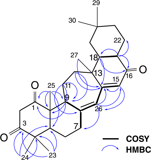
However, to solve the amount of methylene carbons it has been suggested that the C ring was a seven-membered ring. This proposal was confirmed by the correlations observed in the heteronuclear multiple bond correlation (HMBC) spectrum (Figure 1) from 3H-23 and 3H-24 to C-3, C-4 and C-5, from 3H-25 to C-1, C-5, C-9 and C-10, from H-11a to C-8 and C-10, from H-11b and 2H-12 to C-9 constructed the 6/6/7 fused A/B/C rings. This also explains the appearance of the signal at δC 160.7 attributed to the quaternary olefinic carbon β-carbonyl C-14 as observed for serratane-type triterpenoid, 3β,21β-dihydroxyserrat-14-en-16-one25 instead of δC 185.2 attributed to the same carbon for 28-nor-taraxarene triterpenoid, loranthone A.26 Thus, the chemical shifts at δC 200.5, 125.7 and 160.7 were assigned to C-16, C-15 and C-14, respectively. These assignments were confirmed by the correlations observed in the HMBC spectrum (Figures S15-S18, SI section) between the signals at δH 2.71 (H-17) to δC 200.5 (C-16), δC 44.9 (CH-18) and δC 21.4 (CH2-22), δH 1.84 (H-18) to δC 200.5 (C-16) and δC 160.7 (C-14), as well between δH 1.51 (H-12b), δH 5.76 (H-15) and δH 1.01 (3H-27) to δC 160.7 (C-14).
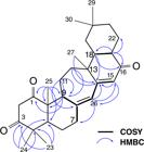
Furthermore, the correlations observed in the correlation spectroscopy (COSY) spectrum (Figures S19-S20, SI section) between the signals at δH 6.04 (m, 1H-26) and δH 5.76 (d, J 1.4 Hz, 1H-15) showed that the olefinic hydrogens were coupling with each other, suggesting the presence of two conjugated double bonds located at ∆8,26 and ∆14,15. These assignments were confirmed by the correlations observed in the HMBC spectrum between δH 5.76 (H-15) to δC 126.1 (CH-26) and δH 6.04 (H-26) to δC 160.7 (C-14), δC 125.7 (CH-15) and δC 36.5 (CH2-7) (Figure 1).
In addition, carbonyl carbon chemical shifts at δC 206.8 (C-1) and δC 209.7 (C-3) in the 13C NMR spectra and a pair of signals for diastereotopic methylene hydrogens at δH 3.43 (d, J 17.0 Hz, 1H-2a) and δH 3.54 (d, J 17.0 Hz, 1H-2b) in the 1H NMR spectra exhibited characteristics of the β-diketone system in the A ring similar to that found for the imberbic acid diketone.27 Although some 1,3-diketone compounds may undergo keto-enol tautomerism, this phenomenon is not well documented in the literature for triterpenoids. Nevertheless, these compounds may predominantly exist in the keto form, as observed for compound 1.28,29 This preference depends on the nature of the substituents, the conformational effects, temperature and polarity of medium.30 The presence of α-hydrogens at C-2, presented as a pair of well-defined doublets, in the 1H NMR spectrum, indicates the absence of enolization, since enol formation would result in the disappearance of these signals and the appearance of a singlet in the olefin region. Furthermore, the 13C NMR resonance of C-2 appears in a characteristic chemical shift of a methylene group flanked by two carbonyls, confirming the α-diketone nature of this carbon and supporting the predominance of the keto form.28,29 These assignments were confirmed by the correlations observed in the HMBC spectrum between δH 2.64 (H-9) and δH 1.00 (H-25) to δC 206.8 (C-1) as well δH 1.15 (H-23) and δH 1.13 (H-24) to δC 209.7 (C-3). All 1H and 13C NMR data are summarized in Table 1.
The relative configuration of 1 was assigned on the basis of nuclear Overhauser effect spectroscopy (NOESY) spectrum recorded in CDCl3 (Figures S21-S23, SI section). The correlations between H-5/H-9, H-5/H3-23, H-9/H3-27 and H-18/H3-27 suggested that these protons were cofacial and were arbitrarily assigned as α-oriented. This assignment is consistent with most triterpenoids derived from the same biosynthetic intermediates described in Scheme 1, where these stereogenic centers are conserved.31,32 The correlations of H3-24/H3-25/H3-30 suggested that these methyls were in the opposite side and thus β-oriented. Nevertheless, some of the NOESY correlations were not well-defined, preventing a fully unambiguous assignment of the relative stereochemistry of compound 1.
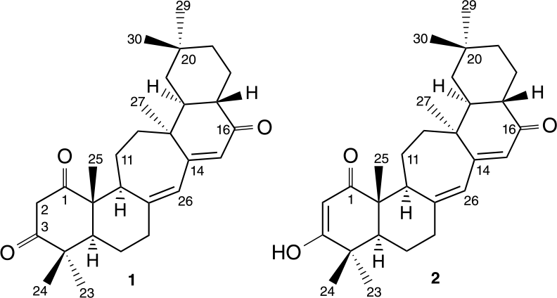
To confirm the suggested stereochemistry for each chiral centers of compound 1, the ML-J-DP4 methodology, proposed by Sarotti and co-workers,18 was applied. ML J DP4 is a hybrid approach that integrates high precision methods (DP4+) with the advantages of 3JHH scalar couplings to constrain conformational sampling, along with the high speed of ML algorithms.17 Quantitative methodologies such as ML-J-DP4, based on quantum mechanical calculations of NMR parameters, are currently considered one of the state-of-the-art methods in structural elucidation, especially in stereochemical determinations in natural product chemistry.33,34 For this application, experimental NMR data, including 13C and 1H NMR chemical shifts and 3JHH-type vicinal coupling constants, were used. Regarding the theoretical chemical shift data, since the isolated triterpene has 6 asymmetric carbons, the total of 64 possible stereoisomers comprises 32 pairs of enantiomers. Since each pair of enantiomers exhibits exactly the same NMR chemical shifts in isotropic media, 32 possible stereoisomers were considered in ML J DP4 approach. Therefore, after evaluating the differences between the simulated and experimental chemical shifts according to conformational sampling constraint criteria obtained from previously reported 3JHH experimental data and using Bayes’ theorem, as expected, ML J DP4 confirmed the NOESY based previously proposed stereochemistry as the most correct among all possible theoretical candidates. The most probable stereoisomer of compound 1, as predicted by the ML-J-DP4 calculations, is shown in Figure 2.
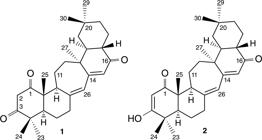
On the basis of these data, the compound 1 was identified as a new nor-triterpene, sapientrione, featuring an unprecedented carbon skeleton, herein trivially named sapienane (Figure 2).
In order to separate the signals at δH 1.00 (C-25) and δH 1.01 (C-27) and thus, to promote a better visualization of the correlations in the HMBC contour map, a 1H NMR spectrum of compound 1 was obtained by using pyridine-d5 as solvent.
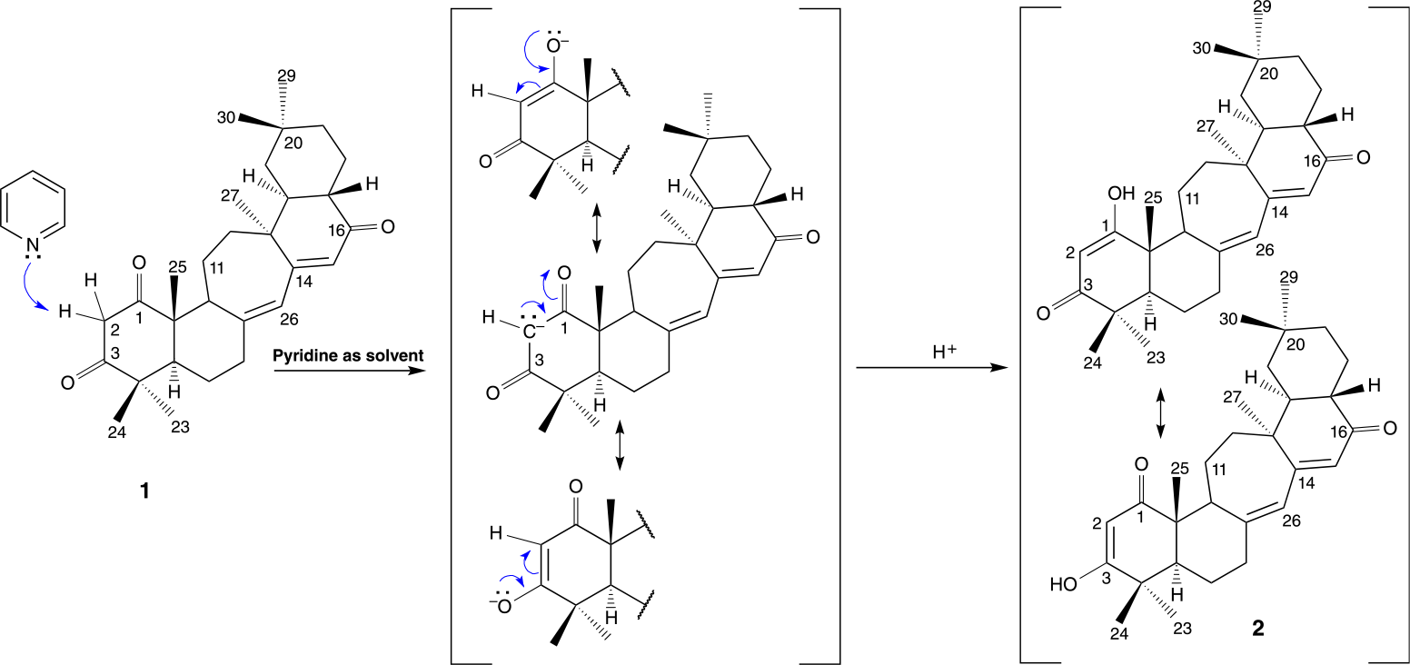
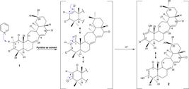
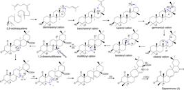
Pyridine is considered a weak Lewis base because the nitrogen atom contains a lone pair of electrons capable of proton abstraction. However, its basicity is reduced by the resonance stabilization of the aromatic ring, which delocalizes electron density and decreases the availability of the nitrogen lone pair for protonation.35 In β-diketone compounds, the α-hydrogens are weakly acidic because their conjugate base, the enolate ion, is stabilized by conjugation with the π orbitals of the adjacent carbonyl groups. Deprotonation at the α-position generates a carbanion, whose lone pair electrons are delocalized onto the electronegative carbonyl oxygens, thereby stabilizing the negative charge of the enolate. The presence of two carbonyl groups enhances this stabilization, increasing the acidity of the α-hydrogens to the point where weak bases, such as pyridine, are sufficient to promote enolate formation.36 Thus, as depicted in Scheme 1, this favored the alteration of the keto form to the enol form, leading to the accidental formation of an analogue called sapienone-1,3 enol (2) which persisted even after complete evaporation of the solvent at room temperature and subsequent dissolution in chloroform (Figure 2).
The chemical structure of this accidental derivative was determined by 1H and 13C NMR, HRESIMS, and IR. The main difference observed in the spectroscopic data of the two compounds was the disappearance of the doublet pair at δH 3.43 and δH 3.54 (H2-2), that corresponded to the α-carbonyl hydrogens, and the appearance of a singlet at δH 5.69 for compound 2, corresponding to an olefinic hydrogen. Similarly, the signal of the α-diketone carbon at δC 52.4 for compound 1 was replaced by a resonance at δC 101.5 for compound 2, characteristic of a less shielded sp2 carbon. Carbon signals that did not appear in the one-dimensional spectrum were identified by HMBC. All data for compound 2 can be seen in the SI section (Table S1 and Figures 24-29).
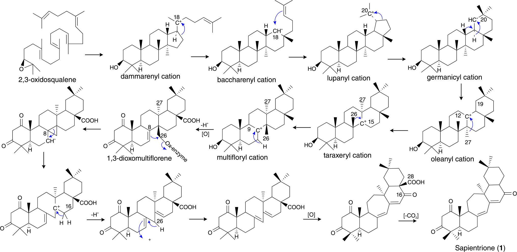
Aiming to rationalize the structural features of compound 1, a plausible biosynthetic pathway is proposed (Scheme 2). In this pathway, 2,3-oxidosqualene undergoes folding into a chair-chair-chair conformation, which initiates the cyclization cascade leading to the formation of the dammarenyl cation. Subsequent ring expansion of the D-ring by way of C-16 migration affords the secondary baccharenyl cation (C-18). This intermediate undergoes annulation at the 18β-position to generate the pentacyclic lupanyl cation. A further C-21 migration promotes expansion to give the germanicyl cation.31,37 From this point, a series of Wagner-Meerwein 1,2-hydride and methyl shifts occurs, successively producing the oleanyl, taraxeryl, and multifloryl cations. Through sequential oxidations, an oxygenase-mediated step may induce the formation of a propanone bridge between C-26 and C-8, concurrent with cleavage of the double bond of 1,3-dioxomultiflorene, yielding the cationic intermediate at C-7. A subsequent 1,2-hydride shift, followed by proton capture, generates two conjugated double bonds at ∆8,26 and ∆14,15. Finally, oxidation at C-16 followed by decarboxylation with the loss of C-28 furnishes the unprecedented nor triterpenoid sapientrione (1).31,37
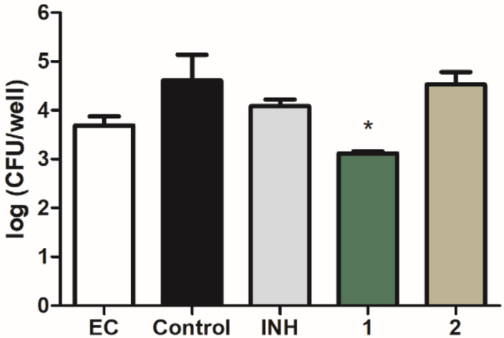
The antitubercular activity of the isolated compounds was evaluated against Mycobacterium tuberculosis H37Ra strain. Sapientrione (1) and its derivative (2) both exhibited MIC values of 100 µg mL-1 (229.2 µM), which corresponds to moderate inhibitory activity. In contrast, the reference drugs isoniazid and moxifloxacin displayed MIC values of 0.3 µg mL-1 (2.2 µM) and 0.09 µg mL-1 (0.2 µM), respectively, under identical conditions (Table S2, SI section). Although the activity of compounds 1 and 2 is considerably lower than that of the standard drugs, this outcome is not unexpected given their natural origin. Criteria for classifying the antimicrobial activity of natural products remain scarce. Several authors38,39 consider a MIC of 100 μg mL-1 as the threshold between moderate and weak activity for purified natural products. In this study, activity levels were defined as follows: significant (MIC < 10 μg mL-1), moderate (10 < MIC ≤ 100 μg mL 1), and low (MIC > 100 μg mL-1). The treatment with compound 1 significantly reduced the bacterial burden compared to the untreated control (P < 0.05) and displayed improved activity relative to isoniazid in assays against nutrient-starved bacteria (Figure 3). This observation allows us to suggest a satisfactory activity of drug 1 in this model of dormant mycobacteria, a condition highly relevant to tuberculosis persistence and relapse.
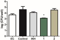
Antimycobacterial activity of compounds 1 and 2 at 50 µg mL-1 against nutrient starved Mycobacterium tuberculosis. Control group was treated with the vehicle, 7H9 medium with 2.5% DMSO. INH, isoniazid; EC, early control, represents the bacterial inoculum in the day treatments were added. *P < 0.05, compared to control. Data were evaluated by ANOVA, followed by Bonferroni post-test.
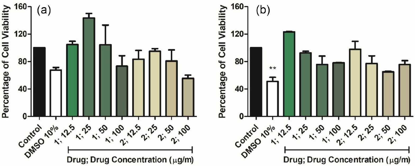
We also have investigated the possible in vitro cytotoxic effects of compounds 1 and 2 on Vero (african green monkey kidney) and HepG2 (human hepatoma) cell lines using the MTT assay. The in vitro incubation of the compounds, at concentrations ranging from 100 to 12.5 µg mL-1 did not significantly affect cell viability of these eukaryotic cell lines (Figure 4). These results indicate a low cytotoxic profile for both compounds and highlight that, despite the relatively high MIC values when compared to standard drugs, the discovery of sapientrione (1), a nor-triterpene with a novel carbon skeleton, represents a valuable contribution to terpenoid research. Its selective activity against dormant mycobacteria, coupled with low cytotoxicity, underscores the promise of this natural product as a scaffold for future medicinal chemistry efforts.
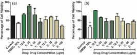
Cytotoxic effects of compounds 1 and 2 on Vero (a) and HepG2 (b) cells. Control: 1% DMSO-treated wells were considered as 100% cell viability. Data were expressed as mean of cell viability ± standard error of mean of three independent experiments performed in triplicate. **P < 0.01 compared to the corresponding control group.
Conclusions
In conclusion, the isolation of sapientrione (1), a nor-triterpene with an unprecedented carbon skeleton, distinguished from other triterpenes by the presence of a seven-membered ring and a double bond at ∆8,26, from S. glandulosum, represents a meaningful contribution in terpenoid chemistry. Despite the vast number of triterpenes that have been reported over past decades, the discovery of structurally unique and chemically diverse members of this class has become increasingly uncommon. Thus, the characterization of sapientrione not only expands the structural diversity known for triterpenoids but also highlights the untapped potential of S. glandulosum as a source of novel bioactive metabolites. Besides, sapientrione significantly reduced the bacterial burden in the nutrient-starved model of M. tuberculosis and showed no significant cytotoxicity in Vero and HepG2 cell lines, thus supporting its pharmacological interest for further investigation.
Supplementary Information
All data (UV, IR, MS, 1D and 2D NMR spectra of compounds 1 and 2) are available free of charge at http://jbcs.sbq.org.br as PDF file.
Acknowledgments
Financial support from Brazilian agencies CAPES (No. 001), CNPq and Fundação de Apoio à Pesquisa do Estado da Paraíba (FAPESQ-PB) are highly acknowledged. We greatly value our collaboration with Rede Norte-Nordeste de Fitoterápicos (INCT-RENNOFITO).
Data Availability Statement
The authors confirm that the data supporting the findings of this study are available within the article and its Supplementary Information.
References
-
1 Flora e Funga do Brasil (Reflora); Euphorbiaceae. [Link] accessed in November 2025
» Link -
2 Wang, H.-B.; Wang, X.-Y.; Liu, L.-P.; Qin, G.-W.; Kang, T.-G.; Chem. Rev. 2015, 115, 2975. [Crossref]
» Crossref -
3 He, Q.; Zhang, L.; Li, T.; Li, C.; Song, H.; Fan, P.; J. Ethnopharmacol 2021, 277, 114206. [Crossref]
» Crossref -
4 Al Muqarrabun, L. M. R.; Ahmat, N.; Aris, S. R. S.; J. Ethnopharmacol 2014, 155, 9. [Crossref]
» Crossref -
5 Flora e Funga do Brasil (Reflora); Sapium [Link] accessed in November 2025
» Link -
6 Díaz, R. F.; Intropica 2020, 15, 81. [Crossref]
» Crossref -
7 Sobottkaa, A. M.; Görick, C.; Melzig, M. F.; Nat. Prod. Res 2015, 30, 1941. [Crossref]
» Crossref -
8 Valle, L. S.; Kaplan, M. A. C.; An. Acad. Bras. Cienc. 2000, 72, 293. [Crossref]
» Crossref -
9 Carvalho, P. E. R.; Espécies Arbóreas Brasileiras: Leiteiro (Sapium glandulatum), vol. 4, 1st ed; Embrapa Informação Tecnológica: Brasília, DF; Embrapa Florestas: Colombo, Brazil, 2010. [Crossref]
» Crossref -
10 Coutinho, D. J. G.; Barbosa, M. O.; de Souza, R. J. C.; da Silva, A. S.; Silva, S. I.; de Oliveira, A. F. M.; Renewable Energy 2016, 91, 275. [Crossref]
» Crossref -
11 da Silva, C. H. T. P.; Peixoto-Sobrinho, T. J. S.; Castro, V. T. N. A.; Lima, D. C. A.; de Amorim, E. L. C.; Molecules 2011, 16, 4728. [Crossref]
» Crossref -
12 Ocampos, F. M. M.: Estudo Fitoquímico, Toxicológico e Avaliação da Atividade Biológica da Espécie Vegetal Sapium glandulosum (L.) Morong (Euphorbiaceae); MSc Dissertation, Federal University of Paraná, Curitiba, Brazil, 2013. [Link] accessed in November 2025
» Link -
13 Bonin, C. R.; Fochat, R. C.; Leite, I. C. G.; Pereira, T. V.; Fajardo, M. O.; Pinto, C. P. G.; Macedo, R. L.; Silva, M. R.; Menezes, P. P. L.; de Araújo, N. M. J. M.; da Costa, R. R.; Einstein 2020, 18, eAO4620. [Crossref]
» Crossref -
14 World Health Organization (WHO); Global Tuberculosis Report 2024; WHO: Geneva, Switzerland, 2024. [Link] accessed in November 2025
» Link - 15 MestReNova, version 14.2.0-26256; Mestrelab Research S.L.; Santiago de Compostela, Spain, 2020.
-
16 The IUCN Red List of Threatened Species, Sapium glandulosum [Link] accessed in November 2025
» Link - 17 Deppmeier, B. J.; Driessen, A. J.; Hehre, T. S.; Hehre, W. J.; Johnson, J. A.; Klunzinger, P. E.; Leonard, J. M.; Pham, I. N.; Pietro, W. J.; Yu, J.; Spartan’10, version 1.1.0; Wavefunction, Inc. Irvine, CA, 2010.
-
18 Tsai, Y.-H.; Amichetti, M.; Zanardi, M. M.; Grimson, R.; Daranas, A. H.; Sarotti, A. M.; Org. Lett 2022, 24, 7487 [Crossref]; [Link] accessed in November 2025.
» Crossref» Link - 19 Frisch, M. J.; Trucks, G. W.; Schlegel, H. B.; Scuseria, G. E.; Robb, M. A.; Cheeseman, J. R.; Scalmani, G.; Barone, V.; Mennucci, B.; Petersson, G. A.; Nakatsuji, H.; Caricato, M.; Li, X.; Hratchian, H. P.; Izmaylov, A. F.; Bloino, J.; Zheng, G.; Sonnenberg, J. L.; Hada, M.; Ehara, M.; Toyota, K.; Fukuda, R.; Hasegawa, J.; Ishida, M.; Nakajima, T.; Honda, Y.; Kitao, O.; Nakai, H.; Vreven, T.; Montgomery, J. A., Jr.; Peralta, J. E.; Ogliaro, F.; Bearpark, M.; Heyd, J. J.; Brothers, E.; Kudin, K. N.; Staroverov, V. N.; Kobayashi, R.; Normand, J.; Raghavachari, K.; Rendell, A.; Burant, J. C.; Iyengar, S. S.; Tomasi, J.; Cossi, M.; Rega, N.; Millam, N. J.; Klene, M.; Knox, J. E.; Cross, J. B.; Bakken, V.; Adamo, C.; Jaramillo, J.; Gomperts, R.; Stratmann, R. E.; Yazyev, O.; Austin, A. J.; Cammi, R.; Pomelli, C.; Ochterski, J. W.; Martin, R. L.; Morokuma, K.; Zakrzewski,V. G.; Voth, G. A.; Salvador, P.; Dannenberg, J. J.; Dapprich, S.; Daniels, A. D.; Farkas, Ö.; Foresman, J. B.; Ortiz, J. V.; Cioslowski, J.; Fox, D. J. Gaussian 09, Revision A.01; Gaussian, Inc. Wallingford, Ct, USA, 2009.
-
20 Muradás, T. C.; Abbadi, B. L.; Villela, A. D.; Macchi, F.; Bergo, P. F.; de Freitas, T. F.; Sperotto, N. D. M.; Timmers, L. F. S. M.; de Souza, O. N.; Picada, J.; Fachini, J.; da Silva, J. B.; de Albuquerque, N.; Habenschus, M. D.; Carrão, D. B.; Rocha, B. A.; Barbosa-Junior, F.; de Oliveira, A.; Mascarello, A.; Neuenfeldf, P.; Nunes, R. J.; Morbidoni, H. R.; Campos, M. M.; Basso, L. A.; Rodrigues-Junior, V. S.; Plos One 2018, 13, e0202568. [Crossref]
» Crossref -
21 Sidrônio, M. G. S.; Castelo Branco, A. P. O. T.; Abbadi, B. L.; Macchi, F.; Silveira, M. D.; Lock, G. A.; Costa, T. D.; de Araújo, D. M.; Cibulski, S.; Bizarro, C. V.; Machado, P.; Basso, L. A.; Rodrigues-Junior, V. S.; Tuberculosis 2021, 128, 102089. [Crossref]
» Crossref -
22 Martinelli, L. K. B.; Rotta, M.; Villela, A. D.; Rodrigues-Junior, V.; Abbadi, B. L.; Trindade, R. V.; Petersen, G. O.; Danesi, G. M.; Nery, L. R.; Pauli, I.; Campos, M. M.; Bonan, C. D.; de Souza, O. N.; Basso, L. A.; Santos, D. S.; Sci. Rep 2017, 7, 46696. [Crossref]
» Crossref - 23 GraphPad Prism, version 5.00; GraphPad Software Inc.; San Diego, USA, 2007.
- 24 Barbosa, L. C. A.; Espectroscopia no Infravermelho na Caracterização de Compostos Orgânicos, 1st ed; UFV: Minas Gerais, 2013.
-
25 Zhou, H.; Li, Y.; Tong, X; Liu, H.; Jiang, S.; Zhu, D.; Nat. Prod. Res 2006, 18, 453. [Crossref]
» Crossref -
26 Abreu, L. S.; do Nascimento, Y. M.; Costa, R. dos S.; Guedes, M. L. S.; Souza, B. N. R. F.; Pena, L. J.; Costa, V. C. O.; Scotti, M. T.; Braz-Filho, R.; Barbosa-Filho, J. M.; da Silva, M. S.; Velozo, E. S.; Tavares, J. F.; J. Nat. Prod. 2019, 82, 2721. [Crossref]
» Crossref -
27 Rogers, C. B.; Subramony, G.; Phytochemistry 1988, 27, 531. [Crossref]
» Crossref -
28 Somwong, P.; Suttisri, R.; Buakeaw, A.; Fitoterapia 2011, 82, 1047. [Crossref]
» Crossref -
29 Mahato, S. B.; Kundu, A. P.; Phytochemistry 1994, 37, 1517. [Crossref]
» Crossref -
30 Kel’in, A. V.; Curr. Org. Chem 2003, 7, 1691. [Crossref]
» Crossref -
31 Hu, D. Gao, H.; Yao, X.-S. In Comprehensive Natural Products III (Chemistry and Biology), vol. 1, 3rd ed.; Liu, H. W.; Begley, T. P., eds.; Elsevier: USA, 2020. [Crossref]
» Crossref -
32 Camargo, K. C.; de Aguilar, M. G.; Moraes, A. R.; de Castro, R. G.; Szczerbowski, D.; Miguel, E. L. M.; Oliveira, R. O.; Sousa, G. F.; Vidal, D. M.; Duarte, L. P.; Molecules 2022, 27, 959. [Crossref]
» Crossref -
33 Costa, F. L. P.; de Albuquerque, A. C. F.; Fiorot, R. G.; Lião, L. M.; Martorano, L. H.; Mota, G. V. S.; Valverde, A. L.; Carneiro, J. W. M.; dos Santos Jr., F. M.; Org. Chem. Front. 2021, 8, 2019. [Crossref]
» Crossref -
34 e Silva, J. P. R.; Pereira, L. C. O.; Abreu, L. S.; Lins, F. S. V.; de Souza, T. A.; do Espírito-Santo, R. F.; Barros, R. P. C.; Villarreal, C. F.; Melo, J. I. M.; Scotti, M. T.; Costa, V. C. O.; Martorano, L. H.; dos Santos-Jr., F. M.; Braz-Filho, R.; da Silva, M. S.; Tavares, J. F.; J. Nat. Prod 2022, 85, 2184. [Crossref]
» Crossref - 35 Costa, P.; Ferreira, V.; Esteves, P.; Vasconcellos, M.; Ácidos e Bases em Química Orgânica, vol. 1, 1st ed.; Bookman: Porto Alegre, Brazil, 2005.
-
36 LibreTexts (Chemistry); Organic Chemistry. [Link] accessed in November 2025
» Link - 37 Dewick, P. M.; Medicinal Natural Products: A Biosynthetic Approach, vol. 1, 3rd ed.; Wiley: New Jersey, USA, 2009.
-
38 Awouafack, M. D.; McGaw, L. J.; Gottfried, S.; Mbouangouere, R.; Tane, P.; Spiteller, M.; Eloff, J. N.; BMC Complementary Altern. Med. 2013, 13, 289. [Crossref]
» Crossref -
39 Kuete, V.; Planta Med 2010, 76, 1479. [Crossref]
» Crossref
Edited by
-
Editor handled this article:
Hector Henrique F. Koolen (Associate)