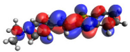
Synthesis and Cytotoxic Activity of 2-Styrylbenzothiazole and 2-Styrylbenzimidazole Derivatives: Insights from DNA/BSA Binding and Molecular Docking Studies
Abstract
Benzothiazole and benzimidazole derivatives are widely recognized as important scaffolds in drug development and pharmaceutical applications. In this study, we report the microwave-assisted synthesis of a series of styrylbenzothiazoles and styrylbenzimidazoles in the presence of sodium bisulfite (NaHSO3), along with the evaluation of their cytotoxic activities against the MDA-MB-231 (human breast carcinoma) and 4T1 (mouse metastatic mammary adenocarcinoma) tumor cell lines, using MCF-10A cells to assess selectivity. With the exception of compound 2e, all thiazole derivatives exhibited cytotoxic activity to varying extents against the MDA-MB-231 cell line, whereas no activity was observed against the 4T1 cell line. Compound 2f, an amino-substituted derivative, displayed the highest cytotoxic activity (IC50 = 4.0 μM), being twice as potent as the reference drug cisplatin (IC50 = 9.6 μM) and 9-25 times more potent than the other derivatives in the series. Styrylbenzimidazole derivatives 3d, 3g, and 3k also showed promising activity against MDA-MB-231 cells, with IC50 values of 8.2, 6.6, and 5.9 μM, respectively. Notably, unlike the thiazole derivatives, styrylbenzimidazoles exhibited cytotoxic activity against the 4T1 cell line, with the methoxy-substituted derivative 3d showing the highest potency (IC50 = 8.4 μM). Competitive deoxyribonucleic acid (DNA)-binding studies involving compounds 2d, 2f, 3d, and 3f with ethidium bromide (EB) or 4’,6-diamidino-2-phenylindole (DAPI) suggested a preferential interaction with DNA via the minor groove, while interaction assays with bovine serum albumin (BSA) confirmed their protein-binding ability. These findings were further supported by molecular docking and density functional theory (DFT) calculations.
Keywords:
2-styrylbenzothiazoles; 2-styrylbenzimidazoles; organic synthesis; sodium bisulfite-mediated reaction; anticancer activity; in silico studies
Introduction
Cancer is a disease that can affect people of any age and remains one of the leading causes of death worldwide. According to data released in 2024 by the American Cancer Society, approximately 20 million new cases of cancer were recorded in 2022 worldwide, resulting in approximately 9.7 million deaths attributed to the disease in the same period.1
Projections indicate that, by 2050, the number of new cancer cases is expected to reach 35 million, representing an estimated increase of 77% compared to the data for 2022.
Among malignant neoplasms, lung cancer remains the most diagnosed globally, with approximately 2.5 million cases and 1.8 million deaths. Breast cancer follows, being more prevalent among women, with more than 2 million new cases recorded annually.2
Breast cancer is the most common type of cancer among women, causing significant impacts on the physical, mental and social health of patients. Diagnosis and treatment, especially interventions such as mastectomy, can compromise self-image and self-esteem, negatively influencing the perception of femininity and body identity. In this context, the development and application of novel pharmacological agents remain a critical therapeutic strategy to improve clinical outcomes and patient quality of life.3,4
Given the high incidence of cancer and its associated mortality rates, the scientific community has intensified efforts to develop novel molecular scaffolds to mitigate these alarming trends. In light of this, heterocyclic compounds have attracted considerable attention due to their significant cytotoxic activity against various cancer cell lines, demonstrating promise as anticancer agents.3,4 For drug discovery, organic substances that present benzothiazole (BT) and benzimidazole (BZI) fragments are of particular interest, as they exhibit interesting pharmacological properties.5,6
Benzothiazole and benzimidazole derivatives are of considerable importance for improving quality of life, given their use as prototypes in drug development and as a source of pharmaceutical raw materials.6,7 Their biological activities have been well-documented in the literature, with notable effects reported across a wide spectrum, including antimicrobial, antifungal, antibacterial, anticancer and antiviral properties, among others.8-18
The most widely employed synthetic route for obtaining 2-substituted benzimidazoles and benzothiazoles involves the reaction of 1,2-phenylenediamines or 2-aminothiophenol with carboxylic acids,19,20 or their derivatives,21 under strongly acidic conditions and at elevated temperatures. Another approach for the synthesis of these compounds involves the condensation of 1,2-phenylenediamines and 2-aminothiophenol with aldehydes under oxidative conditions,22 employing a variety of oxidizing agents and catalysts, such as 1,4-benzoquinone,23 PhI(OAc)2,24 Fe(NO3)3/(2,2,6,6-tetramethylpiperidin-1-yl)oxyl (TEMPO),25 Zn-proline,26 2,3-dichloro-5,6-dicyano-1,4-benzoquinone (DDQ),27 MnO2/granular activated carbon (GAC),28 Pb(OAc)4,29 oxone,30 H2O2/HCl,31 Pd(PPh3)·4/MnO2,32 ceric ammonium nitrate (CAN),33 iodine,34 FeCl3·6H2O,35 In(OTf)3,36 Yb(OTf)3,37 Sc(OTf)3,38 Cu(OTf)2,39 KHSO4,40 ZrOCl2·8H2O,41 ZrCl4,42 VOSO4,43 boron trifluoride etherate,44 and cobalt complexes.45
However, many of these methods require long reaction times and have low yields, in addition to demanding expensive and difficult-to-handle reagents or catalysts. Among the limitations, the use of stoichiometric reagents, prolonged reaction times, and the difficulty in separating the products and recovering the catalyst from the reaction mixture stand out. In this context, the use of mild, practical, stable, inexpensive, recyclable, and environmentally friendly heterogeneous additives represents a highly desirable strategy for organic synthesis. Sodium bisulfite (NaHSO3) is a low-cost and low-toxicity compound that has proven to be a promising alternative for the synthesis of benzimidazoles and benzothiazoles. In a study published in 2015, Araujo et al.46 demonstrated that NaHSO3 is capable of promoting the synthesis of these classes of compounds in high yields, through reactions between aromatic aldehydes and o-phenylenediamine or o-aminothiophenol, carried out in N,N-dimethylacetamide (DMA) under microwave irradiation. Thus, due to the aforementioned factors and as part of our ongoing efforts to develop more efficient and environmentally friendly synthetic methodologies, we report herein a one-pot cyclocondensation-oxidation reaction between 1,2-phenylenediamine or 2-aminothiophenol and various α,β-unsaturated aryl aldehydes for the synthesis of 2-styrylbenzimidazole and 2-styrylbenzothiazole derivatives. The synthesized compounds were also evaluated for their antitumor activity against two highly aggressive breast cancer cell lines, MDA-MB-231 and 4T1.
Experimental
General procedure for the synthesis of compounds 1g-1k
The α,β-unsaturated aldehydes were synthesized via the Wittig reaction, with adaptations of the methodology described by Doyle et al.47 In a 25 mL round-bottom flask, (triphenylphosphoranylidene)acetaldehyde (5 mmol) and toluene (10 mL) were added. After complete solubilization of the reagent, the corresponding aldehyde was added to the reaction mixture. The mixture was maintained under constant stirring and heating at 90 °C. After 2 h of reaction, the progress was monitored by thin-layer chromatography, confirming the formation of the desired product. Subsequently, the solvent was removed under reduced pressure using a rotary evaporator, and the resulting residue was purified by column chromatography. Purification was carried out using a glass column (4.5 cm diameter × 36 cm height), with silica gel (100 g, 70-230 mesh) as the stationary phase. A hexane/ethyl acetate mixture (9:1, v/v), previously distilled, was employed as the mobile phase. The compounds (E)-3-(4-fluorophenyl) acrylaldehyde47 (1g), (E)-4-(3-oxoprop-1-en-1-yl) benzonitrile47 (1h), (E)-3-(4-(methylthio)phenyl) acrylaldehyde48 (1i), (E)-3-(4-bromophenyl) acrylaldehyde47 ( 1j), and (E)-3-(4-(trifluoromethyl)phenyl) acrylaldehyde49 (1k) were confirmed by nuclear magnetic resonance (NMR) spectroscopy, and their signals and chemical shifts were compared with data reported in the literature.
General procedure for the synthesis of compounds 2a-2k
Sodium bisulfite (NaHSO3) (2 eq.) was weighed into a 50 mL round-bottomed flask, to which 5 mL of N,N-dimethylacetamide (DMA) and aldehyde (1.5 mmol) were added. Then, o-aminothiophenol (1.0 mmol) was added, and the reaction mixture was placed under microwave irradiation (MWI) in an open system at 80 °C with a maximum power of 300 watts, a ramp time of 5 min and a reaction time of 60 min, under cooling. After completion of the reaction, the reaction mixture was allowed to cool to room temperature and poured into ice-cold water. The solid formed was filtered under reduced pressure and purified by column chromatography, using hexane and ethyl acetate as the mobile phase, in specific proportions for each compound, followed by recrystallization when necessary.
(E)-2-Styrylbenzo[d]thiazole (2a)
Yield 67%; mp 106-108 ºC; 1H NMR (400 MHz, CDCl3) δ 8.00 (d, J 8.0 Hz, 1H), 7.86 (d, J 8.0 Hz, 1H), 7.62-7.50 (m, 3H), 7.50-7.33 (m, 6H); 13C NMR (100 MHz, CDCl3) δ 167.2, 138.2, 135.3, 129.6, 129.0, 127.5, 126.5, 125.5, 122.8, 121.8, 126.6; high-resolution mass spectrometry (HRMS) (ESI) m/z, calcd. for C15H12NS [M + H]+: 238.0685; found: 238.0686; Fourier-transformed infrared spectroscopy (FTIR) ν / cm-1 3058, 1630, 1446, 1316, 1192, 956, 754, 748, 723, 684.
(E)-2-(4-Methoxystyryl)benzo[d]thiazole (2b)
Yield 74%; mp 145-147 ºC; 1H NMR (400 MHz, CDCl3) δ 8.00 (d, J 8.2 Hz, 1H), 7.87 (d, J 8.2 Hz, 1H), 7.59-7.44 (m, 4H), 7.38 (t, J 7.4 Hz, 1H), 7.34-7.26 (m, 1H), 6.96 (d, J 8.6 Hz, 2H), 3.88 (s, 3H); 13C NMR (100 MHz, CDCl3) δ 167.5, 160.8, 153.9, 137.4, 134.2, 128.9, 128.2, 126.3, 125.1, 122.8, 121.5, 119.9, 114.5, 55.4; HRMS (ESI) m/z, calcd. for C16H14NOS [M + H]+: 268.0791; found: 268.0788; FTIR ν / cm-1 2995, 1601, 1514, 1257, 1174, 1028, 956, 808, 805, 757, 729.
(E)-2-(4-Nitrostyryl)benzo[d]thiazole (2c)
Yield 58%; mp 250-252 ºC; 1H NMR (400 MHz, CDCl3) δ 8.28 (d, J 8.3 Hz, 2H), 8.06 (d, J 8.2 Hz, 1H), 7.90 (d, J 8.2 Hz, 1H), 7.73 (d, J 8.3 Hz, 2H), 7.64-7.48 (m, 3H), 7.74 (t, J 7.4 Hz, 1H); 13C NMR (100 MHz, CDCl3) δ 141.6, 134.7, 127.9, 126.8, 126.1, 125.9, 124.3, 123.4, 121.7; HRMS (ESI) m/z, calcd. for C15H11N2O2S [M + H]+: 283.0536; found: 283.0535; FTIR ν / cm-1 1599, 1518, 1339, 1313, 943, 766, 743, 686.
(E)-2-(2-Methoxystyryl)benzo[d]thiazole (2d)
Yield 69%; mp 265-267 ºC; 1H NMR (400 MHz, CDCl3) δ 7.98 (d, J 8.2 Hz, 1H), 7.87-7.79 (m, 2H), 7.62-7.57 (m, 1H), 7.52 (d, J 16.4 Hz, 1H), 7.48-7.42 (m, 1H), 7.38-7.30 (m, 2H), 7.03-6.96 (m, 1H), 6.93 (d, J 8.2 Hz, 1H), 3.93 (s, 3H); 13C NMR (100 MHz, CDCl3) δ 168.2, 157.8, 153.9, 134.4, 133.4, 130.6, 128.1, 126.2, 125.2, 124.3, 122.9, 121.5, 120.9, 111.1, 55.5; HRMS (ESI) m/z, calcd. for C16H14NOS [M + H]+: 268.0791; found: 268.0790; FTIR ν / cm-1 2995, 1601, 1514, 1257, 1174, 1028, 757.
(E)-2-(2-Nitrostyryl)benzo[d]thiazole (2e)
Yield 54%; mp 250-253 ºC; 1H NMR (400 MHz, CDCl3) δ 8.08-7.97 (m, 3H), 7.89 (d, J 8.0 Hz, 1H), 7.79 (d, J 8.0 Hz, 1H), 7.67 (t, J 7.6 Hz, 1H), 7.55-7.46 (m, 2H), 7.45-7.36 (m, 2H); 13C NMR (100 MHz, CDCl3) δ 166.0, 153.8, 148.1, 134.7, 133.6, 132.5, 131.3, 129.6, 128.6, 127.1, 126.5, 125.9, 125.1, 123.4, 121.7; HRMS (ESI) m/z, calcd. for C15H11N2O2S [M + H]+: 283.0536; found: 283.0535; FTIR ν / cm-1 3056, 1570, 1515, 1342, 1314, 1192, 950, 861, 754, 720.
(E)-2-(4-Dimethylaminostyryl)benzo[d]thiazole (2f)
Yield 61%; mp 205-208 ºC; 1H NMR (400 MHz, CDCl3) δ 7.94 (d, J 8.1 Hz, 1H), 7.82 (d, J 8.1 Hz, 1H), 7.52-7.39 (m, 4H), 7.32 (t, J 7.6 Hz, 1H), 7.20 (d, J 16.2 Hz, 1H), 6.76-6.66 (m, 2H), 3.03 (s, 6H); 13C NMR (100 MHz, CDCl3) δ 168.2, 154.0, 151.2, 138.3, 134.1, 130.7, 128.9, 126.1, 124.7, 123.4, 122.4, 121.4, 117.3, 112.1, 111.8, 40.3; HRMS (ESI) m/z, calcd. for C17H17N2S [M + H]+: 281.1107; found: 281.1104; FTIR ν / cm-1 2891, 1595, 1524, 1474, 1361, 1226, 1181, 1168, 960, 804, 756.
(E)-2-(4-Fluorostyryl)benzo[d]thiazole (2g)
Yield 82%; mp 161-163 ºC; 1H NMR (400 MHz, CDCl3) δ 8.00 (d, J 8.2 Hz, 1H), 7.85 (d, J 8.2 Hz, 1H), 7.59-7.44 (m, 4H), 7.41-7.35 (m, 1H), 7.28 (d, J 16.2 Hz, 1H), 7.10 (t, J 8.5 Hz, 2H); 13C NMR (100 MHz, CDCl3) δ 166.8, 163.3 (d, JC-F 250.0 Hz), 153.9, 136.3, 134.3, 131.7 (d, JC-F 3.4 Hz), 129.2 (d, JC-F 8.3 Hz), 126.4, 125.4, 123.0, 121.9 (d, JC-F 2.5 Hz), 121.5, 116.1 (d, JC-F 22.0 Hz); HRMS (ESI) m/z, calcd. for C15H11FNS [M + H]+: 256.0591; found: 256.0589; FTIR ν / cm-1 3061, 3039, 3001, 1598, 1509, 1222, 952, 816, 758.
(E)-4-(2-(Benzo[d]thiazol-2-yl)vinyl)benzonitrile (2h)
Yield 57%; mp 192-193 ºC; 1H NMR (400 MHz, CDCl3) δ 8.03 (d, J 8.0 Hz, 1H), 7.89 (d, J 8.0, 1H), 7.73-7.61 (m, 4H), 7.57-7.38 (m, 4H); 13C NMR (100 MHz, CDCl3) δ 165.6, 153.8, 139.8, 135.0, 134.6, 132.7, 127.7, 126.7, 125.9, 125.4, 123.4, 121.7, 118.6, 112.4; HRMS (ESI) m/z, calcd. for C16H11N2S [M + Na]+: 285.0457; found: 285.0455; FTIR ν / cm-1 3063, 2998, 2223, 1606, 1478, 1410, 1314, 1188, 1123, 942, 811, 762, 728, 667, 607.
(E)-2-(4-(Methylthio)styryl)benzo[d]thiazole (2i)
Yield 75%; mp 206-207 ºC; 1H NMR (400 MHz, CDCl3) δ 7.99 (d, J 8.0 Hz, 1H), 7.85 (d, J 8.0 Hz, 1H), 7.53-7.43 (m, 4H), 7.40-7.32 (m, 2H), 7.29-7.22 (m, 2H), 2.52 (s, 3H); 13C NMR (100 MHz, CDCl3) δ 167.1, 153.9, 140.7, 137.1, 134.3, 132.1, 127.8, 126.3 (2C), 125.3, 122.9, 121.5, 121.2, 15.3; HRMS (ESI) m/z, calcd. for C16H14NS2 [M + H]+: 284.0562; found: 284.0562; FTIR ν / cm-1 1628, 1591, 1500, 1435, 1091, 951, 802, 758, 729.
(E)-2-(4-Bromostyryl)benzo[d]thiazole (2j)
Yield 83%; mp 231-234 ºC; 1H NMR (400 MHz, CDCl3) δ 8.00 (d, J 8.0 Hz, 1H), 7.87 (d, J 8.0 Hz, 1H), 7.56-7.52 (m, 2H), 7.51-7.42 (m, 4H), 7.42-7.35 (m, 2H); 13C NMR (100 MHz, CDCl3) δ 166.5, 153.8, 136.2, 134.4 (2C), 132.2, 128.8, 126.5, 125.5, 123.5, 123.1, 122.7, 121.6; HRMS (ESI) m/z, calcd. for C15H11BrNS [M + H]+: 315.9790; found: 315.9793; FTIR ν / cm-1 1478, 1399, 1235, 1190, 1070, 957, 943, 808, 753, 722.
(E)-2-(4-(Trifluoromethyl)styryl)benzo[d]thiazole (2k)
Yield 55%; mp 162-163 ºC; 1H NMR (400 MHz, CDCl3) δ 8.03 (d, J 8.1 Hz, 1H), 7.88 (d, J 8.1 Hz, 1H), 7.70-7.64 (m, 4H), 7.58-7.37 (m, 4H); 13C NMR (100 MHz, CDCl3) δ 166.1, 153.8, 138.8, 135.7, 134.5, 130.9 (qt, JC-F 32.0 Hz), 127.5, 126.6, 126.0 (qt, JC-F 270.0 Hz), 125.9 (qt, JC-F 4.0 Hz), 125.8, 124.4, 123.2, 121.6; HRMS (ESI) m/z, calcd. for C16H11F3NS [M + H]+: 306.0559; found: 306.0553; FTIR ν / cm-1 3071, 3000, 2934, 1614, 1557, 1483, 1413, 1316, 1158, 1104, 1066, 1013, 948, 865, 821, 760, 730, 666, 617.
General procedure for the synthesis of compounds 3a-3k
Sodium bisulfite (NaHSO3) (2 eq.) was weighed into a 50 mL round-bottomed flask, to which 5 mL of N,N-dimethylacetamide (DMA) and aldehyde (1.5 mmol) were added. Then, o-1,2-phenylenediamine (1.0 mmol) was added, and the reaction mixture was placed under microwave irradiation (MWI) in an open system at 100 °C with a maximum power of 300 watts, a ramp time of 5 min and a reaction time of 60 min, under cooling. After completion of the reaction, the reaction mixture was allowed to cool to room temperature and poured into ice-cold water. The resulting solid was filtered under reduced pressure and purified by column chromatography using hexane, chloroform, ethyl acetate, and/or methanol as the mobile phase, in specific proportions for each compound, followed by recrystallization in methanol.
(E)-2-Styryl-1H-benzo[d]imidazole (3a)
Yield 82%; mp 201-203 ºC; 1H NMR (400 MHz, CDCl3) δ 7.65 (d, J 16.0 Hz, 1H), 7.59-7.53 (m, 1H), 7.45-7.37 (m, 2H), 7.34-7.24 (m, 4H), 7.24-7.19 (m, 2H), 7.11 (d, J 16.0 Hz, 1H); 13C NMR (100 MHz, DMSO-d6) δ 151.1, 136.1, 135.4, 129.5, 129.1, 127.6, 122.9, 117.5, 115.2; HRMS (ESI) m/z, calcd. for C15H13N2 [M + H]+: 221.1073; found: 221.1071; FTIR ν / cm-1 3059, 1645, 968, 756, 743, 699, 598.
(E)-2-(4-Methoxystyryl)-1H-benzo[d]imidazole (3b)
Yield 88%; mp 214-217 ºC; 1H NMR (400 MHz, DMSO-d6) δ 7.80 (d, J 16.6 Hz, 1H), 7.72-7.64 (m, 4H), 7.39-7.33 (m, 2H), 7.14 (d, J 16.6 Hz, 1H), 7.04 (d, J 8.6 Hz, 2H), 3.83 (s, 3H); 13C NMR (100 MHz, DMSO-d6) δ 161.3, 150.4, 139.1, 135.5, 129.9, 127.9, 124.4, 115.1, 114.6, 111.3, 111.4, 55.9; HRMS (ESI) m/z, calcd. for C16H15N2O [M + H]+: 251.1179; found: 251.1175; FTIR ν / cm-1 3059, 2933, 2836, 1642, 1601, 1511, 1455, 1246, 1174, 1028, 970, 818, 744.
(E)-2-(4-Nitrostyryl)-1H-benzo[d]imidazole (3c)
Yield 34%; mp 267-269 ºC; 1H NMR (400 MHz, DMSO-d6) δ 8.28 (d, J 8.5 Hz, 2H), 7.95 (d, J 8.5 Hz, 2H), 7.79 (d, J 16.2 Hz, 1H), 7.63-7.57 (m, 2H), 7.47 (d, J 16.2 Hz, 1H), 7.27-7.21 (m, 2H); 13C NMR (100 MHz, DMSO-d6) δ 149.9, 147.7, 142.4, 134.2, 132.1, 131.0, 128.8, 125.8, 124.7, 123.9, 120.7, 114.4, 113.7; HRMS (ESI) m/z, calcd. for C15H10N3O2 [M - H]-: 264.0779; found: 264.0778; FTIR ν / cm-1 3076, 2631, 1593, 1513, 1341, 1110, 971, 831, 749, 734, 703.
(E)-2-(2-Methoxystyryl)-1H-benzo[d]imidazole (3d)
Yield 73%; mp 187-189 ºC; 1H NMR (400 MHz, DMSO-d6) δ 7.92 (d, J 16.7 Hz, 1H), 7.70 (d, J 8.0 Hz, 1H), 7.58-7.51 (m, 2H), 7.37 (t, J 8.0 Hz, 1H), 7.27-7.15 (m, 4H), 7.11 (d, J 8.0 Hz, 1H), 7.03 (t, J 7.8 Hz, 1H), 3.92 (s, 3H); 13C NMR (100 MHz, DMSO-d6) δ 157.6, 151.7, 139.2, 130.8, 130.3, 129.9, 128.6, 127.8, 124.5, 122.7, 121.3, 117.9, 115.2, 112.2, 111.2, 56.1; HRMS (ESI) m/z, calcd. for C16H14N2O [M - H]-: 249.1033; found: 249.1032; FTIR ν / cm-1 3061, 2836, 1598, 1491, 1245, 1108, 1025, 974, 743, 603.
(E)-2-(2-Nitrostyryl)-1H-benzo[d]imidazole (3e)
Yield, 54%, mp 214-217 ºC; 1H NMR (400 MHz, CDCl3) δ 8.03-7.99 (m, 1H), 7.96 (d, J 16.5 Hz, 1H), 7.77-7.72 (m, 1H), 7.67-7.60 (m, 3H), 7.51-7.45 (m, 1H), 7.32-7.29 (m, 2H), 7.23 (d, J 16.5 Hz, 1H); 13C NMR (100 MHz, CDCl3) δ 149.8, 147.9, 133.6, 131.4, 129.4, 129.3, 128.4, 125.0, 123.6, 122.4; HRMS (ESI) m/z, calcd. for C15H12N3O2 [M + H]+: 266.0924; found: 266.0925; FTIR ν / cm-1 3076, 2924, 1592, 1512, 1340, 1110, 970, 831, 749, 734, 702.
(E)-2-(4-Dimethylaminostyryl)-1H-benzo[d]imidazole (3f)
Yield 100%; mp 259-261 ºC; 1H NMR (400 MHz, DMSO-d6) δ 7.71 (d, J 16.5 Hz, 1H), 7.64-7.58 (m, 2H), 7.54 (d, J 8.5 Hz, 2H), 7.35-7.29 (m, 2H), 6.94 (d, J 16.5 Hz, 1H), 6.78 (d, J 8.5 Hz, 2H), 3.01 (s, 6H); 13C NMR (100 MHz, DMSO-d6) δ 151.9, 151.2, 139.7, 135.7, 129.7, 124.0, 122.7, 114.3, 112.5, 107.9, 40.2; HRMS (ESI) m/z, calcd. for C17H18N3 [M + H]+: 264.1495; found: 264.1492; FTIR ν / cm-1 3057, 2888, 1640, 1595, 1563, 1528, 1436, 1366, 1186, 1168, 1009, 806, 744.
(E)-2-(4-Fluorostyryl)-1H-benzo[d]imidazole (3g)
Yield 74%; mp 221-223 ºC; 1H NMR (400 MHz, DMSO-d6) δ 7.90-7.77 (m, 3H), 7.74-7.69 (m, 2H), 7.44-7.38 (m, 2H), 7.37-7.23 (m, 3H); 13C NMR (100 MHz, DMSO-d6) δ 163.5 (d, JC-F 251.0 Hz), 149.7, 138.5, 135.0, 131.8 (d, JC-F 3.0 Hz), 130.6 (d, JC-F 8.7 Hz), 124.9, 116.7 (d, JC-F 22.4 Hz), 114.7, 113.5; HRMS (ESI) m/z, calcd. for C15H12FN2 [M + H]+: 239.0979; found: 239.0978; FTIR ν / cm-1 3055, 2615, 1645, 1600, 1509, 1421, 1301, 1229, 1160, 1095, 972, 819, 742, 617.
(E)-4-(2-(1H-Benzo[d]imidazol-2-yl)vinyl)benzonitrile (3h)
Yield 52%; mp 220-222 ºC; 1H NMR (400 MHz, DMSO-d6) δ 7.94-7.87 (m, 4H), 7.79 (d, J 16.6 Hz, 1H), 7.67-7.63 (m, 2H), 7.44 (d, J 16.6 Hz, 1H), 7.34-7.29 (m, 2H); 13C NMR (100 MHz, DMSO-d6) δ 149.9, 140.3, 135.0, 133.4, 133.2, 128.5, 123.9, 119.7, 119.2, 115.3, 111.6; HRMS (ESI) m/z, calcd. for C16H12N3 [M + H]+: 246.1026; found: 246.1026; FTIR ν / cm-1 3094, 2866, 2621, 2227, 1604, 1505, 1458, 1415, 1229, 1064, 824, 744, 611.
(E)-2-(4-(Methylthio)styryl)-1H-benzo[d]imidazole (3i)
Yield 64%; mp 260-264 ºC; 1H NMR (400 MHz, DMSO-d6) δ 7.66 (d, J 16.6 Hz, 1H), 7.63 (d, J 8.5 Hz, 2H), 7.59-7.56 (m, 2H), 7.33 (d, J 8.5 Hz, 2H), 7.25-7.20 (m, 2H), 7.19 (d, J 16.6 Hz, 1H), 2.53 (s, 3H); 13C NMR (100 MHz, DMSO-d6) δ 151.1, 140.3, 135.4, 132.5, 128.1, 126.4, 123.0, 116.1, 115.1 (2C), 14.9; HRMS (ESI) m/z, calcd. for C16H15N2S [M + H]+: 267.0950; found: 267.0948; FTIR ν / cm-1 3056, 2982, 2920, 1641, 1592, 1494, 1423, 1314, 1275, 1095, 1014, 967, 808, 743, 615.
(E)-2-(4-Bromostyryl)-1H-benzo[d]imidazole (3j)
Yield 49%; mp 261-263 ºC; 1H NMR (400 MHz, DMSO-d6) δ 7.79 (d, J 16.6 Hz, 1H), 7.73-7.60 (m, 6H), 7.41-7.235 (m, 2H), 7.32 (d, J 16.6 Hz, 1H); 13C NMR (100 MHz, DMSO-d6) δ 149.8, 137.5, 135.8, 134.6, 132.6, 130.0, 124.6, 123.6, 115.2, 114.9; HRMS (ESI) m/z, calcd. for C15H12BrN2 [M + H]+: 299.0178; found: 299.0178; FTIR ν / cm-1 3059, 2921, 1644, 1487, 1458, 1404, 1071, 1009, 810, 742, 615.
(E)-2-(4-(Trifluoromethyl)styryl)-1H-benzo[d]imidazole (3k)
Yield 61%; mp 215-217 ºC; 1H NMR (400 MHz, DMSO-d6) δ 12.72 (s, 1H), 7.90 (d, J 8.1 Hz, 2H), 7.79 (d, J 8.1 Hz, 2H), 7.74 (d, J 16.5 Hz, 1H), 7.67-7.46 (m, 2H), 7.39 (d, J 16.5 Hz, 1H), 7.26-7.14 (m, 2H); 13C NMR (100 MHz, DMSO-d6) δ 149.4, 139.3, 137.0, 135.8, 130.1 (qt, JC-F 31.6 Hz), 128.7, 126.5, 125.9 (qt, JC-F 3.0 Hz), 124.3 (qt, JC-F 270.0 Hz), 117.2, 115.0; HRMS (ESI) m/z, calcd. for C16H10F3N2 [M - H]-: 287.0802; found: 287.0802; FTIR ν / cm-1 3072, 2952, 2916, 2840, 1616, 1434, 1414, 1326, 1162, 1106, 1070, 1038, 952, 822, 734, 638, 594.
Cytotoxicity assays
The cytotoxicity of the BT and BZI compounds was evaluated in two different breast cancer cell lines: MDA-MB-231 (human breast carcinoma) and 4T1 (mouse metastatic mammary adenocarcinoma). To evaluate the selectivity indexes, the healthy human breast cell line MCF-10A was used under identical conditions. Cells were collected and seeded into Roswell Park Memorial Institute (RPMI 1640) culture medium (pH 7.4), supplemented with 10% fetal bovine serum (FBS), at varying cell densities depending on the cell line: 0.8 × 10³ and 1 × 10³ cells in 100 µL per well in a 96-well plate. The cells were then incubated at 37 °C in a humidified atmosphere with 5% CO2 for 24 h to ensure optimal adhesion. Stock solutions of all compounds in dimethyl sulfoxide (DMSO) were serially diluted (100-0.1 µM) in a cell culture medium (DMSO concentration < 1%). Following 72 h of drug exposure, cells were incubated with 3-(4,5-dimethylthiazol-2-yl)-2,5-diphenyltetrazolium bromide (MTT, 5 µg in 10 µL per well) for 4 h. MTT is metabolized by viable cells, forming a violet formazan product, which, after being dissolved in 100 µL of DMSO per well, can be quantified using a colorimetric assay with a plate reader (absorbance at 570 nm).50 Cisplatin was used as a positive control for these cell lines. The raw data were normalized to the cell viability of the negative control (culture medium without compounds), which was set at 100%. Half-maximal inhibitory concentration (IC50) values were calculated using four-parameter nonlinear regression in GraphPad Prism 9.0 (GraphPad Software Inc., San Diego, USA, 2020), as described above.
Deoxyribonucleic acid (DNA) interaction studies
Competition assays between BT and BZI compounds and ethidium bromide (EB) were conducted by monitoring changes in fluorescence intensity associated with the occupation of interaction sites with calf thymus DNA (ctDNA) via intercalation between nitrogenous bases. In addition, the contact energy transfer assay was employed to evaluate the binding mode of the 4’,6-diamidino-2-phenylindole (DAPI) probe to DNA, based on the excitation spectra of DAPI in the presence and absence of ctDNA, as well as of free ctDNA, over the wavelength range of 220 to 350 nm. The concentrations used were 10 μM for EB and DAPI and 100 μM for ctDNA. Instrumental parameters for molecular fluorescence measurements were defined according to the maximum emission wavelengths of EB (λem = 458 nm) and DAPI (λem = 461 nm). Solutions of ctDNA (Sigma-Aldrich) were employed, and the DNA concentration was determined by UV-Vis spectroscopy, considering that the molar absorptivity (ε) of ctDNA at 260 nm is 6,600 L mol–1 cm–1, using the Beer-Lambert law (A = εbc), where A is the absorbance, b is the optical path length (cm), and c is the concentration (mol L–1). The experiment was carried out by titration of a ctDNA solution (1.11 × 10⁻³ mol L–1) in phosphate buffer (PBS, pH 7.4) containing 8.43 × 10⁻³ mol L–1 of EB; the same procedure was applied for DAPI, using a solution of BT and BZI (1 × 10⁻³ mol L–1), with aliquots of 5 μL added until reaching a 1:3 ratio (DNA:compound). Fluorescence emission spectra were recorded over the range of 530 to 750 nm.
Evaluation of glutathione interaction by NMR spectroscopy
For the NMR studies involving glutathione, each sample was prepared with a total volume of 500 μL, containing 10 mg of compounds 2a dissolved in DMSO-d6. Subsequently, 100 μL of a 100 mM glutathione (GSH) stock solution prepared in PBS were added to the mixture. Initially, 1H NMR spectra of the individual compounds were acquired. After the addition of the GSH solution, new 1H NMR spectra were recorded. Additional measurements were performed after 30 min and 24 h. Prior to analysis, each sample was thoroughly homogenized and transferred to a standard 5 mm NMR tube.51,52
BSA interaction studies
A solution of BSA (bovine serum albumin, Sigma-Aldrich) was prepared in phosphate-buffered saline (PBS), pH 7.4, composed of 1.4 mM sodium chloride (NaCl), 8 mM disodium phosphate (Na2HPO4), and 2 mM monosodium phosphate dihydrate (NaH2PO4·2H2O). The concentration of the BSA solution was determined by UV-Vis spectroscopy, considering that the molar absorptivity (ε) of BSA at 280 nm is 44,300 L mol–1 cm–1 and applying the Beer-Lambert law (A = εbc). Fluorescence emission spectra were recorded over the range of 285 to 550 nm, with an excitation wavelength (λexc) of 280 nm. Initially, the fluorescence spectrum of the buffer containing BSA was recorded. Subsequently, the interaction between the sample and BSA was evaluated. For this purpose, 2,925 μL of PBS buffer (pH 7.4) and 75 μL of a BSA stock solution (1.39 × 10⁻⁴ mol L–1) were added to the cuvette, resulting in an initial BSA concentration of 3.48 × 10⁻⁶ mol L–1 in the cuvette. Successive additions of the compounds dissolved in DMSO were then performed until a 1:5 ratio (BSA: compound) was reached.53,54
Density functional theory (DFT) calculations
The molecular geometries were initially constructed using the Avogadro 1.2.0 (Avogadro Project, international open-source initiative, 2012) and subsequently optimized through density functional theory (DFT) calculations using the ORCA 6.0.1 (Max Planck Institute for Chemical Energy Conversion, Germany, 2024). Geometry optimizations in the ground state were performed employing the hybrid functional B3LYP combined with the def2-SVP basis set. Long-range dispersion corrections were considered using Grimme’s D3 model with Becke-Johnson damping (D3BJ).
Results and Discussion
Chemistry
Synthesis of 2-styrylbenzothiazoles and 2-styryl-benzimidazoles derivatives
The synthesis of 2-styrylbenzothiazoles (BTs) was initially investigated by performing a series of reactions between o-aminothiophenol and cinnamaldehyde in the presence of sodium bisulfite (NaHSO3) (Scheme 1), with the objective of identifying the most suitable conditions for the preparation of compound 2a. N,N-Dimethylacetamide (DMA) was used as the solvent, and the reactions were carried out for 15, 30, and 60 min under microwave irradiation (MWI) at 80, 100, or 120 °C, employing 1, 2, or 3 molar equivalents of NaHSO3.

Synthesis of 2-styrylbenzothiazole (BT) and 2-styrylbenzimidazole (BZI) derivatives. (i) N,N-Dimethylacetamide, microwave irradiation (MWI) at 80 °C and 2 molar equivalents of NaHSO3. (ii) N,N-Dimethylacetamide, microwave irradiation (MWI) at 100 °C and 2 molar equivalents of NaHSO3.
The highest yield (67%) was achieved after 60 min at 80 °C using 2 equivalents of NaHSO3. These optimized conditions were subsequently applied to the synthesis of a series of 2-styrylbenzothiazole derivatives (2a-2k) to assess the generality and robustness of the methodology. Various α,β-unsaturated aldehydes bearing electron-donating or electron-withdrawing substituents were employed. Under these previously unexplored reaction conditions for the construction of 2-styrylbenzothiazoles, NaHSO3, an inexpensive and environmentally friendly reagent, proved to be a highly effective additive. Furthermore, microwave irradiation optimized product formation, whereas conventional heating required reaction times exceeding 72 h.
This general protocol was also applied to the synthesis of 2-styrylbenzimidazoles (BZI), as illustrated in Scheme 1. In this case, 2 equivalents of sodium bisulfite, 1 mmol of o-phenylenediamine, and 1.5 mmol of cinnamaldehyde were subjected to microwave irradiation for 60 min. However, owing to the lower nucleophilicity of nitrogen relative to sulfur, optimization of the reaction temperature was necessary. At 80 °C, only the corresponding imine intermediate was observed, with no formation of the desired 2-styrylbenzimidazole. In contrast, increasing the temperature to 100 °C afforded the product in 82% yield. These optimized conditions were then applied to the synthesis of a series of 2-styrylbenzimidazoles (3a-3k). The obtained results are presented in Table 1. The α,β-unsaturated aldehydes 1a-1f were commercially available, whereas aldehydes 1g-1k were obtained via Wittig reaction between substituted aromatic aldehydes and (triphenylphosphoranylidene)acetaldehyde in toluene.47
Isolated yields of synthesized 2-styrylbenzothiazoles 2a-2k and 2-styrylbenzimidazoles 3a-3k
Biological activity
Cytotoxicity
The cytotoxic activity of BT and BZI derivatives was evaluated in two tumor cell lines, MDA-MB-231 (human triple-negative breast cancer) and 4T1 (murine highly metastatic mammary carcinoma), as well as in the non-tumorigenic epithelial cell line MCF-10A to assess compound selectivity. Cells were exposed to the derivatives for 72 h, and viability was measured using the spectrophotometric MTT assay. Results are presented in Table 2.
In vitro cytotoxic activity (IC50 ± SD), selectivity index (SI), lipophilicity (LogP), and Lipinski’s rule evaluation of compounds 2a-2k and 3a-3k against tumor (MDA-MB-231 and 4T1) and non-tumor (MCF-10A) cell lines
Among the BT-derived compounds, 2f stood out by exhibiting the highest activity against the MDA-MB-231 cell line, being 9-25 times more potent than the other compounds in the series. This compound also displayed a selectivity index (SI) of 4.3, which is higher than that of the reference drug cisplatin (SI = 1.5). Structurally, compound 2f bears a dimethylamino group at the para position of the aromatic ring, which may be related to the higher activity observed for this compound. Replacement of this group with other moieties, either electron-donating (compounds 2b and 2i) or strongly electron-withdrawing (compounds 2c and 2k), resulted in a substantial loss of both activity and selectivity. For this series of compounds, no direct correlation could be established between cytotoxic activity and the presence of electron-donating or electron-withdrawing groups. Interestingly, none of the thiazole derivatives evaluated exhibited cytotoxic activity against the 4T1 tumor cell line.
Overall, the styrylbenzimidazole derivatives exhibited a superior cytotoxic profile, with compounds 3d, 3g, and 3k displaying IC50 values below 10 µM against the MDA-MB-231 cell line, comparable to or even exceeding that of the reference drug cisplatin (IC50 = 9.6 µM). Moreover, unlike BT-derived compounds, several BZI derivatives also demonstrated significant activity against the 4T1 cell line, indicating a broader antitumor spectrum. In particular, compound 3d, an ortho-methoxylated derivative, exhibited an IC50 of 8.4 µM against 4T1 cells, only twofold less potent than cisplatin. By contrast, its para-substituted analogue, 3b, was approximately three times less active against both tumor cell lines, highlighting that both the presence and the position of the –OCH3 substituent on the aromatic ring significantly influence the cytotoxic activity of this class of compounds. It is also noteworthy that compounds 3e and 3c, which contain a –NO2 group at the ortho and para positions, respectively, exhibited low activity against all tested cell lines, both tumor and normal. A similar trend was observed for 3h, which contains a –CN group, a moderately electron-withdrawing substituent. In contrast, compounds with para-positioned substituents such as –CF3 (3k) and –F (3g) displayed high cytotoxic activity and greater selectivity toward 4T1 tumor cells, indicating that the electron-withdrawing nature of the substituent alone is not the sole determinant of biological activity in this series. Similar results were reported by Min et al.,55 who described IC50 values for benzimidazole carboxamide derivatives against MDA-MB-436 cells ranging from 17.4 to over 100 µM. The authors observed that substitutions on the aryl ring, whether electron-donating or electron-withdrawing, did not significantly influence the IC50 values, suggesting a limited impact of these electronic modifications on the anticancer activity of these compounds against this cell line.
In this context, it is important to highlight that ortho- and para-substituted compounds are not directly comparable in structure-activity relationship (SAR) analyses, as the ortho position is adjacent to the main functional group and is therefore more susceptible to steric and conformational effects, whereas the para position is more spatially accessible. Nevertheless, as observed for compound 3d, substitution at the ortho position may lead to enhanced cytotoxic activity, possibly due to specific interactions established between the ortho substituent and the biological target.
Analysis of the physicochemical descriptors (Table 2) reveals that lipophilicity, expressed through LogP values, functions as a fundamental modulating parameter rather than the sole determinant of cytotoxic potency in these series. Data obtained via the SwissADME platform highlight a clear distinction between the studied chemical classes. The benzothiazole derivatives in series 2 exhibit a markedly lipophilic character with LogP values ranging from 3.48 to 5.17, whereas the benzimidazole analogues in series 3 occupy a range of higher relative hydrophilicity with values between 2.65 and 4.34.
This lipophilic divergence correlates non-linearly with the biological activity profile. The BZI series exhibits a broader spectrum of action despite its more moderate LogP values, maintaining efficacy against both human MDA-MB-231 and murine 4T1 cell lines. In contrast, the higher lipophilicity observed in the BT series appears to confer a pronounced selectivity toward MDA-MB-231 cells, which is accompanied by a loss of activity against the 4T1 model.
These observations suggest that LogP values exceeding a certain threshold, as seen in compound 2k, may shift the pharmacodynamic equilibrium of the molecule. In complex biological systems, excessive lipophilicity often correlates with a higher rate of entrapment within lipid membranes or an exacerbated affinity for metabolic enzymes such as the cytochrome P450 complex. Such interactions can reduce the free fraction of the drug available to engage with specific intracellular targets. This mechanism justifies the variance in response between lineages possessing distinct enzymatic profiles and membrane compositions.
The relevance of LogP as a supporting factor is further underscored by comparing 3d and 3b. The near-absolute parity in their lipophilicity, contrasted with a threefold difference in biological potency, demonstrates that activity is governed by conformational factors and the peripheral substitution pattern once an ideal cell permeability window is achieved. Consequently, LogP should be interpreted as a prerequisite for cellular bioavailability, while the magnitude of the cytotoxic response resides in the structural complementarity between the ligand and its biological microenvironment. Moreover, with the exception of compound 2k, all derivatives comply with Lipinski’s rule of five, indicating that their physicochemical parameters fall within the range typically associated with adequate oral absorption and permeability.
It was observed that none of the BT-derived compounds exhibited activity against the murine triple-negative breast cancer cell line (4T1), in contrast to what was observed for the human MDA-MB-231 cell line. This difference may be related to metabolic particularities between these cell lines, as discussed in Table 3.
In Table 3, it is evident that the MDA-MB-231 cell line exhibits increased levels of glutathione and glycerophosphocholine (GPC), both associated with chemical resistance and oxidative stress, whereas this phenomenon is not observed in the 4T1 cell line. To explore this difference, assays were performed with styrylbenzothiazole 2a to assess its interaction with reduced glutathione (Figures S2 and S3, Supplementary Information section). ¹H NMR spectral analysis revealed significant changes in the aromatic region around 8.5 ppm, which could not be attributed to either compound 2a or glutathione alone. Moreover, an increase in signal intensity at 4.5 ppm was observed in the spectra of mixtures containing both 2a and glutathione, providing evidence of a chemical reaction with detectable structural modifications. This interaction likely alters the local chemical environment, inducing conformational and electronic changes reflected in the observed spectral variations. These results indicate that compound 2a, a representative styrylbenzotiazole derivative, can interact to some extent with glutathione. Consequently, the absence of a comparable increase in glutathione levels in 4T1 cells may contribute to the lower biological activity observed in this cell line.
Overall, the IC50 values indicate that BZI derivatives exhibit higher biological activity compared to BT derivatives. This behavior may be attributed to the bioisosteric replacement of the sulfur atom (S) with a nitrogen atom (NH) in the heterocyclic ring. This modification increases the acidity of the compounds and enhances its potential for hydrogen bonding, thereby strengthening its interactions within the biological target’s microenvironment.60
Based on the results obtained so far, compounds 2d, 2f, 3d, and 3f (Figure 1) were selected for further studies on interactions with biomolecules (DNA and BSA). Compounds 2f and 3d were selected because they demonstrated the highest antiproliferative activity against MDA-MB-231 and 4T1 cell lines, respectively. Therefore, these derivatives were chosen as representative active compounds for further interaction analyses. Additionally, compounds 3f and 2d were included to evaluate how the replacement of the heteroaromatic core (thiazole vs. imidazole) influences the interaction process. Specifically, 3f is the imidazole analogue of 2f, while 2d is the thiazole analogue of 3d. This selection strategy allowed us to compare the most active derivatives with their corresponding heteroaromatic counterparts, exploring the influence of the thiazole or imidazole nucleus on the interaction behavior.

DNA interaction studies
To gather additional information on the binding affinities of benzothiazole and benzimidazole derivatives to ctDNA, competitive binding assays based on fluorescence quenching techniques were performed. EB and DAPI (Tables 4 and 5, Figures 2 and 3) were used as fluorescent probes to investigate interaction modes through intercalation and minor groove binding, respectively.61,62
Values of quenching constant (KSV), bimolecular quenching rate constant (Kq), and percentage of fluorescence quenching (ΔQ) of the competition of 2d, 2f, 3d, and 3f compounds with de adduct EB-ctDNA, in PBS buffer (pH 7.4; 2.0% DMSO), 25 °C
Values of quenching constant (KSV), bimolecular quenching rate constant (Kq), and percentage of fluorescence quenching (ΔQ) of the competition of 2d, 2f, 3e, and 3f compounds with the adduct DAPI-ctDNA, in PBS buffer (pH 7.4; 0.3% DMSO), 25 °C

Fluorescence spectra of EB-ctDNA (5:50 μM, λexc = 526 nm) in the absence and presence of increasing concentrations of the compounds (a) 2f, (b) 2d, (b) 3f and (d) 3d every 5 min, in buffer PBS (pH 7.4; 2.0% DMSO). Linear regression F0/F × [Q].

Fluorescence spectra of DAPI-ctDNA (5:50 μM, λexc = 526 nm) in the absence and presence of increasing concentrations of the compounds (a) 3f, (b) 3e, (c) 3f and (d) 3e every 5 min, in buffer PBS (pH 7.4; 2.0% DMSO). Linear regression F0/F × [Q].
EB is a well-known fluorescent probe that interacts with DNA via intercalation between aromatic rings and nitrogenous bases. In its free form, it exhibits low fluorescence emission, which increases significantly upon interaction with DNA. When excited at 526 nm, this system displays an intense emission band with a maximum at 602 nm.63 The addition of molecules that also intercalate into DNA to the EB-DNA system may result in a decrease in fluorescence intensity, as part of the EB returns to its free form. Thus, competition studies were conducted between the compounds 2d, 2f, 3d, and 3f and EB for the DNA intercalation site.
The Stern-Volmer quenching constant (KSV) was determined using equation 1, where F0 and F correspond to the relative fluorescence intensities in the absence and presence of ctDNA, respectively. The bimolecular quenching rate constant (Kq) was calculated using equation 2, where τ0 is the average fluorescence lifetime of the excited EB-ctDNA adduct (23.0 × 10⁻⁹ s).61,63 The apparent binding constant (Kapp) was not calculated because fluorescence quenching did not exceed 50%. The graphs and values are presented in Figure 2 and Table 4.
The emission band with a maximum at 602 nm is attributed to the EB-ctDNA adduct. As aliquots of the compounds were added, the emission of this band exhibited a slight decrease in fluorescence suppression (< 50%, Table 4). These results indicate that the compounds are not able to efficiently displace EB and, therefore, intercalation is not the predominant mechanism of interaction between these compounds and DNA.
In studies by Debia et al.64 on benzothiazole derivatives, the KSV and Kq values were on the order of 102-103 M–1 and 1010-1011 M–1 s–1, respectively, similar to those found in this work. For benzimidazole derivatives, Huang et al.65 reported KSV values of approximately 103 M–1, comparable to those obtained in this study for 3f and 3e, indicating that these compounds interact with DNA, competing with EB. Furthermore, based solely on the quenching rate constant (Kq) values obtained at room temperature, it can be inferred that this weak interaction occurs through a static mechanism involving ground-state association between the compounds and the DNA structure.66
The low constant values may be attributed to the molecular structure of the compounds, which lack the appropriate symmetry for intercalation, as the aryl ring may be rotated, disrupting the planarity of the molecule. This effect was observed by Sontakke et al.,67 who introduced an anthracene group at the site of the chain containing the aryl ring. Through X-ray crystallography, they demonstrated a 60° rotation of the anthracene ring relative to benzimidazole, which hindered the interaction.68
In studies of ligand-DNA interactions, the contact energy transfer method constitutes a robust tool for elucidating the binding mode involved. This approach is applied in the investigation of systems whose association mechanism with the macromolecule has not yet been clearly established, as it allows comparison with reference probes that exhibit well-characterized binding modes in the literature, such as DAPI. Unlike ethidium bromide, DAPI interacts with DNA by binding selectively in the minor groove, particularly favoring AT-rich sequences, without intercalating between base pairs. This groove binding involves hydrogen bonding and electrostatic interactions, allowing DAPI to form fluorescent complexes primarily with double-stranded DNA rather than single-stranded DNA or ribonucleic acid (RNA). Its binding does not significantly alter the DNA secondary structure or viscosity, indicating a non-disruptive mode of interaction.69,70 In general, the efficiency of energy transfer between DNA nitrogenous bases and a ligand depends on factors such as spectral overlap, intermolecular distance, the relative orientation of transition dipoles, and the base-pair composition of the nucleic acid. In the case of intercalating ligands, structural proximity and favorable alignment between the ligand and the base pairs promote more efficient energy transfer, manifested as an increase in fluorescence intensity. In contrast, ligands associated with the grooves of the macromolecule are located at greater distances from the nitrogenous bases and adopt less favorable orientations, resulting in a significant decrease in the efficiency of the energy transfer process.71 Table 5 presents the quenching constants (KSV, Kq) and fluorescence quenching (DQ) for the evaluated compounds with the DAPI-ctDNA adduct. The fluorescence quenching data reveal that compound 3f exhibits the highest quenching constant (KSV = 1.5 × 10⁵ M–1) and bimolecular quenching rate constant (Kq = 6.8 × 10¹³ M–1 s–1), along with the greatest percentage of fluorescence quenching (ΔQ = 69.5%), indicating that 3f competes more effectively with DAPI for binding to the ctDNA complex compared to compounds 2d, 2f, and 3e. These results suggest that 3f has a higher affinity for the DNA minor groove, effectively displacing DAPI, which is consistent with groove-binding behavior rather than intercalation. However, although these interaction data indicate that 3f has greater potential to bind DNA, this does not correlate with the observed IC50 values. Therefore, it is not possible to directly relate DNA interaction to the cytotoxic activity of these compounds.
Interaction with bovine serum albumin (BSA)
Studies involving interactions with BSA play an important role in the context of cytotoxic activity, as they provide valuable insights into the pharmacokinetic behavior of bioactive compounds. Serum albumins are the most abundant transport proteins in the bloodstream and are responsible for the binding, distribution, and bioavailability of a wide range of endogenous and exogenous molecules, including drug candidates. Therefore, evaluating the interaction of cytotoxic compounds with BSA can help predict their stability in circulation, binding affinity, and potential impact on biodistribution and delivery to target tissues. Additionally, such studies may offer indirect information about how protein binding influences the therapeutic efficacy and toxicity profile of these compounds, making BSA a useful and widely accepted model for preliminary investigations of drug-protein interactions. In this context, interaction assays with BSA were conducted in an exploratory manner to assess whether compounds 2d, 2f, 3d, and 3f are capable of interacting efficiently with the protein.
The magnitude of fluorescence quenching of BSA in the presence of studied compounds was calculated using the Stern-Volmer equation (equation 1), where F0 and F represent the relative fluorescence intensities of BSA in the absence and presence of the compounds ([Q]), respectively, and KSV is the Stern-Volmer constant.72 The values of F0 and F were corrected for the Internal Filter Effect (IFE). The KSV values for the BT and BZI compounds were 105 and 104, respectively (Table 6), indicating that the quenching constant was higher for the benzothiazole derivatives.
Values of quenching constant (KSV), intrinsic binding constant (Kb), number of binding sites (n) and percentage of fluorescence quenching (ΔQ) of the interaction of the compounds 2d, 2f, 3d, and 3f with BSA, in PBS buffer (pH 7.4; 2.0% DMSO)
In studies on the interaction between benzothiazole-derived compounds and BSA, Debia et al.64 observed KSV constants on the order of 104, with quenching between 37 and 62%, indicating that these compounds can affect the chromophore environment of the BSA protein.
The modification in fluorescence intensity (Figure 4) may result from different interaction mechanisms. To verify the nature of the fluorescence quenching mechanism induced by the compounds, the Stern-Volmer relationship was evaluated (equation 2, BSA – τ0 = 1.00 × 10⁻⁸ s).72 The results showed a good linear relationship, and the Stern-Volmer quenching constants (KSV and Kq) were determined at room temperature (Table 6).

Fluorescence spectra of BSA (4 μM, λexc = 280 nm) in the absence and presence of increasing concentrations of the compounds (a) 2f, (b) 2d, (c) 3f and (d) 3d every 2 min, in buffer PBS (pH 7.4; 2% DMSO). Linear regression F0/F × [Q].
In studies with benzoxazole and benzothiazole derivatives, Kroetz et al.73 observed constants similar to those found in this work, indicating that the interaction mechanism is likely static. The values of the bimolecular quenching rate constant (Kq ca. 1012-1013 M–1 s–1) are three to four orders of magnitude higher than the diffusion rate constants (Kdiff ca. 7.40 × 10⁹ M–1 s–1), according to the Smoluchowski-Stokes-Einstein theory, confirming that quenching occurs through a static mechanism.74
This is consistent with the findings of Ferreira et al.75 and Rahman et al.,76 who reported that Kq values on the order of 1012 L mol–1 s–1 indicate that the interaction occurs through a static mechanism rather than a dynamic one, involving the formation of a BSA-ligand adduct.
Additionally, the intrinsic binding constants (Kb) and the number of binding sites (n) were calculated using the Scatchard equation77 (equation 3). It was observed that the 2f and 3f compounds, which contain amine substitutions at the para position of the aryl ring, exhibited higher binding constants than those with methoxy substitution at the ortho position. This enhanced interaction may be attributed to the ability of the para-position amine groups (-N(CH3)2) to form favorable hydrogen bonds with specific residues in BSA, facilitating stronger binding. This interpretation is further supported by the higher n values observed for these compounds, suggesting multiple binding sites or cooperative interactions.
In silico studies
Molecular docking
Molecular docking studies represent a widely employed in silico approach for predicting interactions between bioactive molecules and target proteins.78,79 This method enables the visualization of the ligand-receptor complex conformation and provides detailed information regarding binding affinity, preferential interaction sites, and the types of forces involved. It is particularly valuable as a complementary tool to experimental data obtained through spectroscopic techniques.
In the present study, the interactions of the compounds 2d, 2f, 3d, and 3f with BSA were investigated. BSA is a well-studied protein due to its capacity to bind a wide variety of ligands. Structurally, BSA consists of three homologous domains (I, II, and III), each subdivided into two subdomains (A and B). Subdomains IIA and IIIA, also known as Sudlow sites I and II, are the main binding sites for several endogenous and exogenous molecules, as illustrated in Figure 5.80

BSA contains two tryptophan residues: Trp134, located in subdomain IB, and Trp213, situated in subdomain IIA (site I). Initially, the docking protocol was validated through redocking of the co-crystallized ligand (2-hydroxy-3,5-diiodobenzoic acid) into the crystal structure of BSA (PDB ID: 4JK4), using the GOLD 2022.3.0 software. A root mean square deviation (RMSD) value below 2 Å confirmed the reliability of the method employed. Subsequently, the compounds were submitted to molecular docking simulations. The results indicated that the compounds exhibit a higher affinity for site 1 (IIA), showing stronger interactions with Trp213 compared to Trp134, which was found to be distant from the binding poses generated during the docking study. Analysis of the binding cavity revealed that the molecules are favorably accommodated, interacting with several surrounding residues, including Arg194, Leu197, Arg198, Phe205, Arg208, Ala209, Leu210, Ala212, Trp213, Ser214, Leu346, Lys350, Ser453, Leu454, Ser479, Leu480, Val481, Ser201, Ser343, Arg347, Asn482, Arg483, Arg484, Pro485, Arg217, Lys221, Ala290, Glu291, Arg435, and Asp450.
The binding of the compounds near Trp213 explains the observed reduction in the intrinsic fluorescence of BSA, since direct interaction with this residue is known to affect its emission.79,80 Figure 6 illustrates the molecular interaction mode of each compound with BSA. In all cases, π-π stacking interactions with the Trp213 residue were observed, highlighting the importance of this site in molecular recognition. However, some relevant differences were noted among the compounds. For example, compound 3d establishes the π-π stacking interaction through the methoxyphenyl subunit rather than the benzimidazole ring, which may result in a more efficient contribution to this specific interaction. This can be attributed to the electron-donating nature of the methoxy group (–OCH3), possibly compensating for the lower number of interaction points observed in this derivative.

Molecular interaction modes of: (a) 2d, (b) 2f, (c) 3d, (d) 3f, and (e) surface representation of the interaction mode of 2f at site IIA. Images were generated using the PyMOL 3.1.3 (Schrödinger, Inc., USA, 2024).
Compounds 2f and 3f, exhibit very similar interaction profiles, both in terms of positioning and in the quantity and nature of interactions formed. For both, stronger binding affinities were observed, consistent with the presence of multiple hydrophobic and polar interactions, particularly involving residues such as Lys350, Arg208, and Val481, which significantly contribute to the stabilization of the ligand-protein complex.
DFT calculations - HOMO-LUMO gap
All calculations were carried out in the gas phase, without the explicit inclusion of solvation effects. Standard convergence criteria for geometry optimization were applied. Following optimization, vibrational frequency calculations were performed to confirm the energetic minimum nature of the optimized structures, ensuring the absence of imaginary frequencies. The energies of the frontier molecular orbitals (highest occupied molecular orbital (HOMO) and lowest unoccupied molecular orbital (LUMO)) (Table 7) were directly extracted from the optimization results for further analysis of global reactivity descriptors.
The global reactivity descriptors, which provide information about the stability and reactivity of the molecule based on the energies of the frontier molecular orbitals (HOMO and LUMO), are: electronegativity (χ) = –½ × (EHOMO + ELUMO), chemical potential (μ) = – χ, chemical hardness (η) = ½ × (ELUMO – EHOMO), chemical softness (S) = 1 / (2η), electrophilicity index (ω) = μ2 / (2η).
Density Functional Theory (DFT)-based approaches
DFT-based approaches were employed in this study to investigate the electronic and structural properties of the molecules 2d, 2f, 3d, and 3f. The adopted methodology enabled a detailed analysis of the chemical reactivity, kinetic stability, and optoelectronic characteristics of the molecules. The electron-donating and electron-accepting capacities were evaluated based on the energies of the frontier molecular orbitals (HOMO and LUMO), while the chemical stability was related to the energy gap between these orbitals.
Based on Koopmans’ theorem, several global reactivity descriptors were calculated, including the chemical potential (μ), chemical softness (S), chemical hardness (η), and the electrophilicity index (ω). According to this theorem, the negative values of the HOMO and LUMO orbital energies can be associated with the ionization energy and electron affinity of the molecules, respectively.81
The concept of chemical potential, proposed by Parr et al.,82 was applied to describe the tendency of molecules to donate or accept electrons, with higher values of this parameter indicating a greater donating ability. Chemical hardness (η) represents the resistance to changes in electron density and is associated with the stability of the system: molecules with larger HOMO-LUMO gaps tend to be more stable and “harder”. The electrophilicity index (ω), in turn, quantifies the stabilization energy resulting from the acceptance of additional electronic charge; lower values indicate a greater nucleophilic character (Table 8).81,82
Based on the energy values of the frontier orbitals (HOMO and LUMO) obtained through DFT calculations, the main global reactivity descriptors of the studied molecules were estimated. These parameters were correlated with experimental interaction data with BSA, such as the fluorescence quenching constants (Ksv) and binding constants (Kb), presented in Table 6.
One of the main evaluated electronic indicators was the energy gap (ΔE) between the HOMO and LUMO orbitals, which is directly related to molecular stability and reactivity. In general, compounds with a lower ΔE tend to be more reactive, as they require less energy to promote electronic excitation. This parameter also influences other descriptors, such as chemical hardness (η) which represents the resistance of the system to changes in electron density, and chemical softness (S) its inverse, associated with the electronic adaptability of the molecule in interactive environments, such as the BSA binding site.
The compound 2f stood out for having the lowest ΔE (3.390 eV) among all evaluated molecules, suggesting greater electronic reactivity and responsiveness to external stimuli. Moreover, it exhibited the highest softness (S = 0.295 eV–1) and an elevated electrophilicity (ω = 3.521 eV), indicating not only electronic flexibility but also the potential to accept additional charge. This characteristic may favor specific aromatic interactions, such as π-π stacking with the Trp213 residue, contributing to the stabilization of the ligand-protein complex. These factors coincide with the highest experimental values of quenching constant (Ksv = 4.6 × 10⁵ M–1) and binding constant (Kb = 5.4 × 10³ M–1), suggesting that 2f has a pronounced affinity for BSA, favored by hydrophobic interactions (with residues such as leucine and alanine) and electrostatic interactions with basic residues like arginine and lysine present at the BSA binding site.
In contrast, the compound 2d, although presenting the highest ΔE (3.659 eV) among the BT derivatives, suggesting lower chemical reactivity, also had the highest hardness (η = 1.829 eV) and the low softness (S = 0.27 eV–1) within the group. These characteristics indicate reduced electronic flexibility, which may hinder the adaptation of a molecule to the electrostatically diverse regions of the BSA binding site. Despite exhibiting the highest electrophilicity (ω = 4.121 eV), this profile was not sufficient to ensure strong interaction, as shown by the lower Ksv (2.9 × 10⁵ M–1) and Kb (3.4 × 10³ M–1) values. This apparent contradiction can be explained by the fact that, although 2d possesses a high electrophilic character, its electronic rigidity impairs optimal coupling with the protein, resulting in lower affinity, as evidenced by the significantly reduced number and nature of interactions observed in the docking study. Although this argument is not conclusive, it represents a plausible explanation for the observed discrepancy, providing a coherent basis for interpreting the results and highlighting the importance of considering structural and electronic factors in an integrated manner when analyzing the compound’s behavior.
Regarding the derivatives containing the BZI core, the compounds 3d and 3f presented intermediate ΔE values (3.530 and 3.706 eV, respectively), reflecting a balance between electronic stability and reactivity. The compound 3f exhibited elevated softness (S = 0.283 eV–1) and an electrophilicity of 3.075 eV, consistent with its intermediate binding behavior (Kb = 2.5 × 10³ M–1), demonstrating a stable yet moderate interaction with BSA and outperforming 3d. Derivative 3d, although possessing the highest chemical hardness among all evaluated (η = 1.853 eV) and the lowest softness (S = 0.270 eV–1), still demonstrated intermediate interaction values (Ksv = 3.1 × 10⁵ M–1; Kb = 3.1 × 10³ M–1), suggesting that the contribution of the BZI core and the electrophilicity profile (ω = 3.701 eV) partially compensate for its electronic rigidity. This is also supported by the π-π interaction with Trp213 involving the methoxyphenyl subunit, as shown in the docking studies, an interaction that considerably favors affinity despite the overall reduced number of interactions.
These results demonstrate that the combined analysis of global reactivity descriptors derived from DFT is essential for understanding the interaction trends between small molecules and proteins. Among the evaluated parameters, S and the ΔE were especially useful in explaining the binding affinity pattern with BSA, as they reflect the ability of the molecule to electronically adapt to the active site environment and its predisposition toward stable interaction.
Conclusions
This study presents an improved synthetic strategy employing NaHSO3 under microwave irradiation, enabling the efficient production of 2-styrylbenzothiazoles and 2-styrylbenzimidazoles in high yields. Although the antitumor activity of azole derivatives, including benzimidazoles and benzothiazoles, has already been reported in the literature, our results describe, for the first time, the evaluation of the antineoplastic activity of these classes of styrylbenzimidazoles and styrylbenzothiazoles against the highly aggressive breast cancer cell lines MDA-MB-231 and 4T1, thereby highlighting the innovative character of this work. Interaction studies involving compounds 2d, 2f, 3d, and 3f with DNA and BSA elucidated fundamental molecular features underlying their pharmacological potential, findings that were supported by in silico analyses. Taken together, these results highlight the relevance of these heterocycles as new structural platforms for the discovery of novel anticancer agents, contributing to the advancement of medicinal chemistry and the development of more effective therapeutic strategies.
Supplementary Information
Supplementary information is available free of charge at http://jbcs.sbq.org.br as PDF file.
Supplementary PDF
Acknowledgments
This work was made possible by FAPEMIG grants No. APQ-01661-18, and the INCT on Urease Inhibitors of Agricultural and Medicinal Interest and the Network of Biostimulants and Increased Efficiency Fertilizers (CNPq; grant No. 406744/2022-0, FAPEMIG grants No. RED-00082-23 and APQ-04111-24, and CAPES grants No. 88887.954439/2024-00). The authors acknowledge the Núcleo de Extensão e Prestação de Serviços (NEPS-DQ) and the Laboratório de Ressonância Magnética de Alta Resolução (LAREMAR) at the Universidade Federal de Minas Gerais, Belo Horizonte, MG, for the support with (bio)chemical analyses during this work. HS and CMS are recipients of research fellowship from CNPq/Brazil. ChatGPT (OpenAI, version 5.2) was used solely for grammatical and stylistic revision of the manuscript.
Data Availability Statement
The authors state that all data are available in the main text and in the supplementary material.
References
-
1 American Cancer Society. [Link] accessed in April 2026
» Link -
2 World Health Organization. [Link] accessed in April 2026
» Link -
3 Xia, Y.; Sun, M.; Huang, H.; Jin, W. L.; Signal Transduction Targeted Ther. 2024, 9, 92. [Crossref]
» Crossref -
4 Bhardwaj, N.; Kaliya, K.; Yadav, S. K.; Saneja, A.; Med. Chem. Res. 2025, 34, 1974. [Crossref]
» Crossref -
5 Morak-Mlodawska, B.; Pluta, K.; Latocha, M.; Jelen, M.; Med. Chem. Res. 2016, 25, 2425. [Crossref]
» Crossref -
6 Chhajed, M.; Shrivastava, A. K.; Taile, V.; Med. Chem. Res. 2014, 23, 3049. [Crossref]
» Crossref -
7 Singh, M.; Singh, S. K.; Anticancer Agents Med. Chem. 2014, 14, 127. [Crossref]
» Crossref -
8 Lee, Y. T.; Tan, Y. J.; Oon, C. E.; Acta Pharm. Sin. B 2023, 13, 478. [Crossref]
» Crossref -
9 Rep Kaulic, V.; Racané, L.; Leventic, M.; Šubaric, D.; Rastija, V.; Glavaš-Obrovac, L.; Raic-Malic, S.; Int. J. Mol. Sci. 2022, 23, 15843. [Crossref]
» Crossref -
10 Keri, R. S.; Patil, M. R.; Patil, S. A.; Budagumpi, S.; Eur. J. Med. Chem. 2015, 89, 207. [Crossref]
» Crossref -
11 Ibrahim, A. A.; Said, E. G.; AboulMagd, A. M.; Amin, N. H.; Abdel-Rahman, H. M.; RSC Adv. 2025, 15, 22097. [Crossref]
» Crossref -
12 Tarek, A.; Jaballah, M. Y.; Elrazaz, E. Z.; Samir, N.; Future J. Pharm. Sci. 2025, 11, 109. [Crossref]
» Crossref -
13 Durcik, M.; Cruz, C. D.; Scorciapino, M. A.; Ilaš, J.; Tammela, P.; Ceccarelli, M.; Mašic, L. P.; Tomašic, T.; RSC Adv. 2024, 14, 2905. [Crossref]
» Crossref -
14 Dzoyem, J. P.; Tsemeugne, J.; Pone Kamdem, B.; Foyou Meupiap, R.; Kuate, B. A.; Mkounga, P.; Fekam Boyom, F.; McGaw, L. J.; PLoS One 2025, 20, e0318135. [Crossref]
» Crossref -
15 Ibrahim, A. A.; Said, E. G.; AboulMagd, A. M.; Amin, N. H.; Abdel-Rahman, H. M.; RSC Med. Chem. 2025, 16, 3291. [Crossref]
» Crossref -
16 Cheekatla, S. R.; Chemistry 2025, 7, 118. [Crossref]
» Crossref -
17 Yadav, K. P.; Rahman, M. A.; Nishad, S.; Maurya, S. K.; Anas, M.; Mujahid, M.; Intell. Pharm. 2023, 1, 122. [Crossref]
» Crossref -
18 Asiri, Y. I.; Alsayari, A.; Muhsinah, A. B.; Mabkhot, Y. N.; Hassan, M. Z.; J. Pharm. Pharmacol. 2020, 72, 1459. [Crossref]
» Crossref -
19 Shen, W.; Kohn, T.; Fu, Z.; Jiao, X.; Lai, S.; Schmitt, M.; Tetrahedron Lett. 2008, 49, 7284. [Crossref]
» Crossref -
20 Weires, N. A.; Boster, J.; Magolan, J.; Eur. J. Org. Chem. 2012, 2012, 6508. [Crossref]
» Crossref -
21 Karimi-Jaberi, Z.; Amiri, M.; J. Chem. 2012, 9, 167. [Crossref]
» Crossref -
22 Rivera, A.; Nerio, L. S.; Quevedo, R.; Tetrahedron Lett. 2015, 56, 6059. [Crossref]
» Crossref -
23 Meroni, G.; Rajabi, M.; Ciana, P.; Maggi, A.; Santaniello, E.; Arkivoc 2010, vi, 53. [Crossref]
» Crossref -
24 Saha, M.; Mukherjee, P.; Das, A.; Tetrahedron Lett. 2017, 58, 1099. [Crossref]
» Crossref -
25 Yu, J.; Xia, Y.; Lu, M.; Synth. Commun. 2014, 44, 3019. [Crossref]
» Crossref -
26 Varala, R.; Ramu, E.; Kotra, V.; Rao, A.; Chem. Pharm. Bull. 2007, 55, 1254. [Crossref]
» Crossref -
27 Bose, D. S.; Idrees, M.; Srikanth, B.; Synthesis 2007, 2007, 819. [Crossref]
» Crossref -
28 Ferreiro, C.; Villota, N.; Rivero, M.; Zúñiga, V.; Rituerto, J.; Materials 2021, 14, 5207. [Crossref]
» Crossref -
29 Qadir, T.; Amin, A.; Salhotra, A.; Sharma, P.; Jeelani, I.; Abe, H.; Curr. Org. Chem. 2021, 26, 189. [Crossref]
» Crossref -
30 Sharma, S.; Pathare, R. S.; Maurya, A. K.; Gopal, K.; Roy, T. K.; Sawant, D. M.; Pardasani, R. T.; Org. Lett. 2016, 18, 356. [Crossref]
» Crossref -
31 Swami, M.; Patil, S. G.; Mathapati, S.; Ghuge, H. G.; Jadhavc, A. H.; Ind. J. Chem. 2015, 7, 533. [Link] accessed in April 2026
» Link -
32 Joyce, L. L.; Batey, R. A.; Org. Lett. 2009, 11, 2792. [Crossref]
» Crossref -
33 Bahrami, K.; Khodaei, M. M.; Naali, F.; J. Org. Chem. 2008, 73, 6835. [Crossref]
» Crossref -
34 Wang, R.; Ding, Y.; Liu, H.; Peng, S.; Ren, J.; Li, L.; Tetrahedron Lett. 2014, 55, 945. [Crossref]
» Crossref -
35 Lee, H.; Yung, K. F.; Kwong, F. Y.; Synlett 2014, 25, 2743. [Crossref]
» Crossref -
36 Sharma, S.; Malakar, C. C.; Singh, V.; Asian J. Org. Chem. 2020, 9, 1857. [Crossref]
» Crossref -
37 Ilichev, V. A.; Balashova, T. V.; Polyakova, S. K.; Rogozhin, A. F.; Kolybalov, D. S.; Bashirov, D. A.; Konchenko, S. N.; Yablonskiy, A. N.; Rumyantcev, R. V.; Fukin, G. K.; Bochkarev, M. N.; Russ. Chem. Bull. 2022, 71, 298. [Crossref]
» Crossref -
38 Thangamalar, S.; Srinivasan, K.; J. Org. Chem. 2023, 88, 3903. [Crossref]
» Crossref -
39 Borpatra, P. J.; Dutta, M. M. In Five Membered Bioactive N and O-Heterocycles: Models and Medical Applications; IGI Global, 2025, 255. [Crossref]
» Crossref -
40 Xiangming, H.; Huiqiang, M.; Yulu, W.; Arkivoc 2007, 2007, 150. [Link] accessed in May 2026
» Link -
41 Mohammadpoor-Baltork, I.; Khosropour, A. R.; Hojati, S. F.; Catal. Commun. 2007, 8, 1865. [Crossref]
» Crossref -
42 Bi, B.; Mirjalili, F.; Akrami, S. Q.; Org. Chem. Res. 2024, 10, 113. [Crossref]
» Crossref -
43 Digwal, C. S.; Yadav, U.; Sakla, A. P.; Sri Ramya, P. V.; Aaghaz, S.; Kamal, A.; Tetrahedron Lett. 2016, 57, 4012. [Crossref]
» Crossref -
44 Nagawade, R. R.; Shinde, D.; Chin. Chem. Lett. 2006, 17, 453. [Crossref]
» Crossref -
45 Chari, M. A.; Zaied, S. M. J.; Shobha, D.; Malayalama, S.; Int. J. Org. Chem. 2013, 3, 243. [Crossref]
» Crossref -
46 Araujo, D. P.; Morais, V. S. S.; de Fátima, Â.; Modolo, L. V.; RSC Adv. 2015, 5, 28814. [Crossref]
» Crossref -
47 Doyle, A. A.; Krämer, T.; Kavanagh, K.; Stephens, J. C.; Results Chem. 2019, 1, 100013. [Crossref]
» Crossref -
48 Meisner, J. S.; Ahn, S.; Aradhya, S. V.; Krikorian, M.; Parameswaran, R.; Steigerwald, M.; Venkataraman, L.; Nuckolls, C.; J. Am. Chem. Soc. 2012, 134, 20440. [Crossref]
» Crossref -
49 Jiang, R.; Shen, F.; Zhang, M.; Mulati, S.; Wang, J.; Tao, Y.; Zhang, W.; Molecules 2023, 28, 7309. [Crossref]
» Crossref -
50 Mosmann, T.; J. Immunol. Methods 1983, 65, 55. [Crossref]
» Crossref -
51 Kubal, G.; Meyer, D. J.; Norman, R. E.; Sadler, P. J.; Chem. Res. Toxicol. 1995, 8, 780. [Crossref]
» Crossref -
52 Rhieu, S. Y.; Urbas, A. A.; Lippa, K. A.; Reipa, V.; Anal. Bioanal. Chem. 2013, 405, 4963. [Crossref]
» Crossref -
53 Lehrer, S. S.; Biochemistry 1971, 10, 3254. [Crossref]
» Crossref -
54 Kongkamnerd, J.; Milani, A.; Cattoli, G.; Terregino, C.; Capua, I.; Beneduce, L.; Gallotta, A.; Pengo, P.; Fassina, G.; Monthakantirat, O.; Umehara, K.; De-Eknamkul, W.; Miertus, S.; SLAS Discovery 2011, 16, 755. [Crossref]
» Crossref -
55 Min, R.; Wu, W.; Wang, M.; Tang, L.; Chen, D.; Zhao, H.; Zhang, C.; Jiang, Y.; Molecules 2019, 24, 1901. [Crossref]
» Crossref -
56 Johnstone, C. N.; Pattison, A. D.; Gorringe, K. L.; Harrison, P. F.; Powell, D. R.; Lock, P.; Baloyan, D.; Ernst, M.; Stewart, A. G.; Beilharz, T. H.; Anderson, R. L.; Dis. Models Mech. 2018, 11, dmm032250. [Crossref]
» Crossref -
57 Pulaski, B. A.; Ostrand-Rosenberg, S.; Curr. Protoc. Immunol. 2000, 39, 20.2.1. [Crossref]
» Crossref -
58 Arroyo-Crespo, J. J.; Armiñán, A.; Charbonnier, D.; Deladriere, C.; Palomino-Schätzlein, M.; Lamas-Domingo, R.; Forteza, J.; Pineda-Lucena, A.; Vicent, M. J.; Int. J. Cancer 2019, 145, 2267. [Crossref]
» Crossref -
59 Kobayashi, H.; Watanabe, R.; Choyke, P. L.; Theranostics 2013, 4, 81. [Crossref]
» Crossref - 60 Shahar Yar, M.; Haider, K.; Advances of Benzimidazole Derivatives as Anticancer Agents: Bench to Bedside; IntechOpen: London, 2019.
-
61 Mukherjee, A.; Singh, B.; J. Lumin. 2017, 190, 319. [Crossref]
» Crossref -
62 Qais, F.; Abdullah, K. M.; Alam, M. M.; Naseem, I.; Ahmad, I.; Int. J. Biol. Macromol. 2017, 97, 392. [Crossref]
» Crossref -
63 Izumrudov, V.; Zhiryakova, M.; Goulko, A.; Langmuir 2002, 18, 10348. [Crossref]
» Crossref -
64 Debia, N.; Prado, J. J.; Silveira, C.; Chaves, O.; Iglesias, B.; Rodembusch, F.; Lüdtke, D.; J. Mol. Liq. 2020, 309, 113092. [Crossref]
» Crossref -
65 Huang, X.; Zhan, J.; Huang, Y.; Chen, H.; Liang, Z.; Gan, C.; New J. Chem. 2022, 46, 9331. [Crossref]
» Crossref -
66 Gil, E.; da Silva, C. B.; Nogara, P. A.; da Silveira, C. H.; da Rocha, J. B. T.; Iglesias, B. A.; Lüdtke, D. S.; Gonçalves, P. F. B.; Rodembusch, F. S.; J. Mol. Liq. 2019, 297, 111938. [Crossref]
» Crossref -
67 Sontakke, V. A.; Kate, A. N.; Ghosh, S.; More, P.; Gonnade, R.; Kumbhar, N. M.; Kumbhar, A. A.; Chopade, B. A.; Shinde, V. S.; New J. Chem. 2015, 39, 4882. [Crossref]
» Crossref -
68 Sontakke, A. D.; Tiwari, S.; Gupta, P.; Banerjee, S. K.; Purkait, M. K.; Mater. Today Commun. 2024, 38, 108560. [Crossref]
» Crossref -
69 Lai, X.; Lin, Y.; Zhang, C.; Zhou, X.; Anal. Sci. 2013, 29, 435. [Crossref]
» Crossref -
70 Chaires, J. B.; Curr. Opin. Struct. Biol. 1998, 8, 314. [Crossref]
» Crossref -
71 Medeiros, V. G. S.; Vital, C. A.; Guimarães, A. S.; Figueiredo, I. M.; Santos, J. C. C.; Quim. Nova 2025, 48, e-20250237. [Crossref]
» Crossref -
72 Ni, Y.; Zhu, R.; Kokot, S.; Analyst 2011, 136, 4794. [Crossref]
» Crossref -
73 Kroetz, T.; Nogara, P. A.; da Silveira Santos, F.; da Luz, L. C.; Câmara, V. S.; da Rocha, J. B. T.; Gonçalves Dal-Bó, A.; Rodembusch, F. S.; Molecules 2021, 26, 6728. [Crossref]
» Crossref - 74 Montalti, M.; Credi, A.; Prodi, L.; Gandolfi, M. T.; Handbook of Photochemistry, 3rd ed.; CRC Press: Boca Raton, 2006.
-
75 Ferreira, R. C.; Chaves, O. A.; de Oliveira, C. H. C. D. S.; Ferreira, S. B.; Ferreira, V. F.; Sant’Anna, C. M. R.; Cesarin-Sobrinho, D.; Netto-Ferreira, J. C.; Rev. Virtual Quim. 2018, 10, 432. [Crossref]
» Crossref -
76 Rahman, N.; Khalil, N.; Khan, S.; Almutairi, M. H.; Almutairi, B. O.; Alam, M.; J. King Saud Univ., Sci. 2022, 34, 102267. [Crossref]
» Crossref -
77 Tunes, L. G.; Morato, R. E.; Garcia, A.; Schmitz, V.; Steindel, M.; Corrêa-Junior, J. D.; dos Santos, H. F.; Frézard, F.; de Almeida, M. V.; Silva, H.; Moretti, N. S.; de Barros, A. L. B.; do Monte-Neto, R. L.; ACS Infect. Dis. 2020, 6, 1121. [Crossref]
» Crossref -
78 Geromichalos, G. D.; J. BUON 2007, 12, S101. [Link] accessed in April 2026
» Link -
79 Kitchen, D. B.; Decornez, H.; Furr, J. R.; Bajorath, J.; Nat. Rev. Drug Discovery 2004, 3, 935. [Crossref]
» Crossref -
80 Zhang, R. J.; Kou, S. B.; Hu, L.; Li, L.; Shi, J. H.; Jiang, S. L.; J. Mol. Liq. 2022, 354, 118831. [Crossref]
» Crossref -
81 Rani, P.; Kiran; Chahal, S.; Priyanka; Kataria, R.; Kumar, P.; Kumar, S.; Sindhu, J.; J. Mol. Struct. 2022, 1270, 133939. [Crossref]
» Crossref -
82 Parr, R. G.; Pearson, R. G.; J. Am. Chem. Soc. 1983, 105, 7512. [Crossref]
» Crossref