New Advancements in Production, Purification, and Current Biological Activities of Violacein
Abstract
Violacein is one of the very attractive biomolecules and a purple pigment known worldwide because of its pharmacological potential as an antibacterial, antifungal, antiviral, anticancer, antiulcerogenic, trypanocide, and immunomodulatory drug. It is mainly produced through biosynthesis using bacterial strains, recombinant strategies, and synthetic biology. There are also a few total organic synthesis proposals. Nevertheless, low yields, difficulties with bacterial cell growth inhibition, and laborious biosynthetic and total synthesis pathways have impaired the upscaling of violacein fabrication. Another major challenge is the purification strategy that still has not been solved despite efforts and improvements, a reason for the very high costs, impairing the violacein-wide applications even as a drug. The current review aims to address different aspects of violacein, such as its production, purification, biological activities, and toxicity, helping to enhance its viability for biotechnological applications.
Keywords:
violacein; purification; biological activities; applications
1. Introduction
Many modern drugs come from nature-inspired metabolites that plants can produce or are microbially synthesized.1-9 Different microorganism strains adapted to local niches synthesize important metabolites with attractive active biological entities.6 One of those compounds is violacein (Figure 1), discovered as a natural pigment.7-9

Violacein structure. Violacein is a bisindole-derived pigment composed of two tryptophan molecules condensed into a complex framework that includes: two indole rings, a central five-membered heterocyclic ring (oxazolidinone), and various functional groups like hydroxyl (C5’, pink). Tryptophan-derived indole ring systems are common in many natural products. Indirubin and isatin derivatives, which are indole alkaloids, share structural motifs with violacein (particularly the indole and keto/imino features). International Union of Pure and Applied Chemistry (IUPAC) name for violacein follows (3E)-3-[5-(5-hydroxy-1H-indol-3-yl)-2-oxo-1,2-dihydro-3H-pyrrol-3-ylidene]-1,3-dihydro-2H-indol-2-one.
Chromobacterium violaceum (C. violaceum) synthesizes violacein, similar to other microorganisms,2,9-11 which are listed in Table 1. Knowledge of violacein biosynthesis was extensively investigated, and it was discovered that five enzymes, VioA, VioB, VioE, VioC, and VioD in this order (VioABEDC), take part in it.2,9 Violacein biosynthesis is an orchestrated enzymatic process, primarily found in bacteria.2,9 The pathway starts from L-tryptophan and proceeds through a series of oxidative and condensation reactions catalyzed by five key enzymes encoded by the vio gene cluster. The violacein biosynthesis pathway starts with the VioA enzyme that oxidizes tryptophan to indole-3-pyruvic acid imine in a flavin-dependent process. Then, the enzyme VioB, a heme-containing enzyme, catalyzes the condensation of two imines to form a dimerized intermediate. The third reaction involves cyclization and rearrangement, and the VioE enzyme, a unique protein with no known homology, catalyzes rearrangement to form the proviolacein intermediate. The fourth (4th) reaction is conducted by VioD, a flavin adenin dinucleotide (FAD)-dependent monooxygenase, which introduces a hydroxyl group that is required for the full violacein structure, catalyzing oxidation to deoxyviolacein. The final step is oxidation to violacein catalyzed by the action of the enzyme VioC, an α-ketoglutarate-dependent dioxygenase, to yield violacein. The violacein biosynthesis pathway produces several subproducts (or side products) depending on enzyme availability, activity levels, or mutations in the vioABCDE gene cluster. These subproducts: deoxyviolacein, proviolacein, oxyviolacein, chromopyrrolic acid (CPA)-like compounds, hydroxyviolacein variants, and indirubin-like compounds are not just byproducts; they often have distinct biological properties and are of interest in synthetic biology and drug discovery. Each violacein pathway byproduct can have different colors, solubilities, and biological activities. Synthetic biology platforms (like Escherichia coli or yeast engineered with the VioABCDE gene cluster) are used to tune the pathway to favor specific subproducts. This enables custom production of bioactive molecules for pharmaceuticals or industrial use.
Understanding violacein biosynthesis has raised scientific interest in developing efficient communities to establish effective routes to produce it, exploring wild-type, genetically engineered microbial strains, or in vitro synthesis in synthetic biology efforts. Violacein has also been synthesized in vitro using purified VioABCDE enzymes in a cell-free system, bypassing the issues of mixed products caused by endogenous tryptophan.12
Nevertheless, the total chemical synthesis of violacein has been achieved;13,14 it is labor-intensive, involves multiple steps, and is not practical for large-scale production compared to biosynthesis. In 2013, a method utilizing a one-pot ruthenium alkylidene-catalyzed tandem ring-closing metathesis/isomerization/nucleophilic addition sequence was applied to synthesize violacein.13 This approach provided a new entry for the total synthesis of violacein. Additionally, in 2014, a three-component microwave-mediated cyclization method was developed to prepare the 3,5-disubstituted 2H-pyrrol-2-one core of violacein.14 A one-pot ruthenium alkylidene-catalyzed tandem ring-closing metathesis (RCM)/isomerization/nucleophilic addition sequence13 is a clever synthetic method that combines multiple transformations in a single reaction vessel with no need to isolate intermediates between steps. This saves time, resources, and often increases overall yield. Nevertheless, the authors13 have obtained 0.066 g of violacein as a black blue solid, with 49% of purity. Violacein has a fused heterocyclic structure that can be challenging to build in a lab. In a synthetic approach using this one-pot ruthenium-catalyzed method, the process involves RCM, starting with a diene (molecule with two double bonds) that undergoes RCM using a ruthenium alkylidene catalyst (often Grubbs-type catalysts), forming a cyclic olefin. The following step is an isomerization, where a newly formed double bond can be shifted (moved along the carbon chain), setting up the molecule for the next step. This includes nucleophilic addition, where a nucleophile (such as an amine or alcohol) is added, reacting with an electrophilic center generated after isomerization to form the final heterocyclic core structure. Violacein’s scaffold includes a fused indole-pyrrole system. This tandem sequence helps efficiently construct the core heterocyclic framework of violacein from simpler, open-chain precursors. It is especially valuable for forming the pyrrole part and attaching the necessary side chains.
On the other hand, the three-component microwave-mediated cyclization method14 is a more modular and efficient way to construct the 3,5-disubstituted 2H-pyrrol-2-one core, which is a key part of the violacein molecule. In this three-component reaction, mixing three key building blocks, i.e., usually an amine, a keto acid or ester, and a carbonyl compound (like an aldehyde), which are submitted to microwave irradiation. This accelerates the cyclization by providing uniform and rapid energy input, which often drastically reduces reaction time (from hours to minutes) and improves yields. The components undergo condensation and cyclization to form a 2H-pyrrol-2-one ring, which serves as the core of the violacein scaffold, specifically the part at positions 3 and 5 that defines the substitution pattern. This way, the authors14 prepared deoxyviolacein (0.080 g, 62% of yield), and deoxyviolacein and violacein derivatives, with yields up to 50%.
Various research results highlighted violacein’s pharmacological properties and outstanding antibacterial, antifungal, antitumor, antiviral, anti-parasite, such as trypanocide, antiulcerogenic, and immunomodulatory effects.9-11,15-17
Important discoveries led to an improved comprehension of the mechanism of action of violacein, and insights into the toxicity rates helped improve its biological and pharmacological functions. Therefore, violacein and its activities have gained a lot of attention and have been extensively investigated over the last 20 years.9-11,46,47 The current study revisited several violacelin aspects based on reviews conducted by Durán et al.9,11 and Choi et al.47
Nevertheless, low production and difficulty in isolation of pure violacein brought many drawbacks to its application as a drug, despite its numerous biological and interesting physicochemical properties, accounting for the high prices and inviability of violacein’s massive use.
2. Violacein Production
The present review summarizes the latest research advancements concerning violacein, which is produced by C. violaceum and many other wild-type species. Even though wild-type producers are still used with success for violacein synthesis, exploring other hosts designed throughout metabolic engineering focused on improving violacein production, and the following heterologous hosts were investigated: Escherichia coli, Citrobacter freundii, Corynebacterium glutamicum, and Yarrowia lipolytica.47 The goals of such research were to advance this pigment into real clinical and industrial applications by exploring the intrinsic properties of violacein and the applicability of biosynthetic intermediates.48
Wild-type production of violacein is a more natural and straightforward method, but may be less scalable and efficient compared to heterologous production. On the other hand, heterologous systems can be more easily optimized and scaled up, though they require more engineering and may face challenges related to toxicity and metabolic limitations. The choice between the two approaches depends on the intended application, production scale, and available resources. Comparison between wild-type and heterogeneous biosynthesis is illustrated in Table 2.
Wild-type (wt) production of violacein is regulated by specific genes and pathways, particularly the VioABCDE gene cluster,9 which encodes enzymes that lead to violacein biosynthesis. The main advantages of the wt production follows: (i) naturally optimized for producing violacein, wt strains have the necessary genetic pathways and regulatory systems in place, (ii) when grown under ideal conditions (appropriate temperature, pH, nutrients), wt bacteria can produce violacein in substantial quantities, (iii) there is no need for genetic modification or expensive reagents, just the bacteria and suitable growth conditions, (iv) since violacein production is natural for the organism, it typically does not cause harm or stress to the producing bacteria, which can lead to more stable production. There are also some disadvantages of the wt production of violacein, such as limited scale-up: growing large volumes of Chromobacterium in industrial-scale fermenters can be challenging, as these bacteria may have specific environmental or nutritional requirements that are difficult to meet at large scale. The rate of violacein production might not be as fast or high as desired for industrial purposes. Genetic variation and other natural factors may affect the output. The fermentation process may be susceptible to contamination by other microorganisms, which could reduce the purity or quantity of violacein produced. However, the main difficulties during the fermentation process are gas exchange in the fermenter, pressure changes with tank height, “null cells” production (unproductive), and the accumulation of toxic byproducts.
Sessile bacteria, such as C. violaceum, Duganella spp., Iodobacter spp., Janthinobacterium spp., Massilia spp., Pseudoalteromonas spp., and others, synthesize violacein as a toxic agent against other microbes and a quorum sensing (QS) molecule involved in biofilm formation. Genetic regulation, QS mechanism, and the VioABCDE gene cluster sequence involved in violacein biosynthesis processes have been investigated, along with identifying important producing strains. Bioengineering was based on heterologous violacein production using genetically modified host strains, with E. coli or Citrobacter freundii standing out as the most explored microorganisms. In this sense, the VioABCDE gene cluster expressions, optimization of metabolic pathways, and improvements of the gene regulation processes brought some advantages to the production of violacein. Furthermore, VioD and VioE crystallography data were highlighted, and mass production through enzyme engineering was boosted.
Strict aerobic microorganisms rarely synthesize violet and purple pigments, and a search for strains able to yield high quantities of violacein is very active. In this regard, we can cite the discovery of two new microorganism strains from Lake Winnipeg freshwater (Manitoba, Canada), both with violet-to-deep-purple colony coloration.34 Growing conditions change including various carbon sources, pH, and temperatures, were successfully investigated for improving the violacein yield. While pigments were purified using a combination of polar organic solvents, and structurally featured as violacein and deoxyviolacein based on mass spectrometry and nuclear magnetic resonance results. It was found that one of the strains (P117) produced higher deoxyviolacein concentrations when compared to pigments obtained in P102 cultures (Figure 2).

Deoxyviolacein is a close analog of violacein, and its structure and biosynthesis also originate from tryptophan-derived indole compounds. Like violacein, it contains: two indole rings (tryptophan derivatives), a central heterocyclic scaffold (though lacking an oxygen atom that’s present in violacein). The name “desoxy” refers to the absence of a hydroxyl group at position C5’ of the molecule compared to violacein. This slight change leads to subtle differences in planarity, hydrogen bonding, and biological activity (often slightly less potent than violacein). IUPAC name for deoxyviolacein follows (3E)-3-[5-(1H-indol-3-yl)-2-oxo-1,2-dihydro-3H-pyrrol-3-ylidene]-1,3-dihydro-2H-indol-2-one.
Strain P102 was identified as J. lividum based on the sequence similarity (16S rDNA) and used for the synthesis of violacein in a liquid-rich organic medium, pH 8, and 20 °C. The other strain (P117) was annotated as Massilia violacea igra.34 In addition, both strains produced violacein under similar optimal conditions, with the highest pigment concentrations obtained at 15 °C.
One of the difficulties in growing the violacein producers is finding the best media for growing them because of their low capability of forming rich colony-forming units (CFU).34 The other difficulty is to identify and select Gram-negative species that show violacein production ability. Just 5.5 and 6.3% of yield of all eubacterial and archaeal strains in coastal waters and Lake Winnipeg sediments, respectively, belonged to the purple pigment producers, evidencing violacein producers in the aquatic freshwater system.
Regarding the broths and media for violacein production, some attempts to improve violacein yields accounted for the addition of formic acid (FA) to a culture medium.49 Such production was increased by 20% compared to the no-FA-addition group. It is believed that the quorum-sensing (QS)-related gene (cviI) was induced by the addition of FA, and evidenced the VioABCDE gene cluster activation for violacein production purposes. Formic acid addition to the culture medium promoted C. violaceum QS, and, consequently, it promoted violacein synthesis. Subsequently, this promoting effect was also assessed in the bioreactor system.50
Different environments and the skin of amphibians were the niches in which most of the known violacein-producing bacteria have been isolated. For example, nine strains, 6 violacein-producing and 3 non-violacein-producing, were identified to belong to Chromobacterium, Aquitalea, Iodobacter, Duganella, Massilia, and Janthinobacterium based on the discovered genomic features, taxonomic, and phylogenetic characteristics. Furthermore, different metrics used for taxonomic assignment in the PopCOGenT software were addressed. Evolutionary or adaptive reasons leading to Janthinobacterium spp. populations inferred by PopCOGenT should be further investigated. Some metabolic tests can be used to define species and biotypes within a given species, and the delimitation of biotypes within a specific group has been found to overall meet the observed phylogenetic structure.51
The biosynthesis of violacein starts from tryptophan in a complex pathway, while tryptophan is prepared from different carbon sources. A useful strategy to optimize violacein production was based on the application of synergic multi- or co-cultures. For example,52 co-culture consortia explored one culture as a tryptophan producer from a provided carbon source, and the co-culture synthesized violacein using tryptophan as a building block. The goal of the co-culture was to divide the biosynthetic pathway as a way to make it less complex. Thus, tryptophan was not used as a gene activator, but as a building block for violacein production. Using the balanced ratio between the two strains, the violacein production was boosted, while the co-culture growth was examined through fluorescence. The synthetic microbial consortium culture conditions were further optimized, and the perfect carbon sources, temperature, and synthetic span improved violacein yield up to 2.8-fold.52
Another strategy for enhancing violacein yields by applying J. lividum was based on using abiotic stresses by promoting bacterial adaptation to stress. Abiotic stresses were achieved using hydrogen peroxide (H2O2), ampicillin (Amp), and acyl homoserine lactone (AHL). For example, first, the violacein-producing bacteria J. lividum were adapted to Amp by increasing the Amp concentrations. Amp-adapted J. lividum was provoked with H2O2, Amp, and AHL, up to the discovery of the optimal conditions for violacein synthesis. On the other side, Amp-adapted J. lividum culture conditions were investigated for determining the optimal air ratio, and the best carbon source - glycerol. It was discovered that H2O2 and Amp stresses have significantly increased violacein production in comparison to the optimized unprovoked biosynthesis. Violacein production with adapted J. lividum under the aforementioned stress rates and AHL reached approximately 1.3 g L-1. The AHL encoding gene (luxI) was repressed by stress and glycerol according to the reverse transcription polymerase chain reaction (RT-PCR). Moreover, Amp influenced the vioA expression by increasing it, while H2O2 did not have a significant effect on vioA expression. Therefore, microbial adaptation to abiotic stress significantly improved violacein production and accounts for one of the cost-effective methods for this pigment production.53
Another successful co-culture strategy for violacein production relied on E. coli consortium.54 In co-cultures, gene activation can occur as a result of the interactions between different microorganisms. Co-cultures are commonly used to study microbial interactions, such as symbiosis, competition, or antagonism, and these interactions can trigger gene expression changes in both microorganisms involved. When it comes to violacein production, co-cultures may influence the activation of genes responsible for violacein synthesis. Interaction among the co-cultures was achieved by exploring a switch, which was based on the ratio between tryptophan and anthranilate, and the anthranilate auxotrophic strain conducted the synthesis of violacein. However, the best temperature for violacein production is found at 20 to 30 °C, which does not meet the optimal E. coli growth temperature (37 °C). For this reason, the violacein synthesis counted on a setup based on two-temperature conditions, providing an exchange of substrates and co-culture interactions. Co-culture productivity showed maximum cell count at 25 °C, bringing great perspectives for violacein biosynthesis and possible scale-up.54 However, the bioprocess scale-up faced a significant impediment, since E. coli does not have the necessary machinery to export specialized metabolites like violacein. Therefore, E. coli accumulated violacein, and violacein purification required lysing of E. coli cells after each batch of biosynthesis. Moreover, the downstream processing is by far more complex, laborious, and expensive.
As said, violacein is produced by several bacteria and is known for presenting extensive pharmaceutical properties. Unfortunately, low productivity observed in natural violacein producers led to inconsistent violacein supply and limited its application as a future therapeutic drug. Heterologous expression systems, such as E. coli and Pichia pastoris, can provide alternative means to produce this high-value specialized metabolite. There were some efforts to develop and enable heterologous violacein production in E. coli. Some introduced methods were based on exploring C. violaceum MTH01 as a gene-holding species. C. violaceum MTH01 was isolated from the limnological grounds of University Sains Malaysia, Penang.56 The violacein gene cluster has 7.3 kb and was successfully amplified using gene-specific primers. It was cloned into the pUC-19 vector (pVio19) and subcloned into pET-3a and pET-11b, producing the recombinant vectors pVio3a and pVio11b, respectively. E. coli transformed cells were investigated thoroughly to determine the optimal parameters for violacein synthesis. Different carbon sources, temperature, inducer concentration, and the addition of tryptophan were tested in heterologous expression. E. coli cells with either pVio3a or pVio11b developed purple colonies, which point to successful violacein synthesis. Synthesized violacein was extracted from purple-pigmented E. coli transformants and characterized using infrared spectroscopy (FTIR) and thin-layer chromatography (TLC). TLC results revealed Rf values comparable to deoxyviolacein, which is an intermediary metabolite in violacein biosynthesis, whereas FTIR data point to amine and carbonyl groups typical for an indole. The described E. coli heterologous system could use either glucose or glycerol as a carbon source, and tryptophan addition to the growth medium was necessary to guarantee successful violacein pathway expression.55
Advances in synthetic biology have enabled more efficient violacein production through engineered strains of E. coli or yeast, along with metabolic pathway optimization. New techniques in synthetic biology, like pathway redesign and directed evolution of enzymes, have allowed researchers to improve yields and decrease production costs.56 Synthetic biology counted on: (i) gene cloning and expression: the violacein biosynthetic genes are cloned into appropriate expression vectors and introduced into microbial hosts (such as E. coli or S. cerevisiae). (ii) Pathway optimization: optimization of precursor supply (e.g., tryptophan), cofactor management (e.g., nicotinamide adenine dinucleotide (NADH), oxygen), and controlling enzyme activity through synthetic biology tools. (iii) Metabolic engineering: altering host metabolism to enhance the carbon flux toward the violacein pathway, and using clustered regularly interspaced short palindromic repeats (CRISPR)-based tools to knock out competing pathways. (iv) Bioreactor systems: scaling up production using bioreactors to maintain high cell density and optimize environmental factors for maximum yield. These examples showcase the powerful techniques and approaches used in synthetic biology to optimize violacein production for various applications.
Violacein synthesis via synthetic biology in an in vitro setting involves intricate manipulation of genetic and metabolic pathways. While the production of violacein in engineered microorganisms holds significant promise for various applications, optimizing the process for high yield and scalability remains a key challenge.
There is not much research regarding total organic synthesis approaches for violacein fabrication. We can cite only two different approaches, the first one with around 75% yield of crude product was based on a one-pot ruthenium alkylidene-catalyzed tandem ring-closing metathesis (RCM)/isomerization/nucleophilic addition sequence.13 This synthetic approach involved ring-closing metathesis (RCM), where a diene underwent RCM using a ruthenium alkylidene catalyst to form a cyclic olefin. Then, a newly formed double bond was isomerized, followed by a nucleophilic addition to form the final heterocyclic core structure.
The second approach explored the three-component microwave-mediated cyclization method to build the 3,5-disubstituted 2H-pyrrol-2-one core. Key building blocks were submitted to a microwave irradiation, promoting the condensation and cyclization to form a 2H-pyrrol-2-one ring, which serves as the core of the violacein scaffold, specifically the part at positions 3 and 5 that define the substitution pattern.14
There is also a combined GenoChemetic method by Lai et al.,56 which was used to design violacein derivatives using a combination of synthesis and synthetic biology. The authors explored E. coli armed with vio-operon supplemented with tryptophan halogenated derivative and synthesized halogenated analogues of violacein and deoxyviolacein. As discussed, violacein can be synthesized using wild-type, genetically modified strains, co-culture synthesis, synthetic biology for an in vitro synthesis, and a few total organic synthesis approaches. The best yields of violacein were achieved using engineered strains, even though around 5.4 g L-1 of crude violacein in a 3 L bioreactor with a productivity of 47 mg L-1 h-1 was obtained. Yields of around 4.4 g L-1 were achieved in E. coli by overexpressing the rate-limiting enzyme VioE in the violacein biosynthetic pathway. Additionally, the strains engineered with a synthetic vio operon, an inducible promoter system, and optimized fermentation conditions are required to enhance violacein production. These achievements represent the highest violacein titer and productivity reported to date and lay the foundation for its commercial production.
3. Violacein Purification
Purple pigments can be isolated from the bulk of several bacteria, such as M. violacea igra, after they are allowed to grow for a week (5-7 days), until turning violet. Cells are obtained as pellets after centrifugation at high speed. Then, the cell pellet is commonly suspended in the appropriate solvent, with the cell debris pelleted through another centrifugation procedure. The supernatant is decanted, and pigments are dissolved in organic solvents, such as methanol, ethanol, or acetone, or solvent mixes with different polarities, often using acetone/methanol at appropriate ratios. Chloroform or diethyl ether was the tested non-polar solvent. Colored products are preferentially solubilized in polar solvents at 4 °C, in the dark, in long extractions (16 h). After removing the cell debris, the supernatant with solubilized pigments is analyzed or isolated after solvent removal. Thin-layer preparative chromatography (TLC) was applied to isolate violacein, with a stationary phase composed of silica gel (250 mm thick) on a polyester backing TLC plate, and a mobile phase containing a benzene and acetone mix. Pigments were obtained from the corresponding bands with 95% high-performance liquid chromatography (HPLC)-grade acetone. Another methodology applied with success was based on a long extraction (18 h) with dichloromethane (DCM) of the dried crude pigment extract from Massilia violaceinigra strain (50.2 mg per 10 mL of DCM). The supernatant turned purple, but some of the crude pigment remained insoluble. Those solid remains were solubilized in acetone. After solvent evaporation, pigments were dissolved in DCM (2 h), and the extractions of the remaining solids were repeated until 3 mg of partly purified material were obtained. Pigment structural analysis was conducted using 1H nuclear magnetic resonance (NMR) and 2D (heteronuclear single quantum coherence (HSQC) and heteronuclear multiple bond correlation (HMBC)). This way, violacein and deoxyviolacein were copurified from Massilia violaceinigra strain.34
Several microorganisms synthesize violacein and deoxyviolacein in the same pathway as bis-indole and structurally very similar pigments. Another study57 described the biosynthesis of a violacein/deoxyviolacein mix applying a production chassis strategy based on genetically modified Y. lipolytica. The pigment mix was obtained from an intracellular content and purified using open column chromatography. Results have evidenced that pigments’ optimal separation took place after using a system of mobile phases with decreased polarities, where cyclohexane contribution in ethyl acetate/cyclohexane increased from 35% (v/v) until violacein and desoxyviolacein started to split and two column bands; then, 60% (v/v) to separate and isolate deoxyviolacein (less polar); and, finally, decrease to 20% (v/v), which allowed recovering violacein. Then, purified pigments were analyzed through thin-layer chromatography and nuclear magnetic resonance.57
Serious issues were observed at the time to separate and purify violacein and deoxyviolacein, which are often produced by bacteria. Thus, a separation and purification protocol was applied to both compounds. Purification was carried out through previous extraction with chloroform, ethanol, acetone, or hexane. Then, fractions were evaporated and separated by TLC or column, and each fraction was characterized by FTIR, NMR, X-ray photoelectron spectroscopy (XPS), X-ray diffraction (XRD) absorption, and fluorescence.57-59
Violacein/deoxyviolacein separation is carried out through column chromatography.34 In a recent study, Nemer et al.57 purified violacein and deoxyviolacein produced by a genetically modified Y. lipolytica strain using silica as a stationary phase and a mixture of ethyl acetate/cyclohexane different ratios as a start mobile phase. The amount of silica used in this separation was huge - 300 g for the separation of 200 mg of the sample, and the distinction between the violacein and deoxyviolacein bands was not very clear. With the goal of improving the separation of the mixtures of these pigments, we perform a separation using acid alumina column chromatography. As to improve the column chromatography results, crude pigments can be prepared using acid alumina and methanol. Then, the dried pigments/alumina can be placed into a glass column filled with acid alumina suspended with acetone/methanol (95:5, v/v) mix (the mobile phase). By increasing the mobile phase polarity until reaching 100% methanol, fractions are collected from the column. Deoxyviolacein eluates first among pigments and violacein as the second compound. The crude pigments have shown the violacein-related band as more intense in the TLC plate, with light purple/pink for deoxyviolacein and dark purple for violacein (Figure 3). The method employed separated violacein and deoxyviolacein with high resolution, showing to be a good alternative.

Illustration of purification steps: (a) collected fractions of deoxyviolacein (D) and violacein (V); (b) TLC plate showing the comparison between the crude material and the purified pigments: a mixture of pigments (MP), deoxyviolacein (D), and violacein (V).
A study60 investigated the extraction and purification of violacein from Y. lipolytica cells using aqueous solutions of surfactants. The researchers tested various surfactants to optimize extraction yields and employed aqueous biphasic systems (ABS) composed of Tween 20 and cholinium-based ionic liquids for purification. The ABS effectively separated violacein from contaminant proteins, with violacein being fully concentrated in the Tween 20-rich phase and 80% of the contaminant proteins removed. While specific studies on membrane-based separation techniques for violacein purification are limited, the concept has been explored in related fields. For instance, membrane filtration and dialysis have been utilized in the purification of other microbial pigments and metabolites. These methods can effectively separate compounds based on size and charge, potentially applicable to violacein purification.
As exposed, purifying violacein from crude extracts typically involves a few core steps, especially since violacein is a pigmented, hydrophobic compound produced by microorganisms. The most commonly used techniques for violacein purification include: solvent extraction, liquid-liquid extraction, silica-gel or alumina column chromatography, high-performance liquid chromatography, and crystallization. In solvent extraction, the most common solvents used are ethanol, methanol, acetone, or chloroform. This way, violacein is extracted from bacterial biomass using an organic solvent due to its hydrophobic nature. Usually, after cell lysis by sonication, freeze-thaw, or mechanical disruption, the crude extract is mixed with solvent, then centrifuged to separate solids from the violacein-rich solvent phase. Liquid-liquid extraction is used to remove polar impurities. Often includes washing the organic phase with water or brine to help purify violacein from water-soluble contaminants. Silica gel column chromatography further purifies violacein based on polarity and often uses a gradient of nonpolar to moderately polar solvents (e.g., hexane, ethyl acetate, or methanol). Some of the important advantages of using column chromatography are cost-effectiveness and scalability for medium-purity needs. HPLC is used in high-resolution purification, analytical and preparative, and quantification. Purification is based on violacein polarity, a reverse-phase (C18 column), typically using a methanol-water or acetonitrile-water gradient. Violacein is detected by UV-Vis absorbance, often at 570-590 nm. Sometimes, violacein is crystallized and recrystallized from ethanol or methanol in the final polishing step to achieve high purity. After solvent evaporation, violacein can sometimes crystallize, allowing for collection and drying. Some newer methods integrate solid-phase extraction (SPE) or membrane-based separation, but these are less common than the core techniques discussed above.
4. Violacein Biological Activities
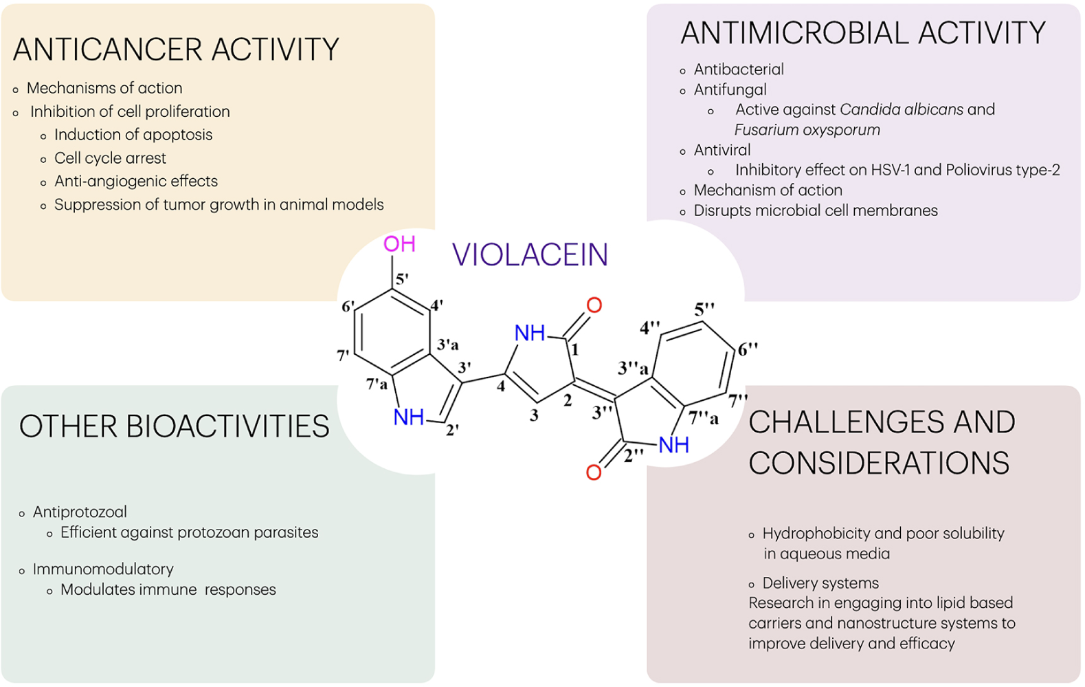
Violacein can be used in numerous end-use applications because of its biological activities,22,61-68 which can be summarized according to the mechanism of action in Table 3, and are illustrated in Scheme 1. It has also been explored for functional fabric development, synthesis of different types of nanoparticles, functional polymer composites, and sunscreen ingredients.49

Illustration of the end-use applications of violacein, which summarized the antimicrobial, anticancer, and other biological activities. There are also important challenges to consider when aiming for a violacein biological applications.
Violacein shows notable antibacterial properties against Gram-positive bacteria, as it has been shown to inhibit the growth of S. aureus, including methicillin-resistant strains (MRSA).61 It can disrupt the cell membrane integrity and interfere with many metabolic processes. It also exhibits strong inhibitory effects on B. subtilis common soil bacterium, often used as a model organism. While Gram-negative bacteria are typically more resistant due to their outer membrane, violacein still shows moderate activity, especially when combined with other agents. Violacein’s activity against P. aeruginosa is weaker, but it can still inhibit biofilm formation, which is key to this bacterium’s pathogenicity. Additionally, violacein has been shown to work synergistically with other antibiotics; for example, it can enhance the effectiveness of drugs like ciprofloxacin or gentamicin against resistant bacteria.62 This synergy is explained by violacein’s action that weakens bacterial membranes, allowing other antibiotics to penetrate more effectively. At last, violacein disrupts the formation of biofilms, which are protective layers created by bacteria to resist antibiotics.63 This is particularly useful against chronic infections involving S. aureus63 or E. coli.
Violacein has shown antiviral potential against several types of viruses.59,69-71 It works mainly by disrupting viral replication inside host cells, interfering with virus entry or attachment to the host cell, and modulating the immune response, enhancing antiviral defenses. Violacein has been reported to reduce Herpes Simplex Virus (HSV-1 and HSV-2) infection in vitro by inhibiting viral replication. Some studies found that violacein and its derivatives, because of the structure similarities with the indole alkaloids, can inhibit virus replications,71 although more research is needed. Additionally, potential inhibitory effects against the Zika and Chikungunya viruses are expected, by affecting their abilities to replicate inside cells, but this is still under investigation.
Among newly studied activities, we can cite violacein and deoxyviolacein applications in solving coronavirus (COVID-19)-related and retrovirus problems. For example, purple pigments obtained from Jantinobacterium sp. were identified as severe acute respiratory syndrome coronavirus (SARS-CoV2) spike protein S1:ACE2 biotin and human immunodeficiency virus (HIV-1) reverse transcriptase binders and inhibitors in vitro.59 Violacein is a protease inhibitor, a useful activity when targeting ACE2 receptor, it also, showed immunomodulatory effects, which is invaluable in treating COVID-19.59 Mpro (also called 3CLpro) and papain-like protease (PLpro) account for the viral polyprotein disruption process, which plays a key role in virus survival and replication processes. Further, the Mpro modulates viral transcription and replication of SARS-CoV, which qualifies it as an attractive pharmacological target.60,61 Known cytotoxic actions of violacein include inhibition of several proteases, applicable to a new target - Mpro. It is known that leukemia progenitor cell death is mediated by calpain (calcium-dependent protease-cysteine protease) suppression and disease-associated protein kinase 1 (DAPK1). Violacein also induced several protein kinase activities, for example, protein kinase A (PKA), pyruvate dehydrogenase kinase (PDK), and protein kinase B (AKT), which were monitored for structural modifications induced by endoplasmic reticulum stress and Golgi apparatus collapse, leading to cell death.62 Because of this activity, it is expected that violacein can greatly add to leukemia treatment.72,73
Another important bioactivity of violacein was discovered when analyzing its effects on different immune cell lines, for example, THP-1, MonoMac 6, murine macrophages (ANA-1, Raw 264.7 cells), and human peripheral blood mononuclear cells PBMCs, among others. TNF-α production stimulation was observed in murine macrophages (ANA-1 and Raw 264.7), and IL-6 and IL-1β secretion was detected in PBMCs. Evidence of the molecular Raw 264.7 cell activation mechanisms was obtained by determining the messenger ribonucleic acid (mRNA) expression pattern upon treatment with violacein. Incubation with violacein led to the activation of pathways related to an inflammatory response.74 Data on the use of TLR-transfected HEK293 cells have indicated that violacein activates the human TLR8 (hTLR8) receptor signaling pathway, but not the human TLR7 (hTLR7) one. Furthermore, the violacein immunostimulatory effect on PBMCs could be suppressed by the specific hTLR8 antagonist, namely: CU-CPT9a. Finally, hTLR8 interaction with violacein in silico was investigated and evidenced that violacein could similarly bind to hTLR8 and to imidazoquinoline compounds. Therefore, based on these results, violacein can be applied in future immunotherapy strategies.74
Violacein has been shown to modulate the immune response in a few notable ways by showing anti-inflammatory effects, suppressing pro-inflammatory cytokines, like TNF-α and IL-6, especially in response to stimuli like lipopolysaccharides (LPS). It reduces activation of nuclear factor kappa B (NF-κB), a key transcription factor in the inflammatory response.73-75 Violacein also influences macrophage polarization, potentially promoting a shift from the pro-inflammatory M1 phenotype to the anti-inflammatory M2 type. This could help treat chronic inflammation or autoimmune diseases. Some studies suggest violacein can inhibit overactive T-cell proliferation, which could be beneficial in diseases where the immune system is overactive. Violacein has also shown potential to stimulate an anti-tumor immune response, possibly by enhancing immune recognition of cancer cells. It does not just fight microbes directly-it also primes the immune system to respond better to infections in some models. Overall, immunomodulatory activity of violacein makes it a promising compound in drug development for inflammation, autoimmune conditions, and possibly cancer immunotherapy.76
Bacteria synthesize antibiotics to control microbial interactions and compete with the microbiome for survival and space. The antibacterial effects in Gram-positive bacteria were studied by Gupta and Ghosh,75 and changes in the molecular composition of lipid were observed under violacein exposure. Lipids’ thermodynamic phases are of paramount importance for maintaining bacterial membrane integrity.
One of the greatest threats to human health is antimicrobial resistance, which is inducible by antibiosis in target organisms. Additionally, antibiotics applied at sublethal doses have been shown to globally change gene expression patterns. Accordingly, hygromycin A derived from Streptomyces sp. strain 2AW induces C. violaceum ATCC 31532 to produce the purple antibiotic known as violacein.77 Likewise, sublethal doses of other antibiotics that similarly target the polypeptide elongation step of translation-induced violacein production, unlike antibiotics with different targets. C. violaceum biofilm formation and virulence against Drosophila melanogaster were also induced by translation-inhibiting antibiotics.77
Moreover, this study identified an antibiotic-induced response (air) two-component regulatory system that is required for these responses.77 Genetic analyses have evidenced a connection among the air system, quorum-dependent signaling, and negative regulator VioS; this finding led to the proposal of a violacein production-induction model. This study suggested a new interspecies interaction mechanism, according to which a given bacterium produces an antibiotic in response to inhibition by another bacterium and supports the role played by antibiotics as signal molecules.77 An interesting study78 has shown that 0.6 mg L-1 violacein from C. violaceum was 12.8-fold more effective against membrane-disrupting Staphylococcus aureus under simulated microgravity than under normal gravity conditions. This finding suggested that an increased rate of anteiso fatty acids (due to microgravity) led to higher membrane disorder and fluidity.78 A ligature-induced periodontitis study conducted with C. violaceum or violacein prevented bone resorption by periodontitis and suggested their application as a new probiotic or antibacterial agent, respectively.79
Violacein/superparamagnetic iron oxide nanoparticles co-encapsulation with polylactic acid nanoparticles had an antimicrobial effect on multidrug-resistant S. aureus and E. coli, besides showing anticancer activity against glioblastoma and melanoma.80
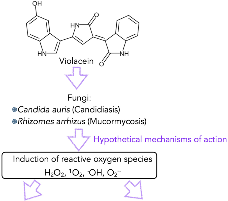
Violacein exhibits strong antifungal properties,69,81,82 especially against pathogenic fungi such as Aspergillus fumigatus, Candida albicans, Cryptococcus neoformans, Fusarium spp., among others. There are some mechanistic aspects of the antifungal effects of violacein: it can integrate into the fungal cell membrane, causing leakage of cellular contents and leading to cell death. Violacein can also increase the production of reactive oxygen species (ROS) inside fungal cells, damaging proteins, lipids, and deoxyribonucleic acid (DNA). Some studies64,69 suggest violacein can trigger programmed cell death in fungi, similar to apoptosis in human cells. In fungi like Candida, violacein interferes with biofilm development, making them more vulnerable to the immune system or other antifungals. Because of these effects, violacein is being explored as a potential alternative or complementary agent to traditional antifungal drugs, especially in the face of rising antifungal resistance. Infections caused by drug-resistant fungi are highly concerning since these infections are comorbidities associated with SARS-CoV-2 infection.
Violacein has the potential to be used as a new strategy against mucormycosis (caused by Rhizopus arrhizus), which is an opportunistic disease capable of affecting individuals’ respiratory tract and the skin, as well as against candidiasis (caused by Candida auris),60,68,69 which presents different manifestations ranging from local skin and mucosal lesions to severe outspread. These two important infections must be taken into consideration in severe COVID-19 cases.69
Violacein is a hydrophobic bis-indole (i.e., XLogP3-AA = 2.7) delivered to aqueous environments through an efficient transport route called outer membrane vesicles (OMVs). OMVs are small, spherical segments detached from the outer membrane of Gram-negative bacteria. C. violaceum OMV secretions are controlled by a mechanism called quorum sensing system CviI/CviR, which enables cell-to-cell communication, as well as regulating different virulence factors, such as biofilm formation and violacein biosynthesis. Another virulence factor bacterial type 3 secretion system (T3SS) is divided into two types: Cpi-1 and Cpi-2. Cpi-1’s needle and rod effector proteins are likely recognized by nucleotide binding domain and leucine rich repeat containing (NLR) family of apoptosis inhibitory proteins (NAIP) receptors in humans and mice; they activate the NLRC4 inflammasome cascade, effectively clear spleen infections via pyroptosis10,83 and cytotoxicity mediated by IL-18-driven Natural killer (NK) cells in the liver.
Violacein activity on biofilms84,85 was extensively investigated through quorum-controlled biofilm formation, violacein delivery by OMVs and T3SS effector protein production, and host-mediated immunological effects against the Cpi1 of T3SS. This finding suggested a research path with natural bioactive molecules, such as palmitic acid, which can act as an anti-quorum agent by reducing the expression of virulence factors, and as an immunomodulatory agent capable of improving innate immune defense through NLRC4 inflammasome hyperactivation and, consequently, of significantly purging C. violaceum infections.73
Violacein is capable of interacting with fungi by stopping and interfering with the hyphal development and biofilm formation processes, due to ROS generation induction in fungal film cells (Scheme 2).59 This pigment also prevents food intoxication by inhibiting bacterial growth and toxin production, as in the case of Bacillus cereus, as recently reported.85

Additionally, it was observed that low violacein doses used in the treatment of patients subjected to intestinal diseases can benefit the host due to changes observed in the gut biome.86
Beyond its medicinal potential, violacein has a variety of other applications, particularly in biotechnology and industry.87 Some potential uses include applications as a natural dye and in bioremediation.88,89 The vibrant purple color of violacein has made it an interesting compound for use as a natural dye. Unlike synthetic dyes, violacein is biodegradable, relatively non-toxic, and could be used in a variety of applications, including textile, food, and cosmetics industries. Its antimicrobial properties could also make it useful in products that require preservation or antibacterial properties. Additionally, it has been studied in the context of bioremediation, the process of using microorganisms or their products to clean up environmental contaminants. Since violacein-producing bacteria like M. violacea are often found in soil and water, they might be used to degrade environmental pollutants. For instance, some studies suggest that violacein-producing bacteria can break down toxic organic compounds, making them useful in cleaning up oil spills or other chemical pollutants.
Although violacein has shown promise in terms of therapeutic benefits, its toxicity to humans, especially at higher concentrations (mmol L-1), needs to be carefully studied. While some studies show that violacein has relatively low toxicity,65 further research is required to determine safe dosage levels for medical applications. It is very important to note that violacein exhibits cytotoxicity that varies across different cell types and concentrations.64-67 It has been shown to induce mitochondrial membrane hyperpolarization, leading to cell death in various cell lines, including MRC-5 and HeLa cells.64 In Caco-2 cells, violacein mediates ROS production, which activates caspase-3, releases cytochrome c, and causes calcium release into the cytosol, culminating in apoptosis.64 The cytotoxic effects of violacein vary among different cell types. For instance, while Caco-2 cells exhibit significant ROS production and apoptosis, HT29 cells do not show increased ROS levels, indicating that the effects of violacein are cell-type specific. Some cytotoxicity data are summarized in Table 4.
While it demonstrates promising therapeutic potential, its toxicity profile must be carefully considered before being applied for any purpose.
Violacein demonstrates significant cytotoxicity across various cell lines, with effects ranging from mitochondrial dysfunction to oxidative stress-induced apoptosis. Its toxicity profile varies depending on the cell type and concentration, highlighting the need for careful consideration in therapeutic applications. Further studies are essential to fully understand its mechanisms and to develop strategies to mitigate potential toxic effects.
5. Conclusions
Violacein has been investigated for its pharmacological activity, as well as for its applications, for several decades. It is well-known for its antibacterial, antifungal, anticancer, antiulcerogenic, antiviral, trypanocide, and immunomodulatory properties. While violacein holds a lot of promise, there are some challenges to be addressed for its widespread use in medical and industrial applications. One of those are linked to production costs, where the natural production of violacein can be inefficient, so researchers are working on optimizing biosynthetic pathways to make violacein more easily and cheaply produced, potentially through genetic engineering or fermentation processes. Difficulties faced in violacein production and purification processes have challenged experts for a long time. Currently, some important advancements have been made in violacein production from several bacteria. The bacterial species J. lividum, for example, was capable of significantly increasing violacein yield. However, the violacein purification process still faces some challenges that are yet to be solved, although some simple processes based on the high content of pure violacein and deoxviolacein derived from C. violaceum were reported.
Another challenge is linked to toxicity and side effects, while some studies show that violacein has relatively low toxicity against cell lines used as control (healthy), such as Vero cells, further research is required to determine safe dosage levels for medical applications. At last, its formulation challenges for therapeutic use, such as in cancer treatment or infection control, must be delivered in a way that ensures it reaches the target site effectively. This could involve encapsulating the compound in nanoparticles or using drug delivery systems that release violacein in a controlled manner.
Ongoing research focuses on optimizing violacein production through biotechnological processes (e.g., recombinant DNA technology) to increase yields and lower costs. There is important research dealing with pharmacokinetics and drug delivery systems to make violacein more effective as a drug. Additionally, understanding the full spectrum of the biological activity of violacein, including its immune-modulating effects, interactions with other drugs, and potential synergies with other therapies is another crucial area of research.
In the current review, we revisited the most relevant violacein-related aspects, such as production, purification, and actual biological activities, pointing to side effects and toxicity issues, aiming to enhance its viability for biotechnological applications and turn it into a new and important product for its technical use as a biological agent. Violacein is not just a pretty pigment; it is a compound with a wide range of biological properties, from antimicrobial and anticancer effects to potential uses in bioremediation and dyeing industries. Its full potential is still being explored, but this natural compound could play a significant role in future therapies and applications.
Data Availability Statement
All data are available in the text.
Acknowledgments
The authors acknowledge the São Paulo Research Council (FAPESP grant number No. 2018/10052-1), the Brazilian National Council for Scientific and Technological Development (CNPq grant number No. 552120/2011-1), and INCTBio.
References
-
1 Newman, D. J.; Cragg, G. M.; Snader, K. M.; Nat. Prod. Rep. 2000, 17, 215. [Crossref]
» Crossref -
2 Shinoda, K.; Hasegawa, T.; Sato, H.; Shinozaki, M.; Kuramoto, H.; Takamiya, Y.; Sato, T.; Nikaidou, N.; Watanabe, T.; Hoshino, T.; Chem. Commun. 2007, 40, 4140. [Crossref]
» Crossref -
3 Harvey, A.; Edrada-Ebel, R.; Quinn, R.; Nat. Rev. Drug Discovery 2015, 14, 111. [Crossref]
» Crossref -
4 Newman, D. J.; Cragg, G. M.; J. Nat. Prod. 2016, 79, 629. [Crossref]
» Crossref -
5 Newman, D. J.; Cragg, G. M.; J. Nat. Prod. 2020, 83, 770. [Crossref]
» Crossref -
6 Numan, M.; Bashir, S.; Mumtaz, R.; Tayyab, S.; Rehman, N. U.; Khan, A. L.; Shinwari, Z. K.; Al-Harrasi, A.; 3Biotech 2018, 8, 207. [Crossref]
» Crossref -
7 Durán, M.; Ponezi, A. N.; Faljoni-Alario, A.; Teixeira, M. F. S.; Justo, G. Z.; Durán, N.; Med. Chem. Res. 2012, 21, 1524. [Crossref]
» Crossref -
8 Choi, S. Y.; Kim, S.; Lyuck, S.; Kim, S. B.; Mitchell, R. J.; Sci. Rep. 2015, 5, 15598. [Crossref]
» Crossref -
9 Durán, N.; Justo, G. Z.; Durán, M.; Brocchi, M.; Cordi, L.; Tasic, L.; Castro, G. R.; Nakazato, G.; Biotechnol. Adv. 2016, 34, 1030. [Crossref]
» Crossref -
10 Durán, N.; Fávaro, W. J.; Inter. J. Med. Rev. 2020, 7, 93. [Crossref]
» Crossref -
11 Durán, N.; Nakazato, G.; Durán, M.; Berti, I. R.; Castro, G. R.; Stanisic, D.; Brocchi, M.; Fávaro, W. J.; Halder, C. V. F.; Justo, G. Z.; Tasic, L.; World J. Microbiol. Biotechnol. 2021, 37, 151. [Crossref]
» Crossref -
12 Hooe, S. L.; Thakur, M.; Lasarte-Aragonés, G.; Breger, J. C.; Walper, S. A.; Medintz, I. L.; Ellis, G. A.; ACS Omega 2024, 9, 3894. [Crossref]
» Crossref -
13 Petersen, M.; Nielsen, T.; Org. Lett. 2013, 15, 1986. [Crossref]
» Crossref -
14 McLaughlin, E. C.; Norman, M. W.; Ko Ko, T.; Stolt, I.; ChemInform 2014, 45 [Crossref]; McLaughlin, E. C.; Norman, M. W.; Ko Ko, T.; Stolt, I.; Tetrahedron Lett. 2014, 55, 2609. [Crossref]
» Crossref» Crossref -
15 Justo, G. Z.; Durán, N.; Best. Pract. Res., Clin. Gastroenterol. 2017, 31, 649. [Crossref]
» Crossref -
16 Shen, Y.; Liang, W.-J.; Shi, Y.-N.; Kennelly, E. J.; Zhao, D.-K.; Nat. Prod. Rep. 2020, 37, 763. [Crossref]
» Crossref - 17 Linhares, T. S. T.; de Lima, K. Y. G.; Marques, K. K. G.; de Souza, R. R. M.; Orchardt, H.; Vasconcelos, U. In Themes Focused on Interdisciplinarity and Sustainable Development Wworldwide; Seven Editora Acadêmica: Brazil, 2024, p. 523.
-
18 Ahmad, W. A.; Yusof, N. Z.; Nordin, N.; Zakaria, Z. A.; Rezali, M. F.; Appl. Biochem Biotechnol. 2012, 167, 1220. [Crossref]
» Crossref -
19 Santos, A. B.; Costa, P. S.; do Carmo, A. O.; da Rocha, F. G.; Scholte, L. L. S.; Ruiz, J.; Kalapothakis, E.; Chartone-Souza, E.; Nascimento, A. M. A.; Int. J. Genomics 2018, 1. [Crossref]
» Crossref -
20 Vöing, K.; Harrison, A.; Soby, S. D.; Genome Announc. 2015, 3, 1. [Crossref]
» Crossref -
21 Brazilian National Genome Project Consortium; Haselkorn, R., ed.; University of Chicago: Chicago, IL, 2003, p. 11660. [Crossref]
» Crossref -
22 Sasidharan, A.; Sasidharan, N. K.; Amma, D. B. N. S.; Vasu, R. K.; Nataraja, A. V.; Bhaskaran, K.; J. Microbiol. 2015, 53, 694. [Crossref]
» Crossref -
23 Lamendella, R.; Jude, B. A.; Microbiol. Resour. Announc. 2018, 7, 1. [Crossref]
» Crossref -
24 Doing, G.; Perron, G. G.; Jude, B. A.; Genome Announc. 2018, 6, e01428. [Crossref]
» Crossref -
25 Atalah, J.; Blamey, L.; Munoz-Ibacache, S.; Gutierrez, F.; Urzua, M.; Encinas, M. V.; Páez, M.; Sun, J.; Blamey, J. M.; Extremophiles 2020, 24, 43. [Crossref]
» Crossref -
26 Valdes, N.; Soto, P.; Cottet, L.; Alarcon, P.; Gonzalez, A.; Castillo, A.; Corsini, G.; Tello, M.; Stand. Genomic Sci. 2015, 1. [Crossref]
» Crossref -
27 Woodhams, D. C.; LaBumbard, B. C.; Barnhart, K. L.; Becker, M. H.; Bletz, M. C.; Escobar, L. A.; Flechas, S. V.; Forman, M. E.; Iannetta, A. A.; Joyce, M. D.; Rabemananjara, F.; Gratwicke, B.; Vences, M.; Minbiole, K. P. C.; Microb. Ecol. 2018, 75, 1049. [Crossref]
» Crossref -
28 Avguštin, J. A.; Bertok, D. Z.; Kostanjšek, R.; Avguštin, G.; Antonie van Leeuwenhoek 2013, 103, 763. [Crossref]
» Crossref -
29 Xu, X.; Tian, L.; Zhang, S.; Jiang, L.; Zhang, Z.; Huang, H.; Microb. Pathog. 2019, 128, 178. [Crossref]
» Crossref -
30 Bettina, A. M.; Doing, G.; O’Brien, K.; Perron, G. G.; Jude, B. A.; Genome Announc. 2018, 6, e01426. [Crossref]
» Crossref -
31 Smith, H. J.; Foreman, C. M.; Akiyama, T.; Franklin, M. J.; Devitt, N. P.; Ramaraj, T.; Genome Announc. 2016, 4, e01468. [Crossref]
» Crossref -
32 Mojib, N.; Nasti, T. H.; Andersen, D. T.; Attigada, V. R.; Hoover, R. B.; Yusuf, N.; Bej, A. K.; Int. J. Dermatol. 2011, 5, 1223. [Crossref]
» Crossref -
33 Alem, D.; Marizcurrena, J. J.; Saravia, V.; Davyt, D.; Martinez-Lopez, W.; Castro-Sowinski, S.; World J. Microbiol. Biotechnol. 2020, 36, 120. [Crossref]
» Crossref -
34 Kuzyk, S. B.; Pritchard, A. O.; Ploufe, J.; Sorensen, J. L.; Yurkov, V.; Can. J. Great Lakes Res. 2021, 47, 715. [Crossref]
» Crossref -
35 Myeong, N. R.; Seong, H. J.; Kim, H. J.; Sul, W. J.; J. Biotechnol. 2016, 223, 36. [Crossref]
» Crossref -
36 Agematu, H.; Suzuki, K.; Tsuya, H.; Biosci. Biotechnol. Biochem. 2011, 75, 2008. [Crossref]
» Crossref -
37 Cress, B. F.; Erkert, K. A.; Barquera, B.; Kofas, M. A.; Genome Announc. 2013, 1, e0004813. [Crossref]
» Crossref -
38 Thøgersen, M. S.; Delpin, M. W.; Melchiorsen, J.; Kilstrup, M.; Månsson, M.; Bunk, B.; Sproer, C.; Overmann, J.; Nielsen, K. F.; Gram, L.; Front. Microbiol. 2016, 7, 1461. [Crossref]
» Crossref -
39 Dang, H. T.; Yotsumoto, K.; Enomoto, K.; Genome Announc. 2014, 2, e01346. [Crossref]
» Crossref -
40 Wu, Y. H.; Cheng, H.; Xu, L.; Jin, X. B.; Wang, C. S.; Xu, X. W.; PLoS One 2017, 12, e0179997. [Crossref]
» Crossref -
41 Aye, A. M.; Bonnin-Jusserand, M.; Brian-Jaisson, F.; Ortalo-Magne, A.; Culioli, G.; Nevry, R. K.; Rabah, N.; Blache, Y.; Molmeret, M.; Microbiology 2015, 161, 2039. [Crossref]
» Crossref -
42 Puranik, S.; Talkal, R.; Qureshi, A.; Khardenavis, A.; Kapley, A.; Purohit, H. J.; Genome Announc. 2013, 1, e01115. [Crossref]
» Crossref -
43 Jude, B. A.; Microbiol. Resour. Announc. 2019, 8, e00683. [Crossref]
» Crossref -
44 Chernogor, L.; Bakhalova, K.; Belikova, A.; Belikov, S.; Microorganisms 2022, 10, 1071. [Crossref]
» Crossref -
45 Bektas, K. I.; Nalcaoglu, A.; Kati, H.; Ceylan, E.; Nalcacioglu, R.; Belduz, A. O.; Canakci, S.; FEMS Microbiol. Lett. 2022, 370, 1. [Crossref]
» Crossref -
46 Kothari, V.; Sharma, S.; Padia, D.; Asian Pac. J. Trop. Med. 2017, 10, 744. [Crossref]
» Crossref -
47 Choi, S. Y.; Lim, S.; Yoon, K. H.; Lee, J. I.; Mitchell, R. J.; J. Biol. Eng. 2021, 15, 10. [Crossref]
» Crossref -
48 Ahmed, A.; Ahmad, A.; Li, R.; AL-Ansi, W.; Fatima, M.; Mushtaq, B. S.; Basharat, S.; Li, Y.; Bai, Z.; J. Microbiol. Biotechnol. 2021, 31, 1465. [Crossref]
» Crossref -
49 Park, H.; Park, S.; Yang, Y. H.; Choi, K. Y.; Crit. Rev. Biotechnol. 2021, 41, 879. [Crossref]
» Crossref -
50 Cheng, K.-C.; Hsiao, H.-C.; Hou, Y.-C.; Hsieh, C.-W.; Hsu, H.-Y.; Chen, H.-Y.; Lin, S.-P.; Antioxidants 2022, 11, 849. [Crossref]
» Crossref -
51 De León, M. E.; Wilson, H. S.; Jospin, G.; Jonathan, A.; Eisen, J. A.; Microb. Genomics 2023, 9, 000971. [Crossref]
» Crossref -
52 Da, G.; Seo, E.; Lee, J. W.; Biotechnol. Bioproc. 2023, 28, 1005. [Crossref]
» Crossref -
53 Bagoghl, N.; Hosseini-Abari, A.; World J. Microbiol. Biotechnol. 2024, 40, 149. [Crossref]
» Crossref -
54 Müller, T.; Schick, S.; Klemp, J. S.; Sprenge, G. A.; Takors, R.; Bioproc. Biosys. Eng. 2024, 47, 713. [Crossref]
» Crossref -
55 Mohamad, S.; Kamarulzaman, E. E.; Minggu, M. M.; Mal. J. Med. Health Sci. 2024, 27. [Crossref]
» Crossref -
56 Lai, H.-E.; Obled, A. M. C.; Chee, S. M.; Morgan, R. M.; Lynch, R.; Sharma, S. V.; Moore, S. J.; Polizzi, K. M.; Goss, R. J. M.; Freemont, P. S.; ACS Chem. Biol. 2021, 16, 2116. [Crossref]
» Crossref -
57 Nemer, G.; Louka, N.; Blandin, P. R.; Maroun, R. G.; Vorobiev, E.; Rossignol, T.; Nicaud, J.-M.; Guénin, E.; Koubaa, M.; Molecules 2023, 28, 4292. [Crossref]
» Crossref -
58 e Lima, L. O.; Alexandre, H. M. S.; Bezerra, C. A. A. S.; da Silva, L. A.; Loureiro, P. B. A.; Vanderley, S. E. R.; Borchardt, H.; Vasconcelos, U.; Violacein - Part II: From Significance to Perspectives; Seven Editora, 2024, p. 531-543. [Crossref]
» Crossref -
59 Doganci, M. A.; Ay Sal, F.; Guler, H. I.; Katı, H, Ceylan, E.; Belduz, A. O.; Bozdal, G.; Yaylı, N.; Canakcı, S.; World J. Microbiol. Biotechnol. 2022, 38, 161. [Crossref]
» Crossref -
60 Kholany, M.; Trébulle, P.; Martins, M.; Ventura, S. P.; Nicaud, J.-M.; Coutinho, J. A.; J. Chem. Technol. Biotechnol. 2020, 95, 1126. [Crossref]
» Crossref -
61 Aruldass, C. A.; Masalamany, S. R. L.; Venil, C. K.; Ahmad, W. A.; Environ. Sci. Pollut. Res. Int. 2018, 25, 5164. [Crossref]
» Crossref -
62 Dodou, H. V.; de Morais Batista, A. H.; Sales, G. W. P.; de Medeiros, S. C.; Rodrigues, M. L.; Nogueira, P. C. N.; Silveira, E. R.; Nogueira, N. A. P.; J. Appl. Microbiol. 2017, 123, 853. [Crossref]
» Crossref -
63 Batista, A. H. M.; Moreira, A. C. D.; de Carvalho, R. M.; Sales, G. W. P.; Nogueira, P. C. N.; Grangeiro, T. B.; Medeiros, S. C.; Silveira, E. R.; Nogueira, N. A. P.; Molecules 2017, 22, 1534. [Crossref]
» Crossref -
64 Leal, A. M. S.; de Queiroz, J. D. F.; de Medeiros, S. R. B.; Lima, T. K. S.; Agnez-Lima, L. F.; BMC Microbiol. 2015, 15, 115. [Crossref]
» Crossref -
65 Neroni, B.; Zingaropoli, M. A.; Radocchia, G.; Ciardi, M. R.; Mosca, L.; Pantanella, F.; Schippa, S.; Oncology Lett. 2022, 23, 132. [Crossref
» Crossref -
66 Santhosh, V. T.; Muthusamy, P.; Asian J. Pharm. Clin. Res. 2017, 10, 227. [Crossref]
» Crossref -
67 Mehta, T.; Vercruysse, K.; Johnson, T.; Ejiofor, A. O.; Myles, E.; Quick, Q. A.; Mol. Med. Rep. 2015, 12, 1443. [Crossref]
» Crossref -
68 Dahlem, C.; Chanda, S.; Hemmer, J.; Schymik, H. S.; Kohlstedt, M.; Wittmann, C.; Kiemer, A. K.; Front. Oncol. 2022, 12, 872223. [Crossref]
» Crossref -
69 Durán, N.; Castro, G. R.; Portela, R. W. D.; Favaro, W. J.; Durán, M.; Tasic, L.; Nakazato, G.; Appl. Microbiol. 2022, 75, 796. [Crossref]
» Crossref -
70 Durán, N.; Justo, G. Z.; Fávaro, W. J.; Nakazato, G.; Inter. J. Med. Rev. 2022, 9, 351. [Crossref]
» Crossref -
71 Rivero Berti, I.; Gantne, M. E.; Rodriguez, D. S.; Islan, G. A.; Fávaro, W. J.; Talevi, S. A.; Castro, G. R.; Durán, N.; Front. Nanotechnol. 2023, 5, 1186386. [Crossref]
» Crossref -
72 Monsalve-Escudero, L. M.; Loaiza-Cano, V.; Pájaro-González, Y.; Oliveros-Díaz, A. F.; Diaz-Castillo, F.; Quiñones, W.; Robledo, S.; Martinez-Gutierrez, M.; BMC Complement. Med. Ther. 2021, 21, 216. [Crossref]
» Crossref -
73 Queiroz, K. C.; Milani, R.; Ruela-de-Sousa, R. R.; Fuhler, G. M.; Justo, G. Z.; Zambuzzi, W. F.; Durán, N.; Diks, S. H.; Spek, C. A.; Ferreira, C. V.; Peppelenbosch, M.; PLoS One 2012, 7, e45362. [Crossref]
» Crossref -
74 Venegas, F. A.; Köllisch, G.; Wibke, K. M. K.; Diederich, Kaufmann, A.; Bauer, S.; Max, Chavarría, M.; Araya, J. J.; García-Piñeres, A. J.; Sci. Rep. 2019, 9, 13661. [Crossref]
» Crossref -
75 Gupta, R.; Ghosh, S. K.; Biochim. Biophys. Acta, Biomembr. 2023, 1865, 184130. [Crossref]
» Crossref -
76 Verinaud, L.; Lopes, S. C.; Prado, I. C.; Zanucoli, F.; Alves da Costa, T.; Di Gangi, R.; Issayama, L. K.; Carvalho, A. C.; Bonfanti, A. P.; Niederauer, G. F.; Duran, N.; Costa, F. T.; Oliveira, A. L.; Höfling, M. A.; Machado, D. R.; Thomé, R.; PLoS One 2015, 10, e0125409. [Crossref]
» Crossref -
77 Lozano, G. L.; Guan, C.; Cao, Y.; Borlee, B. R.; Broderick, N. A.; Stabb, E. V.; Handelsman, J.; mBio 2020, 11, e00948. [Crossref]
» Crossref -
78 Jang, H.; Choi, S. Y.; Mitchell, R. J.; Cells 2023, 12, 1907. [Crossref]
» Crossref -
79 Barbirato, D. S.; Fogacci, M. F.; Guimarães, T. C.; de Carvalho, D. P.; Rurr, J. C.; Takiya, C. M.; Scharfstein, J.; da Costa Leitão, A. A.; Clin. Oral Investig. 2023, 27, 2175. [Crossref]
» Crossref -
80 Kanelli, M.; Saleh, B.; Webster, T. J.; Vouyiouka, S.; Topakas, E.; J. Biomed. Nanotechnol. 2022, 18, 729. [Crossref]
» Crossref -
81 Nakazato, G.; Fávaro, W. J.; Castro, G. R.; Durán, N.; AIRJ Preprints, Article Id: 337, 2021. [Crossref]
» Crossref -
82 Nakazato, G.; Fávaro, W. J.; Durán, N.; AIJR Preprints, Article Id: 330, 2021 [Crossref]
» Crossref -
83 Durán, N.; Fávaro, W. J.; arXiv:2006.15777, 2020. [Crossref]
» Crossref -
84 Venkatramanan, M.; Nalini, E.; Front. Microbiol. 2024, 15, 1303595. [Crossref]
» Crossref -
85 Lou, X.; Zhou, Q.; Jiang, Q.; Lin, L.; Zhu, W.; Mei, X.; Xiong, J.; Gao, Y.; Int. J. Food Microbiol. 2025, 426, 110917. [Crossref]
» Crossref -
86 Pauer, H.; Hardoim, C. C. P.; Teixeira, F. L.; Miranda, K. R.; Barbirato, D. D. S.; de Carvalho, D. P.; Antunes, L. C. M.; Leitão, Á. A. D. C.; Lobo, L. A.; Domingues, R. M. C. P.; PLoS One 2018, 13, e0203748. [Crossref]
» Crossref -
87 Kanelli, M.; Mandic, M.; Kalakona, M.; Vasilakos, S.; Kekos, D.; Nikodinovic-Runic, J.; Topakas, E.; Front. Microbiol. 2018, 9, 1495. [Crossref]
» Crossref -
88 Ramesh, C.; Vinithkumar, N. V.; Kirubagaran, R.; Venil, C. K.; Dufossé, L.; Microorganisms 2019, 7, 186. [Crossref]
» Crossref
Edited by
-
Editor handled this article:
Hector Henrique F. Koolen (Associate)