Stability Assessment of HKUST-1 Metal-Organic Framework with Gold Nanoparticles
Abstract
HKUST-1 is a metal-organic framework (MOF) with the formula [Cu3(BTC)2(H2O)3]n (BTC: benzene-1,3,5-tricarboxylate), which exhibits relevant properties due to its high porosity. However, HKUST-1 is highly sensitive to water, collapsing in aqueous media or under high-humidity conditions. The modification of this MOF with metal nanostructures has seen significant growth in recent years, with gold nanoparticles (AuNPs) among the most widely used. This article reports the association between gold nanoparticles (AuNPs) and MOF HKUST-1 and investigates the influence of water from AuNPs on the degradation of HKUST-1, in addition to proposing a modification that maintains the stable structure, based on the transfer of gold nanoparticles to a non-aqueous medium (N,N-dimethylformamide) before the modification of HKUST-1. An analysis of the influence of the ethanol washing process on gold-modified HKUST-1 samples was also performed.
Keywords:
HKUST-1; gold nanoparticles; stability; degradation; Raman spectroscopy
Introduction
Metal-organic frameworks (MOFs) are a class of coordination polymers with porous dimensions1,2 that have attracted scientific and technological interest due to their hybrid composition, which allows a broad scope for adjusting their chemistry, microporosity, and pore system through the rigorous selection of their precursors.1,3,4 The most relevant characteristics of MOFs are high surface area, high crystallinity, permanent porosity, and good thermal stability. The strong metal-ligand bonding gives MOFs permanent porosity that is kept intact even after solvent molecules are removed from the inner parts of the pores.5 Due to their properties, MOFs have been used in a wide range of applications, such as gas separation and storage,6,7 permanent magnets,8 luminescence,9 heterogeneous catalysis,10 drug loading,11 and others.12
HKUST-1 (Hong Kong University of Science and Technology), first synthesized in 1999,13 is a MOF with the formula [Cu3(BTC)2(H2O)3]n (BTC = benzene-1,3,5-tricarboxylate), composed of square paddlewheel clusters of Cu2 connected by BTC ligands.14 The adsorption properties of HKUST-1 were already well described in the literature.15 It has also been described both experimentally16,17 and computationally18,19 that the crystalline structure of HKUST-1 undergoes considerable structural changes during continuous exposure to water in either liquid or vapor forms.
One of the fastest-growing areas in recent years is the association between metallic nanoparticles (NPs) and MOF.20 This approach aims to provide methods for integrating the properties of MOFs and NPs, specifically the microporosity and guest selectivity of MOFs, with the sensing capabilities or catalytic activities of metallic nanoparticles. In the literature, several methods are available for obtaining hybrid systems composed of MOFs doped with NPs. As defined by Xiang et al.,21 there are three main methods to obtain a MOF system with nanoparticles: ship-in-bottle, bottle-around-ship, and one-step synthesis.
The ship-in-bottle method involves forming metal nanoparticles within the MOF cavity. Initially, the nanoparticle precursor is deposited within the pores of the MOF.22 After the precursor enters the pores, a reductor forms the metallic NPs. This method is convenient and straightforward, but it is challenging to control the size and morphology of the NPs in the pore cavity; the NPs can occasionally grow on the outer surface of the MOF. In the bottle-around-ship method, metal nanoparticles are encapsulated within the MOF in two steps: first, the metal nanoparticles are obtained, and then the NPs are added to a solution containing the MOF precursors, which encapsulate the metal nanoparticles during the self-assembly process.23 The bottle-around-ship method (also called in situ modification) provides improved control over size and morphology of the NPs impregnated within the MOF. However, the introduction of NPs can, in some cases, hinder MOF growth due to a high interfacial energy barrier.24 In addition, aggregation of the NPs may occur during the MOF nucleation process. One-step synthesis involves directly mixing NPs and MOF precursor solutions, followed by their simultaneous growth to form the NP/MOF association. This preparation strategy is straightforward but requires a rigorous balance of precursors quantities for both nucleation and growth of the NPs and the MOF to occur.25 Another strategy for modifying MOFs is post-synthesis modification (PSM), in which the MOF and nanoparticles are synthesized separately and subsequently combined. In this approach, the nanoparticles surface can be functionalized to enhance their interaction with the MOF. Representative examples include the work of Liu et al.,26 who modified the MIL-101 MOF with AuNPs to obtain the MIL-101/Au composite, and the study by Fu et al.,27 who applied the PSM method to incorporate AuNPs into UiO-66, resulting in the UiO-66/AuNPs material.
The modification of HKUST-1 with AuNPs has been widely investigated. For instance, Cao et al.28 and Zho et al.29 used the ship-in-bottle method to prepare AuNP-modified HKUST-1, whereas Jiang et al.30 used a one-step synthesis involving reagent mixing in sealed Teflon® vessels followed by microwave-assisted synthesis. The interaction of HKUST-1 with AuNPs, however, can compromise the integrity of the MOF structure due to the presence of water in colloidal suspensions, which promotes partial or total degradation of the framework.31 It has been shown that regeneration of HKUST-1 is possible through treatments such as ethanol washing, which can partially restore the original crystallinity and porosity by removing coordinated water molecules and re-establishing metal-ligand interactions.32
In this context, this work investigates the influence of water ingress on the structural integrity of HKUST-1 as a function of different modification methods with AuNPs, as well as the effect of the ethanol washing process on the restoration of the HKUST-1 framework.
Furthermore, this study proposes an innovative strategy for modifying HKUST-1 with AuNPs by employing N,N-dimethylformamide (DMF) as the dispersion medium for the nanoparticles, replacing the conventionally used aqueous colloidal systems. This approach overcomes one of the main limitations of AuNPs modification of HKUST-1, namely the structural degradation induced by water exposure, thereby preserving the crystalline integrity and stability of the MOF. By minimizing water entrance into the framework and maintaining intact metal-ligand interactions, the proposed method enables the efficient immobilization of AuNPs without compromising the porosity and intrinsic properties of the HKUST-1 structure.33
Experimental
Materials and reagents
Tetrachloroauric acid (HAuCl4) (99.9%, Sigma-Aldrich), sodium citrate (99%, Sigma-Aldrich), sodium borohydride (NaBH4) (99%, Fluka), N,N dimethylformamide (DMF) (99.8%, Sigma-Aldrich), ethanol (99%, Vetec), tetradecyltrimethylammonium bromide (MTAB) (Sigma-Aldrich), copper nitrate trihydrate (98%, Dinamica), benzene-1,3,5-tricarboxylic acid (98%, Sigma-Aldrich) were all used without further purification. The solvents used were ethanol (P.A., Sigma-Aldrich) and deionized water from a Millipore Synergy-UV deionizer (ρ (resistivity) = 18.2 MΩ cm).
Instrumentation
The Raman spectra were obtained in a Bruker SENTERRA Raman spectrometer equipped with an Olympus BX51 optical microscope. The samples presented a high sensitivity to the laser beam. To mitigate this problem, Raman spectra were obtained by mapping 25 sample points using 0.2 mW of 633 nm laser power, with 15 or 30 s accumulation time, and a 20× (numerical aperture (NA) = 0.40) objective lens. The Raman spectra presented in this work are the average of the spectra from the 25 points considered in each mapping; this procedure allowed to obtain a signal-to-noise ratio high enough to allow an unambiguous interpretation without sample degradation.
Fourier transform infrared spectroscopy (FTIR) spectra were obtained on a Bruker Vertex70 infrared spectrometer attached to a Hyperion3000 module. Measurements were made using the transmission method with the material dispersed in KBr pellets with a resolution of 4 cm-1 and a spectral range between 400 and 4000 cm-1 using a diamond ATR (attenuated total reflectance) accessory, with a resolution of 4 cm-1 and a spectral range between 400 and 4000 cm-1.
Powder X-ray diffraction (PXRD) patterns were obtained on a Bruker D8 Advance DaVinci with Bragg-Brentano θ-θ geometry and using Cu-Kα radiation (λ = 1.54186 Å), detector LynxEye worked at 40 kV and 40 mA. The second instrument used was a Bruker D8 Advanced with Bragg-Brentano θ-θ geometry and using Co-Kα radiation (λ = 1.78897 Å), a LynXeye detector operating at a voltage of 35 kV and a current of 40 mA; both instruments used an angular range of 5 to 70o.
To evaluate the thermal behavior of the samples, thermogravimetric (TG) and differential thermal analysis (DTA) experiments, TG/DTA simultaneously, were carried out on a Shimadzu 60H thermobalance. The thermal energy effects were evaluated during heating at a rate of 10 ºC min-1 between 25 and 600 ºC under a constant airflow (50 mL min-1). First-order TG curves were derived to confirm the range of thermal phenomena. For the thermal decomposition kinetics experiments, 15 and 20 ºC min-1 rates were also performed under the same operational conditions.
Scanning electron microscopy (SEM) images of the samples were obtained using an FEI Quanta 250 instrument operated at 30 kV. Energy-dispersive X-ray spectroscopy (EDS) was employed to perform a qualitative analysis of the elemental composition of the materials. For this analysis, a Hitachi TM3030 benchtop SEM equipped with a SwiftED3000 EDS module was used, operating at 15.0 kV with an acquisition time of 10 s.
UV-Vis-NIR absorption and diffuse reflectance measurements were obtained on an Ocean Optics USB 2000 + XR1-ES, NIR 256-2.1 spectrophotometer in the 200 to 2800 nm range. Dynamic light scattering (DLS) measurements and ζ-potential measurements were performed using Malvern Panalytical Zetasizer Nano ZEN 3500 equipment; the data presented represent the average of three measurements.
Gold nanoparticles (AuNPs) synthesis
Citrate-coated gold nanoparticles (AuNP-Cit) were synthesized by the reflux method following the Frens methodology.34 Initially, 50 mL of a 1.0 × 10⁻²% (m/v) aqueous solution of tetrachloroauric acid (HAuCl4) was prepared and transferred to a two-neck round-bottom flask. The solution was heated to boiling under reflux, after which 1.0 mL of a 1.0% (m/v) aqueous sodium citrate solution was added. The mixture was heated for approximately 5 min after the color changed from pale yellow to ruby red, indicating the formation of gold nanoparticles.
AuNPs synthesized with sodium borohydride (AuNP Boro) were prepared following the Creighton, Blatchford, and Albrecht methodology.35 Initially, 5.0 mL of a 2.3 mmol L-1 aqueous solution of HAuCl4 were prepared, transferred to a flask, and placed in an ice bath. Subsequently, 15 mL of a 1.0 mmol L-1 NaBH4 solution were added under manual stirring, during which bubble formation was observed. After stirring, the resulting solution was stored at 4 ºC for 12 h until gas evolution ceased, yielding a dark red suspension of gold nanoparticles.
Modification of AuNP-Cit with DMF
The dispersion medium of the citrate-stabilized nanoparticles (AuNP-Cit) was changed from water to DMF. For this purpose, 10 mL of AuNP-Cit were mixed with 1 mL of a MTAB solution (0.001 mg mL-1). The mixture was stirred overnight, then centrifuged for 12 min at 12396 g. The supernatant was discarded, and the resulting pellet was resuspended in 10 mL of DMF. The resulting colloid was named AuNP-Cit-DMF.
Several attempts were made to stabilize the AuNP Boro colloid during solvent exchange by changing MTAB concentrations; however, aggregation could not be prevented, and a stable dispersion in DMF was not achieved. Consequently, the solvent exchange procedure was not applied to AuNP-Boro.
HKUST-1 synthesis and modification with gold nanoparticles
Scheme 1 summarizes the HKUST-1 synthesis and the MOF modifications performed in situ and post-synthesis with different gold nanoparticles in addition to washing with ethanol. The synthesis of the MOF HKUST-1 was based on the work of Hassan et al.,36 where initially, 260 mg (1.24 mmol) of benzene-1,3,5-tricarboxylic acid were solubilized in a flask containing 20.0 mL of ethanol; 298 mg (1.28 mmol) of copper nitrate trihydrate were solubilized in another flask containing 20.0 mL of ethanol. The two solutions were mixed and stirred for 10 min. The resulting product was centrifuged for 5 min, and the supernatant was discarded. The final product was washed with ethanol and left to dry at room temperature. The HKUST-1 MOF was modified with either AuNP-Cit or AuNP-Boro using two methods: during synthesis (in situ modification) and post-synthesis modification (PSM).

The in situ modification of HKUST-1 was also based on the work of Hassan et al.,36 where initially, the procedure described in that study was followed to prepare the reaction mixture containing the BTC ligand and the CuII precursor. The reaction mixture was stirred for 10 min, and 10.0 mL of an aqueous AuNPs colloidal suspension were added after 5 min of stirring. The supernatant was discarded, and the final product was stored at room temperature. AuNP-Cit or AuNP-Boro colloids in aqueous suspension were used to modify the MOF. HKUST-1 with AuNP-Cit resulted in the material called HKUST-AuCit; modification by AuNP Boro resulted in the material called HKUST AuBoro.
Post-synthesis modification was performed using the previously synthesized HKUST-1 MOF. For this purpose, a heat-treatment step was used, in which HKUST-1 was placed in an oven at 150 ºC for 12 h under vacuum; this procedure is referred to as MOF activation. Subsequently, 50 mg of activated HKUST-1 were added to 5 mL of an aqueous colloidal AuNPs suspension, and the mixture was stirred for 10 min. The final mixture was centrifuged, the supernatant was discarded, and the resulting material was dried at room temperature. HKUST-1 modified with AuNP-Cit was named HKUST-AuCit-PSM, and the MOF modified with AuNP-Boro was named HKUST-AuBoro-PSM.
To evaluate the influence of water on the structure, the PSM procedure for HKUST-1 was carried out using deionized water instead of AuNPs. The HKUST-1 obtained after this procedure was referred to as HKUST-water-PSM.
HKUST-1 was modified with AuNP-Cit-DMF using the same in situ and PSM methodologies described above. The resulting materials were named HKUST-AuCit-DMF and HKUST-AuCit-PSM-DMF, respectively.
Washing with ethanol
The HKUST-1 samples, modified either in situ or PSM with AuNP-Cit or AuNP-Boro, were subjected to ethanol washing. In this process, 5.0 mg of each sample were suspended in 2.0 mL of absolute ethanol, stirred overnight at ambient conditions, centrifuged, and the supernatant was discarded. The resulting solid was left to dry at room temperature. The washed products were designated HKUST-AuCit-Washed, HKUST-AuBoro-Washed, HKUST-AuCit-PSM-Washed, and HKUST-AuBoro-PSM-Washed, corresponding to the ethanol-washed versions of HKUST-AuCit, HKUST-AuBoro, HKUST-AuCit-PSM, and HKUST-AuBoro-PSM, respectively.
Results and Discussion
In situ and post-synthesis modification
The PXRD patterns of pristine HKUST-1 and HKUST-1 modified in situ with AuNPs are shown in Figure 1a. The PXRD results in Figure 1a for HKUST-AuCit and HKUST-AuBoro samples show a diffraction pattern that holds some similarity to pristine HKUST-1, with the main HKUST-1 peaks in the low-angle regions present in the diffractogram of modified MOF, specifically the first 3 characteristic MOF peaks assigned to the (200), (220), and (222) planes.37,38 Two weak peaks were identified in HKUST-AuCit and HKUST-AuBoro at 38.40° and 44.35°, which can be attributed to the (111) and (200) planes of gold, respectively, indicating the presence of AuNPs in the MOF structure. However, the diffraction region in the 17-35° range showed several changes that cannot be easily associated with the original diffractogram of HKUST-1, especially in the 20-25° range. Those changes were more pronounced at HKUST-AuBoro and indicated the onset of a phase change in the MOF with the in situ modification.
The diffractogram of HKUST-AuCit-PSM and HKUST AuBoro-PSM, Figure 1b, showed significant changes in the diffraction pattern in the entire angular range. The peaks related to the (111) and (200) planes of gold were still observable. Still, the main peaks of HKUST-1 could not be identified in the diffractogram of the MOF after modification, meaning that PXRD showed the formation of a new phase different from HKUST-1. Figure S1 (see the Supplementary Information (SI) section) presents more details on the diffractograms of the modified HKUST-1, highlighting the gold peaks.
Figure S2 (see the SI section) shows the EDS spectra of pure HKUST-1 samples and those modified with AuNPs under different conditions. All spectra exhibit the characteristic signals of the original MOF, with intense peaks attributed to Cu, C, and O.39 In the modified HKUST-1, all samples exhibit additional peaks assignable to Au, providing evidence of the successful association of gold nanoparticles with the MOF. Figure S3 (SI section) shows the EDS elemental distribution maps of pristine HKUST-1 and the samples modified with gold nanoparticles (AuNPs) prepared using different strategies. In all cases, the presence of carbon (C), oxygen (O), and copper (Cu), which are characteristic elements of the HKUST-1 framework, is clearly observed. For the AuNP modified HKUST-1 samples, the elemental maps reveal the presence of Au homogeneously distributed over the HKUST-1 surface, confirming successful immobilization of AuNPs.
Figure S4 (see SI section) presents the reflectance spectra of pristine HKUST-1 and the samples modified with AuNPs via in situ and post-synthesis methods using AuNP Cit or AuNP-Boro. In all modified samples (HKUST-AuCit, HKUST-AuCit-PSM, HKUST-AuBoro, and HKUST-AuBoro-PSM), an additional band centered around 540 nm is observed and attributed to the plasmonic extinction of AuNPs, as demonstrated in the UV-Vis spectra in Figure S5 (SI section). This band indicates the association of HKUST-1 with AuNPs. This intense and broad band at 540 nm is also observed for HKUST-1 modified with AuCit DMF, as well as for samples regenerated with ethanol after in situ and post-synthesis modification.
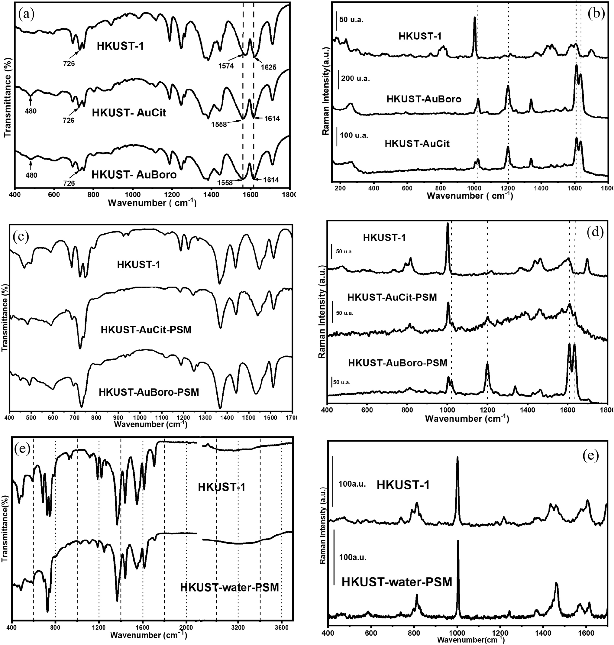
Figure 2 shows the Raman and FTIR spectra of HKUST-1 and the four materials obtained by modification with AuNP-Cit and AuNP-Boro. In Figure 2a, the FTIR spectra of the in situ modified samples are compared to HKUST-1. The most relevant bands of HKUST-1 were identified in the FTIR spectra of the AuNPs-modified HKUST-1 samples, such as the band at 480 cm-1 assigned to ν(Cu-O) and two bands at 726 and 752 cm-1 (β(C-H)). In all obtained spectra, the bands attributed to νas(COO-) and νs(COO-) appeared at 1445 and 1382 cm-1, respectively.40,41 The bands at 1573 and 1625 cm-1 in the infrared spectrum of unmodified HKUST-1, ν(CC) of the benzene ring, were identified at 1562 and 1615 cm-1, respectively, in the spectrum of AuNPs-modified HKUST-1. The shift of these bands in the FTIR spectra of AuNPs-modified HKUST-1 indicates the interaction of the nanoparticles with the MOF. In the infrared spectra of both HKUST-AuCit-PSM and HKUST-AuBoro-PSM in Figure 2c, the bands at 1220, 1573, and 1625 cm-1 in the infrared spectrum of unmodified HKUST-1 shifted similarly to the in situ modified HKUST-1. The infrared spectral similarities among the several materials indicate regular changes with the modification.42

PXRD diffractograms (λ = 1.78897 Å) of pure HKUST-1 and HKUST-1 containing AuNPs obtained by (a) in situ modification and (b) post-synthetic modification. (c) PXRD (λ = 1.54186 Å) patterns of pure HKUST-1 and HKUST-water-PSM. (D) PXRD (λ = 1.54186 Å) patterns of HKUS water-PSM, HKUST-AuCit-PSM, and HKUST-AuBoro-PSM.

FTIR spectrum in KBr pellets (a) and Raman spectra (b) of HKUST-1 and HKUST-1 modified in situ with AuNPs. FTIR (KBr pellets) (c) and Raman spectra (d) of HKUST-1 and HKUST-1 modified via PSM with AuNPs. FTIR (ATR) (e) and Raman spectra (f) of HKUST-1 and HKUST-water-PSM.
The Raman spectra (Figures 2b and 2d) of HKUST-1 showed similar modifications of the spectral profiles, regardless of whether it was modified by AuNPs in situ or in a PSM. The band at 1006 cm-1, the most intense in the Raman spectrum of HKUST-1, assigned to the breathing mode of the benzene ring, was observed as a shoulder to a band at 1025 cm-1 in the Raman spectra of HKUST-AuCit and HKUST-AuBoro; on the other hand, for HKUST-AuCit-PSM and HKUST-AuBoro-PSM, the most intense of the two features is the band at 1006 cm-1 and the band at 1025 cm-1 is observed as a shoulder to it. However, both bands can be assigned to the ring-breathing mode in different coordination states; this indicates that the 1025 cm-1 band in Raman spectra of the HKUST-1 materials containing AuNPs can be attributed to some extent to the change in coordination of the aromatic ring in the presence of the AuNPs. It should also be mentioned that the band assigned to νas(COO-) appeared at 1460 cm-1 in the spectrum of HKUST-1, while it presented very low intensity in the case of AuNPs-modified HKUST-1. The band assigned to νs(COO-) was observed at 1382 cm-1 in the spectrum of HKUST-1 but was not identified in the spectrum of the AuNPs-modified MOF. In the HKUST-1 spectrum, the bands at 1580 and 1605 cm-1 are assigned to the ν(CC) of the aromatic ring, while in the spectra of the AuNPs-modified HKUST-1, these bands shifted to higher wavenumbers at 1610 and 1630 cm-1, respectively.41-43 Tables S1, S2, and S3 provide further details on the assignments of the vibrational modes of pure HKUST-1 and HKUST-1 modified with AuNPs.
The above discussion indicates greater spectral changes in the Raman spectra than in the FTIR spectra. The more significant changes, including the enhancement of some specific bands of the Raman spectrum of HKUST-1, may be associated with the surface-enhanced Raman spectroscopy (SERS) effect caused by the presence of the plasmonic AuNPs associated with the HKUST-1 structure. The occurrence of the SERS effect would enhance bands absent in the spectrum of HKUST-1 under normal Raman conditions, caused by the enhancement of bands associated with modes of chemical groups that are closer to the surface of the nanoparticles,28 resulting in the SERS effect acting as a site-specific probe for the interaction between the AuNPs and the MOF structure. Thus, the band shifts in the Raman spectra discussed above can be attributed to specific interactions of AuNPs with chemical moieties of the MOF structure in direct contact with the nanoparticle surface, which are enhanced by the SERS effect.44,45 The changes observed in the Raman and infrared spectra indicate structural modifications in the MOF upon association with gold nanoparticles, suggesting interfacial interactions between the nanoparticles and the MOF. The work of Hassan et al.36 was used as a reference for the preparation of this hybrid material, and according to that study, the interaction between AuNPs and the MOF occurs predominantly at the MOF surface. In this context, AuNPs interact with HKUST-1 primarily via surface adsorption during MOF formation, mediated by interactions between the functional groups of the BTC ligand and the AuNPs surface. This results in the association of the nanoparticles with the MOF matrix without encapsulation within the intrinsic pores. For the PSM samples, the spectroscopic data indicate a similar type of interaction.
The ζ-potential results presented in Figure S6 demonstrate that the AuNP-Cit and AuNP-Boro nanoparticles used to modify HKUST-1 exhibit good colloidal stability associated the negative surface potential. DLS analyses, showed in Figure S7 (SI section) indicate that the AuNPs have dimensions larger than 10 nm (with an average size of 20 nm), which are incompatible with the pore size of HKUST-1 (ca. 1 nm).36 From a structural perspective, the cavity size of HKUST-1 is significantly smaller than the nanoparticle dimensions, preventing their accommodation within the MOF pores. Therefore, the most probable configuration involves nanoparticle interaction at the external surface of the MOF. According to the classification proposed by Li et al.,46 this type of interaction is defined as guest-on-MOF.
No significant differences were observed between the Raman spectra of in situ modified HKUST-1 and HKUST-1 modified via post-synthetic modification. However, significant differences were observed in the PXRD of these materials. The changes observed in HKUST-1 after the in situ and post-synthesis modifications (Figures 1a and 1b) were attributed to the intrusion of water from the nanoparticle dispersion into the MOF structure. HKUST-1 is known to be sensitive to water, and among the two approaches, the post-synthesis modification method proved to be more aggressive. To obtain further information on the role of AuNPs in the spectral changes, a blank experiment was performed for the HKUST-1 PSM. The same modification was used, except that the AuNPs colloid was substituted by deionized water, resulting in the HKUST-water-PSM sample.
The PXRD pattern of pristine HKUST-1 and HKUST-water-PSM is shown in Figure 1c. There is a significant change in the MOF diffraction pattern after modification, characterized by the disappearance of the main HKUST-1 peaks. The diffraction profile of the HKUST-water-PSM material is very similar to the HKUST-AuCit-PSM and HKUST-AuBoro-PSM samples (a comparison of the diffractogram for the three materials is presented in Figure 1d). This change was primarily detectable by PXRD analysis. As shown in Figure S8 (SI section), no peaks corresponding to HKUST-1 precursors, such as copper nitrate trihydrate or the trimesic acid ligand, were identified in the XRD patterns of the MOF modified in situ or via PSM. A possible factor contributing to this more pronounced degradation is the stirring, which may accelerate water ingress into the MOF structure and promote degradation to a great extent. One possible factor contributing to this more pronounced degradation in PSM is the agitation process, which may accelerate water ingress into the MOF structure and promote its degradation.
Figures 2e and 2f show the FTIR and Raman spectra of the pristine HKUST-1 samples and the water-modified counterpart, HKUST-water-PSM. There was a slight change in the spectral profile of the MOF after modification under both techniques. In the Raman spectrum, the position of the band at 1006 cm-1 remained unchanged, and the relative intensity of the bands at 1550 and 1618 cm-1 were inverted in HKUST-water-PSM compared to HKUST-1. The FTIR spectra also exhibited only relatively minor changes, with the most relevant alteration being the shift of the band from 1220 to 1246 cm-1, mirroring the behavior observed in the HKUST AuCit-PSM and HKUST-AuBoro-PSM materials. As shown in Figure 1d, the diffractograms of the HKUST-AuCit-PSM and HKUST-AuBoro-PSM materials exhibit the same profile as that of HKUST-Water-PSM. Based on the above results, there is an indication that the PSM using aqueous AuNPs leads to the degradation of the MOF in a manner similar to that observed in the presence of solely water.
The PXRD pattern of the HKUST-water-PSM material is very similar to that documented in the study by Majano et al.,32 where the structure of HKUST-1 collapsed after exposure to 77% humidity for 2 months. In the present study, direct exposure of HKUST-1 to liquid water from the AuNPs colloids seems to have accelerated the degradation process. Consistent with results in the literature, prolonged exposure of HKUST-1 to water can lead to complete collapse, resulting in the formation of a hybrid material composed of BTC and various copper oxide species,31,47,48 which is likely the result of the modification process; however, the PXRD comparison with the diffractograms of the cited species could not account for the structural changes induced by the modification with aqueous AuNPs.
Figure 3 presents the SEM images of pure HKUST-1 and AuNPs-modified HKUST-1. The HKUST-1 microcrystals presented octahedral morphology (Figure 3a). After AuNPs entered the structure, the SEM micrographs (Figures 3b 3e) presented intense changes in the morphology of the microcrystals, which started to show irregular shapes and edges with much less clarity. The changes in microcrystal morphology upon association with AuNPs, as evidenced by SEM analysis, indicate MOF degradation following modification with AuNPs.

SEM images of HKUST-1 (a), HKUST-AuCit (b), HKUST-AuBoro (c), HKUST-AuCit-PSM (d) HKUST-AuBoro-PSM (e), HKUST AuCit DMF (f) and HKSUT-AuCit-PSM-DMF (g).
Another study carried out was the thermogravimetric analysis of HKUST-1 modified with gold to obtain more information on the influence of water on HKUST-1 structure. Figure 4 shows the thermogravimetric (TG) curves at 10 °C min-1. The thermal stability of all samples is similar, with a mass loss of 75%. Mass loss initiates at room temperature with a significant loss at 300 °C. Figure S9 (see SI section) shows the decomposition curves of the HKUST-1 samples with DTA and DTG results, which show that the events are exothermic. The exothermic event was not considered when calculating the thermal decomposition kinetics of each sample from the TG curve data. Instead, an interpolated temperature curve was considered solely in this region.

For thermal decomposition kinetic analysis, the TG dynamic curves at heating rates of 10, 15, and 20 °C min-1 were used to determine the conversion degree (α) curves. The isoconversional Vyazovkin method and the multilayer perceptron neural network (MLP) were applied for three heating rates. The activation energy (Ea), frequency factor (A), and mechanism were determined based on Araujo et al.49 As demonstrated in Figure S10 (see the SI section), the association of AuNPs with the MOF structure resulted in a slight increase in the average activation energy of HKUST-1. The contribution models calculated by the MLP network are shown in Figure S11 (see the SI section), with the Avrami-Erofeev model of order n = 2 being the predominant kinetic model across all samples. Adding AuNPs does not appear to have affected the physical mechanism underlying HKUST-1 degradation. This suggests that water is the primary factor responsible for the changes observed in AuNPs-modified HKUST-1.
Influence of ethanol washing on the HKUST-1 AuNPs-modification process
The previous section presented evidence that water promotes the degradation of HKUST-1 during the modification by aqueous AuNPs. The influence of water on the structural degradation of the MOF has been reported in the literature14,18,31 and attributed mainly to strong affinity for Cu2+ ions, which can lead to the breaking of metal-ligand bonds and, consequently, loss of the characteristic structure of the MOF.
HKUST-1 is composed of paddlewheel-type clusters in which two Cu2+ ions are coordinated to four carboxylate groups of the BTC (benzene-1,3,5-tricarboxylate) ligand, leaving each copper ion with an open axial site that can coordinate with water molecules.14 These water molecules bind to the open metal sites, releasing significant adsorption energy due to the strong interaction between the copper ion and the oxygen atom of the water molecule. This generates enough energy to cleave the Cu-O carboxylate bonds of the paddlewheel secondary building unit (SBU),50 exposing new metal sites that coordinate with additional water molecules in a cascade process. This self-amplifying mechanism promotes the progressive replacement of the organic ligands by water molecules, ultimately compromising the connectivity between the metal clusters and the ligands.14,31
Several studies involving the modification of HKUST-1 with gold nanoparticles reported using ethanol at different stages of the process, such as during the preparation of colloidal suspensions,42 washing,51 or redispersion of the material.42 In the present study, the influence of ethanol on the regeneration of HKUST-1 after modification with AuNPs using two methodologies, in situ and PSM, was evaluated. Figure 5 shows the diffractograms comparing pure HKUST-1 samples with AuNPs-modified samples before and after ethanol washing. For HKUST-1 modified with AuNPs in the in situ process (Figure 5a), it was possible to observe complete regeneration of the MOF, evidenced by the disappearance of the peaks between 20 25°. Figure 5b shows PXRD diffractograms comparing the PSM modification of HKUST-1. It can be observed that, after the washing process, the most characteristic peaks of HKUST-1 reappeared, such as those between 5-15°, but some peaks associated with the degraded framework remained observable, including those between 17-20° and the peaks at 22° and 27°. The presence of these residual peaks indicates that the washing process was insufficient to fully restore the structure of the MOF modified via the PSM method, compared to the in situ modified MOF. This corroborates the observation that the post-synthetic modification (PSM) process is more aggressive than the in situ process. The infrared spectra presented in Figure S12 (SI section) indicate that no significant structural changes occurred after washing the AuNPs-modified HKUST-1. An improvement in the signal-to-noise ratio was observed, which can be attributed to the removal of coordinated water molecules from the framework.

PXRD (λ = 1.54186 Å) comparing HKUST-1 modified with AuNPs before and after ethanol washing for in situ modification (a) and PSM (b).
Majano et al.32 proposed that the regeneration of degraded HKUST-1 occurs via a solvent-assisted process in which ethanol acts as a mediator. After degradation, the original structure collapses, yielding a mixture of partially coordinated trimesate and copper-oxo species. When the degraded material is exposed to ethanol, the solvent breaks hydrogen interactions and increases the mobility of trimesic acid, thereby allowing it to recoordinate to copper ions. Thus, the porous and crystalline structure of HKUST-1 is partially or almost completely restored, recovering the original structure. According to the authors, this process exploits a “memory effect” of the material because, after degradation, the trimesic acid and copper species remain intimately mixed and partially organized at the local scale; then, when ethanol is introduced, the building blocks reassemble into the original architecture of HKUST-1. The results agree with those observations. However, the ability of ethanol to restore the HKUST-1 structure is limited when the structure is strongly affected, as in the case of the PSM modification.
In situ and post-synthesis modification of HKUST-1 with AuNP-Cit-DMF
As shown above, the water present in AuNPs suspensions influences the structure of HKUST-1; therefore, ethanol is used to prevent water from entering the system or to regenerate the material after nanoparticle modification. An alternative approach to modifying HKUST-1 with AuNPs, avoiding structural degradation, is to employ nanoparticles dispersed in a medium that does not compromise the MOF structure, such as AuNPs dispersed in DMF. To perform the modification in the proposed medium, the dispersion medium of the AuNP-Cit colloid was changed by centrifugation of the aqueous suspension and redispersion in DMF; the resulting material was designated AuNP-Cit-DMF. As shown in Figure S4 (see SI section), a slight shift and broadening in the localized surface plasmon resonance (LSPR) band were observed following the solvent exchange; the broadening may be associated with partial aggregation (however, no deposition was observed) and the shift was associated to the change in the medium refractive index change, as the refractive index of DMF is 1.430, higher than water (1.333).52 Despite the slight changes, the nanoparticles remained stable in DMF medium.
Figure 6a presents the Raman spectra of HKUST-1 after in situ and PSM using AuNP-Cit-DMF. No significant changes were observed in the Raman spectral after the association between gold nanoparticles (AuNPs) and the MOF structure. The main vibrational bands were observed in the same positions, including the BTC ring-breathing mode band at 1006 cm-1.

Raman spectra (a), PXRD (λ = 1.54186 Å) patterns (b), and FTIR spectra acquired using the ATR method in the fingerprint region (400 1800 cm 1) (c) and in the high-wavenumber region (2600-4000 cm-1) (d) of HKUST-1, HKUST-AuCit-DMF, and HKUST-AuCit-PSM-DMF.
Figures 6c and 6d show the FTIR spectra of HKUST-1 modified with AuNP-Cit-DMF via both modification methodologies. The overall spectral profile remained unchanged after the modification by AuNPs. However, new bands at 659, 1256, 1678, and 2935 cm-1 were identified, which were associated with the presence of DMF within the modified-MOF material.53,54
Figure 6b shows the diffractograms comparing pure HKUST-1 and AuNP-Cit-DMF-modified HKUST-1. No changes were observed in the diffraction pattern after association the metal nanoparticles, indicating that the MOF structure remained intact and that HKUST-1 was not degraded. The EDS analysis (Figure S1, SI section) qualitatively confirmed the presence of AuNPs within the HKUST-1 structure. Furthermore, the UV-Vis diffuse reflectance spectra presented in Figure 3e also support the successful association of AuNP-Cit-DMF with the MOF.
Regarding the MOF morphology, as shown in the SEM micrographs of the modified material (Figures 3f-3g), no significant morphological changes were observed, unlike the alterations seen in the aqueous AuNP-Cit-modified MOF. The morphology of HKUST-AuCit-DMF is comparable to that of HKUST-AuCit-PSM-DMF. Thus, Raman, FTIR, XRD, and SEM analyses indicate that there was no significant structural change between the in situ and PSM methods when AuNP-Cit-DMF was used to modify HKUST-1.
The spectroscopic, diffraction, and microscopy evidence indicate that exchanging the solvent of the AuNPs to DMF allowed obtaining the AuNP-modified HKUST-1, preserving the MOF structure intact. That improvement may be understood considering that, although DMF can be adsorbed within the MOF’s pores,55 it does not exhibit an affinity for copper ions as strong as water does and, therefore, does not induce structural degradation. Water acted as a destructive agent for HKUST-1 due to its ability to cleave metal-ligand bonds via hydrolysis, whereas DMF, despite being involved in the synthesis process, does not exert the same destabilizing effect on the MOF.54
Conclusions
This study investigated the modification of HKUST-1 with AuNPs using two distinct strategies: in situ modification and PSM. PXRD analysis revealed that both approaches induced partial degradation of the HKUST-1 framework, with PSM causing more pronounced structural alterations than the in situ method. This greater extent of degradation was attributed to the increased of water introduced into the MOF structure during the PSM. Importantly, these structural changes were detectable only by PXRD analysis. Thermogravimetric and thermal decomposition kinetic analyses further confirmed that water ingress into the HKUST-1 framework was the main factor responsible for its degradation. Given the increasing number of studies focusing on the use of nanoparticles to modify HKUST-1 for diverse applications, these results underscore the importance of detailed structural characterization to verify the integrity of the MOF framework after modification.
Building on these observations, the use of ethanol to mitigate or reverse structural degradation proved to be moderately effective, as the higher water content associated with PSM resulted in partial rather than complete recovery of the MOF structure after ethanol washing. In contrast, employing AuNPs suspended in DMF enabled successful modification of HKUST-1 without detectable structural degradation. This finding was supported by PXRD, as well as spectroscopic analyses (UV-Vis-NIR, FTIR, and Raman) and SEM, establishing the DMF-based approach as a reliable strategy for modifying HKUST-1 with AuNPs while preserving its structural integrity.
Supplementary Information
Supplementary material 1
Supplementary information is available free of charge at http://jbcs.sbq.org.br as PDF file.
Acknowledgments
The authors thank FAPEMIG, CNPq, and CAPES for their financial support. Laboratório Multiusuário de Bioprodutos e Bioprocessos (CENTRALBIO) for its support in carrying out the DLS/ζ-potential, and the EDS measurements.
Data Availability Statement
All data are available in the text.
References
-
1 Li, C.; Zhang, L.; Chen, J.; Li, X.; Sun, J.; Zhu, J.; Wang, X.; Fu, Y.; Nanoscale 2021, 13, 485. [Crossref]
» Crossref -
2 Khan, N. A.; Hasan, Z.; Jhung, S. H.; Coord. Chem. Rev. 2018, 376, 20. [Crossref]
» Crossref -
3 Ma, T.; Li, H.; Ma, J.-G.; Cheng, P.; Dalton Trans. 2020, 49, 1712. [Crossref]
» Crossref -
4 Stock, N.; Biswas, S.; Chem. Rev. 2012, 112, 933. [Crossref]
» Crossref -
5 Férey, G.; Serre, C.; Chem. Soc. Rev. 2009, 38, 1380. [Crossref]
» Crossref -
6 Evans, A.; Luebke, R.; Petit, C.; J. Mater. Chem. A 2018, 6, 10570. [Crossref]
» Crossref -
7 Silva, F. H.; Flores, L. S.; da Silva, A. F. M.; Ronconi, C. M.; Leitão, A. A.; Corrêa, C. C.; New J. Chem. 2024, 48, 6451. [Crossref]
» Crossref -
8 Ye, Z.; Padilla, J. A.; Xuriguera, E.; Brillas, E.; Sirés, I.; Appl. Catal. B 2020, 266, 118604. [Crossref]
» Crossref -
9 Hao, J.; Xu, X.; Fei, H.; Li, L.; Yan, B.; Adv. Mater. 2018, 30, 1705634. [Crossref]
» Crossref -
10 Chen, Y.; Zhang, R.; Jiao, L.; Jiang, H.; Coord. Chem. Rev. 2018, 362, 1. [Crossref]
» Crossref -
11 Sha, H.; Yan, B.; Anal. Chim. Acta 2023, 1272, 341525. [Crossref]
» Crossref -
12 Zhang, J.; Zhu, E.; Li, R.; Wang, X.; Zou, T.; Wang, Y.; Liu, Y.; Yang, L.; Guan, X.; ACS Omega 2024, 47, 46643. [Crossref]
» Crossref -
13 Chui, S. S.-Y.; Lo, S. M.-F.; Charmant, J. P. H.; Orpen, A. G.; Williams, I. D.; Science 1999, 283, 1148. [Crossref]
» Crossref -
14 Xue, W.; Zhang, Z.; Huang, H.; Zhong, C.; Mei, D.; Phys. Chem. C 2020, 124, 1991. [Crossref]
» Crossref -
15 Castillo, J. M.; Vlugt, T. J. H.; Calero, S.; J. Phys. Chem. C 2008, 112, 15934. [Crossref]
» Crossref -
16 Liu, P.; Zhao, T.; Cai, K.; Chen, P.; Liu, F.; Tao, D. J.; Chem. Eng. J. 2022, 437, 135364. [Crossref]
» Crossref -
17 Gu, J.; Li, J.; Ma, Q.; Nanomaterials 2022, 12, 1134. [Crossref]
» Crossref -
18 Domán, A.; Czakkel, O.; Porcar, L.; Madarász, J.; Geissler, E.; László, K.; Appl. Surf. Sci. 2019, 480, 138. [Crossref]
» Crossref -
19 Fan, H.; Yang, C.; Zhou, Y.; Phys. Rev. B 2022, 106, 085417. [Crossref]
» Crossref -
20 Shirkhodaie, M.; Seidi, S.; Shemirani, F.; Zaroudi, F.; TrAC, Trends Anal. Chem. 2023, 164, 117109. [Crossref]
» Crossref -
21 Xiang, W.; Zhang, Y.; Lin, H.; Liu, C.; Molecules 2017, 22, 2103. [Crossref]
» Crossref -
22 Hu, Y.; Liao, J.; Wang, D.; Li, G.; Anal. Chem. 2014, 86, 3955. [Crossref]
» Crossref -
23 Ma, Y.-C.; Zhu, Y.-H.; Tang, X.-F.; Hang, L.-F.; Jiang, W.; Li, M.; Khan, M. I.; You, Y.-Z.; Wang, Y.-C.; Biomater. Sci. 2019, 7, 2740. [Crossref]
» Crossref -
24 Cheetham, A. K.; Kieslich, G.; Yeung, H. H.-M.; Acc. Chem. Res. 2018, 51, 659. [Crossref]
» Crossref -
25 Yang, Q.; Xu, Q.; Jiang, H.-L.; Chem. Soc. Rev. 2017, 46, 4774. [Crossref]
» Crossref -
26 Liu, H.; Zhao, B.; Zhang, C.; Charact. Appl. Nanomater. 2022, 5, 30. [Crossref]
» Crossref -
27 Fu, J.; Lai, H.; Zhang, Z.; Li, G.; Anal. Chim. Acta 2021, 1161, 338464. [Crossref]
» Crossref -
28 Cao, X.; Hong, S.; Jiang, Z.; She, Y.; Wang, S.; Zhang, C.; Li, H.; Jin, F.; Jin, M.; Wang, J.; Analyst 2017, 142, 2640. [Crossref]
» Crossref -
29 Zhou, H.; Zhu, A.; Wang, C.; Guo, X.; Ying, Y.; Wu, Y.; Liu, X.; Wang, F.; Wen, Y.; Yang, H.; Spectrochim. Acta, Part A 2024, 304, 123280. [Crossref]
» Crossref -
30 Jiang, Y.; Zhang, X.; Dai, X.; Zhang, W.; Sheng, Q.; Zhuo, H.; Xiao, Y.; Wang, H.; Nano Res. 2017, 10, 876. [Crossref]
» Crossref -
31 McHugh, L. N.; McPherson, M. J.; McCormick, L. J.; Morris, S. A.; Wheatley, P. S.; Teat, S. J.; McKay, D.; Dawson, D. M.; Sansome, C. E. F.; Ashbrook, S. E.; Stone, C. A.; Smith, M. W.; Morris, R. E.; Nat. Chem. 2018, 10, 1096. [Crossref]
» Crossref -
32 Majano, G.; Martin, O.; Hammes, M.; Smeets, S.; Baerlocher, C.; Pérez-Ramírez, J.; Adv. Funct. Mater. 2014, 24, 3855. [Crossref]
» Crossref -
33 Heravi, M. M.; Ghavidel, M.; Mohammadkhani, L.; RSC Adv. 2018, 8, 27832. [Crossref]
» Crossref -
34 Frens, G.; Nature, Phys. Sci. 1973, 241, 20. [Crossref]
» Crossref -
35 Creighton, J. A.; Blatchford, C. G.; Albrecht, M. G.; J. Chem. Soc. 1979, 75, 790. [Crossref]
» Crossref -
36 Hassan, M. H.; El-Basha, O.; Haikal, R. R.; Ibrahim, A. H.; Alkordi, M. H.; ACS Appl. Mater. Interfaces 2018, 10, 32942. [Crossref]
» Crossref -
37 Sagar, V.; Kukkar, D.; Environ. Monit. Assess. 2023, 195, 1056. [Crossref]
» Crossref -
38 Chen, Y.; Mu, X.; Lester, E.; Wu, T.; Prog. Nat. Sci.:Mater. Int. 2018, 28, 584. [Crossref]
» Crossref -
39 Lestari, W. W.; Ni’maturrohmah, D.; Arrozi, U. S. F.; Suwarno, H.; IOP Conf. Ser. Mater. Sci. Eng. 2018, 299, 012031. [Crossref]
» Crossref -
40 Dhumal, N. R.; Singh, M. P.; Anderson, J. A.; Kiefer, J.; Kim, H. J.; J. Phys. Chem. C 2016, 120, 3295. [Crossref]
» Crossref -
41 Todaro, M.; Alessi, A.; Sciortino, L.; Agnello, S.; Cannas, M.; Gelardi, F. M.; Buscarino, G.; J. Spectrosc. 2016, 2016, ID 8074297. [Crossref]
» Crossref -
42 Chen, S.; Wang, C.; Wang, M.; Pan, L.; Xu, D.; Li, J.; J. Environ. Chem. Eng. 2024, 12, 113448. [Crossref]
» Crossref -
43 Hadjiivanov, K. I.; Panayotov, D. A.; Mihaylov, M. Y.; Ivanova, E. Z.; Chakarova, K. K.; Andonova, S. M.; Drenchev, N. L.; Chem. Rev. 2021, 121, 1286. [Crossref]
» Crossref -
44 Guselnikova, O.; Postnikov, P.; Elashnikov, R.; Miliutina, E.; Svorcik, V.; Lyutakov, O.; Anal. Chim. Acta 2019, 1068, 70. [Crossref]
» Crossref -
45 Wang, H.-L.; You, E.-M.; Panneerselvam, R.; Ding, S.-Y.; Tian, Z.-Q.; Light Sci. Appl. 2021, 10, 161. [Crossref]
» Crossref -
46 Li, H.; Liu, N.; Liu, Y.; Guo, Y.; Jiang, Z.; Energy Technol. 2025, 13, 2400178. [Crossref]
» Crossref -
47 Peterson, G. W.; Wagner, G. W.; Balboa, A.; Mahle, J.; Sewell, T.; Karwacki, C. J.; J. Phys. Chem. C 2009, 113, 13906. [Crossref]
» Crossref -
48 Álvarez, J. R.; Sánchez-González, E.; Pérez, E.; Schneider-Revueltas, E.; Martínez, A.; Tejeda-Cruz, A.; Islas-Jácome, A.; González-Zamora, E.; Ibarra, I. A.; Dalton Trans. 2017, 46, 9192. [Crossref]
» Crossref -
49 Araujo, N. R. S.; Sebastião, R. C. O.; Freitas-Marques, M. B.; da Nova Mussel, W.; Yoshida, M. I.; Virtuoso, L. S.; Therm. Sci. Eng. Prog. 2022, 36, 101490. [Crossref]
» Crossref -
50 Gentile, F. S.; Pannico, M.; Causà, M.; Mensitieri, G.; Di Palma, G.; Scherillo, G.; Musto, P.; J. Mater. Chem. A 2020, 8, 10796. [Crossref]
» Crossref -
51 Butova, V. V.; Kirichkov, M. V.; Budnyk, A. P.; Guda, A. A.; Soldatov, M. A.; Lamberti, C.; Soldatov, A. V.; Polyhedron 2018, 154, 357. [Crossref]
» Crossref -
52 Bozorova, D.; Gofurov, S.; Ziyayev, M.; Kokhkharov, A.; Kadirova, Z.; Abdiraimova, O.; Ismailova, O.; Latv. J. Phys. Tech. Sci. 2025, 62, 14. [Crossref]
» Crossref -
53 Sharma, A.; Kaur, S.; Mahajan, C. G.; Tripathi, S. K.; Saini, G. S. S.; Mol. Phys. 2007, 105, 117. [Crossref]
» Crossref -
54 Alfonso-Herrera, L. A.; Rodríguez-Girón, J. S.; de Sampedro, H. I. G.; Sánchez-Martínez, D.; Navarrete-López, A. M.; Beltrán, H. I.; Chempluschem 2024, 89, e202300579. [Crossref]
» Crossref -
55 Ediati, R.; Setyani, M. A.; Sulistiono, D. O.; Santoso, E.; Hartanto, D.; Al Bakri Abdullah, M. M.; J. Water Process Eng. 2021, 39, 101670. [Crossref]
» Crossref
Edited by
-
Editor handled this article: Juliano Alves Bonacin (Associate)