
 Influence of Halide Identity and Ligand Side-Chain Length on the Structural, Spectroscopic and Preliminary Antibacterial Properties of CuI-Aminoquinoline Complexes
Abstract
Two 7-chloro-4-substituted aminoquinoline ligands, 2-((7-chloroquinolin-4-yl)amino)ethanol (ACQ12OH) and 3-((7-chloroquinolin-4-yl)amino)propan-1-ol hemihydrate (ACQ13OH), and six CuI complexes containing chloride, bromide, or iodide were synthesized and characterized in order to evaluate the influence of halide identity and ligand side-chain length on their structural, spectroscopic, and preliminary antibacterial properties. Characterization involved elemental, vibrational, electronic, and diffraction analyses, including single-crystal X-ray diffraction for the iodide complex of the ACQ13OH series. Small structural modifications led to significant changes in stoichiometry, coordination mode, solid-state organization, and preliminary antibacterial response. The chloride and bromide derivatives of the ACQ12OH series showed isostructural hydrated trinuclear arrangements, whereas the iodide complexes displayed distinct architectures. Under the minimum inhibitory concentration (MIC) assays, ACQ13OH was more active than ACQ12OH in the free form, whereas coordination to CuI generally led to lower MIC values on a molar basis, particularly for the chlorido and bromido derivatives. Among the investigated systems, the bromido complexes showed the most favorable antibacterial response considering Gram-negative species, including Klebsiella pneumoniae American Type Culture Collection (ATCC) 700603, Acinetobacter baumannii ATCC 19606, Escherichia coli ATCC 25922, and Gram-positive species, including Staphylococcus aureus ATCC 33591, Staphylococcus epidermidis ATCC 12228 species.
Keywords:
CuI complexes; aminoquinoline ligands; crystal structure; vibrational spectroscopy; antibacterial activity
Introduction
Bacterial infections remain a major global public health concern, particularly in light of the increasing spread of antimicrobial resistance. Global estimates indicate that bacterial antimicrobial resistance was directly responsible for 1.27 million deaths and associated with 4.95 million deaths worldwide in 2019, highlighting the urgent need for new antibacterial agents capable of expanding current therapeutic options.1,2
Within this scenario, skin and soft tissue infections deserve particular attention, as they are among the most common bacterial infections encountered in clinical practice and may range from superficial manifestations to severe and potentially life-threatening conditions.3 Their clinical relevance becomes even greater in patients with comorbidities, such as diabetes,4 and in hospital settings, where treatment is often complicated by resistant pathogens.5
This concern is also reflected in recent international priority-pathogen frameworks. The 2024 bacterial priority pathogens list comprises 24 pathogens distributed across 15 families and highlights bacteria resistant to last-resort antibiotics, as well as clinically relevant species such as Pseudomonas aeruginosa and Staphylococcus aureus.6,7 In this context, the search for new compounds with antibacterial potential remains highly relevant.
From a molecular standpoint, aminoquinoline compounds represent a promising class for the development of new bioactive molecules.8 The quinoline nucleus is widely recognized as a relevant scaffold in medicinal chemistry, and 4-aminoquinoline derivatives, Figure 1, stand out because of their structural versatility.9 In particular, modifications in the quinoline ring and in the side chain attached to the amino group may influence properties such as basicity, lipophilicity, and molecular interaction profile, making this framework attractive for the design of new compounds with antibacterial potential.10

In addition to the intrinsic interest of these ligands, copper(I) complexes have also attracted attention in bioinorganic chemistry because of the biological potential associated with this class of compounds.11,12 In the literature,13,14 the antibacterial activity of copper-based systems has been related to multiple structure-dependent mechanisms, including interactions with copper-sensitive cellular proteins and metalloproteins, induction of oxidative stress, membrane perturbation, and possible damage to biomolecules such as deoxyribonucleic acid (DNA). When incorporated into coordination compounds, the properties of the metal center may be modulated by the ligand environment, influencing parameters such as stability, lipophilicity, reactivity, and interaction with biomolecules. Therefore, CuI complexes constitute promising systems for the development of new bioactive compounds, especially in the antibacterial field, where small changes in the coordination sphere may lead to significant differences in activity.15,16
In this context, the present work investigates copper(I) complexes derived from 7-chloro-4-substituted aminoquinolines, with emphasis on the influence of halide identity and ligand side-chain length on the structural, spectroscopic, and preliminary antibacterial properties of the resulting systems.
Experimental
Synthesis and general features
The general procedures for the synthesis of the 7-chloro-4-aminoquinoline ligands and the corresponding CuI complexes are illustrated in Schemes 1 and 2, respectively. All reagents and solvents were obtained from commercial sources and used without further purification.

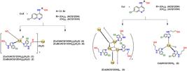
Synthesis of copper(I) 7-chloro-4-aminoquinoline derivative complexes. Acetonitrile is used in all routes as solvent. All reactions were performed under ambient atmosphere, and no evidence of oxidation was observed under the adopted synthetic conditions.
Synthesis of the 7-chloro-4-aminoquinoline derivatives
The ligands 2-((7-chloroquinolin-4-yl)amino)ethanol (ACQ12OH) and 3-((7-chloroquinolin-4-yl)amino)propan-1-ol hemihydrate (ACQ13OH) were synthesized by adding 2-aminoethanol (5.00 mmol, 0.301 mL) or 3-amino-1-propanol (5.00 mmol, 0.383 mL), previously dissolved in ethanol (EtOH, 10 mL), to an ethanolic solution of 4,7-dichloroquinoline (5.00 mmol, 990 mg, in 10 mL of EtOH). The reaction mixture was maintained under reflux at approximately 78 °C for 48 h under constant stirring, while the reaction progress was monitored by thin-layer chromatography (TLC), using CH2Cl2/MeOH (methanol), (9:1) eluent. After completion, the reaction mixtures were treated with 50 mL of 1 mol L-1 aqueous ammonium hydroxide (NH4OH) and stirred at room temperature (RT) for 2 h. The resulting solids were isolated by simple filtration. After solvent evaporation, the polycrystalline materials were purified by washing with hexane/chloroform (8:2 v/v, 100 mL). The purified ligands, see Supplementary Information (SI) section (Figures S20-S23), were then characterized by elemental, spectroscopic, and biological analyses.
Synthesis of CuI complexes
The copper(I) complexes were prepared by reacting the appropriate copper halide with the corresponding aminoquinoline ligand (ACQ12OH or ACQ13OH) in a 1:3 molar ratio. CuCl (1.00 mmol, 99.0 mg), CuBr (1.00 mmol, 143 mg), or CuI (1.00 mmol, 190 mg) was dissolved in CH3CN (10 mL) and added to a CH3CN solution (10 mL) containing ACQ12OH (3.00 mmol, 668 mg) or ACQ13OH (3.00 mmol, 737 mg). The reaction mixture was maintained under stirring at RT. After formation of the precipitate, the suspension was stirred for an additional 1 h. The resulting solids were then isolated by filtration and purified by successive washings with MeOH and CH3CN (1:1 v/v, 50 mL), and this procedure was repeated three times.
The CuI complexes were poorly soluble in CHCl3, CH2Cl2, and CH3CN. Although dissolution was observed in dimethyl sulfoxide (DMSO), it was accompanied by copper disproportionation within 5 min. Because dissolution in this solvent alters the oxidation state of the metal center, solution-state spectroscopic analyses were not carried out for these systems, since the species present in solution do not correspond to those in the solid state. Accordingly, nuclear magnetic resonance (NMR) spectra were recorded only for the free ligands ACQ12OH and ACQ13OH to confirm their formation. The structural discussion of the copper(I) complexes was therefore based on solid-state characterization data, including elemental analysis, vibrational spectroscopy, diffuse reflectance, and diffraction analyses.
Data for ACQ12OH
Yield 60% (0.668 g); UV-Vis λ / nm 260-340 (π-π*); FTIR ATR ν / cm-1 1579 ν(CC + CN), 1078 (νCCl + dCH), 572 d(OH); FT Raman ν / cm-1 1575 ν(CC + CN), 1081 (νCCl + dCH); 1H NMR (500 MHz, DMSO-d6) d 8.38 (d, 1H, J 5.4 Hz, H(2)), 8.25 (d, 1H, J 9.0 Hz, H(5)), 7.78 (d, 1H, J 2.2 Hz, H(8)), 7.44 (dd, 1H, J 9.0 Hz, 2.3, H(6)), 7.26 (t, 1H, J 5.6 Hz, H(11)), 6.49 (d, 1H, J 5.5 Hz, H(3)), 4.87 (t, 1H, J 5.6 Hz, H(14)), 3.66 (q, 2H, J 5.8 Hz, H(13)), 3.35 (q, 2H, J 5.9 Hz, H(12)); 13C{1H} NMR (125 MHz, DMSO-d6) d 151.93 C(2), 150.29 C(4), 149.10 C(9), 133.42 C(7), 127.49 C(8), 124.08 C(5)/C(6), 117.49 C(10), 98.74 C(3), 58.82 C(13), 45.15 C(12); anal. calcd. for C11H11ClN2O: C 59.33, H 4.98, N 12.58; found: C 59.23, H 4.69, N 12.46.
Data for ACQ13OH
Yield 55% (0.651 g); UV-Vis λ / nm 260-340 (ππ*); FTIR ATR ν / cm-1 1583 ν(CC + CN), 1089 (νCCl + dCH), 538 d(OH); FT Raman ν / cm-1 1581 ν(CC + CN), 1081 (νCCl + dCH); 1H NMR (500 MHz, DMSO-d6) d 8.39 (d, 1H, J 5.5 Hz, H(2)), 8.25 (d, 1H, J 9.0 Hz, H(5)), 7.78 (d, 1H, J 2.3 Hz, H(8)), 7.43 (dd, 1H, J 9.0 Hz, 2,3, H(6)), 7.30 (t, 1H, J 5.4 Hz, H(11)), 6.46 (d, 1H, J 5.4 Hz, H(3)), 4.62 (s, 1H, (0.5 H2O)), 3.54 (t, 1H, J 6.1 Hz, H(14)), 3.36 (s, 1H, H(15)), 3.33 (qt, 2H, J 6.5 Hz, H(12)), 1.82 (q, 2H, J 6.7 Hz, H(13)); 13C{1H} NMR (125 MHz, DMSO-d6) d 151.93 C(2), 150.14 C(4), 149.09 C(9), 133.33 C(7), 127.49 C(8), 124.04 C(5), 123.99 C(6), 117.47 C(10), 98.60 C(3), 58.57 C(14), 39.61 C(12), 31.06 C(13); anal. calcd. for C12H14ClN2O1.5: C 58.66, H 5.74, N 11.40; found: C 58.75, H 5.58, N 11.35.
Data for (1)
Yield 15% (0.248 g); UV-Vis λ / nm 260-375 (π-π*), 375-500 [metal-halide-to-ligand charge transfer/metal-to-ligand charge transfer - (MX)LCT/MLCT]; FTIR ATR ν / cm-1 1579 ν(CC + CN), 601 d(OH); FT Raman ν / cm-1 1583 ν(CC + CN), 495 ν(CuO), 366 ν(CuN); anal. calcd. for C66H68Cl9Cu3N12O7: C 48.54, H 4.07, N 10.29; found: C 49.05, H 4.42, N 10.35.
Data for (2)
Yield 17.3% (0.309 g); UV-Vis λ / nm 260-375 (π π*), 375-535 ((MX)LCT/MLCT); FTIR ATR ν / cm-1 1579 ν(CC + CN), 603 d(OH); FT Raman ν / cm-1 1581 ν(CC + CN), 485 ν(CuO), 366 ν(CuN); anal. calcd. for C66H68Br3Cl6Cu3N12O7: C 45.42, H 3.84, N 10.42; found: C 45.51, H 4.07, N 10.16.
Data for (3)
Yield 71% (0.422 g); UV-Vis λ / nm 260-375 (π π*); FTIR ATR ν / cm-1 1589 ν(CC + CN); FT Raman ν / cm-1 1589 ν(CC + CN), 383 ν(CuN); anal. calcd. for C11H11ClCuIN2O: C 31.98, H 2.68, N 6.78; found: C 31.76, H 2.68, N 6.94.
Data for (4)
Yield 21.7% (0.372 g); UV-Vis λ / nm 260-350 (π π*), 350-550 ((MX)LCT/MLCT); FTIR ATR ν / cm-1 1579 ν(CC + CN), 555 d(OH); FT Raman ν / cm-1 1583 ν(CC + CN), 460 ν(CuO), 379 ν(CuN); anal. calcd. for C72H79Cl9Cu3N12O7: C 49.87, H 4.59, N 9.69; found: C 50.12, H 4.41, N 9.76.
Data for (5)
Yield 17% (0.315 g); UV-Vis λ / nm 260-350 (ππ*), 350-550 ((MX)LCT/MLCT); FTIR ATR ν / cm-1 1579 ν(CC + CN), 565 d(OH); FT Raman ν / cm-1 1581 ν(CC + CN), 445 ν(CuO), 377 ν(CuN); anal. calcd. for C72H80Br3Cl6Cu3N12O7: C 46.28, H 4.32, N 9.00; found: C 46.51, H 4.07, N 9.16.
Data for (6)
Yield 25% (0.225 g); UV-Vis λ / nm 260-350 (ππ*), 350-560 ((MX)LCT/MLCT); FTIR ATR ν / cm-1 1575 ν(CC + CN), 549 d(OH); FT Raman ν / cm-1 1579 ν(CC + CN), 379 ν(CuN); anal. calcd. for C36H39Cl3CuIN6O3: C 48.01, H 4.37, N 9.33; found: C 47.77, H 4.09, N 9.12.
Physical measurements
Fourier transform infrared attenuated total reflectance (FTIR ATR) spectra were recorded for all compounds at 298 K on a Bruker Vertex 70 spectrophotometer equipped with a diamond ATR accessory, in the 4000-400 cm-1 range, with 4 cm-1 resolution and 1024 scans. Fourier transform Raman (FT Raman) spectra were obtained at 298 K on a Bruker RFS 100 spectrophotometer using an Nd:YAG laser (λ = 1064 nm), in the 4000-50 cm-1 region, with 4 cm-1 resolution, 10 mW laser power, and 1024 scans. Spectral data were processed and the figures were prepared with OriginPro 2019b (OriginLab Corporation, Northampton, MA, USA).
Diffuse reflectance ultraviolet-visible (UV-Vis) spectra were obtained at 298 K on an Ocean Optics USB2000 fiber-optic spectrophotometer equipped with a deuterium-halogen light source, in the 200-1100 nm range, using an integration time of 100 ms. The spectra were processed and plotted with OriginPro 2019b.
Elemental analyses of carbon, hydrogen, and nitrogen (CHN) were carried out on a PerkinElmer Series II 2400 CHNS/O analyzer.
Single-crystal X-ray diffraction (SCXRD) studies
Single-crystal X-ray diffraction (SCXRD) data for complex (6) were acquired on an Agilent SuperNova diffractometer using CuKα radiation (λ = 1.54059 Å) at 291.15 K. Data reduction, including integration and intensity scaling, was carried out with CrysAlis PRO, version 1.171.41.93a (Rigaku Oxford Diffraction, 2020). The structure was solved by intrinsic phasing with SHELXT 2018/217 (University of Göttingen, G. M. Sheldrick) and refined by full-matrix least-squares on F2 using SHELXL 2018/3.18 The crystallographic representations of complex (6) presented in the section “Illustrations and crystallographic tables” of the SI were prepared with OLEX2, version 1.3 (OlexSys Ltd., Durham, UK).19 Additional images included in the SI section (Figures S47-S50, and Tables S8 and S9) were generated with the POV-Ray rendering tool implemented in Mercury, version 2022.3.0 (Cambridge Crystallographic Data Centre (CCDC), Cambridge, UK).20
Powder X-ray diffraction (PXRD) structural characterization
Powder X-ray diffraction (PXRD) techniques were employed to investigate the structural features of the polycrystalline materials. Complete diffraction patterns were collected on a Bruker AXS D8 da Vinci diffractometer. Crystallographic models were established and refined using advanced laboratory powder diffraction methods.21,22
Briefly, the polycrystalline samples (1-3) were gently ground in an agate mortar to obtain a fine powder, which was then mounted on a low-background glass sample holder free of Bragg reflections. Data were collected overnight over the 5-105° 2θ range, with a step size of 0.02 at 298 K. The D8 da Vinci diffractometer is equipped with Ni-filtered CuKα radiation and a Lynxeye linear position-sensitive detector. The following optical components were used: primary beam Soller slit (2.94°), fixed divergence slit (0.3°), and anti-scatter slit (8.09 mm). The operating conditions were 40 kV and 40 mA.
The unit-cell parameters were refined over the 4-50° 2θ interval using the Pawley method, through the indexing of 22 reflection planes.23,24 Structure solution was subsequently carried out by simulated annealing,25 as implemented in the TOPAS25,26 (Topas-R, 2009) package. In these models, the CuI and halide ions were treated as freely floating species within the unit cell. The ACQ12OH ligand was introduced as rigid body, modeled through the Z-matrix formalism on the basis of available single-crystal data,27,28 and defined with six degrees of freedom, namely three translational and three rotational parameters, as well as torsion angles, as described in Figure 2.

Sketch of ACQ12OH, ACQ13OH and numbering (carbon, nitrogen, oxygen and hydrogen) used for structural labels in the present article. The torsion angles refined in the PXRD analysis of complexes (1-3) are indicated as τn. The hydrogens bonded to carbon atoms are omitted for clarity.
Final structural refinement was performed by the Rietveld method,29 including background modelling with a Chebyshev polynomial function and refinement of optical and unit-cell parameters. The rigid-body constraints applied during the structure solution step were retained throughout the refinement procedure. Isotropic displacement parameters (Beq) were assigned to all light atoms, whereas copper(I) and halide atoms were refined with Beq + 2.0 Å2. The final Rietveld plots are presented in SI section (Figures S34, S39 and S44).
Antibacterial assays
Due to the limited solubility of the complexes in common organic solvents and their rapid disproportionation in DMSO, the antibacterial evaluation was performed using the agar dilution method, in which the compounds were incorporated into molten Mueller-Hinton (MH) agar,30,31 as recommended by the Clinical and Laboratory Standards Institute (CLSI) M7-A6 guideline32 for minimum inhibitory concentration (MIC) determination, as this method has demonstrated advantages over broth-based dilution techniques for evaluating compounds with limited aqueous solubility.33
MH agar was used as culture medium and was previously sterilized in an autoclave at 121 °C for 15 min. After sterilization, the molten medium was maintained in a water bath at 40 °C until use. The ligands and copper(I) complexes were previously finely ground in a mortar to obtain a finely particulate material, which was then added to the molten culture medium at final concentrations of 4.0, 2.0, 1.0, 0.5, and 0.25 g L-1. The suspensions were thoroughly homogenized by vortex mixing immediately before plate preparation to promote a homogeneous distribution of the compounds throughout the culture medium. The thickness of MH agar was standardized in 5 mm at Petri dishes.
The following bacterial strains were used in the assays: Klebsiella pneumoniae ATCC 700603, Staphylococcus aureus ATCC 33591, Acinetobacter baumannii ATCC 19606, Staphylococcus epidermidis ATCC 12228, Escherichia coli ATCC 25922, and Pseudomonas aeruginosa Instituto Nacional de Controle de Qualidade em Saúde (INCQS) 2942. Bacterial inocula were standardized to 0.5 on the McFarland scale, corresponding to approximately 1.5 × 10⁸ colony-forming units (CFU) mL-1 and subsequently diluted 10-fold in sterile saline solution. The inocula were simultaneously applied onto the agar surface using a manual multipoint inoculator equipped with 3 mm pins, delivering 2 µL of each inoculum, resulting in approximately 104 CFU per spot. The plates were subsequently incubated under aerobic conditions at 37 °C for 24 h.
A growth control, consisting of MH agar inoculated with the bacterial strains in the absence of the tested compounds, was included to confirm the viability of the microorganisms under the assay conditions. A sterility control, consisting of uninoculated culture medium, was used to verify the absence of contamination. In addition, ciprofloxacin at a final concentration of 0.001 g L-1 was included as a positive antibacterial control. All assays were performed in sextuplicate.
The lowest tested concentration at which no visible bacterial growth was observed under the adopted solid-medium conditions was taken as the MIC for comparative discussion.
Results and Discussion
Vibrational characterization, electronic absorption and structural correlations
The FTIR ATR and FT Raman spectra provide relevant information on the coordination behavior of the ACQOH ligands in the CuX-ACQOH series and establish spectroscopic trends that are consistent with the structural features discussed in the section Halide-dependent structural diversity. In particular, the vibrational data support coordination of the ligands to the CuI center through the quinoline nitrogen atom and, for the chlorido and bromido derivatives, also indicate participation of the alcohol oxygen atom in metal binding. The most relevant spectroscopic data of the complexes are summarized in Table 1, whereas Figure 3 compares the spectra of the complexes containing the ACQ12OH ligand, highlighting the main vibrational similarities and differences among these compounds. The complete set of FTIR ATR and FT Raman spectra is provided in the SI section, Figures S1 S19.
The main bands observed in the FTIR and FT Raman spectra and their assignments for free ligands and complexes (1-6)

Upon complexation, the band assigned to the ν(CC + CN) stretching mode of the free ligands undergoes only minor variations, depending on the ligand series and the coordinated halide. In contrast, the band associated with the alcohol-containing side chain is more strongly perturbed in the chlorido and bromido derivatives, indicating a more pronounced involvement of the alcohol group in coordination to the CuI center. Additional support is provided by the FT Raman spectra, in which ν(CuN) bands are observed for all complexes, whereas ν(CuO) bands are detected only for these two derivatives. Thus, the vibrational data support coordination through the quinoline nitrogen atom in all complexes and through the alcohol oxygen atom mainly in the Cl- and Br- containing systems.34-37
Overall, the vibrational data are consistent with a limited perturbation of the CuI coordination environment upon chloride to bromide exchange, as indicated by the close ν(CC + CN), ν(CuO), and ν(CuN) frequencies observed for the corresponding derivatives. Conversely, the iodido complexes show more evident changes, particularly in quinoline-associated vibrations and in the absence of ν(CuO) bands, indicating that the alcohol oxygen atom is not involved in metal coordination in the same way as observed for the chlorido and bromido analogues.
The bands observed in the 250-375 nm region, which are also present in the free ligands, were mainly assigned to intraligand π→π* and/or n→π* transitions.38 Upon coordination, most complexes displayed a broadening of the absorption profile up to ca. 500 nm, suggesting the possible contribution of additional metal-associated charge-transfer transitions. However, in the absence of computational or complementary spectroscopic evidence, these bands cannot be unambiguously assigned and are therefore discussed only as possible metal-to-ligand charge transfer (MLCT) and/or metal-halide-to-ligand charge transfer ((MX)LCT) contributions. In copper(I) complexes, broad and low-intensity absorptions at longer wavelengths are commonly associated with charge-transfer states.39,40 In agreement with the vibrational results, which indicate coordination through the quinoline nitrogen and, in some cases, participation of the alcohol group, these spectral changes support the formation of distinct coordination environments of the complexes in the ACQ12OH and ACQ13OH series. In addition, the reflectance profiles of the chlorido and bromido complexes derived from ACQ13OH were similar to that observed for (2), suggesting related electronic environments and, possibly, analogous coordination arrangements. Although this spectral similarity alone does not allow a direct structural assignment, when considered together with the analytical, vibrational, and powder diffraction data, it supports the proposal that the CuX-ACQ13OH systems (X = Cl, Br) may exhibit structural organizations similar to those observed for the corresponding complexes of the ACQ12OH series.
In contrast to (6), the complex (3) did not exhibit an absorption band above 550 nm in the diffuse reflectance spectrum. This difference is consistent with the visual appearance of the solids, since (3) is white, whereas (6) is yellow. Taken together with the vibrational results and the distinct structural arrangements discussed in section Halide-dependent structural diversity, this behavior suggests that, in the case of (6), an additional low-energy absorption, possibly associated with charge-transfer character, may contribute to the visible-region band. The complete set of diffuse reflectance spectra is provided in the SI section, Figures S24-S29.
Overall, the FTIR ATR, FT Raman, and diffuse reflectance data indicate that the iodido complexes are not simply heavier analogues of their lighter halide counterparts. The similar vibrational and electronic profiles observed for the Cl- and Br- containing systems suggest that, despite their different masses, both halides are still able to preserve comparable CuI coordination environments within the ACQOH series. In these compounds, the presence of both ν(CuN) and ν(CuO) bands indicates that the quinoline nitrogen atom and the alcohol oxygen atom participate in metal coordination.
The iodido derivatives behave differently because I- combines a larger ionic radius, higher polarizability, lower charge density, and a more diffuse donor character. These features favor longer and more covalent Cu-I interactions and can reduce the relative contribution of the alcohol oxygen atom to the copper(I) coordination sphere, as supported by the absence of ν(CuO) bands in the Raman spectra.41,42 In addition, the high polarizability and diffuse nature of iodide can modify the electronic distribution around the CuI center, affecting the energy and intensity of charge-transfer transitions in the diffuse reflectance spectra.43,44 This effect is particularly evident for complex (6), which displays an additional absorption in the visible region. Thus, the combined vibrational and electronic data show that iodide substitution induces a more pronounced perturbation of the CuI coordination and electronic environment than the chloride-to-bromide exchange.42,45
Halide-dependent structural diversity
The solid-state structures provide a direct structural framework for the spectroscopic trends discussed above and demonstrate how halide identity and ligand side-chain length govern the coordination motifs adopted by the CuI ACQOH complexes.
The complexes (1) and (2) are isostructural and display a 1:2 stoichiometric ratio (CuX:ACQ12OH). In these systems, the halide ligand and one ACQ12OH molecule bind monodentately to the metal center through the quinoline nitrogen atom, whereas the second ACQ12OH molecule acts as a bridging ligand, coordinating simultaneously through the quinoline nitrogen atom and the alcohol oxygen atom. This arrangement gives rise to a trinuclear coordination assembly in which each copper(I) center adopts a distorted seesaw geometry, with τ4 values of 0.46 for the chlorido complex and 0.40 for the bromido analogue, as shown in Figures 4a and 4b. Moreover, the chlorido and bromido derivatives exhibited only small differences in their unit-cell parameters and unit-cell volume, further supporting the conclusion that replacement of chloride by bromide does not lead to significant changes in the overall structural organization of these complexes. A lattice water molecule is also present at the special position (0, 0, 0), located on a crystallographic C3 axis, in agreement with the elemental analysis and the proposed formulation for these compounds. In the isostructural compounds, supramolecular stabilization is promoted by non-classical -NH/OH···Cl hydrogen bonds, with distances in the range 1.812-2.694 Å. In the bromido complex, an additional OH···Br hydrogen bond (2.697 Å) is observed between the lattice water molecule and the coordinated halide.

(a) Asymmetric unit, (b) trinuclear structure of the complex (1), (c) two-dimensional packing along the a axis for (3). Color code: Cu, indigo; C, gray; H, white; O, red; N, blue; Cl, green; I, purple. Hydrogen atoms were omitted for clarity in (b).
In contrast, (3) (Figure 4c) exhibits a two-dimensional crystal organization consisting of a double-ladder coordination polymer, similar to the copper(I)-dichloroquinoline system.28 In this structure, the CuI centers are interconnected by μ3-I bridges and are additionally coordinated to the ACQ12OH ligand through one nitrogen donor atom, resulting in a local tetrahedral coordination environment. This arrangement leads to a 1:1 CuX:L ratio, giving rise to an electrically neutral system. The supramolecular structure is stabilized by intermolecular π···π interactions between ACQ12OH ligands from adjacent units, with a distance of 3.415 Å, as well as by non-classical -NH···I hydrogen bonds between adjacent polymeric chains, with a distance of 2.975 Å.
The main crystallographic parameters for the complexes (1-3) are summarized in Table 2, whereas selected bond lengths and angles are listed in SI section, Table S7. Additional crystallographic details for the complexes (1-3) are provided in the SI section (Figures S30-S44 and Tables S1-S7).
The complex (6) exhibits a 1:3 stoichiometric ratio (CuI:ACQ13OH), in which the iodide ligand and three ACQ13OH molecules coordinate monodentately to the CuI center through the quinoline nitrogen atom. As a consequence, a tetrahedral metal center is formed, as shown in Figure 5. In addition, both the copper(I) ion and the coordinated iodide ligand lie on a crystallographic C3 axis, so that the full structure is generated by symmetry operations. It is also worth noting that the aliphatic chain of the ACQ13OH ligand exhibits positional disorder, which was modeled over two orientations with refined occupancies of 0.527 and 0.473. This disorder is consistent with the conformational flexibility of the hydroxyalkyl substituent in the crystal structure. Owing to this disorder, intermolecular contacts involving atoms from this fragment were interpreted with caution and were not considered as definitive supramolecular interactions. In the crystal packing, supramolecular stabilization is promoted by a weak non-classical -NH···I hydrogen bonds (3.195 Å), as well as by intermolecular π···π interactions between ACQ13OH ligands from adjacent units (3.601 Å). The main crystallographic parameters for (6) are summarized in Table 2, whereas selected bond lengths and angles are listed in SI section, Table S9. Additional crystallographic details for this complex are provided in the SI (Table S8).

Discrete structure of the complex (6). Color code: Cu, indigo; C, gray; H, white; O, red; N, blue; Cl, green; I, purple.
Taken together, the results show that the architecture of the CuI complexes is controlled by both halide identity and ligand steric features. Within the lighter-halide subset, chloride to bromide exchange causes only minor structural changes and preserves the main coordination motif, as shown by the isostructural behavior of compounds (1) and (2). This interpretation is supported by the very small differences observed in the unit-cell parameters and unit-cell volume of these derivatives. Although the crystal structures of (4) and (5) were not determined, their powder X-ray diffraction patterns are highly similar (see SI section, Figures S45 and S46), and the elemental, vibrational, electronic, and diffraction data are consistent with an arrangement closely related to that found for (1) and (2). This behavior agrees with the known flexibility of copper(I) halide systems, in which both halide identity and subtle steric changes in the ligand can affect the final coordination pattern.46,47 In contrast, the iodido derivatives show a clear structural divergence. This behavior is consistent with the larger ionic radius, higher polarizability, lower charge density, and more diffuse donor character of I-, which favor longer Cu-I bonds and more flexible bridging modes than those commonly observed for chloride and bromide derivatives.45,48,49 As a consequence, iodide can promote substantial changes in nuclearity and dimensionality, as observed for (3), where τ3-I bridges generate a two-dimensional double-ladder coordination polymer with a 1:1 CuI:ACQ12OH ratio. However, the structure of (6) demonstrates that the iodide effect is also modulated by the ligand side chain: the longer hydroxypropyl substituent in ACQ13OH favors a discrete tetrahedral species with a 1:3 CuI:ACQ13OH ratio rather than an extended polymeric arrangement. Therefore, the iodido complexes differ from the chlorido and bromido analogues not only because of the intrinsic coordination versatility of iodide, but also because the ligand side-chain length changes the steric and conformational conditions that define the final solid-state assembly.49,50
Preliminary antibacterial activity of the ligands and CuI complexes
The choice of the agar dilution method was justified by the physicochemical characteristics of the tested compounds, which do not exhibit adequate solubility in either aqueous media or conventional organic solvents. Incorporation of the compounds directly into molten agar allows for a more uniform distribution within the solid matrix, minimizing issues related to localized precipitation and variability in effective concentration. Furthermore, agar dilution is recognized by the CLSI as a reference method for MIC determination, particularly in situations requiring greater experimental control.33,51 Therefore, its use in this study is appropriate to ensure improved reliability and standardization of the obtained results.
Under the adopted solid-medium screening conditions, the antibacterial response, Table 3, depended on ligand structure, copper(I) coordination, and halide identity. When the data are considered on a mass basis, the systems derived from ACQ13OH generally exhibited lower MIC values than their ACQ12OH analogues, indicating that subtle elongation of the aminoalcohol side chain has a marked influence on the preliminary biological response.10,52-54 Among the free ligands, ACQ13OH was consistently more active than ACQ12OH against all tested strains, confirming the favorable effect of the longer side chain under the adopted assay conditions. These trends, however, should be interpreted together with the values expressed in mmol L-1, since the formula weights of the ligands and complexes differ substantially.
MIC values of the ligands and copper(I) complexes against Gram-positive and Gram-negative bacterial strains
For the ACQ12OH derived systems, coordination to CuI was clearly associated with lower MIC values relative to the free ligand, particularly when the data are analyzed on a molar basis. This effect was especially evident for S. aureus, for which ACQ12OH showed no detectable inhibitory effect under the tested conditions, whereas all corresponding complexes exhibited measurable activity, with complex (2) providing the lowest values within the series. A similar improvement was observed for E. coli, K. pneumoniae, and A. baumannii. These results indicate that, for the ACQ12OH series, metal coordination strongly enhances the preliminary antibacterial response under the adopted conditions.52,53,55
A more nuanced behavior was observed for the ACQ13OH derived systems. On a mass basis, the free ligand remained highly competitive and, for some strains, displayed values comparable to or lower than those of the complexes. However, when the data are analyzed in mmol L-1, complexes (4) and especially (5) generally exhibit lower MIC values than ACQ13OH for most of the tested strains, indicating that coordination to CuI is also beneficial in this series. Thus, the ACQ13OH ligand already possesses favorable structural features for antibacterial response, but these features can be further modulated by coordination, particularly in the chlorido and bromido derivatives.
Comparison among the halides further shows that the bromido complexes are the best-performing systems in both series. In the ACQ12OH series, complex (2) consistently outperformed the chlorido and iodido analogues, whereas in the ACQ13OH series complex (5) displayed the lowest molar inhibitory endpoint values for most strains, emerging as the most effective system of the set under the adopted conditions. In contrast, the iodido complexes (3) and (6) were generally less favorable than the corresponding chlorido and bromido derivatives, reinforcing the conclusion that halide identity plays a decisive role in modulating the preliminary biological response.47,56
The antibacterial response of the present CuI-ACQOH complexes can be contextualized by comparison with related CuI systems containing quinoline-based and other N-donor ligands. Directly comparable CuI-aminoquinoline antibacterial systems are still scarce; however, CuI complexes bearing quinoline-derived ligands have been previously reported. For example, [Cu(qbtz)(PPh3)(X)] complexes, where qbtz = 2-(2-quinolyl)benzothiazole, PPh3 = triphenylphosphine and X = Br or I, showed antibacterial activity against S. aureus in agar diffusion assays, although the results were reported as inhibition zones rather than MIC values.57 Since these studies differ in methodology, concentration scale, and bacterial panel, a direct comparison is not appropriate. Nevertheless, they support the biological relevance of quinoline-containing copper(I) complexes. In CuI-phosphine/diimine systems, the response against S. aureus is strongly ligand-dependent, with reported MIC values of 0.080-0.320 mmol L-1 for phenanthroline derivatives and up to ca. 2.56 mmol L-1 for less active bipyridine analogues.58,59 These comparisons indicate that the antibacterial activity of CuI complexes is strongly governed by the ligand framework, coordination environment, solubility, halide identity, and assay conditions. Within this context, the present CuI-ACQOH complexes exhibit moderate but clear structure-dependent antibacterial activity, with the bromido derivatives showing the best overall performance under the adopted solid-medium conditions.
The bacterial panel investigated in this study encompasses species of high clinical relevance, including members of the ESKAPE group (Enterococcus faecium, Staphylococcus aureus, Klebsiella pneumoniae, Acinetobacter baumannii, Pseudomonas aeruginosa and Enterobacter spp.), which are recognized for their ability to “escape” the effects of antimicrobial agents and are strongly associated with hospital-acquired infections.60 Notably, several of these pathogens are classified among the critical and high-priority organisms by the World Health Organization due to their high levels of multidrug resistance and limited therapeutic options.6 From a microbiological standpoint, the observed activity does not follow a simple Gram-positive/Gram-negative distinction. While S. epidermidis and S. aureus were susceptible to several compounds, relevant inhibition was also observed for Gram-negative strains such as E. coli, K. pneumoniae, and A. baumannii. In contrast, P. aeruginosa INCQS 2942 remained the most resistant organism tested, which is consistent with its well-established intrinsic resistance mechanisms and clinical behavior.
Although ciprofloxacin was included as a positive antibacterial control in the revised assays, the antibacterial evaluation should still be regarded as preliminary. Therefore, the observed antibacterial trends and MIC values reported here are intended for comparative discussion under the adopted solid-medium screening conditions and within a clinically relevant panel of susceptible and resistant organisms, rather than as definitive antibacterial rankings. Further studies using additional reference drugs and standardized susceptibility assays are required to confirm the antibacterial profile of these compounds.
Conclusions
This study demonstrated that both halide identity and ligand side-chain length play decisive roles in controlling the stoichiometry, coordination mode, solid state organization, spectroscopic features, and preliminary antibacterial response of the CuI-aminoquinoline systems. Even subtle structural modifications were sufficient to produce significant changes in stoichiometry, coordination mode, and supramolecular organization. In particular, the chlorido and bromido derivatives of the ACQ12OH series were found to be isostructural hydrated trinuclear assemblies, whereas the iodide complexes displayed distinct structural arrangements. A similar trend was observed for the ACQ13OH series, for which the available analytical, spectroscopic, and powder diffraction data support closely related structures for the chlorido and bromido systems and a singular structural behavior for the iodido derivative. These findings show that both iodide coordination and side-chain elongation play decisive roles in defining the final architecture of the copper(I) complexes. The vibrational and electronic spectroscopic results further support these structural differences, indicating a closer relationship between the chlorido/bromido pairs and a distinct behavior for the iodido compounds. Coordination through the quinoline nitrogen atom was supported for all complexes, whereas participation of the alcohol oxygen atom was indicated mainly for the chlorido and bromido derivatives. From a biological perspective, the MIC assays showed that the antibacterial response under the adopted conditions depends on ligand structure, CuI coordination, and halide identity. ACQ13OH was more active than ACQ12OH in the free form, confirming the influence of side-chain length on the preliminary biological response. Upon coordination, both series showed improved activity when the data were analyzed on a molar basis, although this effect was particularly pronounced for the ACQ12OH series. Among the complexes, the bromido derivatives generally exhibited the lowest MIC values (mmol L-1), with complexes (2) and (5) emerging as the best performing systems in the ACQ12OH and ACQ13OH series, respectively. In contrast, P. aeruginosa INCQS 2942 remained the most resistant strain under the evaluated conditions. Taken together, these results show that modulation of halide identity and ligand side-chain length is an effective strategy for tuning the structural, spectroscopic, and preliminary antibacterial properties of these CuI complexes. Among the evaluated systems, the bromido derivatives emerged as the most relevant targets for further investigation. However, the antibacterial findings should be regarded as preliminary comparative screening data obtained under the adopted solid-medium conditions, rather than definitive antibacterial rankings. Further studies using additional reference drugs and standardized susceptibility assays, together with evaluations of chemical stability, permeation, cytotoxicity, and local tolerability, are required to confirm the potential of these systems.
-
1
H and 13C{1H} NMR spectra were recorded at 298 K on a Bruker Avance III HD 500 MHz spectrometer in DMSO-d6, using ca. 20 mg of sample. Chemical shifts (d) are given in ppm relative to tetramethylsilane (TMS) and coupling constants (J) are reported in Hz. Signal multiplicities are designated as s (singlet), d (doublet), dd (doublet of doublets), t (triplet), qt (quartet), and q (quintet). Spectra were processed and the figures were prepared with MestReNova v14.1.2-25024 (Mestrelab Research, Santiago de Compostela, Spain).
Supplementary Information
Supplementary Information (FTIR and FT Raman spectra, 1H, 13C{1H}, NMR spectra, UV-Vis spectra, supramolecular interactions, Rietveld refinement plot and packing diagrams) is available free of charge at http://jbcs.sbq.org.br as PDF file.
Supplementary PDF
CCDC deposition numbers 2546095, 2546098, 2546191, and 2558358 contain the supplementary crystallographic data (excluding structure factors) for the structure of complexes (1), (2), (3) and (6) respectively, are reported in this paper. These data can be obtained free of charge from the Cambridge Crystallographic Data Center (CCDC), via https://www.ccdc.cam.ac.uk/structures/.
Acknowledgments
This study was financed by FAPEMIG - Minas Gerais, Brazil (APQ-04458-23). Also, the authors would like to thank the Brazilian agencies CNPq and CAPES for financial support. K. A. D. thanks the Chemistry Department of the Federal University of Juiz de Fora and Núcleo de Espectroscopia e Estrutura Molecular for the technical support in spectroscopy measurements, C. C. Corrêa for technical support in elemental analysis, T. A. A. Guedes and S. F. Oliveira from the Physics Department of the Federal University of Juiz de Fora for the technical support.
The authors used ChatGPT (OpenAI) to assist with language editing, including grammar correction and stylistic refinement. All content was subsequently reviewed and revised by the authors, who take full responsibility for the final version and its scientific integrity.
Data Availability Statement
All data are available in the text.
References
-
1 Antimicrobial Resistance Collaborators; Lancet 2022, 399, 629. [Crossref]
» Crossref -
2 World Health Organization; Global Antibiotic Resistance Surveillance Report 2025; World Health Organization: Geneva, Switzerland, 2025. [Link] accessed in August 2026
» Link -
3 Stevens, D. L.; Bisno, A. L.; Chambers, H. F.; Dellinger, E. P.; Goldstein, E. J. C.; Gorbach, S. L.; Hirschmann, J. V.; Kaplan, S. L.; Montoya, J. G.; Wade, J. C.; Clin. Infect. Dis. 2014, 59, e10. [Crossref]
» Crossref -
4 Holt, R. I. G.; Cockram, C. S.; Ma, R. C. W.; Luk, A. O. Y.; Diabetologia 2024, 67, 1168. [Crossref]
» Crossref -
5 World Health Organization; Global Report on Infection Prevention and Control 2024; World Health Organization: Geneva, Switzerland, 2024. [Link] accessed in August 2026
» Link -
6 World Health Organization; WHO Bacterial Priority Pathogens List, 2024: Bacterial Pathogens of Public Health Importance to Guide Research, Development and Strategies to Prevent and Control Antimicrobial Resistance; World Health Organization: Geneva, Switzerland, 2024. [Link] accessed in August 2026
» Link -
7 Sati, H.; Carrara, E.; Savoldi, A.; Hansen, P.; Garlasco, J.; Campagnaro, E.; Boccia, S.; Castillo-Polo, J. A.; Magrini, E.; Garcia-Vello, P.; Wool, E.; Gigante, V.; Duffy, E.; Cassini, A.; Huttner, B.; Pardo, P. R.; Naghavi, M.; Mirzayev, F.; Zignol, M.; Cameron, A.; Tacconelli, E.; WHO Bacterial Priority Pathogens List Advisory Group; Lancet Infect. Dis. 2025, 25, 1033. [Crossref]
» Crossref -
8 Ravindar, L.; Hasbullah, S. A.; Rakesh, K. P.; Raheem, S.; Agustar, H. K.; Ismail, N.; Lau, Y. L.; Hassan, N. I.; Eur. J. Med. Chem 2024, 264, 116043. [Crossref]
» Crossref -
9 Delgado, F.; Benítez, A.; Gotopo, L.; Romero, A. H.; Front. Chem. 2025, 13, 1553975. [Crossref]
» Crossref -
10 Laumaillé, P.; Dassonville-Klimpt, A.; Peltier, F.; Mullié, C.; Andréjak, C.; Da-Nascimento, S.; Castelain, S.; Sonnet, P.; Pharmaceuticals 2019, 12, 91. [Crossref]
» Crossref -
11 Anastasiadou, D.; Psomas, G.; Kalogiannis, S.; Geromichalos, G.; Hatzidimitriou, A. G.; Aslanidis, P.; J. Inorg. Biochem. 2019, 198, 110750. [Crossref]
» Crossref -
12 Aldabaldetrecu, M.; Parra, M.; Soto, S.; Arce, P.; Tello, M.; Guerrero, J.; Modak, B.; Molecules 2020, 25, 3183. [Crossref]
» Crossref -
13 Macomber, L.; Imlay, J. A.; Proc. Natl. Acad. Sci. U. S. A. 2009, 106, 8344. [Crossref]
» Crossref -
14 Chillappagari, S.; Seubert, A.; Trip, H.; Kuipers, O. P.; Marahiel, M. A.; Miethke, M.; Mol. Microbiol. 2010, 192, 317. [Crossref]
» Crossref -
15 Evangelinou, O.; Hatzidimitriou, A. G.; Velali, E.; Pantazaki, A. A.; Voulgarakis, N.; Aslanidis, P.; Polyhedron 2014, 72, 122. [Crossref]
» Crossref -
16 Tsiaggali, M. A.; Andreadou, E. G.; Hatzidimitriou, A. G.; Pantazaki, A. A.; Aslanidis, P.; J. Inorg. Biochem. 2013, 121, 121. [Crossref]
» Crossref -
17 Sheldrick, G. M.; Acta Crystallogr., Sect. A 2015, 71, 3. [Crossref]
» Crossref -
18 Sheldrick, G. M.; Struct. Chem. 2014, 71, 3. [Crossref]
» Crossref -
19 Dolomanov, O. V.; Bourhis, L. J.; Gildea, R. J.; Howard, J. A. K.; Puschmann, H.; J. Appl. Crystallogr. 2009, 42, 339. [Crossref]
» Crossref -
20 Macrae, C. F.; Sovago, I.; Cottrell, S. J.; Galek, P. T. A.; McCabe, P.; Pidcock, E.; Paltiel, M.; Shields, G. P.; Stevens, J. S.; Towler, M.; Wood, P. A.; J. Appl. Crystallogr. 2020, 53, 226. [Crossref]
» Crossref -
21 D’Oliveira, K. A.; Souza, I. O.; Glanzmann, N.; da Silva, A. D.; Bruzeguini, C. E. T.; Ribeiro, M. A.; Canales, C. S. C.; Roque-Borda, C. A.; Pavan, F. R.; Cuin, A.; J. Coord. Chem. 2025, 78, 409. [Crossref]
» Crossref -
22 D’Oliveira, K. A.; Glanzmann, N.; da Silva, A. D.; Bruzeguini, C. E. T.; Ribeiro, M. A.; Canales, C. S. C.; Roque-Borda, C. A.; Pavan, F. R.; da Silva, D. F. M.; Pereira, D. H.; Cuin, A.; ACS Omega 2026, 11, 2114. [Crossref]
» Crossref -
23 Pawley, G. S.; J. Appl. Crystallogr. 1981, 14, 357. [Crossref]
» Crossref -
24 Wolff, P. M. A. D.; J. Appl. Crystallogr. 1968, 1, 108. [Crossref]
» Crossref -
25 Coelho, A. A.; J. Appl. Crystallogr. 2003, 36, 86. [Crossref]
» Crossref - 26 TOPAS-R, v. 4.2; Bruker AXS: Karlsruhe, Germany, 2009.
-
27 Gama, I. L.; de Souza, M. V.; Wardell, J. L.; Tiekink, E. R.; Acta Crystallogr., Sect. E 2014, 70, o385. [Crossref]
» Crossref -
28 D’Oliveira, K. A.; Peixoto, L. P. F.; da Silva, D. F. M.; Pereira, D. H.; da Silva, A. D.; Cuin, A.; J. Braz. Chem. Soc. 2026, 37, e-20260036. [Crossref]
» Crossref - 29 Young, R. A.; The Rietveld Method; IUCr Monograph 5; Oxford University Press: New York, 1981.
-
30 Ericsson, H. M.; Sherris, J. C.; Acta Pathol. Microbiol. Scand. B 1971, Suppl. 217, 1. [Link] accessed in August 2026
» Link - 31 Jorgensen, J. H.; Turnidge, J. D.; Washington, J. A. In Manual of Clinical Microbiology, 7th ed.; Murray, P. R.; Baron, E. J.; Pfaller, M. A.; Tenover, F. C.; Yolken, R. H., eds.; American Society for Microbiology: Washington, DC, 1999.
- 32 Clinical and Laboratory Standards Institute; Approved Standard, NCCLS document M7-A6; Methods for Dilution Antimicrobial Susceptibility Tests for Bacteria That Grow Aerobically, 6th ed.; Clinical and Laboratory Standards Institute: Wayne, PA, 2003.
-
33 Hu, X. X.; Sun, L.; Nie, T. Y.; Yang, Y.; Wang, X. K.; Pang, J.; Lu, X.; Li, X.; Lu, Y.; Li, C.; Yang, X.; Meng, Y.; Li, G.; You, X. F.; Antibiotics 2022, 11, 1392. [Crossref]
» Crossref -
34 Yuan, Y.; Yao, J.; Lu, J.; Zhang, Y.; Gu, R.; Inorg. Chem. Commun. 2005, 8, 1014. [Crossref]
» Crossref -
35 Elwell, C. E.; Gagnon, N. L.; Neisen, B. D.; Dhar, D.; Spaeth, A. D.; Yee, G. M.; Tolman, W. B.; Chem. Rev. 2017, 117, 2059. [Crossref]
» Crossref -
36 Sharma, J.; Jiang, Z.; Bhutani, A.; Behera, P.; Shoemaker, D. P.; CrystEngComm 2019, 21, 2718. [Crossref]
» Crossref -
37 Morzyk-Ociepa, B.; Szmigiel-Bakalarz, K.; Nentwig, M.; Oeckler, O.; Malik-Gajewska, M.; Turlej, E.; Wietrzyk, J.; Michalska, D.; Inorg. Chim. Acta 2019, 490, 68. [Crossref]
» Crossref -
38 Mondal, R.; Lozada, I. B.; Davis, R. L.; Williams, J. A. G.; Herbert, D. E.; Inorg. Chem. 2018, 57, 4966. [Crossref]
» Crossref -
39 Bruschi, C.; Gui, X.; Salaeh-arae, N.; Barchi, T.; Fuhr, O.; Lebedkin, S.; Klopper, W.; Bizzarri, C.; Eur. J. Inorg. Chem. 2021, 39, 4074. [Crossref]
» Crossref -
40 Gusev, A.; Kiskin, M.; Braga, E.; Zamnius, E.; Kryukova, M.; Karaush-Karmazin, N.; Baryshnikov, G.; Minaev, B.; Linert, W.; RSC Adv. 2023, 13, 3899. [Crossref]
» Crossref -
41 Vlasova, V.; Toikka, Yu. N.; Suslonov, V. V.; Smirnov, A. S.; Bokach, N. A.; Russ. J. Gen. Chem. 2024, 94, 1682. [Crossref]
» Crossref -
42 Kuzovlev, A. S.; Zybalov, M. D.; Kharin, S. V.; Burlakov, S. S.; Martynov, A. G.; Kubasov, A. S.; Chernyshev, V. V.; Tafeenko, V. A.; Aslanov, L. A.; Tyurin, V. S.; Dunaev, S. F.; Inorg. Chem. 2025, 64, 24646. [Crossref]
» Crossref -
43 Yang, H.; Kuang, X.; Mi, Y.-H.; Wang, M.-M.; Zhao, Y.; Lin, F.; Lu, C.-Z.; New J. Chem. 2024, 48, 10646. [Crossref]
» Crossref -
44 Lv, Y.; Yang, J.; Li, H.; Liu, W.; Ouyang, G.; Mater. Adv. 2024, 5, 1234. [Crossref]
» Crossref -
45 Sánchez-Férez, F.; Solans-Monfort, X.; Rodríguez-Santiago, L. A.; Calvet, T.; Font-Bardia, M.; Pons, J.; J. Solid State Chem. 2024, 333, 124639. [Crossref]
» Crossref -
46 Peng, R.; Li, M.; Li, D.; Coord. Chem. Rev. 2010, 254, 1. [Crossref]
» Crossref -
47 Mensah, A.; Shao, J.-J.; Ni, J.-L.; Li, G.-J.; Wang, F.-M.; Chen, L.-Z.; Front. Chem. 2021, 9, 816363. [Crossref]
» Crossref -
48 Hei, X.; Teat, S. J.; Li, M.; Bonite, M.; Li, J.; J. Mater. Chem. C 2023, 11, 3086. [Crossref]
» Crossref -
49 Boivin, L.; Schlachter, A.; Fortin, D.; Lescop, C.; Harvey, P. D.; Molecules 2023, 28, 7781. [Crossref]
» Crossref -
50 Schlachter, A.; Scheel, R.; Fortin, D.; Strohmann, C.; Knorr, M.; Harvey, P. D.; Inorg. Chem. 2022, 61, 11306. [Crossref]
» Crossref -
51 Wiegand, I.; Hilpert, K.; Hancock, R.; Nat. Protoc. 2008, 3, 163. [Crossref]
» Crossref -
52 Frei, A.; Zuegg, J.; Elliott, A. G.; Baker, M.; Bräse, S.; Brown, C.; Chen, F.; Dowson, C. G.; Dujardin, G.; Jung, N.; King, A. P.; Mansour, A. M.; Massi, M.; Moat, J.; Mohamed, H. A.; Renfrew, A. K.; Rutledge, P. J.; Sadler, P. J.; Todd, M. H.; Willans, C. E.; Wilson, J. J.; Cooper, M. A.; Blaskovich, M. A. T.; Chem. Sci. 2020, 11, 2627. [Crossref]
» Crossref -
53 Ngece, K.; Khwaza, V.; Paca, A. M.; Aderibigbe, B. A.; Antibiotics 2025, 14, 516. [Crossref]
» Crossref -
54 Jonet, A.; Dassonville-Klimpt, A.; Sonnet, P.; Mullié, C.; J. Antibiot. 2013, 66, 683. [Crossref]
» Crossref -
55 Suzuki, R.; Inoue, Y.; Murata, I.; Nomura, H.; Isshiki, Y.; Hashimoto, M.; Kudo, Y.; Kitagishi, H.; Kondo, S.; Kanamoto, I.; J. Mol. Struct 2019, 1194, 216. [Crossref]
» Crossref -
56 Kalia, S. B.; Kaushal, G.; Kumar, M.; Cameotra, S. S.; Sharma, A.; Verma, M. L.; Kanwar, S. S.; Braz. J. Microbiol 2009, 40, 916. [Crossref]
» Crossref -
57 Meghdadi, S.; Amirnasr, M.; Mirhashemi, A.; Amiri, A.; Polyhedron 2015, 97, 234. [Crossref]
» Crossref -
58 Starosta, R.; Pharmaceuticals 2023, 16, 766. [Crossref]
» Crossref -
59 Starosta, R.; Florek, M.; Król, J.; Puchalska, M.; Kochel, A.; New J. Chem. 2010, 34, 1441. [Crossref]
» Crossref -
60 Rice, L. B.; J. Infect. Dis. 2008, 197, 1079. [Crossref]
» Crossref
Edited by
-
Editor handled this article:
Izaura Cirino Nogueira Diógenes (Executive)


