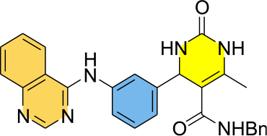
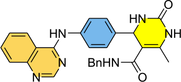
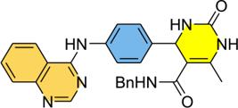
Quinazoline-Dihydropyrimidinone Hybrids as Potent Antichagasic and Antileishmanial Agents: Synthesis, Biological Evaluation and Molecular Docking Studies
Abstract
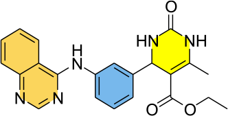
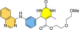
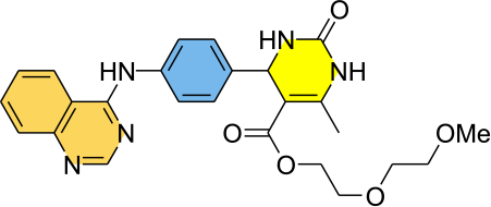
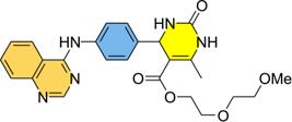
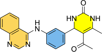
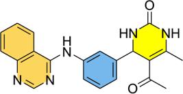
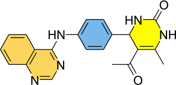
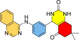
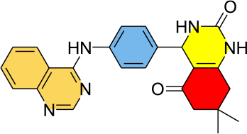
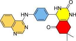
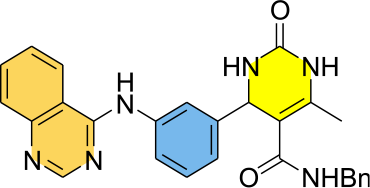
This study presents the design and synthesis of a series of quinazoline-dihydropyrimidinone hybrid compounds with potential antichagasic and antileishmanial activities, addressed to fight against neglected tropical diseases (NTDs). All compounds were evaluated against the epimastigote and trypomastigote forms of Trypanosoma cruzi and the promastigote forms of Leishmania infantum, Leishmania braziliensis, Leishmania amazonensis, and Leishmania major. Among them, hybrids 7a and 7c showed significant activity against all parasites, surpassing the potency of the reference drugs miltefosine and benznidazole. Molecular docking simulation indicated strong interactions of hybrids 7a and 7c with key parasitic enzymes - such as cruzain from T. cruzi and sterol 14-alpha demethylase from L. infantum; dihydroorotate dehydrogenase from L. braziliensis; nucleoside diphosphate kinase (NDK) from L. amazonensis; and N-myristoyltransferase from L. major - which may underlie their observed in vitro efficacy.
Keywords:
quinazolines; dihydropyrimidinones; molecular hybridization; antileishmanial; antichagasic; Trypanosoma cruzi
Introduction
Neglected tropical diseases (NTDs) are a group of infectious diseases that affect approximately 1.7 billion people in tropical and subtropical countries. These diseases are often associated with areas of poverty and limited health resources (income less than US$ 1.90 per day), including remote rural communities, urban slums, and displaced populations.1,2 Leishmaniasis and Chagas disease are part of the group of NTDs and are caused by protozoa of the order Kinetoplastida and the family Trypanosomatidae, characterized by the presence of a flagellum and a kinetoplast.3 However, they differ in terms of the genus and species of the causative parasites: Chagas disease is caused by Trypanosoma cruzi, while leishmaniasis is caused by several species of the genus Leishmania, which are divided into two subgenera (Leishmania and Viannia) based on the development of the parasite in the intestine of the vector.4 Both diseases caused by trypanosomatids are characterized by significant health problems.
Chagas disease is a multifactorial infectious disease, with clinical manifestations characterized by cardiac and gastrointestinal diseases that lead to molecular and morphological alterations in various types of tissues,5 with approximately 7 in 10 patients possibly unaware of their infection, with a mortality rate of up to 12,000 people per year.6 It is commonly treated with drugs benznidazole and nifurtimox.7 Launched over 50 years ago, they are effective in the acute phase of the infection and limited in the chronic phase (less than 25% efficacy), even though this is the most prevalent and severe form of the infection.8
Leishmaniasis is a group of diseases that affect approximately 12 million people around the world. Annually, 1.3 million new cases per year are recorded in almost 100 endemic countries. This condition represents the second most common cause of death and the fourth disease among tropical infections.9 These diseases manifest themselves mainly in two clinical forms: cutaneous leishmaniasis, which affects the skin and mucous membranes, and visceral leishmaniasis, the most severe form, which causes infection in organs such as the lymph nodes, liver and spleen, and can be lethal if not properly treated.10 The currently options available for the treatment of leishmaniasis include the use of pentavalent antimonials, amphotericin B, miltefosine and pentamidine.11 All of these drugs have significant adverse effects, often leading to discontinuation of therapy. Furthermore, prolonged use of the same drugs over decades has resulted in the emergence of resistant strains, reinforcing the need for new therapies.12 The present status of leishmaniasis and Chagas disease as NTDs highlights the need for accessible, safe and effective treatments, since current options are inadequate or inaccessible.
Nitrogen-containing heterocyclic compounds are constantly mentioned in literature, which are employed in fighting tropical diseases such as malaria, Chagas disease and leishmaniasis.13-15 Quinazoline derivatives are described as selective inhibitors of the enzymes dihydrofolate reductase (DHFR) and pteridine reductase (PTR), with effect on amastigote strains.16,17 Several recent reviews18-21 have been dedicated to explore the development of drugs against leishmaniasis, including quinazoline scaffolds. A set of synthesized 2,3 dihydroquinazolin 4(1H) ones was evaluated in vitro against promastigotes of Leishmania donovani and show significant bioactivity.22 Anti-leishmanial activity of 2-aminoquinazolines against amastigote cells of L. donovani was also reported.23 In another recent report, Birhan and co-workers24 observed a strong activity of quinazoline derivatives against L. donovani, being 150 times more active than the standard drug miltefosine. Furthermore, quinazoline derivatives also were active against T. cruzi.25 Perry and co-workers26 reported a high toxicity in in vitro and in vivo models exhibited by 2-aryl-4-aminoquinazolines against T. cruzi infection. Similarly, 2-aryl-4-(hydrazine-furylidene)quinazolines showed potent against epimastigotes from T. cruzi.27 Additionally, 2,4-diaminoquinazolines exhibited strong cruzain inhibition,28 while 2,4,6-triaminoquinazolines displayed notable selectivity against the highly virulent T. cruzi Queretaro strain controlling the relative growth for epimastigotes and trypomastigotes.29 Hybrid quinazolines have been investigated for their activity against Chagas disease. Indole-containing pyrazino-quinazolines 3,6-diones displayed high toxicity against T. brucei with high selectivity index.30
On the other hand, dihydropyrimidinones (DHPMs) obtained via the Biginelli reaction exhibit a plethora of bioactivities.31 However, their activity against Leishmaniasis has been less explored. It was found that dihydropyrimidinones (thiones) induce apoptosis-like cell death in L. donovani, while monastrol showed inhibitory activity of pteridine reductase enzyme isolated from L. donovani.32,33 The antileishmanial activity of carboxamide-DHPMs were investigated34,35 against Leishmania major promastigotes with reduction of cutaneous lesion in vivo. More recently, a hybrid indole-tetrahydropyrimidine was reported36 as active compound against promastigotes and amastigotes forms from L. donovani with low half maximal inhibitory concentration (IC50) values and good selectivity index.
While there are several reports on the activity of DHPMs against leishmaniasis, the same cannot be said about the DHPM’s activity against Chagas disease. To the best of our knowledge, no investigation of the activity of DHPMs against T. cruzi have been reported. The study performed by Inoue and co-workers37 regarding the synthesis of T. cruzi dihydroorotate dehydrogenase inhibitors, allowed the insightful information about the activity of structurally analogues of dihydropyrimidinones. Recently, Palchykov and co-workers38 performed the in silico screening of synthesized O-, S- and Se-containing Biginelli adducts, suggesting a potential activity against epimastigotes, trypomastigotes and amastigotes forms of T. cruzi.
Despite the lack of studies proving the trypanocidal action of DHPM compounds, their heterocyclic structure suggests they could be promising bioactive candidates, especially considering their antiproliferative, anti-HIV (human immunodeficiency virus), antimicrobial, antibacterial, antifungal and antiviral properties. The molecular hybridization strategy has gained prominence within the chemical space,39,40 and opened new opportunities to prospect the quinazolines and dihydropyrimidinones as a single hybrid entity.41,42 Hybrid compounds can generate new properties or synergistic effects from both units, leading to a potency increase greater than the sum of the individual activities.43
Due to the limited number of studies involving DHPMs in combating leishmaniasis or Chagas disease, we postulate that the association of a DHPM with structurally simple quinazolines, in the form of hybrid compounds, could bring some synergism in antiprotozoal activity for the specific control of these NTDs.
Two recent studies involving quinazoline-DHPMs have appeared in literature. The first focused on combating Plasmodium falciparun and Plasmodium vivax.44 The second explored the quinazolinone-dihydropyrimidinone hybrids as a potential anti-diabetic agent.45 Accordingly, the structural design of our quinazoline-DHPM hybrids is not only structurally distinct from those previously reported, but also are the only one that evaluate the activity against leishmaniasis and Chagas disease. In this context, fourteen quinazoline-dihydropyrimidinone hybrid compounds were designed and synthesized, and their activities against the NTDs, such as leishmaniasis and Chagas disease, were evaluated.
Results and Discussion
Synthesis of quinazoline-DHPM hybrids
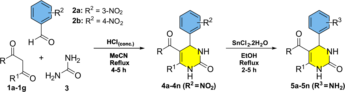
For the synthesis of dihydropyrimidinone pharmacophore, the multicomponent Biginelli reaction between different β-dicarbonyl compounds 1a-1g, nitro-benzaldehydes 2a-2b and urea (3) were performed in presence of drops of concentrated hydrochloric acid to afford the corresponding nitro-DHPMs 4a-4n (Scheme 1).46 The nitro-DHPMs were obtained in yields ranging from 55-89%, as shown in Table 1.
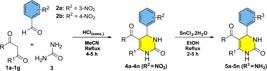
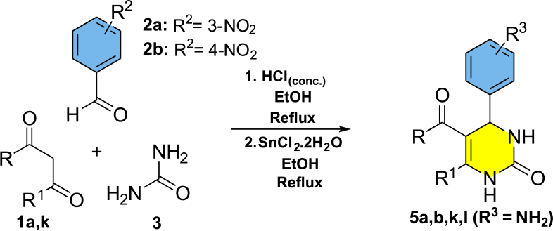
The transformation of nitro-DHPMs 4a-4n into the amino-DHPMs 5a-5n were achieved using SnCl2.2H2O as reducing agent (see Scheme 1). The amino-DHPM derivatives were obtained in yields ranging from 66 to 85% (see Table 1). To reduce steps of synthesis and avoid complex purification steps, we evaluated a one-pot nitro-DHPM synthesis followed by in situ reduction (Scheme 2). Table 2 compares the yields of amino-DHPMs 5a, 5b, 5j, 5k from the conventional and one-pot methods.
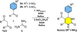
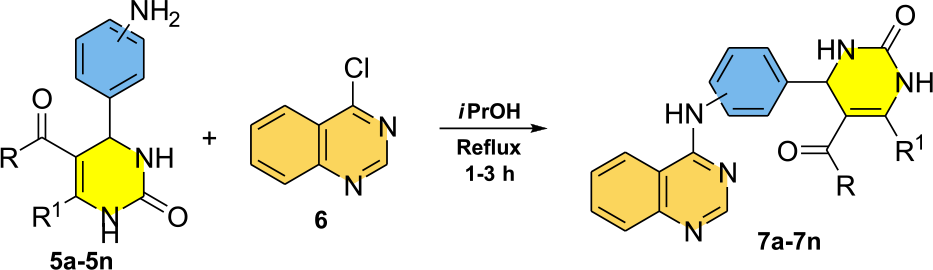
The yields obtained from the one-pot experiment were higher than those obtained in two stages in all examples, and this protocol was used as standard. Next, the synthesis of quinazoline-DHPM hybrids 7a-7m involved the nucleophilic aromatic substitution reactions of amino-DHPMs 5a-5m on the 4-chloroquinazoline (6), in isopropanol under reflux conditions (Scheme 3).47
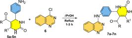
The reagents were rapidly consumed, and the formation of a solid product was observed. All quinazoline-DHPM hybrids were obtained in reasonable to good yields (60 85%) after recrystallization from an EtOH/H2O solvent mixture (Table 3).
Antileishmanial activity of hybrid compounds 7a-7n
The antileishmanial activity of quinazoline-DHPM hybrids 7a-7n against the promastigote forms of the species L. amazonensis, L. major, L. braziliensis and L. infantum was evaluated in vitro. The cytotoxicity against the macrophages RAW 264.7 cell was evaluated, once it serves as in vitro model for the study of host-pathogen interactions, due to the functional stability and ability to accomplish the phagocytosis and pinocytosis processes.48
The hybrids 7a and 7c showed strong antileishmanial activity against all species of parasites. These two compounds were most potent against the specie L. braziliensis with low IC50 of 1.58 µM (7a) and 1.12 µM (7c), respectively (Table 4, entries 1 and 3, respectively). Additionally, the hybrids 7a and 7c were more potent against all leishmania species than miltefosine, used as the reference drug. The IC50 values ranged from 14.68 to 19.75 µM for inhibition of parasite growth (see entry 18). Unfortunately, all other hybrids showed IC50 values higher than 50μM and the exact value was not determined. Both 7a and 7c hybrids presented high selectivity index (SI) values due to their low cytotoxicity against RAW 264.7 macrophages, with cytotoxic concentration 50% (CC50) of 274.3 and 289.31 µM, respectively, regarding the protozoa L. braziliensis species (entries 1 and 3, respectively). The SI, defined as the ratio between CC50 and IC50, was used as a tool for analyzing the biological efficacy of antiparasitic compounds. According to Ramírez-Macíase et al.,49 a SI ≥ 20 is considered effective, indicating greater selectivity to the parasite when compared to heathy cells.
Activity of quinazoline-dihydropyrimidinone hybrids against promastigote form in different Leishmania species
Compounds 7a and 7c show promising in vitro activity and warrant further evaluation, including intracellular amastigote assays and target validation. It should be noted that the isolated amino-DHPMs 5a or 5c showed the IC50 values > 50 μM (Table 4, entries 15 and 16, respectively). In the same way, the chloroquinazoline 6 also showed high IC50 values > 50 μM (entry 17) suggesting that the hybridization strategy was necessary for the high activity exhibited by compounds 7a and 7c.
It is essential to interpret SI values within the context of the specific experimental frameworks employed. While the high SI values observed for compounds 7a and 7c (e.g., SI = 173.61 for 7a) are promising, we acknowledge the inherent limitations of cross-study comparisons. Variations in parasite strains such as the differential drug sensitivity between Leishmania sp. as well as the distinct metabolic profiles of host cells (e.g., primary macrophages versus immortalized lineages) can significantly influence IC50 and CC50 determinations. Furthermore, the choice of viability markers, such as the enzymatic reduction of 3-(4,5-dimethylthiazol-2-yl)-2,5-diphenyltetrazolium bromide (MTT) compared to Resazurin-based assays, may yield divergent results based on the interference of the compound with cellular redox states. Consequently, the SI reported herein serves as a robust preliminary indicator of safety and efficacy in vitro, which warrants further validation through upcoming in vivo pharmacokinetic and toxicological assessments.
Anti-trypanocidal activity of hybrids 7a-7n
The trypanocidal activity of hybrids 7a-7n against the epimastigote and trypomastigote forms of T. cruzi, as well as the cytotoxic profile against Vero cells were evaluated (Table 5). Compounds 7a and 7c were again the most effective, presenting IC50 values of 2.15 and 1.94 µM for the epimastigote form (entries 1 and 3, respectively) and 3.22 and 2.20 µM for the trypomastigote form (entries 1 and 3, respectively).
Activity of quinazoline-dihydropyrimidinone hybrids against epimastigote and trypomastigote forms of T. cruzi
As observed earlier for Leishmania, other hybrids presented IC50 values higher than 50 μM. The potency of the most active compounds was superior to the reference drug benznidazole, which showed the IC50 value of 294.86 µM for epimastigotes and 235.80 µM for trypomastigotes. The most active hybrids exhibited low cytotoxicity against the Vero cell with CC50 values of 198.62 µM for 7a and 189.92 µM for 7c (Table 5, entries 1 and 3, respectively). Thus, the hybrid compounds present a high SI value exhibiting cellular cytotoxicity only at high concentrations.
Hybrids 7a and 7c demonstrated high selectivity profiles; specifically, compound 7a was 92.38-fold and 61.68-fold more toxic to epimastigotes and trypomastigotes, respectively, than to normal cells. Similarly, compound 7c exhibited a toxicity toward these parasitic forms that was 97.89-fold and 86.32-fold higher than its toxicity to host cells. These values are higher than those of reference compound benznidazole, which presented SI of 0.90 and 1.13, respectively. The activity of both isolated DHPM scaffolds 5a and 5c (Table 5, entries 15 and 16, respectively) as well as the 4-chloroquinazoline (6) (entry 17) were evaluated against T. cruzi. However, all these three individual pharmacophores showed the IC50 value > 50 μM, highlighting again, the importance of molecular hybridization for the observation of trypanocidal activity.
In silico studies
Molecular docking simulations
To gain some insights into the possible factors justifying the promising activity of hybrid compounds against the T. cruzi and Leishmania protozoa, the quinazoline DHPM derivatives were subjected to molecular docking simulations with proteins that are essential for maintaining the survival of the parasites.
The cruzain protein from T. cruzi (Protein Data Bank (PDB): 1EWM) was targeted due to the activity as cysteine protease, responsible for protein digestion, host cell invasion and evasion of the parasite immune system.50 The sterol 14-alpha demethylase (CYP51) from L. infantum (PDB: 3L4D) was targeted since it is essential for the synthesis of ergosterol, a vital component of the parasite membrane, and is also relevant in the study of drug resistance.51,52 The dihydroorotate dehydrogenase from L. braziliensis (PDB: 4WZH) is an essential enzyme for the synthesis of pyrimidine nucleotides, which are fundamental for the reproduction of the parasite.53 Nucleoside diphosphatase kinase (NDK) protein from L. amazonensis (PDB: 5GO1) is an essential enzyme for the metabolism of the parasite and responsible for maintaining the balance of nucleotides necessary for the synthesis of deoxyribonucleic acid (DNA), ribonucleic acid (RNA) and energy.54 The target N-myristoyltransferase from L. major (PDB: 4CGO) is essential for the survival and proliferation of the parasite, since it performs the lipid modification N-myristoylation in proteins, crucial for its cellular functioning and for the correct localization and activity of the proteins within the parasite.55
Prior to performing the molecular docking simulations, the validation process of targets was carried out by redocking the co-crystallized ligand with the investigated targets. This simulation evaluates whether there are structural differences between the co-crystallized ligand and its most stable pose. This metric is evaluated by means of the root mean square deviation (RMSD) values, with a value of up to 2.0 Å considered the appropriate value.56,57 The redocking calculation for the target nucleoside diphosphatase kinase (NDK) protein from L. amazonensis was not performed, since it does not have co-crystallized ligand. All redocking values for the studied proteins were within the determined limit, validating the method and program used. (see Tables S1 S3 and S5 in the Supplementary Information section).
To avoid the occurrence of false-positive results, consensus analyses were performed using the normalized score obtained from the binding energy values referring to the scoring functions from MolDock, Rerank and PLANTS.58-60 The normalized score was calculated by dividing the individual score value for each compound by the lowest score obtained for each scoring function used. The total normalized score was then determined by the sum of the normalized values divided by the total number of observations. The complex with the highest affinity was the one with the highest normalized score value.
The binding affinity values for hybrids 7a and 7c showed negative energy, indicating interaction for all targets (see Tables S1-S5 in the Supplementary Information section). Both compounds also had normalized scores higher than at least one control. Notably, both scored above 0.74, with compound 7a scoring higher than 7c. It is worth mentioning that the results of the molecular docking simulations represent putative binding modes that require experimental validation in future studies.
Molecular interaction of compounds 7a and 7c with the target cruzain from Trypanosoma cruzi
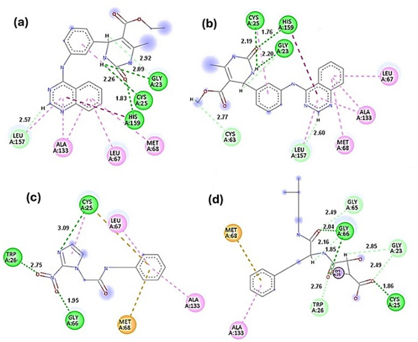
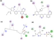
For the cruzain target, the complex formed by the co-crystallized ligand, the homophenyl-alanylamino derivative ligand (Figure 1d) had the highest affinity with a normalized score of 1 (see Table S1 in the Supplementary Information section). The hybrids 7a and 7c achieved normalized scores of 0.828 and 0.783 respectively to cruzain enzyme from T. cruzi, denoting higher affinity scores than the positive control benznidazole (0.754), emphasize that these results are predictive in nature. Compounds 7a and 7c (Figures 1a and 1b, respectively) exhibited significant hydrogen bond interactions with dihydropyrimidinone moiety and hydrophobic interactions involving the cysteine 25 residue, which is crucial for the enzymatic activity. Compound 7a had a length of 2.26 Å, and compound 7c measured 2.19 Å. The positive control nifurtimox (Figure 1c) displayed lengths of 3.09 and 1.86 Å for the PDB ligand (Figure 1d). Although the lengths of hybrids are greater when compared to the homophenyl-alanylamino derivative ligand (Figure 1d), they are shorter than the length shown for the positive control nifurtimox (Figure 1c).
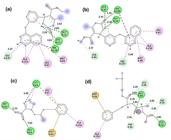
2D interactions of compounds 7a (a), 7c (b), positive control nifurtimox (c), and homo phenyl-alanyl amino derivative PDB ligand (d) with the target cruzain from T. cruzi. Interactions: conventional hydrogen interaction (dark green dashed line); carbon-hydrogen interaction and van der Waals interaction (light green dashed line); π-alkyl interaction (light pink dashed line); π-stacked amide interaction (dark pink dashed line); π-sulfur interaction (orange dashed line); covalent interaction (small grey circle). Residues: Leu (leucine), Ala (alanine), Met (methionine), His (histidine), Cys (cysteine), Gly (glycine) and Trp (tryptophan).
Molecular interaction of compounds 7a and 7c with the target sterol 14-alpha demethylase from Leishmania infantum
The ligand derived from difluorophenyl-triazole, originally co-crystallized in the PDB structure (3L4D), showed the highest normalized score (0.836) for the sterol 14-alpha demethylase (CYP51) enzyme from L. infantum. Compound 7a demonstrated a similarly high affinity (0.826), followed by compound 7c (0.741). Both compounds exhibited a higher normalized docking score than the reference drug miltefosine (0.704), but these results are only predictive (see Table S2 in the Supplementary Information).
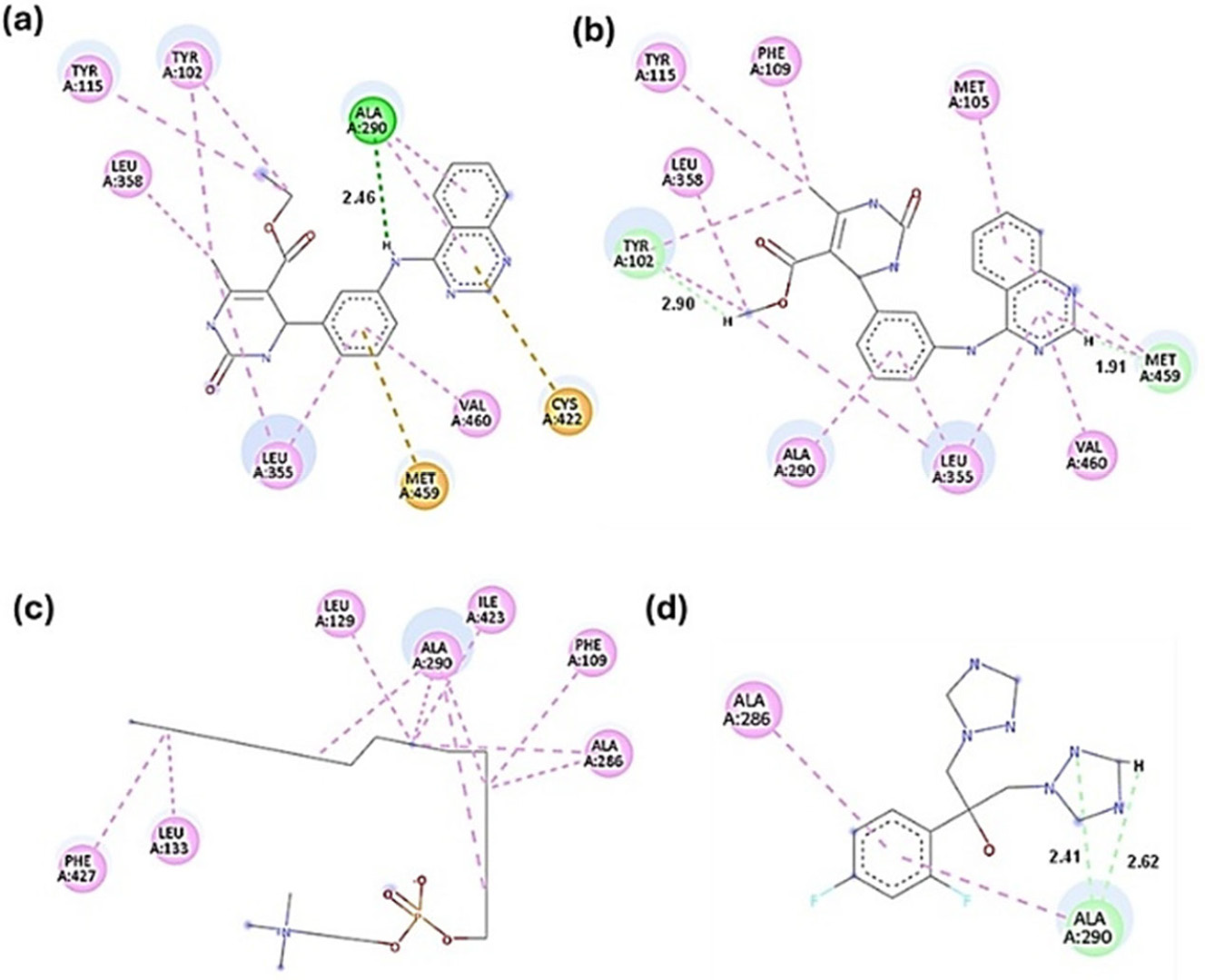
Figure 2 highlights the key molecular interactions between compounds 7a and 7c (Figures 2a and 2b, respectively) and the enzyme, which are crucial for their inhibitory activity. Compound 7a exhibited a significant π-sulfur interaction with cysteine 422 (Cys422), an important residue for heme cofactor binding. Additionally, both 7a and 7c formed interactions with tyrosine residues 102 (Tyr102) and 115 (Tyr115), located on the porphyrin ring within the active site of the enzyme. All tested compounds also showed hydrophobic interactions with alanine 290 (Ala290), suggesting they may bind to the same region within the enzyme. The complex corresponding to the positive control miltefosine (Figure 2c) showed a greater contribution of hydrophobic interactions of the alkyl and pi-alkyl type (dashed line in light pink) through residues Phe427, Leu133, Leu129, Ala290 (4 interactions), Ile423, Phe109 and Ala286 (2 interactions). The co-crystallized ligand difluoro-phenyl-triazole derivative (Figure 2d) presented two carbon-hydrogen interactions (dashed line in light green) through residue Ala290 and two hydrophobic interactions of the π-alkyl type through residues Ala286 and Ala290. It was observed that the hydrophobic interaction established by residue Ala290 was observed in all four complexes under study.
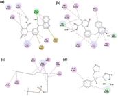
2D interactions of compounds 7a (a), 7c (b), positive control miltefosine (c) and PDB ligand difluoro-phenyl-triazole derivative (d) with the target sterol 14-alpha demethylase (CYP51) from L. infantum. Interactions: conventional hydrogen interaction (dark green dashed line); carbon-hydrogen interaction (light green dashed line); alkyl and π-alkyl interaction (light pink dashed line); π-sulfur interaction (orange dashed line). Residues: Leu, Tyr (tyrosine), Ala, Cys, Val (valine), Met, Phe (phenylalanine) and Ile (isoleucine).
Molecular interaction of compounds 7a and 7c with the target dihydroorotate dehydrogenase from Leishmania braziliensis
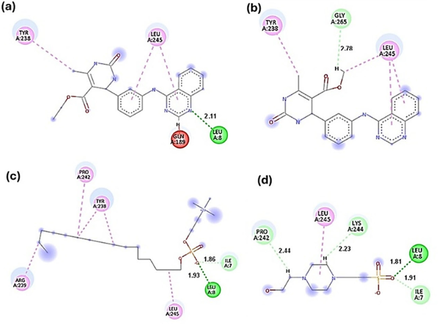
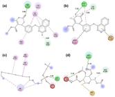
For the dihydroorotate dehydrogenase enzyme from Leishmania braziliensis (PDB: 4WZH), compounds 7a and 7c showed the higher affinities, with normalized scores of 0.873 and 0.848, respectively (see Table S3 in the Supplementary Information), than the positive control miltefosine (0.783) and the PDB ligand piperazine derivative (0.696), emphasizing that these results are predictive in nature. Figure 3 highlights the hydrophobic interactions (dashed pink line) mainly in the dihydropyrimidinone moiety. Compound 7a interacts with residues Tyr233 and Leu245 (2 interactions) through the alkyl and π-alkyl types.
2D interactions of compounds 7a (a), 7c (b), positive control miltefosine (c) and piperazine derivative PDB ligand (d) with the target dihydroorotate dehydrogenase from L. braziliensis. Interactions: conventional hydrogen interaction (dark green dashed line); carbon-hydrogen interaction (light green dashed line); alkyl and π-alkyl interaction (light pink dashed line); unfavorable interaction (red dashed line). Residues: Tyr, Leu, Gln (glutamine), Gly, Arg (arginine), Ile, Pro (proline).
Additionally, the quinazoline group shows an unfavorable interaction with residue Gln189 (red dashed line) and a polar hydrogen bond with residue Leu8 (dark green dashed line). The compound 7c (Figure 3b) showed carbon-hydrogen interaction through Gly265 in the dihydropyrimidinone moiety. Three hydrophobic interactions were also seen involving Tyr238 and Leu245 residues. All compounds displayed hydrophobic interaction with Leu245. It was also observed that the complex related to the control compound miltefosine (Figure 3c) presented a greater contribution of hydrophobic interactions of the alkyl type (dashed line in light pink) through residues Arg239, Pro242, Tyr238 (2 interactions) and Leu245, as well as a conventional hydrogen type interaction (dashed line in dark green) through residue Leu8 and a carbon-hydrogen type interaction (dashed line in light green) through residue Ile7. The ligand complex related to the co-crystallized piperazine derivative ligand (Figure 3d) presented a greater contribution of polar interactions corresponding to the conventional hydrogen interaction (dashed line in dark green) through residue Leu8 and carbon-hydrogen interactions through residues Pro242, Lys244 and Ile7, as well as a hydrophobic interaction of the π-alkyl type was observed through residue Leu245. It is important to note that the hydrophobic interaction Leu245 was observed in the four complexes under study.
Molecular interaction of compounds 7a and 7c with the target nucleoside diphosphatase kinase (NDK) protein from Leishmania amazonensis
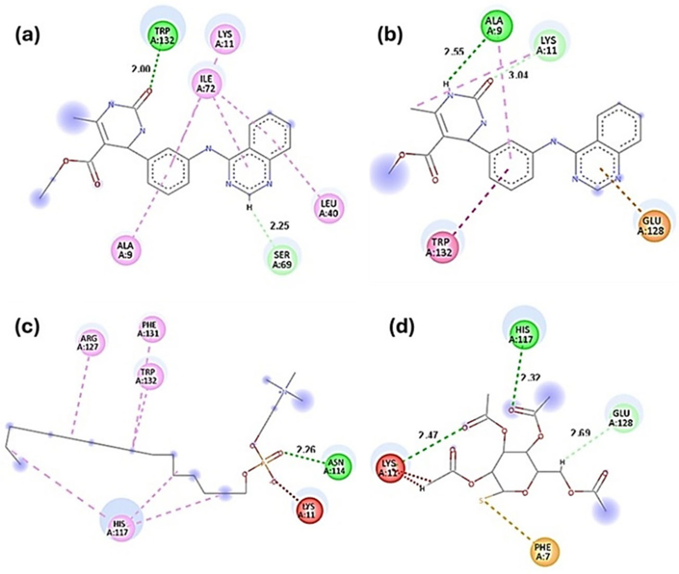
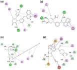
For the nucleoside diphosphatase kinase (NDK) target protein from L. amazonensis (PDB: 5GO1), the positive control miltefosine exhibited the highest affinity, with a normalized score of 0.978 (see Table S4 in the Supplementary Information). Compound 7a showed a normalized score of 0.931, closely trailing miltefosine, while compound 7c followed with a score of 0.915. Both compounds 7a and 7c demonstrated higher affinity scores than the literature control auranofin (0.726), indicating consistent and favorable host-guest interactions (Figure 4), emphasize that these results are predictive in nature. Hybrids 7a and 7c (Figures 4a and 4b) established both hydrophobic interactions (represented by pink dashed lines) and polar interactions (depicted by green dashed lines) with lysine 11 (Lys11), a key residue situated in the enzyme active site and essential for its function.61
2D interactions of compounds 7a (a), 7c (b), positive control miltefosine (c) and literature inhibitor auranofin (d) with the target nucleoside diphosphatase kinase (NDK) protein from L. amazonensis. Interactions: conventional hydrogen interaction (dark green dashed line); carbon-hydrogen interaction (light green dashed line); alkyl and π-alkyl interaction (light pink dashed line); π-π T-shaped interaction (dark pink line); unfavorable interaction (red dashed line); π-sulfur interaction (orange dashed line). Residues: Trp, Lys (lysine), Ile, Leu, Ser (serine), Ala, Glu (glutamic acid), Arg, Phe, His and Asn (asparagine).
The complex corresponding to the positive control compound miltefosine (Figure 4c) presented a greater contribution of alkyl-type hydrophobic interactions (light pink dashed line) through residues Arg127, Phe131, Trp132 and His117 (3 interactions), as well as an unfavorable interaction (red dashed line) through residue Lys11 and a conventional hydrogen interaction (dark green dashed line) through residue Asn114. The complex corresponding to the inhibitor auranofin (Figure 4d) presented two conventional hydrogen interactions (dark green dashed line) through residues His117 and Lys11, a carbon-hydrogen interaction through residue Glu128, two unfavorable interactions (red dashed line) through residue Lys11 and a π-sulfur interaction (orange dashed line) through residue Phe7. No similar interactions were observed between the complexes under study, indicating that the compounds under study may present different binding sites.
Molecular interaction of compounds 7a and 7c with the target N-myristoyltransferase from Leishmania major
For the target N-myristoyltransferase from L. major (PDB: 4CGO), it was observed that the PDB ligand derived from thienopyrimidine presented the highest affinity, with a normalized score corresponding to 1 (see Table S5 in the Supplementary Information). However, compounds 7a (0.929) and 7c (0.902) presented very close affinity score values, denoting affinity. The control compound miltefosine (0.684) was the compound with the lowest affinity, emphasizing that these results are predictive in nature.
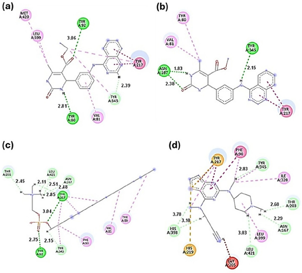
In Figures 5a and 5b, it can be observed that compounds 7a and 7c engaged in key interactions with residues within the active site of the enzyme. These include a hydrophobic π-π T-shaped interaction (indicated by a dark pink dashed line) involving Tyr217, as well as a polar hydrogen bond interaction (represented by a green dashed line) with Tyr345.62 The complex corresponding to the control compound miltefosine (Figure 5c) presented seven hydrophobic interactions of the alkyl-type (dashed line in light pink) through residues Tyr80, Val81, Phe90, Tyr345 and Tyr217 (3 interactions), 6 interactions of the carbon-hydrogen type (dashed line in light green) through residues Thr203, Leu421 (2 interactions), Asn117 and Tyr345 and two conventional hydrogen interactions (dashed line in dark green) through residues Tyr217 and Tyr92. The co crystallized ligand thienopyrimidine derivative (Figure 5d) presented hydrophobic interactions of the alkyl and π-alkyl type (dashed line in light pink) through residues Tyr217, Phe90, Tyr345, Ile328 and Leu399 and of the type π-π T-shaped and π-π stacked interaction (dark pink line) through residues Tyr217 and Phe90, as well as six carbon-hydrogen interactions (light green dashed line) through residues Tyr345, Thr203, Asn167, Leu421 and His398 (2 interactions), one unfavorable interaction (red dashed line) through residue Gly205 and two π-sulfur interactions (orange dashed line) through residues Tyr217 and His219. It was observed that the hydrogen interaction established by Tyr345 is present in the four complexes under study.
2D interactions of compounds 7a (a), 7c (b), positive control miltefosine (c) and the PDB ligand thienopyrimidine derivative (d) with the target N-myristoyltransferase from L. major. Interactions: conventional hydrogen interaction (dark green dashed line); carbon-hydrogen interaction (light green dashed line); alkyl and π-alkyl interaction (light pink dashed line); π-π T-shaped and π-π stacked interaction (dark pink line), unfavorable interaction (red dashed line); π-sulfur interaction (orange dashed line). Residues: Met, Leu, Tyr, Val, Asn, Thr, Phe, Ile, Gly and His.
Molecular dynamics simulations
Molecular dynamics (MD) simulations have been performed to study the behavior of molecules over time, and to provide detailed insight into their motions, interactions and stability at the atomic level, allowing, for example, to assess the stability of protein-ligand complexes and estimate the free energy of binding.63
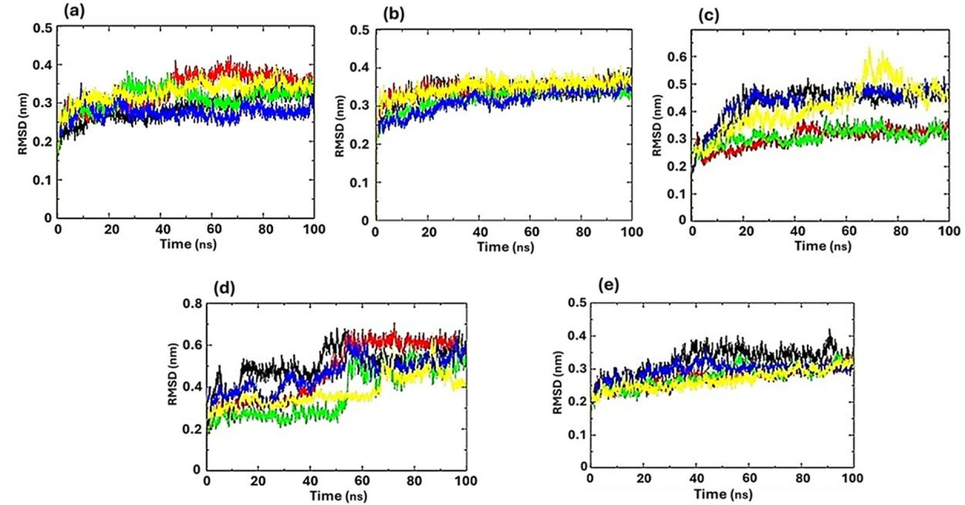
Figure 6 demonstrates the root mean square deviation (RMSD) values of the complexes related to the average difference between the positions of the α-carbon atoms (of the protein backbone) at a given instant of the simulation, compared to a reference structure (usually the initial structure or an experimental structure such as the one in the PDB).64
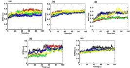
RMSD-protein of the α-carbon. Complex: protein (black line); compound 7a + protein (red line); compound 7c + protein (green line); positive control + protein (blue line) and PDB ligand + protein (yellow line). Protein: (a) cruzain from T. cruzi; (b) sterol 14-alpha demethylase from L. infantum; (c) dihydroorotate dehydrogenase from L. braziliensis; (d) nucleoside diphosphatase kinase (NDK) protein from L. amazonensis and, (e) N-myristoyltransferase from L. major.
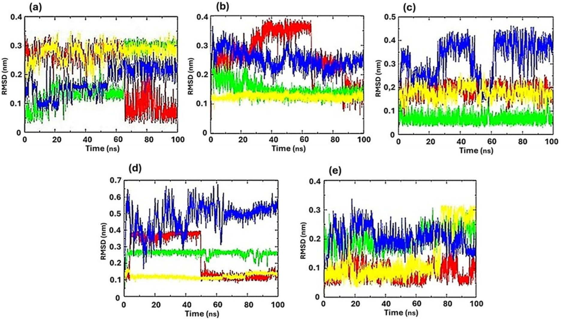
It can be observed that for the proteins cruzain from T. cruzi (Figure 6a), sterol 14-alpha demethylase from L. infantum (Figure 6b), and N-myristoyltransferase from L. major (Figure 6e) exhibited greater stability, since the RMSD values comprised the range of 0 to 3 nm indicating a stable system that reached equilibrium. Also, it was observed that there were not many fluctuations as in protein complex of dihydroorotate dehydrogenase from L. braziliensis (Figure 6c) and nucleoside diphosphatase kinase (NDK) from L. amazonensis (Figure 6d). Specifically, regarding the target sterol 14-alpha demethylase (Figure 6b), it was observed that compounds 7a (red line) and 7c (green line) exhibited similar RMSD values and even lower values compared to the control compounds miltefosine (blue line) and the PDB ligand fluconazole (yellow line), as well as the CYP51 protein complex itself (black line). These results suggest that 7a and 7c demonstrate high stability and a strong ability to remain bound within the enzyme active site. The RMSD analysis of the ligands, which assesses their stability under variations in temperature, pressure, and solvent effects, showed all values below 2 nm (Figure 7). These findings indicate that the compounds maintained stable interactions within the active site throughout the simulation, despite variations in environmental conditions.
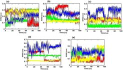
RMSD-protein of the carbono-α. Complex: compound 7a + protein (red line); compound 7c + protein (green line); positive control + protein (blue line) and PDB ligand + protein (yellow line). Protein: (a) cruzain from T. cruzi; (b) sterol 14-alpha demethylase from L. infantum; (c) dihydroorotate dehydrogenase from L. braziliensis; (d) nucleoside diphosphatase kinase (NDK) protein from L. amazonensis and, (e) N-myristoyltransferase from L. major.
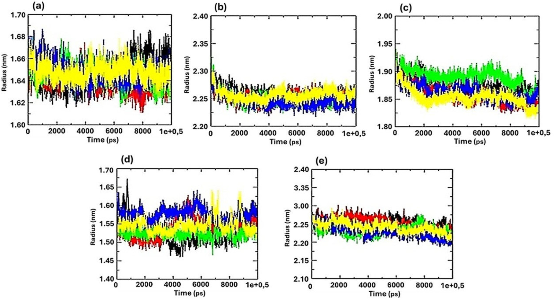
Notably, for the target sterol 14-alpha demethylase from L. infantum (Figure 7b), compound 7c (green line) and the PDB ligand fluconazole (yellow line) displayed the lowest RMSD values, indicating superior stability within the binding site. For the target dihydroorotate dehydrogenase from L. braziliensis (Figure 7c), compound 7c (green line) showed lower RMSD values, around 1 nm, indicating greater stability. Similarly, for the target N-myristoyltransferase from L. major, compound 7a (red line) demonstrated the highest stability, as reflected by its consistently low RMSD values throughout the simulation. The maintenance of protein structural packing was evaluated by the observation of radius of gyration which showed no noticeable structural differences among the complexes (Figure 8). The compounds showed very similar values of radius of gyration, with complete overlap observed between most complexes, indicating that the overall protein structure remained stable throughout the simulations. However, in the case of dihydroorotate dehydrogenase (L. braziliensis) (Figure 8c) and nucleoside diphosphate kinase (NDK, L. amazonensis) (Figure 8d), some complexes displayed noticeable fluctuations in the radius of gyration. Specifically, the 7c complex with dihydroorotate dehydrogenase (green line in Figure 8c) and the positive control miltefosine with NDK (blue line in Figure 8d) showed deviations, suggesting conformational changes in the protein structure. Additionally, root mean square fluctuation (RMSF) analysis was used to measure the flexibility of each residue, showing how much individual atoms (typically C-α atoms of the protein backbone) fluctuate over time relative to their average position during the simulation.65,66
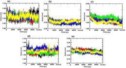
Radius of gyration. Complex: protein (black line); compound 7a + protein (red line); compound 7c + protein (green line); positive control + protein (blue line) and PDB ligand + protein (yellow line). Protein: (a) cruzain from T. cruzi; (b) sterol 14-alpha demethylase from L. infantum; (c) dihydroorotate dehydrogenase from L. braziliensis; (d) nucleoside diphosphatase kinase (NDK) protein from L. amazonensis and, (e) N-myristoyltransferase from L. major.
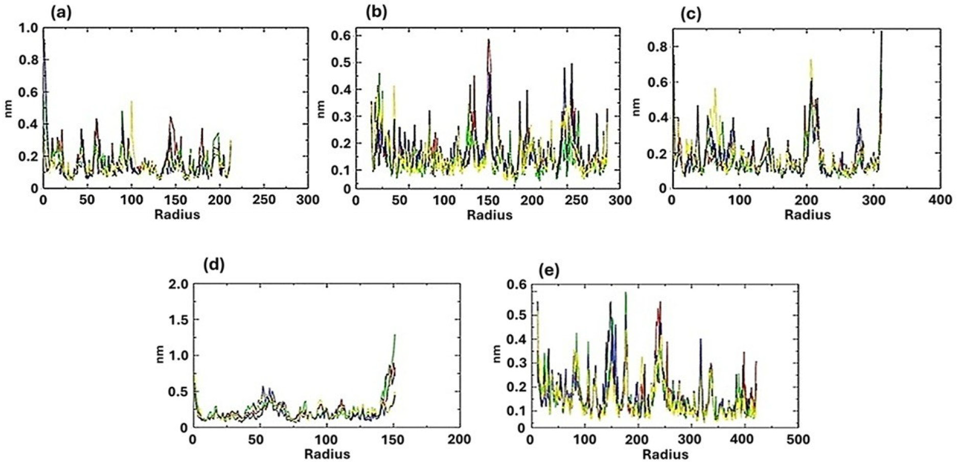
Figure 9 shows that cruzain from T. cruzi (Figura 9a) and sterol 14-alpha demethylase from L. infantum (Figura 9b) exhibited low fluctuations, with most residues showing deviations of up to 0.3 nm. This indicates that both proteins are well-structured and relatively rigid macromolecules. For these targets, only a single residue in each protein displayed flexibility values exceeding the ideal threshold: alanine 1 (Ala1) in cruzain and glycine 29 (Gly29) in sterol 14-alpha demethylase. However, since these residues are not part of the active site, their increased flexibility is unlikely to affect the activity of the enzyme.
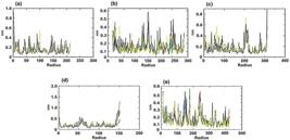
Root mean square fluctuation (RMSF). Complex: protein (black line); compound 7a + protein (red line); compound 7c + protein (green line); positive control + protein (blue line) and PDB ligand + protein (yellow line). Protein: (a) cruzain from T. cruzi; (b) sterol 14-alpha demethylase from L. infantum; (c) dihydroorotate dehydrogenase from L. braziliensis; (d) nucleoside diphosphatase kinase (NDK) protein from L. amazonensis and, (e) N-myristoyltransferase from L. major.
Regarding the target dihydroorotate dehydrogenase from L. braziliensis (Figure 9c), nucleoside diphosphatase kinase (NDK) protein from L. amazonensis (Figure 9d) and N-myristoyltransferase from L. major (Figure 9e), a significant number of complexes with fluctuations above 0.3 nm were observed, characterizing these targets as more flexible and disorganized regions. For the enzyme dihydroorotate dehydrogenase from L. braziliensis (Figure 9c), high fluctuation values were observed in the positions related to the residues aspartic acid 204 (Asp204), valine 205 (Val205), glutamic acid 206 (Glu206), threonine 207 (Thr207), glutamic acid 208 (Glu208), serine 209 (Ser209), lysine 213 (Lys213), glutamine 216 (Gln216), phenylalanine 218 (Phe218), lysine 310 (Lys310), alanine 311 (Ala311) and methionine 312 (Met312), the latter residue is a component of the enzyme active site, and may induce a conformational change in the target. The nucleoside diphosphatase kinase (NDK) protein from L. amazonensis presented high fluctuation values for the residues positions methionine 1 (Met1), aspartic acid 53 (Asp53), serine 56 (Ser56), lysine 57 (Lys57), proline 58 (Pro58), phenylalanine 59 (Phe59), tryptophan 141 (Trp141), glutamine 148 (Gln148), tyrosine 150 (Tyr150) and glutamic acid 151 (Glu151), however the residues mentioned refer to the structure editing performed in the UCSF Chimera 1.17.3 program,67 as they are not constituents of the active site.
For the enzyme N-myristoyltransferase from L. major, high fluctuation values were observed in the positions corresponding to the residues: glutamic acid 150 (Glu150), arginine 176 (Arg176), glutamic acid 177 (Glu177), arginine 179 (Arg179), glutamine 240 (Gln240), lysine 241 (Lys241), phenylalanine 242 (Phe242) and glutamine 243 (Gln243), but they also do not correspond to residues of the active site of the enzyme.
The contribution of Coulomb and Lennard-Jonnes energies for the interactions of hybrid compounds 7a and 7c and enzymes were investigated (see Tables S6-S10 in the Supplementary Information section). The Coulomb energies measure the electrostatic (charge-charge) interactions between atoms in the enzyme and the ligand, arising from the attraction or repulsion between partial charges on the atoms. The negative Coulomb energy is a favorable interaction (attraction between opposite charges, hydrogen bonds with electrostatic contribution) while a positive Coulomb energy means an unfavorable interaction (repulsion between charges). Thus, it reflects how well the charged regions of the ligand and enzyme complement each other (important for polar or charged binding sites) and has importance in host-guest enzyme systems. On the other hand, Lennard-Jones energy measures the non covalent interactions based on distance-dependent forces like van der Waals attractions and steric repulsion. It arises from the weak dispersion forces (important when atoms are at an optimal short distance) and represent attractive interactions. Repulsive interactions have the origin in steric clashes when atoms get too close (due to overlapping electron clouds). This kind of energy is very important in host-guest enzyme systems, since it shows how well the ligand fits physically into the binding pocket in terms of shape complementarity and packing efficiency (important for hydrophobic regions).
Hybrids 7a and 7c showed a greater contribution of Coulomb energies to cruzain from T. cruzi. Hybrid 7c showed energy value of -67.073 kJ mol-1, close to those presented by the PDB ligand, corresponding to -68.267 kJ mol-1. This metric represents a favorable electrostatic interaction between charges.68 Additionally, hybrid 7a presented the Lenard-Jones energy value of -105.856 kJ mol-1, close to the energy value presented by the positive control benznidazole of -114.728 kJ mol-1 (Table S6 in the Supplementary Information section).
For the targets related to the Leishmania species discussed in this work, it seems the all Lennard-Jones metrics contributed more significantly to the interactions with the hybrids than the Coulomb energies. For example, the interaction of compound 7c in the target sterol 14-alpha demethylase from L. infantum showed the energy value close to the positive control miltefosine, corresponding to -199.667 and -205.112 kJ mol-1, respectively. The Coulomb energies for 7a and 7c, while showing negative energy values, are higher than those of miltefosine or the PDB ligand (Table S7 in the Supplementary Information section). The hybrid 7a exhibited the lowest Lenard-Jones energy, at -135.453 kJ mol-1 related to enzyme dihydroorotate dehydrogenase from L. braziliensis, while the miltefosine showed -125.121 kJ mol-1 (Table S8 in the Supplementary Information section). Similarly to the previous case, both hybrids did not present important contributions in interactions with this enzyme. For the protein nucleoside diphosphatase kinase (NDK) from L. amazonensis, the Lenard-Jones energy for positive control miltefosine showed the lowest energy, corresponding to -170.377 kJ mol-1. In this case, both 7a and 7c presented lower energy values compared to the PDB ligand, at -126.817, -134.809, and -110.540 kJ mol-1, respectively. In addition, the values of Coulomb energies of the hybrids were significantly lower when compared to those from the PDB ligand (Table S9 in the Supplementary Information section).
Finaly, for the enzyme N-myristoyltransferase from L. major, it was observed that 7c exhibited the lowest energy, at -163.852 kJ mol-1, which was very close to the value presented by the PDB ligand (-163.354 kJ mol 1). The Coulomb energies for 7a and 7c were again higher than the value for miltefosine (Table S10 in the Supplementary Information section). This data set suggested that electrotactic (Coulomb) energies were more significant in the interactions of hybrids 7a and 7c with cruzain of T. cruzi (Chagas disease). On the other hand, the week van der Waals forces were more significant in the interactions with enzymes related to different species of Leishmania.
Conclusions
In summary, the rational design and synthesis of quinazoline-dihydropyrimidinone hybrids produced compounds in good yields with antileishmanial and antichagasic activities. Among them, hybrids 7a and 7c displayed superior activity compared to the reference drugs miltefosine and benznidazole, underscoring their potential as lead candidates for the development of novel therapeutics against Chagas disease and leishmaniasis. Molecular docking studies further suggest that their efficacy may arise from strong interactions with essential parasitic enzymes, providing valuable insights into their potential mechanisms of action. It is worth mentioning that the results of the molecular docking simulations represent putative binding modes that require experimental validation in future studies. These findings highlight the relevance of hybrid-based strategies for addressing unmet challenges in the treatment of neglected tropical diseases.
Experimental
Chemistry
The reagents were purchased from commercial sources and used without further purification, except ethyl acetate and hexanes, which were purified by simple distillation. Column chromatography was performed using a silica gel 60 Å (Acros Organics, 0.035 0.070 mm). The reactions were monitored using thin layer chromatography (TLC) and visualized under ultraviolet (UV) light. The nuclear magnetic resonance (NMR) spectra were recorded using a Varian VNMRS 300 spectrometer (1H at 300 MHz and 13C at 75 MHz) or Bruker (1H at 400 MHz and 13C at 100 MHz in dimethyl sulfoxide (DMSO)-d6 as solvent). The chemical shifts (d) were reported in parts per million (ppm) units using the residual non deuterated solvent nuclei as references for 1H NMR spectra (d = 2.50 ppm) and the deuterated solvent nuclei for 13C NMR spectra (d = 39.5 ppm). The coupling constants (J) were reported in hertz (Hz) and refered to peak multiplicities, which were described as s for singlet, bs for broad singlet, d for doublet, t for triplet, dd for doublet of doublets, ddd for doublet of doublets of doublets, dt for doublet of triplets, and m for multiplet. Attenuated total reflection Fourier transform infrared (ATR-FTIR) spectra were collected using Bruker Alpha-P equipment. The melting points were obtained on a Buchi melting point M-565 apparatus with a non-calibrated thermometer. The high-resolution mass spectrometry (HR MS) were obtained on a Bruker Impact II (ESI-QTOF-MS) in positive electrospray ionization (ESI) mode using DMSO as solvent. All compounds were purified by silica gel column chromatography (230-400 mesh) prior to NMR and HRMS analysis.
General procedure for the synthesis of nitro-DHPMs 4a-4q46
To a round-bottom flask, it was added 3 mmol of nitro-benzaldehyde, 3.2 mmol of urea, 3 mmol of β-dicarbonyl compound, 5 mL of acetonitrile, and 3 drops of concentrated HCl. The mixture was stirred under reflux and monitored by TLC (4:6 ethyl acetate / hexane v/v) until completion of reaction (4-5 h). After that, the mixture was kept in a freezer for at least 2 h, then filtered in a Büchner funnel and washed with water. The nitro-DHPMs were purified by column chromatography. When dimedone was used as a reagent, it was added in small portions, starting 1 h after adding the catalyst (HCl).
Ethyl 6-methyl-4-(3-nitrophenyl)-2-oxo-1,2,3,4-tetrahydro-pyrimidine-5-carboxylate (4a)69
White solid; mp 240-244 °C; FTIR (ATR) ν / cm-1 3324, 3209, 3076, 2962, 2809, 2364, 1710, 1684, 1627, 1526, 1348, 1221, 1088, 796, 687; 1H NMR (400 MHz, DMSO-d6) d 9.38 (1H, brs), 8.17-8.11 (1H, m), 8.08 (1H, t, J 1.7 Hz), 7.91 (1H, brs), 7.70 (1H, dt, J 7.8, 1.7 Hz), 7.66 (1H, t, J 7.8 Hz), 5.30 (1H, d, J 3.5 Hz), 4.06-3.92 (2H, m), 2.27 (3H, s), 1.10 (3H, t, J 7.0 Hz); 13C NMR (75 MHz, DMSO-d6) d 165.1, 151.8, 149.4, 147.7, 147.0, 133.0, 130.2, 122.4, 121.0, 98.3, 59.4, 53.6, 17.9, 14.0.
Ethyl 6-methyl-4-(4-nitrophenyl)-2-oxo-1,2,3,4-tetrahydro-pyrimidine-5-carboxylate (4b)70
White solid; mp 205-208 °C; FTIR (ATR) ν / cm-1 3322, 3082, 2967, 1698, 1640, 1513, 1348, 1291, 1208, 1081, 770, 694; 1H NMR (300 MHz, DMSO-d6) d 9.58 (1H, brs), 8.43 (2H, d, J 8.8 Hz), 8.12 (1H, brs), 7.92 (1H, bs), 7.74 (2H, d, J 8.8 Hz), 5.52 (1H, d, J 3.3 Hz), 4.22 (2H, q, J 7.0 Hz), 2.50 (3H, s) 1.32 (3H, t, J 7.0 Hz); 13C NMR (75 MHz, DMSO-d6) d 165.1, 152.0, 151.8, 149.4, 146.7, 127.7, 123.8, 98.2, 59.4, 53.7, 17.9, 14.0.
Methyl 6-methyl-4-(3-nitrophenyl)-2-oxo-1,2,3,4-tetrahydro-pyrimidine-5-carboxylate (4c)69
White solid; mp 270-272 °C; FTIR (ATR) ν / cm-1 3356, 3216, 3105, 2959, 2363, 1692, 1604, 1532, 1348, 1228, 1094, 796, 694; 1H NMR (400 MHz, DMSO-d6) d 9.41-9.34 (1H, m), 8.13 (1H, dt, J 7.7, 1.8 Hz), 8.08 (1H, t, J 1.8 Hz), 7.94-7.88 (1H, m), 7.71-7.62 (2H, m), 5.29 (1H, d, J 3.4 Hz), 3.54 (3H, s), 2.28 (3H, s); 13C NMR (100 MHz, DMSO-d6) d 165.6, 151.8, 149.7, 147.8, 146.7, 133.0, 132.9, 130.2, 122.4, 120.9, 98.1, 53.3, 50.9, 17.9.
Methyl 6-methyl-4-(4-nitrophenyl)-2-oxo-1,2,3,4-tetrahydro-pyrimidine-5-carboxylate (4d)71
White solid; mp 214-217 °C; FTIR (ATR) ν / cm-1 3362, 3222, 3114, 2998, 2352, 1717, 1691, 1635, 1520, 1355, 1228, 1094, 802; 1H NMR (400 MHz, DMSO-d6) d 9.39 (1H, brs), 8.21 (2H, d, J 8.9 Hz), 7.92 (1H, m), 7.50 (2H, d, J 8.9 Hz), 5.28 (1H, d, J 3.5 Hz), 3.53 (3H, s), 2.27 (3H, s); 13C NMR (100 MHz, DMSO-d6) d 165.6, 151.8, 149.6, 146.7, 127.6, 123.9, 98.0, 53.5, 50.9, 17.9.
Prop-2-yn-1-yl 6-methyl-4-(3-nitrophenyl)-2-oxo-1,2,3,4-tetra-hydropyrimidine-5-carboxylate (4e)
White solid; mp 199-203 °C; FTIR (ATR) ν / cm-1 3362, 3260, 3082, 2923, 1736, 1698, 1526, 1348, 1215, 1088, 795, 687; 1H NMR (400 MHz, DMSO-d6) d 9.53 9.47 (1H, m), 8.13 (1H, ddd, J 7.9, 2.3, 1.3 Hz), 8.09 (1H, t, J 1.9 Hz), 7.98-7.94 (1H, m), 7.72-7.69 (1H, m), 7.65 (1H, t, J 7.8 Hz), 5.30 (1H, d, J 3.3 Hz), 4.70-4.58 (2H, m), 3.43 (1H, t, J 2.3 Hz), 2.29 (3H, s); 13C NMR (100 MHz, DMSO-d6) d 164.2, 151.6, 150.8, 147.8, 146.6, 133.0, 130.2, 122.4, 121.0, 97.4, 78.6, 77.2, 53.3, 51.1, 18.0; HRMS (ESI) m/z, calcd. for [C15H14N3O5 + H]+: 316.0928, found: 316.0923.
Prop-2-yn-1-yl 6-methyl-4-(4-nitrophenyl)-2-oxo-1,2,3,4-tetrahydropyrimidine-5-carboxylate (4f)72
White solid; mp 182-184 °C; FTIR (ATR) ν / cm-1 3444, 3310, 1660, 1602, 1437, 1329, 1234, 840, 758, 713, 554; 1H NMR (400 MHz, DMSO-d6) d 9.52-9.44 (1H, m), 8.21 (2H, d, J 8.8 Hz), 7.99-7.92 (1H, m), 7.52 (2H, d, J 8.8 Hz), 5.28 (1H, d, J 3.5 Hz), 4.70-4.59 (2H, m), 3.46 (1H, t, J 2.5 Hz), 2.28 (3H, s); 13C NMR (100 MHz, DMSO-d6) d 164.2, 151.6, 150.8, 146.8, 127.6, 123.8, 97.3, 78.6, 77.3, 53.4, 51.1, 18.0.
2-(3-Methoxypropoxy)ethyl 6-methyl-4-(3-nitrophenyl)-2 oxo-1,2,3,4-tetrahydropyrimidine-5-carboxylate (4g)
White solid; mp 190-194 °C; FTIR (ATR) ν / cm-1 3362, 3260, 3082, 2923, 1698, 1736, 1526, 1088, 1348, 1215, 1088, 1348, 1215, 1088, 795, 687; 1H NMR (400 MHz, DMSO-d6) d 9.44-9.32 (1H, m), 8.18-8.10 (1H, m), 8.10-8.04 (1H, m), 7.95-7.87 (1H, m), 7.75-7.68 (1H, m), 7.64 (1H, t, J 7.8 Hz), 5.30 (1H, d, J 3.3 Hz), 4.13-3.98 (2H, m), 3.60-3.47 (2H, m), 3.45-3.39 (2H, m), 3.39-3.33 (2H, m), 3.20 (3H, s), 2.30 (3H, s); 13C NMR (100 MHz, DMSO-d6) d 165.0, 151.8, 149.8, 147.8, 146.9, 133.1, 130.2, 122.4, 121.0, 120.9, 98.2, 71.2, 69.4, 68.3, 62.7, 58.0, 53.5, 17.8; HRMS (ESI) m/z, calcd. for [C17H22N3O7 + H]+: 402.1272, found: 402.1282.
2-(3-Methoxypropoxy)ethyl 6-methyl-4-(4-nitrophenyl)-2 oxo-1,2,3,4-tetrahydropyrimidine-5-carboxylate (4h)
White solid; mp 185-188 °C; FTIR (ATR) ν / cm-1 3228, 3114, 2891, 1729, 1698, 1640, 1520, 1349, 1208, 1088, 770; 1H NMR (400 MHz, DMSO-d6) d 9.41-9.35 (1H, m), 8.21 (2H, d, J 8.8 Hz), 7.93-7.87 (1H, m), 7.52 (2H, d, J 8.8 Hz), 5.28 (1H, d, J 3.5 Hz), 4.12-4.00 (2H, m), 3.59-3.48 (2H, m), 3.47-3.33 (2H, m), 3.40-3.36 (2H, m), 3.22 (3H, s), 2.26 (3H, s); 13C NMR (100 MHz, DMSO-d6) d 164.9, 152.0, 151.8, 149.7, 146.7, 127.6, 123.8, 98.1, 71.2, 69.4, 68.3, 62.7, 58.0, 53.6, 17.8; HRMS (ESI) m/z, calcd. for [C17H22N3O7 + H]+: 402.1272, found: 402.1271.
5-Acetyl-6-methyl-4-(3-nitrophenyl)-3,4-dihydropyrimidin-2(1H)-one (4i)73
White solid; mp 231-233 °C; FTIR (ATR) ν / cm-1 3343, 3266, 1679, 1596, 1526, 1329, 1234, 1107, 967, 764, 687, 573; 1H NMR (400 MHz, DMSO-d6) d 9.35 (1H, bs), 8.14-8.09 (2H, m), 8.01-7.97 (1H, m), 7.67 (1H, dt, J 7.7, 1.6 Hz), 7.64 (1H, t, J 7.7 Hz), 5.40 (1H, d, J 3.3 Hz), 2.32 (3H, s), 2.19 (3H, s); 13C NMR (100 MHz, DMSO-d6) d 194.1, 152.0, 149.2, 147.9, 146.5, 146.4, 133.0, 130.2, 122.3, 121.1, 109.5, 53.0, 30.6, 19.1.
5-Acetyl-6-methyl-4-(4-nitrophenyl)-3,4-dihydropyrimidin-2(1H)-one (4j)49
White solid; mp 249-251 °C; FTIR (ATR) ν / cm-1 3336, 3247, 3139, 1710, 1672, 1608, 1513, 1348, 1329, 1234, 821, 764, 719, 579; 1H NMR (400 MHz, DMSO-d6) d 9.35 9.30 (1H, m), 8.20 (2H, d, J 8.8 Hz), 8.01-7.94 (1H, m), 7.50 (2H, d, J 8.8 Hz), 5.39 (1H, t, J 3.6 Hz), 2.31 (3H, s), 2.18 (3H, s); 13C NMR (100 MHz, DMSO-d6) d 193.9, 152.0, 151.6, 149.1, 146.7, 127.7, 123.8, 109.5, 53.2, 30.6, 19.1.
7,7-Dimethyl-4-(3-nitrophenyl)-4,6,7,8-tetrahydroquinazoline-2,5(1H,3H)-dione (4k)70
White solid; mp 295-299 °C; FTIR (ATR) ν / cm-1 3355, 3222, 3101, 2962, 1691, 1621, 1526, 1374, 1348, 1234, 719, 560; 1H NMR (400 MHz, DMSO-d6) d 9.65 (1H, brs), 8.11 (1H, ddd, J 8.0, 2.3, 1.3 Hz), 8.07 (1H, t, J 2.0 Hz), 7.97-7.92 (1H, m), 7.73-7.68 (1H, m), 7.64 (1H, t, J 7.8 Hz), 5.32 (1H, d, J 2.9 Hz), 2.44 (1H, d, 3J 17.2 Hz), 2.30 (1H, d, 3J 17.2 Hz), 2.22 (1H, d, 3J 16.0 Hz), 2.04 (1H, d, 3J 16.0 Hz), 1.02 (3H, s), 0.89 (3H, s); 13C NMR (100 MHz, DMSO-d6) d 193.0, 153.2, 151.5, 147.7, 146.7, 133.1, 130.1, 122.2, 120.9, 106.4, 51.6, 49.7, 32.3, 28.7, 26.8.
7,7-Dimethyl-4-(4-nitrophenyl)-4,6,7,8-tetrahydroquinazoline-2,5(1H,3H)-dione (4l)72
White solid; mp 295-298 °C; FTIR (ATR) ν / cm-1 3317, 3241, 2962, 1698, 1665, 1621, 1526, 1374, 1341, 1234, 827, 770, 726, 560; 1H NMR (400 MHz, DMSO-d6) d 9.65-9.59 (1H, m), 8.20 (2H, d, J 8.8 Hz), 7.94-7.88 (1H, m), 7.50 (2H, d, J 8.8 Hz), 5.30 (1H, t, J 3.0 Hz), 2.43 (1H, d, 3J 17.2 Hz), 2.28 (1H, d, 3J 17.2 Hz), 2.20 (1H, d, 3J 16.0 Hz), 2.03 (1H, d, 3J 16.0 Hz), 1,02 (3H, s), 0.86 (3H, s); 13C NMR (100 MHz, DMSO-d6) d 192.9, 153.1, 151.8, 151.6, 146.6, 127.6, 123.7, 106.4, 51.8, 49.7, 32.3, 28.6, 26.8.
N-Benzyl-6-methyl-4-(3-nitrophenyl)-2-oxo-1,2,3,4-tetra-hydro-pyrimidine-5-carboxamide (4m)74
White solid; mp 206-209 °C; FTIR (ATR) ν / cm-1 3292, 3211, 3083, 2955, 2665, 2362, 1687, 1566, 1377, 1221, 1088, 764, 682; 1H NMR (400 MHz, DMSO-d6) d 8.78-8.93 (1H, m), 8.21-8.13 (2H, m), 8.10 (1H, t, J 1.9 Hz), 7.71-7.60 (3H, m), 7.19-7.12 (3H, m), 6.98-6.91 (2H, m), 5.45 (1H, d, J 2.6 Hz), 4.29-4.16 (2H, m), 2.06 (3H, s); 13C NMR (75 MHz, DMSO-d6) d 166.0, 152.2, 147.7, 146.4, 139.6, 138.8, 133.4, 130.2, 128.0, 126.9, 126.5, 122.4, 121.3, 103.7, 54.6, 42.1, 17.1.
N-Benzyl-6-methyl-4-(4-nitrophenyl)-2-oxo-1,2,3,4-tetra-hydropyrimidine-5-carboxamide (4n)75
White solid; mp 287-290 °C; FTIR (ATR) ν / cm-1 3292, 3211, 3083, 2955, 2665, 2362, 1687, 1566, 1377, 1221, 1088, 764, 682; 1H NMR (400 MHz, DMSO-d6) d 8.78-8.66 (1H, m), 8.23-8.12 (1H, m), 7.65 (1H, bs), 7.48 (2H, d, J 8.8 Hz), 7.22-7.15 (3H, m), 7.03-6.96 (2H, m), 5.42 (1H, t, J 2.3 Hz), 4.26 (1H, dd, J 15.1, 6.3 Hz), 2.03 (3H, s); 13C NMR (100 MHz, DMSO-d6) d 166.0, 152.3, 151.5, 146.7, 139.6, 138.4, 128.0, 127.8, 127.0, 126.5, 123.7, 103.8, 54.7, 42.1, 17.0.
General procedure for the synthesis of amino-DHPMs 5a-5n76
To a round-bottom flask, it was added 1 mmol of nitro-DHPM, 5 mmol of SnCl2.2H2O, and 10 mL of ethanol. The mixture was stirred under reflux and monitored by TLC (5:5 ethyl acetate / hexane v/v) until completion of reaction (2-5 h). After that, the ethanol was removed in a rotatory evaporator. Around 20 mL of water was added to the mixture, and the pH was adjusted to 10-11 through slow addition of an aqueous solution of 1 M NaOH. The product was extracted with ethyl acetate (4 × 15 mL), then the organic phase was concentrated under reduced pressure.
Ethyl 4-(3-aminophenyl)-6-methyl-2-oxo-1,2,3,4-tetra-hydro-pyrimidine-5-carboxylate (5a)77
Yellow solid; mp 200-204 °C; FTIR (ATR) ν / cm-1 3366, 3209, 3105, 2954, 2955, 1692, 1645, 1521, 1220, 1089, 1442, 1344, 788; 1H NMR (400 MHz, DMSO-d6) d 9.07 (1H, bs), 7.59 (1H, bs), 6.92 (1H, t, J 7.7 Hz), 6.53 6.26 (3H, m), 5.03 (2H, s), 5.00 (1H, d, J 3.1 Hz), 3.99 (2H, q, J 7.0 Hz), 2.22 (3H, s), 1.12 (3H, t, J 7.0 Hz); 13C NMR (75 MHz, DMSO-d6) d 165.6, 152.4, 148.7, 147.9, 145.6, 128.9, 114.0, 113.1, 111.8, 99.7, 59.3, 54.2, 17.9, 14.3.
Ethyl 4-(4-aminophenyl)-6-methyl-2-oxo-1,2,3,4-tetra-hydro-pyrimidine-5-carboxylate (5b)78
Yellow solid; mp 205-206 °C; FTIR (ATR) ν / cm-1 3542, 3380, 3218, 3103, 2976, 1688, 1640, 1229, 1101, 777, 689; 1H NMR (400 MHz, DMSO-d6) d 9.04 (1H, bs), 7.56-7.47 (1H, m), 6.86 (2H, d, J 8.5 Hz), 6.46 (2H, d, J 8.5 Hz), 4.98 (2H, s), 4.96 (1H, d, J 3.3 Hz), 3.97 (2H, q, J 7.1 Hz), 2.22 (3H, s), 1.10 (3H, t, J 7.0 Hz); 13C NMR (100 MHz, DMSO-d6) d 165.5, 152.3, 147.9, 147.3, 132.3, 127.0, 113.5, 100.0, 59.1, 53.5, 17.7, 14.1.
Methyl 4-(3-aminophenyl)-6-methyl-2-oxo-1,2,3,4-tetra-hydro-pyrimidine-5-carboxylate (5c)78
Yellow solid; mp 206-208 °C; FTIR (ATR) ν / cm-1 3292, 3211, 3083, 2955, 2665, 2362, 1687, 1566, 1377, 1221, 1088, 764, 682; 1H NMR (400 MHz, DMSO-d6) d 9.17-9.07 (1H, m), 7.65-7.58 (1H, m), 6.93 (1H, t, J 7.9 Hz), 6.46-6.37 (3H, m), 5.03 (2H, s), 5.00 (1H, d, J 3.4 Hz), 3.54 (3H, s), 2.23 (3H, s); 13C NMR (100 MHz, DMSO-d6) d 166.0, 152.2, 148.7, 148.1, 145.3, 128.8, 113.8,
Methyl 4-(4-aminophenyl)-6-methyl-2-oxo-1,2,3,4-tetra-hydropyrimidine-5-carboxylate (5d)79
White solid; mp 220-223 °C; FTIR (ATR) ν / cm-1 3400, 3209, 3101, 2955, 2358, 1698, 1640, 1424, 1228, 1088, 783, 662, 554; 1H NMR (400 MHz, DMSO-d6) d 9.12 8.99 (1H, m), 7.59-7.48 (1H, m), 6.86 (2H, d, J 8.5 Hz), 6.46 (2H, d, J 8.5 Hz), 4.98 (s, 2H), 4.96 (1H, d, J 3.3 Hz), 3.52 (3H, s), 2.22 (3H, s); 13C NMR (100 MHz, DMSO-d6) d 166.0, 152.2, 147.9, 147.7, 132.0, 126.9, 113.6, 99.7, 53.4, 50.7, 17.8.
Prop-2-yn-1-yl 6-methyl-4-(3-aminophenyl)-2-oxo-1,2,3,4-tetra-hydropyrimidine-5-carboxylate (5e)
Yellow solid; mp 200-202 °C, FTIR (ATR) ν / cm-1 3447, 3367, 3238, 3097, 2935, 1687, 1640, 1377, 1216, 1080, 770, 696, 614; 1H NMR (400 MHz, DMSO-d6) d 9.28-9.19 (1H, m), 7.74-7.67 (1H, m), 8.09 (1H, t, J 1.9 Hz), 6.92 (1H, t, J 7.9 Hz), 6.47-6.37 (3H, m), 5.06 (2H, bs), 4.99 (1H, d, J 3.5 Hz), 4.68 (1H, dd, J 15.9, 2.4 Hz), 4.61 (1H, dd, J 15.9, 2.4 Hz), 3.48 (1H, t, J 2.4 Hz), 2.25 (3H, s); 13C NMR (100 MHz, DMSO-d6) d 164.6, 151.1, 149.3, 148.6, 145.1, 128.8, 113.9, 113.8, 113.0, 111.5, 98.5, 78.9, 77.2, 53.8, 50.9, 17.9; HRMS (ESI) m/z, calcd. for [C15H16N3O3 + H]+: 286.1186, found: 286.1188.
Prop-2-yn-1-yl 6-methyl-4-(4-aminophenyl)-2-oxo-1,2,3,4-tetrahydropyrimidine-5-carboxylate (5f)
White solid; mp 233-236 °C, FTIR (ATR) ν / cm-1 3292, 3211, 3083, 2955, 2665, 2362, 1687, 1566, 1377, 1221, 1088, 764, 682; 1H NMR (400 MHz, DMSO-d6) d 9.18 (1H, bs), 7.60 (1H, bs), 6.88 (2H, d, J 8.4 Hz), 6.47 (2H, d, J 8.4 Hz), 5.16-4.92 (3H, m), 4.69-4.55 (2H, m), 3.46 (1H, t, J 2.4 Hz), 2.23 (3H, s); 13C NMR (100 MHz, DMSO-d6) d 164.6, 152.2, 148.9, 147.9, 131.9, 126.9, 113.6, 99.1, 78.9, 77.2, 53.2, 50.9, 17.9; HRMS (ESI) m/z, calcd. for [C15H16N3O3 + H]+: 286.1186, found: 286.1184.
2-(3-Methoxypropoxy)ethyl 4-(3-aminophenyl)-6-methyl-2 oxo-1,2,3,4-tetrahydropyrimidine-5-carboxylate (5g)
White solid; mp 158-161 °C, FTIR (ATR) ν / cm-1 3358, 3225, 3097, 2928, 1687, 1633, 1451, 1371, 1216, 1080, 777; 1H NMR (400 MHz, DMSO-d6) d 9.12-9.07 (1H, m), 7.64-7.58 (1H, m), 6.92 (1H, t, J 7.7 Hz), 6.47-6.38 (3H, m), 5.06-4.96 (3H, m), 4.11-4.03 (2H, m), 3.58-3.52 (2H, m), 3.50-3.44 (2H, m), 3.43-3.38 (2H, m), 3.23 (3H, s); 13C NMR (75 MHz, DMSO-d6) d 165.5, 152.3, 148.7, 148.2, 145.4, 128.8, 113.9, 112.9, 111.6, 99.4, 71.3, 69.5, 68.4, 62.6, 58.1, 54.0, 17.9; HRMS (ESI) m/z, calcd. for [C17H24N3O5 + H]+: 350.1710, found: 350.1704.
2-(3-Methoxypropoxy)ethyl 4-(4-aminophenyl)-6-methyl-2 oxo-1,2,3,4-tetrahydropyrimidine-5-carboxylate (5h)
White solid; mp 125-129 °C, FTIR (ATR) ν / cm-1 3387, 3211, 3090, 2881, 1694, 1633, 1222, 1074, 777, 663; 1H NMR (400 MHz, DMSO-d6) d 9.07 (1H, brs), 7.54 (1H, bs), 6.89 (2H, d, J 8.3 Hz), 6.47 (2H, d, J 8.3 Hz), 5.06-4.90 (3H, m), 4.04 (1H, t, J 4.7 Hz), 3.59-3.45 (4H, m), 3.43-3.38 (2H, m), 3.24 (3H, s), 2.22 (3H, s); 13C NMR (100 MHz, DMSO-d6) d 165.5, 152.3, 147.8, 147.7, 132.2, 126.9, 113.5, 99.9, 71.3, 69.5, 68.4, 62.5, 58.1, 53.4, 17.7; HRMS (ESI) m/z, calcd. for C17H24N3O5 [M + H]+: 350.1710, found: 350.1705.
5-Acetyl-4-(3-aminophenyl)-6-methyl-3,4-dihydropyrimidin-2(1H)-one (5i)
White solid; mp 223-226 °C, FTIR (ATR) ν / cm-1 3434, 3353, 3279, 1667, 1586, 1330, 1229, 757, 628, 581; 1H NMR (400 MHz, DMSO-d6) d 9.11-9.03 (1H, m), 7.72-7.64 (1H, m), 6.95 (1H, t, J 7.3 Hz), 6.47-6.38 (3H, m), 5.09 (1H, d, J 3.3 Hz), 5.05 (2H, s), 2.26 (3H, s), 2.06 (3H, s); 13C NMR (75 MHz, DMSO-d6) d 164.5, 152.1, 148.8, 147.6, 144.9, 129.0, 114.1, 113.0, 111.8, 109.2, 54.4, 30.1, 18.9; HRMS (ESI) m/z, calcd. for [C13H16N3O2 + H]+: 246.1237, found: 246.1238.
5-Acetyl-4-(4-aminophenyl)-6-methyl-3,4-dihydropyrimidin-2(1H)-one (5j)60
Yellow solid; mp 180-184 °C, FTIR (ATR) ν / cm-1 3447, 3313, 1660, 1600, 1438, 1330, 1235, 757, 709, 548; 1H NMR (400 MHz, DMSO-d6) d 9.05-9.02 (1H, m), 7.63 7.58 (1H, m), 8.01-7.94 (1H, m), 6.88 (1H, d, J 8.5 Hz), 6.48 (1H, d, J 8.5 Hz), 5.06 (1H, t, J 3.3 Hz), 5.01 (2H, bs), 2.25 (3H, s), 2.02 (3H, s); 13C NMR (75 MHz, DMSO-d6) d 194.7, 152.1, 148.0, 147.3, 131.4, 127.3, 113.7, 109.5, 53.8, 30.0, 18.8.
4-(3-Aminophenyl)-7,7-dimethyl-4,6,7,8-tetrahydro-quina-zoline-2,5(1H,3H)-dione (5k)
White solid; mp 248-261 °C; FTIR (ATR) ν / cm-1 3428, 3346, 3279, 2962, 1673, 1593, 1371, 1229, 764; 1H NMR (400 MHz, DMSO-d6) d 9.36 (1H, brs), 7.63 (1H, brs), 6.92 (1H, t, J 7.7 Hz), 6.47 (1H, brs), 6.43-6.37 (2H, m), 5.10-4.93 (3H, m), 2.39 (2H, d, 3J 17.3 Hz), 2.26 (2H, d, 3J 17.3 Hz), 2.18 (2H, d, 3J 16.1 Hz), 2.03 (2H, d, 3J 16.1 Hz), 1.02 (3H, s), 0.93 (3H, s); 13C NMR (100 MHz, DMSO-d6) d 192.8, 152.0, 148.5, 145.3, 128.7, 113.7, 112.7, 111.9, 107.7, 52.1, 49.9, 32.3, 28.8, 27.0; HRMS (ESI) m/z, calcd. for [C16H20N3O2 + H]+: 286.1550, found: 286.1549.
4-(4-Aminophenyl)-7,7-dimethyl-4,6,7,8-tetrahydro-quina-zoline-2,5(1H,3H)-dione (5l)80
Yellow solid; mp 172-175 °C; FTIR (ATR) ν / cm-1 3400, 3353, 3319, 3211, 3097, 2969, 1681, 1667, 1640, 1451, 1377, 1228, 837, 757, 709; 1H NMR (400 MHz, DMSO-d6) d 9.33 (1H, bs), 7.58 (1H, bs), 6.86 (2H, d, J 8.6 Hz), 6.45 (1H, d, J 8.5 Hz), 5.02-4.90 (3H, m), 2.38 (1H, d, 3J 17.1 Hz), 2.24 (1H, d, 3J 17.1 Hz), 2.17 (1H, d, 3J 16.0 Hz), 2.00 (1H, d, 3J 16.0 Hz), 1.01 (3H, s), 0.90 (3H, s); 13C NMR (100 MHz, DMSO-d6) d 192.8, 152.0, 151.7, 147.7, 132.1, 126.9, 113.5, 108.0, 51.5, 49.9, 32.3, 28.9, 26.8.
4-(3-Aminophenyl)-N-benzyl-6-methyl-2-oxo-1,2,3,4-tetra-hydro-pyrimidine-5-carboxamide (5m)
Yellow solid; mp 275-277 °C, FTIR (ATR) ν / cm-1 3292, 3211, 3083, 2955, 2665, 2362, 1687, 1566, 1377, 1221, 1088, 764, 682; 1H NMR (400 MHz, DMSO-d6) d 8.50-8.44 (1H, m), 8.01 (1H, t, J 6.0 Hz), 7.37 (1H, bs), 7.25-7.12 (3H, m), 7.02 (2H, d, J 6.9 Hz), 6.93 (1H, t, J 7.7 Hz), 6.50-6.38 (3H, m), 5.14 (1H, d, J 2.1 Hz), 5.04 (2H, bs), 4.31-4.16 (2H, m), 1.99 (3H, s); 13C NMR (75 MHz, DMSO-d6) d 166.4, 152.6, 148.7, 145.1, 139.8, 136.9, 128.7, 128.1, 126.9, 126.4, 114.1, 112.9, 111.9, 105.1, 55.4, 42.1, 17.0; HRMS (ESI) m/z, calcd. for [C19H21N4O2 + H]+: 337.1659, found: 337.1659.
4-(4-Aminophenyl)-N-benzyl-6-methyl-2-oxo-1,2,3,4-tetrahydropyrimidine-5-carboxamide (5n)
Yellow solid; mp 159-165 °C, FTIR (ATR) ν / cm-1 3292, 3211, 3083, 2955, 2665, 2362, 1687, 1566, 1377, 1221, 1088, 764, 682; 1H NMR (400 MHz, DMSO-d6) d 8.48-8.42 (1H, m), 7.97 (1H, t, J 6.0 Hz), 7.29 (1H, bs), 7.23-7.12 (3H, m), 6.94 (2H, d, J 6.8 Hz), 6.91 (2H, d, J 8.4 Hz), 5.49 (2H, t, J 8.4 Hz), 5.12 (1H, d, J 2.1 Hz), 5.04 (2H, bs), 4.28-4.15 (2H, m), 2.00 (3H, s); 13C NMR (100 MHz, DMSO-d6) d 166.5, 152.5, 148.0, 139.7, 136.8, 131.6, 128.0, 127.4, 126.8, 126.3, 113.6, 105.3, 54.8, 42.0, 16.9; HRMS (ESI) m/z, calcd. for [C19H21N4O2 + H]+: 337.1659, found: 337.1658.
General procedure for the one-pot synthesis of amino-DHPMs
To a round-bottom flask, it was added 1 mmol of nitro-benzaldehyde, 1 mmol of β-dicarbonyl compound, 1.1 mmol of urea, 5 mL of ethanol, and 3 drops of concentrated HCl. The mixture was stirred under reflux and monitored by TLC (4:6 ethyl acetate / hexane v/v) until completion of reaction (4-5 h) (when dimedone was used as a reagent, it was added in small portions, starting 1 h after adding HCl). After that, it was added to the mixture 5 mmol of SnCl2.2H2O and 10 mL of ethanol. The mixture was stirred under reflux and monitored by TLC (5:5 ethyl acetate / hexane v/v) until completion of reaction (2-5 h). The ethanol was removed in a rotatory evaporator. Around 20 mL of water was added to the mixture and the pH was adjusted to 10-11 through slow addition of an aqueous solution of 1 M NaOH. The product was extracted with ethyl acetate (4 × 15 mL), then the organic phase was concentrated under reduced pressure.
General procedure for the synthesis of quinazoline-DHPMs 7a-7n47
To a round-bottom flask, it was added 0.2 mmol of amino-DHPM, 0.2 mmol of 4-chloroquinazoline, and 2 mL of isopropanol. The mixture was stirred under reflux and monitored by TLC (7:3 ethyl acetate / hexane v/v) until completion of reaction (1-3 h). After that, the mixture was maintained under refrigeration for 3 h, then filtered under reduced pressure. The solid was washed with 5 mL of isopropanol and 5 mL of diethyl ether, then dried under reduced pressure.
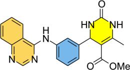
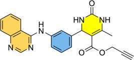
Ethyl 6-methyl-2-oxo-4-(3-(quinazolin-4-ylamino)phenyl)-1,2,3,4-tetrahydropyrimidine-5-carboxylate (7a)
Beige solid; mp 239-244 °C, FTIR (ATR) ν / cm-1 3198, 3083, 2975, 1681, 1566, 1371, 1221, 1088, 757, 687; 1H NMR (400 MHz, DMSO-d6) d 11.63 (1H, bs), 9.27-9.22 (1H, m), 8.90-8.83 (2H, m), 8.13-8.08 (1H, m), 8.00 (1H, dd, J 8.4, 0.9 Hz), 7.89-7.82 (2H, m), 7.66 (1H, ddd, J 8.0, 2.1, 1.1 Hz), 7.58 (1H, t, J 1.8 Hz), 7.45 (1H, t, J 7.9 Hz), 7.22 (1H, dt, J 7.7, 1.3 Hz), 5.22 (1H, d, J 3.4 Hz), 4.01 (2H, q, J 7.0 Hz), 2.27 (3H, s), 1.12 (3H, t, J 7.0 Hz); 13C NMR (100 MHz, DMSO-d6) d 165.3, 159.8, 152.0, 150.9, 148.7, 145.8, 138.8 136.8, 136.1, 128.6, 128.5, 124.9, 124.8, 124.1, 122.4, 119.9, 113.5, 98.9, 59.3, 53.9, 17.9, 14.11; HRMS (ESI) m/z, calcd. for [C22H22N5O3 + H]+: 404.1717, found: 404.1715.
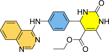
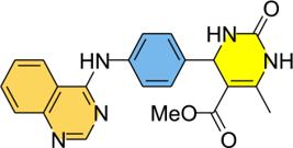
Ethyl 6-methyl-2-oxo-4-(4-(quinazolin-4-ylamino)phenyl)-1,2,3,4-tetrahydropyrimidine-5-carboxylate (7b)
Yellow solid; mp 290-292 °C, FTIR (ATR) ν / cm-1 3319, 3110, 2544, 2362, 1701, 1654, 1640, 1563, 1458, 1377, 1235, 1094, 770; 1H NMR (400 MHz, DMSO-d6) d 11.85 (1H, bs), 9.25 (1H, bs), 9.01 (1H, d, J 8.3 Hz), 8.87 (1H, s), 8.09 (1H, t, J 7.8 Hz), 8.01 (1H, d, J 8.3 Hz), 7.88-7.79 (2H, m), 7.67 (2H, d, J 8.3 Hz), 7.34 (2H, d, J 8.3 Hz), 5.20 (1H, d, J 3.0 Hz), 4.01 (2H, q, J 7.0 Hz), 2.28 (3H, s), 1.13 (3H, t, J 7.0 Hz); 13C NMR (100 MHz, DMSO-d6) d 165.3, 159.8, 152.1, 151.0, 148.6, 143.2, 139.1, 136.0, 135.7, 128.4, 126.5, 125.0, 124.9, 120.0, 113.5, 99.0, 59.2, 53.6, 17.8, 14.1; HRMS (ESI) m/z, calcd. for [C22H22N5O3 + H]+: 404.1717, found: 404.1710.
Methyl 6-methyl-2-oxo-4-(3-(quinazolin-4-ylamino)phenyl)-1,2,3,4-tetrahydropyrimidine-5-carboxylate (7c)
Yellow solid; mp 260-264 °C, FTIR (ATR) ν / cm-1 3292, 3211, 3083, 2955, 2665, 2362, 1687, 1566, 1377, 1221, 1088, 764, 682; 1H NMR (400 MHz, DMSO-d6) d 11.75 (1H, bs), 9.33-9.25 (1H, m), 8.97-8.84 (2H, m), 8.15-8.08 (1H, m), 8.04-7.99 (1H, m), 7.91-7.84 (2H, m), 7.68-7.63 (1H, m), 7.59-7.55 (1H, m), 7.46 (1H, t, J 7.8 Hz), 7.23 (1H, t, J 7.8 Hz), 5.21 (1H, d, J 3.5 Hz), 3.56 (3H, s), 2.28 (3H, s); 13C NMR (100 MHz, DMSO-d6) d 165.8, 160.0, 152.0, 150.7, 149.1, 145.6, 138.4, 136.7, 136.3, 128.8, 128.6, 125.0, 124.9, 124.3, 122.5, 119.5, 113.5, 98.7, 53.7, 50.9, 17.9; HRMS (ESI) m/z, calcd. for [C21H20N5O3 + H]+: 390.1556, found: 390.1561.
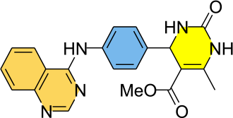
Methyl 6-methyl-2-oxo-4-(4-(quinazolin-4-ylamino)phenyl)-1,2,3,4-tetrahydropyrimi dine-5-carboxylate (7d)
Beige solid; mp 277-280 °C, FTIR (ATR) ν / cm-1 3171, 3076, 2355, 1687, 1633, 1438, 1377, 1229, 1094, 750; 1H NMR (400 MHz, DMSO-d6) d 11.75 (1H, bs), 9.30-9.25 (1H, m), 8.93 (1H, d, J 8.3 Hz), 8.88 (1H, s), 8.13-8.06 (1H, m), 7.98 (1H, d, J 8.1 Hz), 7.90-7.80 (2H, m), 7.65 (2H, d, J 8.6 Hz), 7.34 (2H, d, J 8.6 Hz), 5.20 (1H, d, J 3.3 Hz), 3.56 (3H, s), 2.28 (3H, s); 13C NMR (75 MHz, DMSO-d6) d 165.9, 159.8, 152.1, 151.2, 149.0, 143.1, 139.1, 136.1, 135.8, 128.6, 126.6, 125.1, 124.8, 120.1, 113.5, 98.8, 53.5, 50.9, 17.9; HRMS (ESI) m/z, calcd. for [C21H20N5O3 + H]+: 390.1561, found: 390.1560.
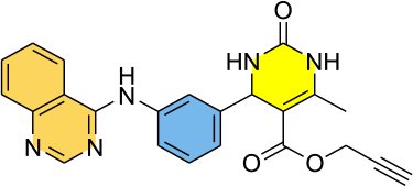
Prop-2-yn-1-yl 6-methyl-2-oxo-4-(3-(quinazolin-4-ylamino)phenyl)-1,2,3,4-tetrahydropyrimidine-5-carboxylate (7e)
Light yellow solid; mp 210-215 °C, FTIR (ATR) ν / cm-1 3204, 3097, 2942, 1694, 1627, 1566, 1377, 1216, 1074, 764, 682; 1H NMR (400 MHz, DMSO-d6) d 11.69 (1H, brs), 9.42 (1H, brs), 8.93-8.83 (2H, m), 8.15-8.07 (1H, m), 8.01 (1H, d, J 8.3 Hz), 7.96-7.91 (1H, m), 7.86 (1H, t, J 7.7 Hz), 7.69-7.61 (1H, brs), 7.45 (1H, t, J 7.7 Hz), 7.29-7.20 (1H, m), 5.21 (1H, d, J 3.3 Hz), 4.74-4.59 (2H, m), 3.47 (1H, t J 2.4 Hz), 2.29 (3H, s); 13C NMR (100 MHz, DMSO-d6) d 164.9, 160.3, 152.4, 151.3, 150.7, 145.9, 139.2, 137.3, 136.6, 129.2, 129.0, 125.3, 125.2, 124.7, 122.8, 120.2, 114.0, 98.5, 79.3, 77.8, 54.0, 51.5, 18.5; HRMS (ESI) m/z, calcd. for [C23H20N5O3 + H]+: 414.1561, found: 414.1555.
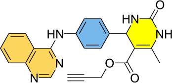
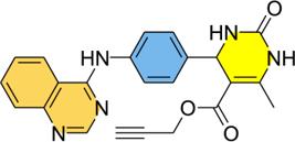
Prop-2-yn-1-yl 6-methyl-2-oxo-4-(4-(quinazolin-4-ylamino)phenyl)-1,2,3,4-tetrahydropyrimidine-5-carboxylate (7f)
Beige solid; mp 220-223 °C, FTIR (ATR) ν / cm-1 3198, 3076, 2942, 2632, 2362, 1687, 1566, 1377, 1222, 1088, 764, 689; 1H NMR (400 MHz, DMSO-d6) d 11.76 (1H, bs), 9.45-9.38 (1H, m), 8.94 (1H, d, J 8.3 Hz), 8.88 (1H, s), 8.10 (1H, t, J 8.1 Hz), 7.98 (1H, d, J 8.1 Hz), 7.93-7.89 (1H, m), 7.85 (1H, t, J 7.6 Hz), 7.66 (2H, d, J 8.3 Hz), 7.35 (1H, d, J 8.3 Hz), 5.20 (1H, d, J 3.3 Hz), 4.67 (2H, d, J 2.3 Hz), 3.51 (1H, t, J 2.3 Hz), 2.29 (3H, s); 13C NMR (75 MHz, DMSO-d6) d 164.5, 159.8, 152.0, 151.1, 150.2, 142.9, 139.1, 136.1, 135.8, 128.6, 126.6, 125.1, 124.8, 120.1, 113.5, 98.1, 78.8, 77.38, 53.5, 51.1, 18.0; HRMS (ESI) m/z, calcd. for [C23H20N5O3 + H]+: 414.1561, found: 414.1563.
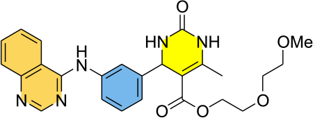
2-(2-Methoxyethoxy)ethyl 6-methyl-2-oxo-4-(3-(quinazolin-4-ylamino)phenyl)-1,2,3,4-tetrahydropyrimidine-5 carboxylate (7g)
White solid; mp 240-244 °C, FTIR (ATR) ν / cm-1 3191, 3097, 2948, 1687, 1627, 1566, 1377, 1216, 1087, 783, 757, 689; 1H NMR (400 MHz, DMSO-d6) d 11.70 (1H, brs), 9.32-9.23 (1H, m), 8.95-8.83 (2H, m), 8.11 (1H, t, J 8.1 Hz), 8.02 (1H, d, J 8.1 Hz), 7.93-7.80 (2H, m), 7.69 7.62 (1H, m), 7.59 (1H, brs), 7.49-7.40 (1H, m), 7.26 (2H, d, J 7.8 Hz), 5.22 (1H, d, J 3.3 Hz), 4.09 (2H, t, J 4.7 Hz), 3.61-3.50 (2H, m), 3.48-3.41 (2H, m), 3.39-3.32 (2H, m), 3.18 (3H, s), 2.27 (3H, s); 13C NMR (100 MHz, DMSO-d6) d 165.2, 159.9, 152.1, 150.8, 149.1, 145.7, 138.6, 136.7, 136.2, 128.7, 128.6, 125.0, 124.9, 124.2, 122.4, 119.7, 113.5, 98.9, 71.2, 69.4, 68.4, 62.7, 58.0, 53.8, 25.5, 17.9; HRMS (ESI) m/z, calcd. for [C25H27N5O5 + H]+: 478.2085, found: 478.2080.
2-(2-Methoxyethoxy)ethyl 6-methyl-2-oxo-4-(4-(quinazolin-4-ylamino)phenyl)-1,2,3,4-tetrahydropyrimidine-5 carboxylate (7h)
Yellow solid; mp 253-257 °C, FTIR (ATR) ν / cm-1 3225, 3090, 2935, 2517, 1708, 1559, 1431, 1371, 1222, 1080, 810, 757, 682; 1H NMR (400 MHz, DMSO-d6) d 11.86 (1H, brs), 9.33-9.29 (1H, m), 8.96 (1H, d, J 8.3 Hz), 8.89 (1H, s), 8.14-8.08 (1H, m), 8.00 (1H, d, J 8.1 Hz), 7.89-7.83 (2H, m), 7.65 (2H, d, J 8.3 Hz), 7.36 (2H, d, J 8.3 Hz), 5.20 (1H, d, J 3.3 Hz), 4.09 (1H, t, J 4.7 Hz), 3.62-3.52 (2H, m), 3.52-3.48 (2H, m), 3.45-3.39 (2H, m), 3.23 (3H, s), 2.27 (3H, s); 13C NMR (100 MHz, DMSO-d6) d 165.3, 159.9, 152.2, 150.9, 149.0, 143.4, 138.5, 136.3, 135.6, 128.6, 126.6, 125.1, 125.0, 119.6, 113.4, 99.0, 71.3, 69.5, 68.4, 62.7, 58.1, 53.5, 17.9; HRMS (ESI) m/z, calcd. for [C25H28N5O5 + H]+: 478.2085, found: 478.2087.
5-Acetyl-6-methyl-4-(3-(quinazolin-4-ylamino)phenyl)-3,4 dihydropyrimidin-2(1H)-one (7i)
Yellow solid; mp 265-267 °C, FTIR (ATR) ν / cm-1 3204, 3097, 2942, 1687, 1613, 1566, 1377, 1229, 757, 682; 1H NMR (400 MHz, DMSO-d6) d 11.75 (1H, brs), 9.27 (1H, bs), 8.97-8.84 (2H, m), 8.15-8.07 (1H, m), 8.01 (1H, t, J 8.3 Hz), 7.95 (1H, brs), 7.86 (1H, t, J 7.6 Hz), 7.65 (1H, d, J 7.8 Hz), 7.56 (1H, brs), 7.46 (1H, t, J 7.8 Hz), 7.29 (1H, t, J 7.7 Hz), 5.33 (1H, d, J 2.9 Hz), 2.31 (3H, s), 2.17 (3H, s); 13C NMR (100 MHz, DMSO-d6) d 194.20, 159.9, 152.1, 150.8, 148.6, 145.2, 138.5, 136.7, 128.8, 128.7, 125.1, 124.9, 124.2, 122.6, 119.6, 113.5, 109.5, 53.6, 30.5, 19.1; HRMS (ESI) m/z, calcd. for [C21H20N5O2 + H]+: 374.1612, found: 374.1607.
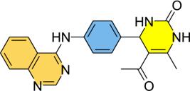
5-Acetyl-6-methyl-4-(4-(quinazolin-4-ylamino)phenyl)- 3,4-dihydropyrimidin-2(1H)-one (7j)
Yellow solid; mp 240-245 °C, FTIR (ATR) ν / cm-1 3198, 3076, 2942, 2632, 2362, 1687, 1566, 1377, 1222, 1088, 764, 689; 1H NMR (400 MHz, DMSO-d6) d 9.81 (1H, brs), 9.19 (1H, m), 8.56 (1H, s), 8.53 (1H, d, J 8.4 Hz), 7.89-7.81 (2H, m), 7.80-7.74 (3H, m), 7.66-7.60 (1H, m), 7.27 (2H, d, J 8.6 Hz), 5.27 (1H, d, J 3.4 Hz), 2.31 (3H, s), 2.13 (3H, s); 13C NMR (100 MHz, DMSO-d6) d 194.4, 157.8, 154.5, 152.1, 149.7, 148.0, 139.7, 138.2, 133.0, 127.8, 126.6, 126.2, 123.0, 122.8, 115.1, 109.6, 30.3, 18.9; HRMS (ESI) m/z, calcd. for [C21H20N5O2 + H]+: 374.1612, found: 374.1610.
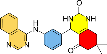
7,7-Dimethyl-4-(3-(quinazolin-4-ylamino)phenyl)-3,4,7,8-tetrahydroquinazoline-2,5(1H,6H)-dione (7k)
White solid; mp 295-299 °C, FTIR (ATR) ν / cm-1 3319, 3097, 2955, 2658, 1681, 1627, 1559, 1438, 1371, 1229, 757, 689; 1H NMR (400 MHz, DMSO-d6) d 11.76 (1H, brs), 9.60-9.52 (1H, m), 8.93 (1H, d, J 8.3 Hz), 8.87 (1H, s), 8.14-8.08 (1H, m), 8.03 (1H, d, J 7.8 Hz), 7.91-7.82 (2H, m), 7.66-7.61 (1H, m), 7.60-7.57 (1H, m), 7.44 (2H, t, J 7.8 Hz), 7.23 (1H, d, J 7.5 Hz), 5.23 (1H, d, J 2.8 Hz), 2.41 (1H, d, 3J 17.1 Hz), 2.33 (1H, d, 3J 17.1 Hz), 2.19 (1H, d, 3J 16.0 Hz), 2.07 (1H, d, 3J 16.0 Hz), 1.02 (3H, s), 0.91 (3H, s); 13C NMR (100 MHz, DMSO-d6) d 192.9, 159.9, 152.7, 151.8, 150.8, 145.6, 138.6 136.6, 136.2, 128.7, 128.6, 124.9, 124.8, 124.0, 122.6, 119.7, 113.5, 107.1, 51.9, 49.8, 32.4, 28.6, 27.1; HRMS (ESI) m/z, calcd. for [C24H24N5O2 + H]+: 414.1925, found: 414.1933.
7,7-Dimethyl-4-(4-(quinazolin-4-ylamino)phenyl)-3,4,7,8-tetrahydroquinazoline-2,5(1H,6H)-dione (7l)
Bright yellow solid; mp 268-271 °C, FTIR (ATR) ν / cm-1 3191, 3090, 2948, 2867, 1687, 1633, 1613, 1566, 1438, 1371, 1229, 764, 682, 561; 1H NMR (400 MHz, DMSO-d6) d 11.73 (1H, brs), 9.58-9.52 (1H, m), 8.94 (1H, d, J 8.4 Hz), 8.87 (1H, s), 8.12-8.05 (1H, m), 7.98 (1H, d, J 7.9 Hz), 7.88-7.81 (2H, m), 7.65 (2H, d, J 8.6 Hz), 7.33 (2H, d, J 8.6 Hz), 5.20 (1H, d, J 2.8 Hz), 2.43 (1H, d, 3J 17.2 Hz), 2.32 (1H, d, 3J 17.1 Hz), 2.21 (1H, d, 3J 16.1 Hz), 2.06 (1H, d, 3J 16.1 Hz), 1.03 (3H, s), 0.92 (3H, s); 13C NMR (100 MHz, DMSO-d6) d 193.0, 159.5, 152.5, 151.9, 151.8, 142.7, 136.0, 135.6, 128.2, 126.6, 124.5, 124.4, 121.4, 115.3, 113.8, 107.2, 51.7, 49.8, 32.4, 28.7, 27.0; HRMS (ESI) m/z, calcd. for [C24H24N5O2 + H]+: 414.1925, found: 414.1917.
N-Benzyl-6-methyl-2-oxo-4-(3-(quinazolin-4-ylamino)phenyl)-1,2,3,4-tetrahydropyrimidine-5-carboxamide (7m)
Yellow solid; mp 219-223 °C, FTIR (ATR) ν / cm-1 3198, 3076, 2942, 2632, 2362, 1687, 1566, 1377, 1222, 1088, 764, 689; 1H NMR (400 MHz, DMSO-d6) d 11.71 (1H, brs), 8.92 (1H, d, J 8.3 Hz), 8.88 (1H, s), 8.62 (1H, brs), 8.26 (1H, t, J 5.6 Hz), 8.11 (1H, t, J 7.8 Hz), 8.01 (1H, d, J 8.3 Hz), 7.86 (1H, t, J 7.7 Hz), 7.72 (1H, d, J 7.9 Hz), 7.60 (1H, brs), 7.43 (1H, t, J 7.8 Hz), 7.22 (1H, d, J 7.6 Hz), 7.19-7.12 (2H, m), 7.10-7.01 (3H, m), 5.37 (1H, brs), 4.25 (2H, d, J 5.6 Hz), 2.05 (3H, s); 13C NMR (100 MHz, DMSO-d6) d 166.3, 159.8, 152.5, 150.9, 145.2, 139.7, 138.7, 137.8, 136.8, 136.2, 128.6 128.5, 128.0, 127.0, 126.3, 125.0, 124.8, 124.0, 122.5, 119.8, 113.5, 104.6, 54.9, 42.2, 17.0; HRMS (ESI) m/z, calcd. for [C27H25N6O2 + H]+: 465.2034, found: 465.2028.
N-Benzyl-6-methyl-2-oxo-4-(4-(quinazolin-4-ylamino)phenyl)-1,2,3,4-tetrahydropyrimidine-5-carboxamide (7n)
Yellow solid; mp 216-218 °C, FTIR (ATR) ν / cm-1 3198, 3076, 2942, 2632, 2362, 1687, 1566, 1377, 1222, 1088, 764, 689; 1H NMR (400 MHz, DMSO-d6) d 11.80 (1H, brs), 8.98 (1H, d, J 8.3 Hz), 8.89 (1H, s), 8.64 (1H, brs), 8.23 (1H, t, J 5.6 Hz), 8.11 (1H, t, J 7.7 Hz), 8.00 (1H, d, J 8.3 Hz), 7.86 (1H, t, J 7.6 Hz), 7.67 (1H, d, J 8.2 Hz), 7.59 (1H, bs), 7.34 (1H, d, J 8.2 Hz), 7.24 (2H, d, J 7.3 Hz), 7.16 (1H, t, J 7.2 Hz), 7.07 (2H, d, J 7.6 Hz), 5.37 (1H, brs), 4.32-4.19 (2H, m), 2.06 (3H, s); 13C NMR (100 MHz, DMSO-d6) d 166.3, 159.8, 152.6, 151.0, 142.7, 139.7, 138.8, 138.1, 136.2, 135.7, 128.6, 128.1, 127.0, 126.5, 124.9, 119.9, 113.5, 104.5, 54.5, 42.2, 17.0; HRMS (ESI) m/z, calcd. for [C27H25N6O2 + H]+: 465.2034, found: 465.2030.
Biology
Maintenance of parasites and cells
The species Leishmania amazonensis (IFLA/BR/67/PH8), Leishmania major (MHOM/BR/1976/JOF), Leishmania infantum (MHOM/BR/1975/M2903) and Leishmania braziliensis (MHOM/BR/1975/M2903) were cultured in the promastigote form in vitro in Schneider medium supplemented with 20% fetal bovine serum (FBS) (Cultilab, Campinas, Brazil), and 1% penicillin (100 U mL-1) and streptomycin (100 μg mL-1) (Sigma-Aldrich, St. Louis, USA), pH 7, at 26 ± 1 °C in a biochemical oxygen demand (BOD) incubator (Eletrolab EL202, São Paulo, Brazil).81 Epimastigote forms of T. cruzi (Y strain) were maintained in liver infusion tryptose (LIT) medium supplemented with 10% FBS and 1% penicillin-streptomycin, kept in BOD at 28 °C.82 Trypomastigote forms (Y strain) were obtained from the supernatant of previously infected Vero cells and cultured in Dulbecco’s modified eagle medium (DMEM) (Sigma-Aldrich, St. Louis, USA) with 1% penicillin (100 U mL-1) and streptomycin (100 μg mL-1) and 10% FBS, kept in an incubator (ThermoScientific series 800 WJ, Waltham, MA, USA) at 37 °C in 5% CO2. Murine macrophages of the RAW 264.7 lineage and Vero cells were used, maintained in DMEM medium (10% FBS, 1% penicillin (100 U mL-1) and streptomycin (100 μg mL 1), pH 7, at 37 °C, 5% CO2 in an incubator.83
Growth inhibition assay of promastigote forms of Leishmania spp.
Promastigotes in the amount of 1 × 10⁶ parasites per well and in logarithmic growth phase were added to a 96-well plate containing supplemented Schneider medium. Quinazoline compounds and the reference drug (miltefosine) were added to the same plate in serial concentrations, ranging from 50 to 1.56 μM for compounds and 200 to 1.56 μM for miltefosine, and cultured for 72 h at 26 °C in a BOD incubator. After this time, 50 μL of DMSO was added per well to dissolve the formazan crystals. The plate was shaken, and the reading was performed at 540 nm in a microplate spectrophotometer (Biosystems ELx800 model, Curitiba, Brazil). The negative control (100% viability) was performed with Schneider medium supplemented and containing 0.2% DMSO.83
Effect on the growth of different evolutionary forms of T. cruzi
Epimastigote and trypomastigote forms were added to a 96-well culture plate containing 100 μL of liver infusion tryptose (LIT) medium supplemented with 1 × 10⁶ parasites per well. Then, test compounds were added in serial concentrations (100-1.56 μM). Benznidazole was used as the positive control in all assays, with concentrations ranging from 12.5 to 400 μM. To ensure experimental consistency, positive controls were run simultaneously on the same plates and under the same experimental conditions as the test compounds. The plate was then shaken and read in a microplate reader at 540 nm. The negative control was performed with complete LIT at 0.5% DMSO, considered as 0% inhibition of parasite growth. The positive control was compared with reference drug benznidazole.82
Cytotoxicity in cells
RAW 264.7 macrophages and Vero cells were seeded in 96-well plates at a density of 1 × 10⁵ cells per well in 100 µL of supplemented DMEM. The plates were incubated at 37 °C with 5% CO2 for 4 h to allow for cell adhesion. Following incubation, cells were washed with PBS and treated with 100 µL of DMEM containing serial dilutions of the test compounds (ranging from 1.56 to 800 µM). Positive controls (miltefosine and benznidazole) were included in every assay plate and evaluated across the same concentration range (1.56-800 µM). All test compounds and positive controls were screened simultaneously under identical experimental conditions to ensure internal consistency. After 72 h of treatment, 10 µL of MTT (5 mg mL-1) was added to each well, and the plates were incubated for an additional 4 h. The culture medium was then aspirated, and 100 µL of DMSO was added to each well to solubilize the formazan crystals. After mechanical agitation, absorbance was measured at 540 nm using a microplate reader. Supplemented DMEM containing 0.5% DMSO served as the negative control, representing 100% cell viability.84
Statistical analysis
Statistical analyses were performed using GraphPad Prism software (version 8.0, GraphPad Software, San Diego, CA, USA). All in vitro assays were conducted in five independent biological replicates (n = 5), each consisting of three technical replicates per concentration per plate. All experimental groups and their respective controls were run on the same plates to minimize inter-assay variability. Normality was assessed using the Kolmogorov-Smirnov test. For parametric data, differences between groups were compared using one-way analysis of variance (ANOVA) followed by Tukey’s post-hoc test with a 95% confidence interval. Results with values p < 0.05 were considered statistically significant. Data were expressed as mean ± standard error of the mean (SEM). The half-maximal inhibitory (IC50) and cytotoxic (CC50) concentrations, along with their 95% confidence intervals (95% CI), were determined via nonlinear regression using a four-parameter logistic model with a variable slope (log(inhibitor) vs. response - variable slope). For the nonlinear regression, the bottom and top of the curves were constrained to 0 and 100, respectively, to ensure consistent IC50 estimation across compounds. For compounds where no significant reduction in viability was observed at the maximum tested concentration, results were reported as > 50 µM.
In silico studies
Molecular docking simulations
Molecular docking simulations were performed with the aim of contributing to the elucidation of the antitrypanosome mechanism of action of quinazoline-dihidropyrimidinone derivatives, through their binding affinity with the targets involved in this effect, which correspond to the following enzymes: cruzain from T. cruzi in complex with WRR-112 (PDB: 1EWM); sterol 14-alpha demethylase (CYP51) from L. infantum in complex with fluconazole (PDB: 3L4D);62 dihydroorotate dehydrogenase from L. braziliensis in complex with the piperazine derivative (PDB: 4WZH);85 nucleoside diphosphatase kinase (NDK) protein from L. amazonensis (PDB: 5GO1)63 and N-myristoyltransferase from L. major (PDB: 4CGO) in complex with pyrimidin-4-yl amino derivative.52 The 3D structures of the enzymes were obtained from the Protein Data Bank (PDB),86 through X-ray diffraction. The synthetic derivative compounds of quinazoline-dihidropyrimidinone and the positive control compounds miltefosine, benznidazole and auranofin were designed in the Marvin Sketch program, version 19.1887 and saved as .sdf files, then the conformational search and minimization of the compounds under study were performed in the Spartan 14 software,88 using the molecular mechanics method,89 and immediately afterwards a second minimization was carried out using the semi-empirical method Recife Model 1 (RM1).90 Molecular docking was performed using the Molegro Virtual Docker (MVD) software, version 6.0.1 (CLC Bio Company).
After removing the water molecules and defining the active site with the co-crystallized ligand, redocking was performed to validate the method using root mean square deviation (RMSD) values, with values up to 2.0 Å being considered adequate.53 The coordinates of the active site of the enzymes under study, according to the co-crystallized ligand, corresponded to: x = 2.24, y = 11.09 and z = 5.97 for the enzyme cruzain from T. cruzi in complex with WRR 112 (PDB: 1EWM); x = 32.02, y = -29.16 and z = -1.27 for the enzyme sterol 14-alpha demethylase (CYP51) from L. infantum in complex with fluconazole (PDB: 3L4D); x = 6.95, y = -6.06 and z = 20.55 for the enzyme dihydroorotate dehydrogenase from L. braziliensis in complex with the piperazine derivative (PDB: 4WZH); x = 5.74, y = 47.82 and z = 60.15 for the enzyme N-myristoyltransferase from L. major (PDB: 4CGO) in complex with pyrimidin-4-yl amino derivative. For enzymes that do not have a co-crystallized ligand, as in the case of the enzyme nucleoside diphosphatase kinase (NDK) protein from L. amazonensis (PDB: 5GO1), the target validation process and active site marking were performed using literature references on key residues for target inhibition, corresponding to the coordinates x = 16.99, y = 11.34 and z = 40.22. Molecular pocket prediction tools, including the ProteinPlus platform,91 were also employed, and auranofin was used as a reference compound. Furthermore, due to the in vitro test, benznidazole was used as control compounds for the cruzain from T. cruzi in complex with WRR-112 (PDB: 1EWM) and miltefosine for targets referring to Leishmania species.
The simulations used the MolDock SE algorithm with specific parameters and analyzed the interactions between the compounds and the enzyme, based on the energy values, using the MolDock (GRID) and PLANTS (GRID) scoring functions to calculate the docking energy values.92-94 Based on this, a consensus calculation for each of the scoring functions under study was performed by dividing the score of the ligand in question by the lowest energy value. After that, a second calculation was performed by summing the normalized score value for each of the functions and dividing it by the total number of observations.58 A total of 30 runs with a maximum of 3,000 interactions using a population of 50 individuals, 2,000 minimization steps for each flexible residue, and 2,000 global minimization steps per run. A GRID was set at 0.3 Å, and the search sphere was set at 18 Å in radius. For the ligand energy analysis, internal electrostatic interactions, internal hydrogen bonds, and sp2-sp2 torsions were evaluated. Interactions were visualized in Discovery Studio Visualizer, version 20.1.0.19295.95 For the enzyme nucleoside diphosphatase kinase (NDK) protein from L. amazonensis (PDB: 5GO1), the 3D structure was edited in UCSF Chimera 1.17.3 software67 to correct regions that did not present amino acid residues.
Molecular dynamics simulations
Molecular dynamics simulations were performed using GROMACS 2023.1,96,97 with topologies generated on the automated topology builder (ATB) platform using the GROMOS54a7 force field.98,99 The system was solvated with the simple point charge (SPC) water model in a cubic box, energy-minimized, heated to 300 K, and equilibrated at 1 atm for 100 ns and 50.000.000 steps. RMSD, RMSF and energy metrics of the C-α atoms were calculated, and the corresponding graphs were generated using the Grace software.100,101
Supplementary Information
Supplementary material 1
Supplementary data (dose / response curves for T. cruzi and Leishmanias; 3D molecular interaction maps of the complexes formed with enzymes from parasite; tables of protein binding energy; tables of Coulomb and Lenard-Jones energies; 1H NMR, 13C NMR and HRMS spectra of compounds) are available free of charge at http://jbcs.sbq.org.br as PDF file.
Acknowledgments
The authors are grateful to the governmental agencies for financial support and fellowships. FAPERGS (D.R. grant No. 19/2551-0001767-7); CNPq (D.R. grant No. 310438/2020-9 and 403260/2021 3). CAPES and FAPERGS (E.B.M., V.V. and E.U.B.A.) for the fellowships.
Data Availability Statement
The data supporting this article is available in the text.
References
-
1 Pino-Marín, A.; Medina-Rincón, G. J.; Gallo-Bernal, S.; Duran-Crane, A.; Duque, A. I. A.; Rodríguez, M. J.; Medina-Mur, R.; Manrique, F. T.; Forero, J. F.; Medina, H. M.; Pathogens 2021, 10, 505. [Crossref]
» Crossref -
2 Hotez, P. J.; Aksoy, S.; Brindley, P. J.; Kamhawi, S.; PLoS Neglected Trop. Dis. 2020, 14, e0007999. [Crossref]
» Crossref -
3 Lin, Y.; Fang, K.; Zheng, Y.; Wang, H. L.; Wu. J.; J. Travel Med. 2022, 29, taac031. [Crossref]
» Crossref -
4 Pereira, L. O. R.; Moreira, R. B.; Oliveira, M. P.; Reis, S. O.; Oliveira Neto, M. P.; Pirmez, C.; Int. J. Infect. Dis. 2017, 57, 132. [Crossref]
» Crossref -
5 Chaves, A. T.; Menezes, C. A. S.; Costa, H. S.; Nunes, M. C. P.; Rocha, M. O. C.; Parasite Immunol. 2019, 41, e12663. [Crossref]
» Crossref -
6 Pan American Health Disease; Chagas Disease [Link] accessed in February 2026
» Link -
7 Revollo, S.; Oury, B.; Vela, A.; Tibayrenc, M.; Sereno, D.; Pathogens 2019, 8, 197. [Crossref]
» Crossref -
8 Benaim, G.; Paniz-Mondolfi, A.; Biomolecules 2024, 14, 406. [Crossref]
» Crossref -
9 Mouloudi, I.; Zitouni, N.; Himi, H.; Injai, U.; Toumi, A.; Rahhou, I.; Legssyer, B.; E3S Web Conf. 2024, 527, 02016. [Link] accessed in February 2026
» Link -
10 World Health Organization; Leishmaniasis [Link] accessed in February 2026
» Link -
11 World Health Organization; Integrating Neglected Tropical Diseases in Global Health and Development: Fourth WHO Report on Neglected Tropical Diseases, 2017. [Link] accessed in February 2026
» Link -
12 Santos, S.; Araújo, R. V.; Giarolla, J.; Seoud, O. E. I.; Ferreira, E. I.; Int. J. Antimicrob. Agents 2020, 55, 105906. [Crossref]
» Crossref -
13 Setshedi, K. J.; Beteck, R. M.; Jesumoroti, O. J.; Ilbeigi, K.; Mabille, D.; Caljon, G.; Van der Kooy, F.; Legoabe, L. J.; Chem. Biol. Drug Des. 2023, 102, 763. [Crossref]
» Crossref -
14 Bouchut, A.; Rotili, D.; Pierrot, C.; Valente, S.; Lafitte, S.; Schultz, J.; Hoglund, U.; Mazzone, R.; Lucidi, A.; Fabrizi, G.; Pechalrieu, D.; Arimondo, P. B.; Skinner-Adams, T. S.; Chua, M. J.; Andrews, K. T.; Mai, A.; Khalife, J.; Eur. J. Med. Chem. 2019, 161, 277. [Crossref]
» Crossref -
15 Welsch, M. E.; Snyder, S. A.; Stockwell. B. R.; Curr. Opin. Chem. Biol. 2010, 14, 347. [Crossref]
» Crossref -
16 Gilbert, I. H.; Biochim. Biophys. Acta, Mol. Basis Dis. 2002, 1587, 249. [Crossref]
» Crossref -
17 Romero, A. H.; Rodríguez, N.; Oviedo, H.; Bioorg. Chem. 2019, 83, 145. [Crossref]
» Crossref -
18 Mahender, T.; Pankaj, W.; Kumar, S. P.; Ankur, V.; Kumar, S. S.; Mini-Rev. Med. Chem. 2022, 22, 743. [Crossref]
» Crossref -
19 Pal, R.; Teli, G.; Akhtar, M. J.; Matada, G. S. P.; Eur. J. Med. Chem. 2023, 258, 115609. [Crossref]
» Crossref -
20 Kalakwade, A.; Kavalapure, R. S.; Gharge, S.; Alegaon, S. G.; Ranade, S. D.; Wong, L. S.; Ramu, R.; Results Chem. 2026, 19, 102932. [Crossref]
» Crossref -
21 Pal, R.; Teli, G.; Akhtar, M. J.; Matada, G. S. P.; Eur. J. Med. Chem. 2024, 263, 115927. [Crossref]
» Crossref -
22 Rode, N. R.; Tantray, A. A.; Shelar, A. V.; Pai, K.; Patil, R. H.; Terdale, S. S.; Res. Chem. Intermed. 2024, 50, 1125. [Crossref]
» Crossref -
23 Das, N.; Roy, J.; Patra, B.; Saunders, E.; Sarkar, D.; Goon, S.; Sinha, B. P.; Roy, S.; Roy, S.; Sarif, J.; Bandopadhyay, P.; Barik, S.; Mukherjee, S.; McNamara, N.; Varghese, S.; Simpson, K.; Baell, J.; McConville, M.; Ganguly, D.; Talukdar, A.; Eur. J. Med. Chem. 2024, 269, 116256. [Crossref]
» Crossref -
24 Seifu, G. W. Y.; Birhan, S.; Beshay, B. Y.; Hymete, A.; Bekhit, A. A.; BMC Chem. 2024, 16, 107. [Crossref]
» Crossref -
25 Bollinia, M.; Bruno, A. M.; Niño, M. E.; Casala, J. J.; Sasiambarrena, L. D.; Valdez, D. A. G.; Battinia, L.; Puente, V. R.; Lombardo; M. E.; Med. Chem. 2019, 15, 265. [Crossref]
» Crossref -
26 Tawaraishi, T.; Ochida, A.; Akao, Y.; Itono, S.; Kamaura, M.; Akther, T.; Shimada, M.; Canan, S.; Chowdhury, S.; Cao, Y.; Condroski, K.; Engkvist, O.; Francisco, A.; Ghosh, S.; Kaki, R.; Kelly, J. M.; Kimura, C.; Kogej, T.; Nagaoka, K.; Naito, A.; Pairaudeau, G.; Radu, C.; Roberts, I.; Shum, D.; Watanabe, N. A; Xie, H.; Yonezawa, S.; Yoshida, O.; Yoshida, R.; Mowbray, C.; Perry, B.; J. Med. Chem. 2023, 66, 1221. [Crossref]
» Crossref -
27 Romero, A. H.; Aguilera, E.; Gotopo, L.; Cabrera, G.; Dávila, B.; Cerecetto, H.; RSC Med. Chem. 2023, 14, 1992. [Crossref]
» Crossref -
28 Silva, E. B.; Rocha, D. A.; Fortes, I. S.; Yang, W.; Monti, L.; Siqueira-Neto, J. L.; Caffrey, C. R.; McKerrow, J.; Andrade, S. F.; Ferreira, R. S.; J. Med. Chem. 2021, 64, 13054. [Crossref]
» Crossref -
29 Vázquez, C.; Matus-Meza, A. S.; Nuñez-Moreno, O.; Barbosa Sánchez, B. M.; Farías-Gutiérrez, V. M.; Mendoza Conde, Hernández-Luis, F.; Saavedra, E.; Molecules 2024, 29, 4501. [Crossref]
» Crossref -
30 Long, S.; Duarte, D.; Carvalho, C.; Oliveira, R.; Santarém, N.; Palmeira, A.; Resende, D. I. S. D. P.; Silva, A. M. S.; Moreira, R.; Kijjoa, A.; Silva, A. C.; Nogueira, F.; Sousa, E.; Pinto, M. M. M.; ACS Med. Chem. Lett. 2022, 13, 225. [Crossref]
» Crossref -
31 Matos, L. H. S.; Masson, F. T.; Simeoni, L. A.; Homem de Mello, M.; Eur. J. Med. Chem. 2018, 143, 1779. [Crossref]
» Crossref -
32 Singh, N.; Kaur, J.; Kumar, P.; Gupta, S.; Singh, N.; Ghosal, A.; Dutta, A.; Kumar, A.; Tripathi, R. P.; Siddiqi, M. I.; Mandal, C.; Dube, A.; Parasitol. Res. 2009, 105, 1317. [Crossref]
» Crossref -
33 Kaur, J.; Sundar, S.; Singh, N.; J. Antimicrob. Chemother. 2010, 65, 1742. [Crossref]
» Crossref -
34 Mohammadi-Ghalehbin, B.; Sepehri, S.; Nejatkhah, N.; Safari, S.; Hosseinali, Z.; Sabour, S.; Razzaghi-Asl, N.; Pharm. Chem. J. 2022, 55, 1050. [Crossref]
» Crossref -
35 Saudi, M. N. S.; El-Semary, M. M. A.; Elbayaa, R. Y.; Jaeda, M. I.; Eissa, M. M.; Amer, E. I.; Baddour, N. M.; Med. Chem. Res. 2012, 21, 257. [Crossref]
» Crossref -
36 Mishra, G.; Jaiswal, A. K.; Kushawaha, A. K.; Kumar, A.; Bhatt, A.; Ansari, A.; Bisen, A. C.; Hansda, R.; Agrawal, S.; Acharjee, P.; Guha, R.; Bhatta, R. S.; Purkait, B.; Sashidhara, K. V.; Eur. J. Med. Chem. 2025, 293, 117667. [Crossref]
» Crossref -
37 Inaoka, D. K.; Iida, M.; Hashimoto, S.; Tabuchi, T.; Kuranaga, T.; Balogun, E. O.; Honma, T.; Tanaka, A.; Harada, S.; Nara, T.; Kita, K.; Inoue, M.; Bioorg. Med. Chem. 2017, 25, 1465. [Crossref]
» Crossref -
38 Dil, K. V.; Palchykov, V. A.; Beilstein J. Org. Chem. 2024, 20, 2143. [Crossref]
» Crossref -
39 Mato, R.; Bournez, C.; Lefebvre, Q.; Synthesis 2024, 35, 3450. [Crossref]
» Crossref -
40 Liu, Y.; Mathis, C.; Bajczyk, M. D.; Marshall, S. M.; Wilbraham, L.; Cronin, L.; Sci. Adv. 2021, 7, eabj2465. [Crossref]
» Crossref -
41 Viegas-Jr., C.; Danuello, A.; Bolzani, V. S.; Barreiro, E. J.; M. Fraga, C. A.; Curr. Med. Chem. 2007, 14, 1829. [Crossref]
» Crossref -
42 Ivasiv, V.; Albertini, C.; Gonçalves, A. E.; Rossi, M.; Bolognesi, M. L.; Curr. Top. Med. Chem. 2019, 19, 1694. [Crossref]
» Crossref - 43 Njogu, P. M.; Okombo, J.; Chibale, K. In Design of Hybrid Molecules for Drug Development; Deker, M., ed.; Elsevier: Amsterdam, Netherlands, 2017, p. 83-136.
-
44 Kaur, P. H.; Persoons, L.; Andrei, G.; Singh, K.; ChemMedChem 2022, 17, e202200031. [Crossref]
» Crossref -
45 Jaiswal, A. K.; Kushawaha, A. K.; Kumar, P.; Ansari, A.; Chhikara, N.; Bhatt, H.; Katiyar, S.; Ahmad, I.; Choudhury, A. D.; Bhatta, R. S.; Tamrakar, A. K.; Sashidhara, K. V.; Eur. J. Med. Chem. 2025, 288, 117366. [Crossref]
» Crossref -
46 Kappe, C. O.; Stadler, A.; Org. React. 2004, 63, 1. [Crossref]
» Crossref -
47 Wang, Z.; Wu, X.; Wang, L.; Zhang, J.; Liu, J.; Song, Z.; Tang, Z.; Bioorg. Med. Chem. Lett. 2016, 26, 2589. [Crossref]
» Crossref -
48 Taciak, B.; Białasek, M.; Braniewska, A.; Sas, Z.; Sawicka, P.; Kiraga, L.; Rygiel, T.; Krol, M.; PloS One 2018, 13, e0198943. [Crossref]
» Crossref -
49 Ramírez-Macías, I.; Maldonado, C. R.; Marín, C.; Olmo, F.; Gutiérrez-Sánchez, R.; Rosales, M. J.; Quirós, M.; Salas, J. M.; Sánchez-Moreno, M.; J. Inorg. Biochem. 2012, 112, 1. [Crossref]
» Crossref -
50 Santos, V. C.; Oliveira, A. E. R.; Campos, A. C. B.; Reis-Cunha, J. L.; Bartholomeu, D. C.; Teixeira, S. M. R.; Lima, A. P. C. A.; Ferreira, R. S.; Sci. Rep. 2021, 11, 18231. [Crossref]
» Crossref -
51 Sousa, N. F.; Souza, H. D. S.; Menezes, R. P. B.; Alves, F. S.; Acevedo, C. A. H.; Nunes, T. A. L.; Sessions, Z. L.; Scotti, L.; Muratov, E. N.; Mendonça-Jr., F. J. B.; Rodrigues, K. A. F.; Athayde Filho, P. F.; Scotti, M. T.; Pathogens 2023, 12, 136. [Crossref]
» Crossref -
52 Leite, F. F.; Rodrigues, L. C.; Oliveira, B. H. M.; Duarte, G. D.; Ferreira, M. D. L.; Sousa, N. F.; Vanderley, S. E. R.; Cardoso, L. L.; Keesen, T. S. L.; Araújo, R. S. A.; Scotti, L.; Scotti, M. T.; Mendonça-Jr., F. J. B.; Nat. Prod. Res. 2026, 40, 196. [Crossref]
» Crossref -
53 Froes, T. Q.; Zapata, L. C. C.; Akamine, J. S.; Castilho, M. S.; Nonato, M. C.; Curr. Top. Med. Chem. 2021, 21, 2134. [Crossref]
» Crossref -
54 Menezes, R. P. B.; Assis, E. B.; Sousa, N. F.; Souza, J. M. S.; Rodrigues, K. A. F.; Scotti, L.; Tavares, J. F.; Silva, M. S.; Scotti, M. T.; Mol. Diversity 2025 [Crossref]
» Crossref -
55 Nascimento, I. J. S.; Cavalcanti, M. D. A. T.; Moura, R. O.; Eur. J. Med. Chem. 2023, 258, 115550. [Crossref]
» Crossref -
56 Stanzione, F.; Giangreco, I.; Cole, J. C.; Prog. Med. Chem. 2021, 60, 273. [Crossref]
» Crossref -
57 Muhammed, M. T.; Aki-Yalcin, E.; Lett. Drug Des. Discovery 2024, 21, 480. [Crossref]
» Crossref -
58 Silva, M. L. Q.; Sousa, G. R.; Sousa, N. F.; Santos, A. T. L.; Coutinho, H. D. M.; Barbosa Filho, J. M.; Morais-Braga, M. F. B.; Process Biochem 2024, 142, 46. [Crossref]
» Crossref -
59 Gandhi, G. R.; Sharanya, C. S.; Jayanandan, A.; Haridas, M.; Hillary, V. E.; Gandhi, S. R.; Vasconcelos, A. B. S.; Montalvão, M. M.; Ceasar, S. A.; Sousa, N. F.; Scotti, L.; Scotti, M. T.; Gurgel, R. Q.; Quintans-Jr., L. J.; J. Biomol. Struct. Dyn. 2024, 42, 3051. [Crossref]
» Crossref -
60 Silva, P. R.; Pazos, N. D. N.; Andrade, J. C.; Sousa, N. F.; Pires, H. F. O.; Lima, J. L. F.; Dias, A. L.; Salvadori, M. G. S. S.; Golzio, A. M. F. O.; Castro, R. D.; Scotti, M. T.; Patil, V. M.; Felipe, C. F. B.; Almeida, R. N.; Scotti, L.; Mini-Rev. Med. Chem. 2024, 24, 1556. [Crossref]
» Crossref -
61 Mishra, A. K.; Singh, N.; Agnihotri, P.; Mishra, S.; Singh, S. P.; Kolli, B. K.; Chang, K. P.; Sahasrabuddhe, A. A.; Siddiqi, M. I.; Pratap, J. V.; J. Comput.-Aided Mol. Des. 2017, 31, 547. [Crossref]
» Crossref -
62 Brannigan, J. A.; Roberts, S. M.; Bell, A. S.; Hutton, J. A.; Hodgkinson, M. R.; Tate, E. W.; Leatherbarrow, R. J.; Smith, D. F.; Wilkinson, A. J.; IUCrJ 2014, 1, 250. [Crossref]
» Crossref -
63 Aghajani, J.; Farnia, P.; Farnia, P.; Ghanavi, J.; Velayati, A. A.; Tanaffos 2022, 21, 1. [Link] accessed in February 2026
» Link -
64 Fukutani, T.; Miyazawa, K.; Iwata, S.; Satoh, H.; Bull. Chem. Soc. Jpn. 2021, 94, 655. [Crossref]
» Crossref -
65 Shukla, R.; Tripathi, T. In Molecular Dynamics Simulation of Protein and Protein-Ligand Complexes; Singh, D. B., ed.; Springer: Singapore, 2020. [Crossref]
» Crossref -
66 Martínez, L.; PloS One 2015, 10, e0119264. [Crossref]
» Crossref -
67 Pettersen, E. F.; Goddard, T. D.; Huang, C. C.; Couch, G. S.; Greenblatt, D. M.; Meng, E. C.; Ferrin, T. E.; J. Comput. Chem. 2004, 25, 1605. [Crossref]
» Crossref -
68 Mehta, N. A.; Levin, D. A.; Phys. Rev. E 2019, 99, 033302. [Crossref]
» Crossref -
69 Khan, M. S.; Nawaz, M. A.; Jalil, S.; Rashid, F.; Hameed, A.; Asari, A.; Mohamad, H.; Rehman, A. U.; Iftikhar, M.; Iqbal, J.; Al-Rashid, M.; Bioorg. Chem. 2022, 118, 105457. [Crossref]
» Crossref -
70 Safaei-Ghomi, J.; Abdulhameed, Y. W.; Alisavari, Z.; Ameen, B. A. H.; Ebrahimi, S. M.; RSC Adv. 2022, 12, 12471. [Crossref]
» Crossref -
71 Mohamadpour, F.; ACS Omega 2022, 7, 8429. [Crossref]
» Crossref -
72 Rao, G. B. D.; Anjaneyulu, B.; Kaushik, M. P.; RSC Adv. 2014, 4, 43321. [Crossref]
» Crossref -
73 Moradi, F.; Abdoli-Senejani, M.; Ramezani M.; Curr. Org. Synth. 2019, 17, 46. [Crossref]
» Crossref -
74 Shaabani, A.; Seyyedhamzeh, M.; Maleki, A.; Hajishaabanha, F.; Tetrahedron 2010, 66, 4040. [Crossref]
» Crossref -
75 Zhang, Z.; Yan, X.; Zhang, G.; Liu, Q.; Ma, N.; Liu, T.; Shi, L.; Tetrahedron 2016, 72, 3077. [Crossref]
» Crossref -
76 Celik, F.; Arslan, M.; Yavuz, E.; Demir, D.; Gençer. N.; J. Enzyme Inhib. Med. Chem. 2014, 29, 18. [Crossref]
» Crossref -
77 Sun, S.; Quan, Z.; Wang, X.; RSC Adv. 2015, 5, 84574. [Crossref]
» Crossref - 78 Bruce, M. A.; Poindexter, G. S.; Johnson, G.; U.S. pat. 5,889,016 1999
-
79 Zhu, L.; Cheng, P.; Lei, N.; Yao, J.; Sheng, C.; Zhuang, C.; Guo, W.; Liu, W.; Zhang, Y.; Dong, G.; Wang, S.; Miao, Z.; Zhang, W.; Arch. Pharm. 2011, 344, 726. [Crossref]
» Crossref -
80 Rafian-Boroujeni, M.; Rezaeinasab, R.; Bahrami, N.; Int. Pharm. Acta 2022, 5, e5. [Crossref]
» Crossref -
81 Rodrigues, K. A. D. F.; Amorim, L. V.; Dias, C. N.; Moraes, D. F. C.; Carneiro, S. M. P.; Carvalho, F. A. D. A.; J. Ethnopharmacol. 2015, 160, 32. [Crossref]
» Crossref -
82 Azeredo, C. M. O.; Santos, T. G.; Maia, B. H. L. N. S.; Soares, M. J.; BMC Complementary Altern. Med. 2014, 14, 309. [Crossref]
» Crossref -
83 Sousa, J. M. S.; Nunes, T. A. L.; Rodrigues, R. R. L.; Sousa, J. P. A.; Val, M. C. A.; Coelho, F. A. R.; Santos, A. L. S.; Maciel, N. B.; Souza, V. M. R.; Machado, Y. A. A.; Sousa, P. S. A.; Araújo, A. R.; Rocha, J. A.; Sousa, D. P.; Silva, M. V.; Arcanjo, D. D. R.; Rodrigues, K. A. F.; Pharmaceuticals 2023, 16, 183. [Crossref]
» Crossref -
84 Rodrigues, R. R. L.; Nunes, T. A. L.; Araújo, A. R.; Marinho Filho, J. D. B.; Silva, M. V.; Carvalho, F. A. A.; Pessoa, O. D. L.; Freitas, H. P. S.; Rodrigues, K. A. F.; Araújo, A. J.; Int. Immunopharmacol. 2021, 90, 107124. [Crossref]
» Crossref -
85 Reis, R. A.; Lorenzato, E.; Silva, V. C.; Nonato, M. C.; Acta Crystallogr., Sect. F: Struct. Biol. Commun. 2015, 71, 547. [Crossref]
» Crossref -
86 Research Collaboratory for Structural Bioinformatics Protein Data Bank. [Link] accessed in February 2026
» Link - 87 Marvin Sketch, version 19.18; Certara Chemaxon Inc., Cambridge, 2019.
- 88 Spartan 14, version 1.1.8; Wavefun Inc., Irvine, CA, USA, 2014.
-
89 Murphy, R. B.; Philipp, D. M.; Friesner, R. A.; J. Comput. Chem. 2000, 21, 1442. [Crossref]
» Crossref -
90 Filho, M. A. M.; Dutra, J. D. L.; Rocha, G. B.; Simas, A. M.; Freire, R. O.; Plos One 2014, 9, e86376. [Crossref]
» Crossref - 91 ProteinPlus Platform, version 2023.04; Zentrum für Bioinformatik (ZBH), Universität Hamburg, Hamburg, 2023.
-
92 Azevedo, J.; Walter F.; Curr. Drug Targets 2010, 11, 327. [Crossref]
» Crossref -
93 Thomsen, R.; Christensen, M. H.; J. Med. Chem. 2006, 49, 3315. [Crossref]
» Crossref -
94 Prakoeswa, C. R. S.; Purwanto, D. A.; Endaryanto, A.; Medico Legal Update 2020, 20, 305. [Crossref]
» Crossref - 95 BIOVIA, version 20.1.0.19295; Dassault Systèmes, Waltham, MA, USA, 2020.
-
96 Abraham, M. J.; Murtola, T.; Schulz, R.; Páll, S.; Smith, J. C.; Hess, B.; Lindahl, E; SoftwareX 2015, 1-2, 19. [Crossref]
» Crossref -
97 Berendsen, H. J. C.; van der Spoel, D.; van Drunen, R.; Comput. Phys. Commun. 1995, 91, 43. [Crossref]
» Crossref - 98 Schmid, N.; Eichenberger, A. P.; Choutko, A.; Riniker, S.; Winger, M.; Mark, E. A.; van Gunsteren, W. F.; GROMOS, version 54a7; ETH Zürich, Switzerland, 2011.
- 99 Abraham, M. J.; Murtola, T.; Schulz, R.; Páll, S.; Smith, J. C.; Hess, B.; Lindahl, E.; GROMACS, version 2023.1; Stockholm University, Stockholm, Sweden, 2023.
- 100 Grace, WYSIWYG 2D plotting tool, version 2025; Grace Development Team, Weizmann Institute of Science, Rehovot, Israel, 2025.
-
101 Bondi, A.; J. Phys. Chem. 1964, 68, 441. [Crossref]
» Crossref
Edited by
-
Editor handled this article:
Giovanni Wilson Amarante (Executive)