Prevalence and factors associated with pulp stones: a systematic review with meta-analysis and meta-regression
Abstract
Introduction Pulp stones are radiopaque calcified masses located within the coronal or radicular pulp tissue. They are typically asymptomatic and detected incidentally on radiographic examination. Global prevalence estimates vary widely, and methodological inconsistencies across studies hinder a comprehensive understanding of their epidemiology. Therefore, this systematic review aimed to answer the following question: “What is the global prevalence of pulp stones, and what factors are associated with their occurrence?” Aim: To systematically review and synthesize the evidence on the global prevalence of pulp stones and to identify factors associated with their occurrence.
Methodology Six electronic databases were searched up to March 2024. Data extraction included participant demographics, diagnostic methods, tooth type, and the presence of systemic diseases. Risk of bias was assessed using the JBI checklist, and prevalence was pooled using random-effects meta-analyses at individual and tooth levels. Meta-regression was performed to explore potential sources of heterogeneity, and the certainty of evidence was assessed using GRADE.
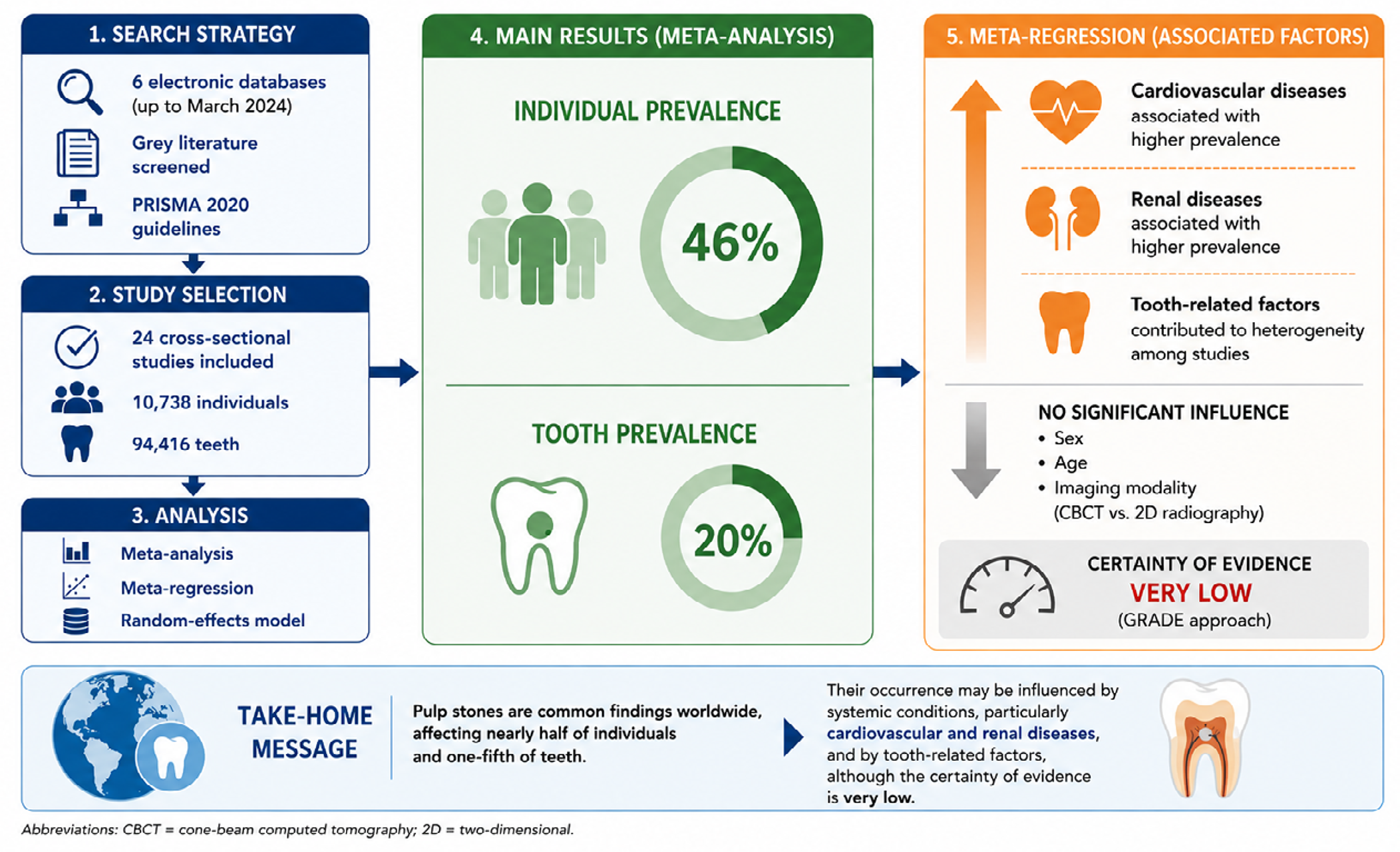
Results Twenty-four studies comprising 10,738 individuals and 94,416 teeth met the inclusion criteria. Pulp stones were present in 46% of individuals and 20% of teeth, with substantial heterogeneity across studies. Meta-regression identified systemic conditions and tooth type as significant contributors to prevalence variability. Individuals with both cardiovascular and renal diseases exhibited a higher prevalence (β=0.31, p=0.04), whereas those with cardiovascular diseases alone showed a lower prevalence (β=-0.33, p=0.04) than healthy participants (R2= 26.0). Risk of bias was moderate in most studies, and the overall certainty of evidence was very low.
Conclusion Pulp stones are highly prevalent, and their occurrence may be influenced by systemic conditions and tooth-related factors. However, these findings are based on very-low-certainty evidence and are limited by heterogeneity and methodological inconsistencies across the included studies.
Keywords:
Dental pulp calcification; Meta-analysis; Prevalence; Pulp stones; Systematic review

Introduction
Pulp stones (PS) are dystrophic calcifications located within the pulp chamber or root canals of teeth. Their etiology remains incompletely understood. Osteopontin1 and calcifying nanoparticles,2 formerly known as nanobacteria, have been suggested to play a role in their formation. Ageing and physiological changes, dental caries and periodontal disease favoring the penetration of bacterial toxins and chronic irritation of the dental pulp, trauma, occlusal forces, orthodontic movements, systemic diseases, genetic syndromes, and idiopathic factors3 have also been associated with PS occurrence.
Pulp stones can be classified according to their anatomical location and histopathological structure. Anatomically, they are categorized as “free” when entirely surrounded by pulp tissue, “embedded” when enclosed within dentin, and “adherent” when continuous with the surrounding dentin.3 Histopathologically, “true” pulp stones resemble dentin in structure and may exhibit a peripheral odontoblastic layer, whereas “false” pulp stones consist of concentric mineral deposits surrounding a central nidus.4
Despite their frequent occurrence, the global prevalence of pulp stones remains poorly understood, with studies reporting highly variable rates across different populations and demographic groups. For instance, prevalence rates have been reported to range from as low as 4.6%5 in some populations to as high as 98.3%6 in others, depending on many factors. This variability highlights the need for a more standardized and comprehensive approach to studying their epidemiology.
Pulp stones can complicate dental procedures, particularly endodontic treatments, by obstructing access to the pulp chamber and root canals. In addition, pulp stones have been associated with pain resembling trigeminal neuralgia, as described in a classic case report.7 Beyond their clinical implications, pulp stones may also serve as potential indicators of broader health issues, such as cardiovascular8-11 and renal diseases.8,12 Therefore, understanding their prevalence is critical for both dental and systemic health management.
Despite these findings, significant gaps remain in the understanding of the global burden of pulp stones. Most studies have been limited to specific populations and have employed inconsistent diagnostic criteria and methodologies. This lack of standardization hinders definitive conclusions regarding their global prevalence and associated risk factors. A previous systematic review13 investigating the global prevalence of pulp stones reported a prevalence of 36.53% at the individual level and 9.57% at the tooth level. However, several additional studies have been published since its publication, warranting an updated analysis. Thus, this study aims to synthesize the available data on the global prevalence of dental pulp stones. By examining their distribution across diverse populations and identifying associated risk factors, this review seeks to address the following focused question: “What is the global prevalence of pulp stones, and what factors are associated with their occurrence?”
Methodology
This systematic review followed the Preferred Reporting Items for Systematic Review and Meta-Analysis (PRISMA) recommendations14 and was registered on the PROSPERO database (CRD42024528752).
Search Strategy
The search was performed independently by two reviewers in the following electronic databases: MEDLINE/PubMed, Cochrane Library, Web of Science, EMBASE, Scopus, and OpenAIRE Graph (grey literature). The search was conducted up to March 2024, with no restrictions on publication year or language. The electronic search strategy was developed using the most cited descriptors from previous publications on this topic, combining Medical Subject Headings (MeSH) and text words (tw.). For each database, the following terms were combined: “Dental Pulp Calcification;” “Calcification, Dental Pulp;” “Pulp Calcification, Dental;” “Dental Pulp Stone;” “Pulp Stones, Dental;” “Stone, Dental Pulp;” “Pulp Stones;” “Dental Pulp Stones;” “Denticles;” “Denticle;” “Pulp Stone, Dental;” “Stones, Dental Pulp;” “Coronal Dentin Dysplasia;” “Dentin Dysplasia, Shields Type 2;” “Anomalous Dysplasia of Dentin;” “Dentin Dysplasia, Type II;” “Dentin Dysplasia, Shields Type II;” “Dentin Dysplasia, Coronal;” “Pulpal Dysplasia;” “Prevalence;” “Period Prevalence;” “Point Prevalence;” “Cross-Sectional Studies;” “Cross Sectional Analysis;” “Disease Frequency Surveys;” Cross-Sectional Survey;” “Surveys, Disease Frequency;” “Analysis, Cross-Sectional;” “Prevalence Studies.” The Boolean operators ‘AND’ and ‘OR’ were applied to combine the search terms. The search strategies for each database and the corresponding results are summarized in Table 1. Furthermore, the reference lists of the selected studies were screened, and related articles were searched in PubMed. All retrieved records were imported into Mendeley© (Mendeley Ltd, London, United Kingdom) to catalogue the references and facilitate duplicate removal.
Eligibility criteria
The eligibility criteria were based on the CoCoPop strategy15,16 as follows:
-
Condition (Co): prevalence of pulp stones;
-
Context (Co): worldwide;
-
Population (Pop): adults only.
Only studies reporting the prevalence of pulp stones in adult populations (≥18 years), regardless of the country in which the study was conducted, were included. Pediatric populations were excluded because pulp stone formation is primarily considered an age-related degenerative phenomenon. Thus, including this demographic could artificially skew the overall prevalence estimates.
Exclusion Criteria
Studies that included pediatric populations (<18 years) and/or evaluated the effects of orthodontic tooth movement on pulp stone prevalence were excluded. Additionally, histological studies, pilot studies, animal studies, systematic reviews (with or without meta-analysis), literature reviews, opinion articles, letters, conference abstracts, case reports, case series, and laboratory studies were excluded.
Study Selection
Two reviewers were responsible for study selection. Duplicate records were identified and removed. Titles and abstracts for potentially eligible studies were screened. When the title and/or abstract did not provide sufficient information to determine eligibility, the full text was retrieved and assessed.
Studies that met the inclusion criteria underwent full-text review. Both reviewers independently assessed the selected studies, and any disagreements were resolved by a third reviewer, a senior investigator with over 30 years of experience in dental research.
Data Extraction
The following information was extracted from each study: author(s), year of publication, country of origin, number of participants, age, sex, teeth evaluated, diagnostic methods, presence of systemic diseases, and main findings. Data extraction was performed independently by two reviewers. Any disagreements were resolved by a third and more experienced reviewer. All extracted data were recorded in an Excel spreadsheet.
Risk of Bias Assessment
The risk of bias of the included studies was assessed using the JBI Critical Appraisal Checklist for Studies Reporting Prevalence Data,17 which consists of nine questions:
Was the sample frame appropriate to address the target population?
Were study participants sampled in an appropriate way?
Was the sample size adequate?
Were the study subjects and the setting described in detail?
Was the data analysis conducted with sufficient coverage of the identified sample?
Were valid methods used for the identification of the condition?
Was the condition measured in a standard, reliable way for all participants?
Was there appropriate statistical analysis?
Was the response rate adequate, and if not, was the low response rate managed appropriately?
Each item was rated as “yes,” “no,” “unclear,” or “not applicable.” The overall risk of bias was determined as follows: each “yes” response received 1 point, while “no,” “unclear,” and “not applicable” responses received 0 points. The total score for each study was then used to classify the risk of bias as follows: 0–3 points indicated high risk, 4–6 points indicated moderate risk, and 7–9 points indicated low risk.
Statistical analyses
Meta-analyses were conducted to estimate the prevalence of pulp stones. Two models were used to estimate the prevalence at the individual and tooth levels. Prevalence rates were analyzed using the metaprop command in Stata (version 14 for Macintosh). The I2 statistic, which ranges from 0 to 100%, was used to quantify inconsistencies across studies. Random-effects models were applied due to high heterogeneity. Publication bias was assessed using Egger’s test. Sensitivity analyses were performed by stratifying studies according to their risk of bias based on the JBI assessment (high, moderate, and low risk). Subgroup analyses were also performed.
Meta-regression was performed to investigate potential sources of heterogeneity in prevalence estimates at the individual level. Model building began with a full model including all variables considered potentially associated with heterogeneity and pulp stone prevalence. Variables were retained in the final model if they had a p-value < 0.05 or acted as confounders, defined as producing a change greater than 35% in the coefficient of another variable. This was assessed by manually removing and re-entering each variable into the model. The following variables were evaluated during model building: sex, inclusion of older adults, country, systemic diseases, tooth type, and diagnostic method. Due to significant inconsistency and variability in the reporting of these data across the primary literature, the categorizations described as follows were considered the only feasible options.
To evaluate the potential influence of sex, studies were initially categorized according to the proportion of male and female participants. However, several studies failed to report this distribution. Therefore, sex was categorized based on whether a statistically significant difference in pulp stone prevalence between sexes was reported in each primary study. Coding was assigned as follows: 0 = no significant difference between sexes; 1 = higher prevalence in males; 2 = higher prevalence in females. A similar approach was applied for age, as multiple studies did not report the age ranges for their samples. Consequently, studies were dichotomized according to the inclusion of older adults. Specifically, this variable was coded as follows: 0 = no individuals aged ≥ 60 years included; 1 = individuals aged ≥60 years included.
The included studies were conducted across eight countries (Brazil, India, Iran, Jordan, Lithuania, Peru, Saudi Arabia, and Turkey). Saudi Arabia (n=8) and Turkey (n=6) were the most frequently represented countries, collectively accounting for just over 50% of the included studies. To accommodate this distribution while maintaining statistical power, countries were categorized into Saudi Arabia (2), Turkey (1), and others (0), based on the geographic distribution of the final search results.
Systemic diseases were categorized based on cardiovascular and renal status of participants in studies that explicitly evaluated this variable. Since only one study included patients with renal disease, it was grouped into a combined category of renal and cardiovascular diseases. Coding was as follows: 0 = no systemic conditions reported, including systemically healthy individuals; 1 = presence of cardiovascular diseases; 2 = presence of both cardiovascular and renal diseases.
Tooth type was categorized based on the range of teeth evaluated: 0 = not specified; 1 = all teeth; 2 = premolars and molars; 3 = molars only. Lastly, the diagnostic method was categorized, considering cone-beam computed tomography (CBCT) as reference imaging modality. Coding was as follows: 1 = diagnostic methods other than CBCT; 2 = use of CBCT. One study was excluded from meta-regression due to missing information regarding the imaging modality used. Therefore, 23 studies were included in the final model.
Strength of evidence
The strength of evidence of the included studies was assessed using the Grading of Recommendations Assessment, Development, and Evaluation (GRADE) approach. The evaluation was performed using the GRADEpro Guideline Development Tool (GRADEpro GDT; McMaster University, 2015, developed by Evidence Prime, Inc.), available at: https://gdt.gradepro.org.18 The GRADE system considers five domains that may lead to downgrading the quality of evidence: risk of bias, inconsistency, indirectness, imprecision, and other considerations (including publication bias, significant effect, plausible confounding, and dose–response gradient) (GRADE Working Group, 2004).
Results
Study Selection
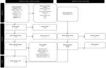
Figure 1 illustrates the flow diagram of the study selection process. Database searches identified 591 potentially relevant studies. After removal of 306 duplicates, 285 studies remained for title and abstract screening. Of these, 235 were excluded, and 50 full-text articles were assessed for eligibility. Twenty-six studies were excluded (reasons for exclusion are listed in Supplementary File). Ultimately, 24 studies5,6,19-40 met the eligibility criteria and were included in the review. No additional studies were identified via manual searches of reference lists.
Data Extraction
Table 2 presents a summary of the descriptive data from the included studies. The total number of participants was 10,738, and the combined tooth sample size comprised 94,416 teeth. Participants’ ages ranged from 18 to 93 years. The overall prevalence of pulp stones ranged from 4.6% to 98.3% among patients and from 3.3% to 52.1% among teeth.
Nine studies6,20,22,23,30,32,34,36,37 used CBCT as the diagnostic method. One study38 employed both CBCT and panoramic radiographs, while the remaining studies utilized panoramic, interproximal, and/or periapical radiographs. Only one study31 did not specify the diagnostic method used for pulp stone evaluation.
Regarding associated diseases, nine studies5,24,25,29,31,33,37,39,40 investigated possible associations between systemic diseases and pulp stones; nine studies6,20,22,23,32,34-36,38 investigated possible associations between oral alterations and pulp stones; three studies21,26,30 examined both systemic and oral alterations in relation to pulp stones; and three studies19,27,28 did not report evaluating any such associations.
Risk of Bias
Table 3 presents the results of the risk of bias analysis. Four studies28,31,33,39 were categorized as having a high risk of bias. Fifteen studies5,6,20-2529,32,34,35,37,38,40 were assessed as having a moderate risk of bias. Finally, five studies19,26,27,30,36 were classified as having a low risk of bias.
Meta-analysis
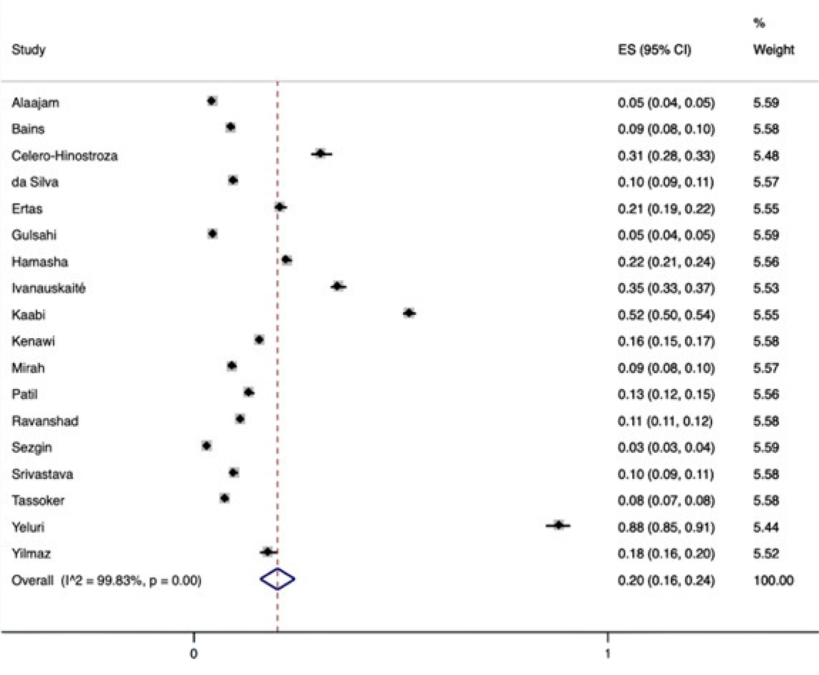
A total of 24 studies with 10,738 individuals were included in the meta-analysis of individual-level prevalence of pulp stones (Figure 2). The pooled prevalence of pulp stones was 46% at the individual level with high heterogeneity observed. At the tooth level (Figure 3), 18 studies comprising 94,416 teeth were included. The pooled prevalence at the tooth level was 20%, also with high heterogeneity. Egger’s test for publication bias at both the individual and tooth levels showed no clear evidence of publication bias (p>0.05).
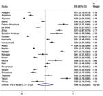
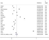
Sensitivity analysis was performed according to the risk of bias of the included studies (Figure 4A–B). The prevalence estimated from high-risk studies was 63%, whereas that from low-risk studies was 36%. Both estimates showed high heterogeneity, and no statistically significant difference was observed between groups (p=0.15), likely due to the small number of studies in each category (Figure 4A). When high-risk studies were excluded, the pooled prevalence of pulp stones was 43%, with persistent high heterogeneity (Figure 4B), indicating that risk of bias did not explain the observed heterogeneity.
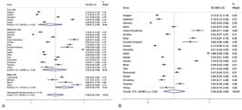
Sensitivity analysis according to risk of bias of the included studies. (A) Comparison of pooled prevalence estimates between high-risk and low-risk studies. (B) Pooled prevalence after exclusion of high-risk studies.
Subgroup meta-analyses
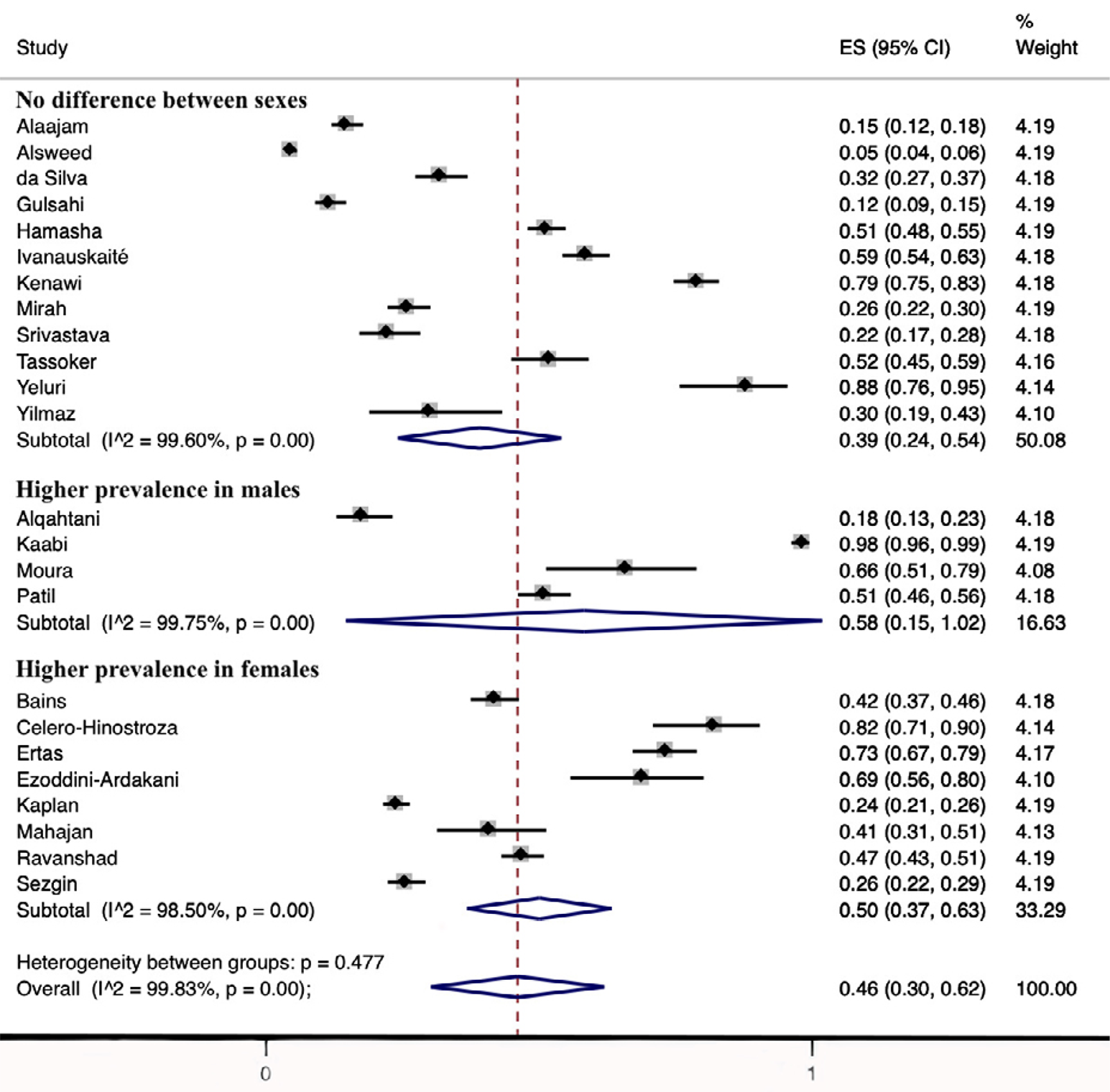
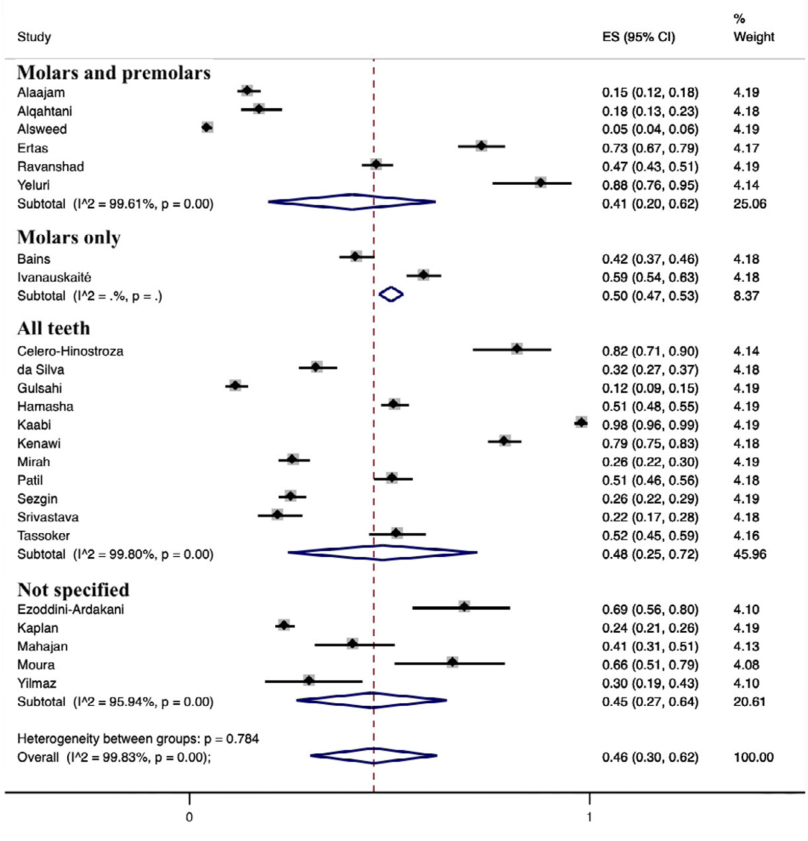
Figures 5–10 show forest plots according to categories of variables explored as potential sources of heterogeneity and possible factors associated with the occurrence of pulp stones. Small, non-significant differences in individual-level pulp stone prevalence were observed across categories of sex (p=0.477; Figure 5), inclusion of older adults in the study sample (p=0.177; Figure 6), tooth type (p=0.784; Figure 7), and diagnostic method (p=0.765; Figure 8).
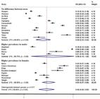
Forest plot of subgroup analysis by sex, showing pooled prevalence of pulp stones at the individual level.
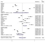
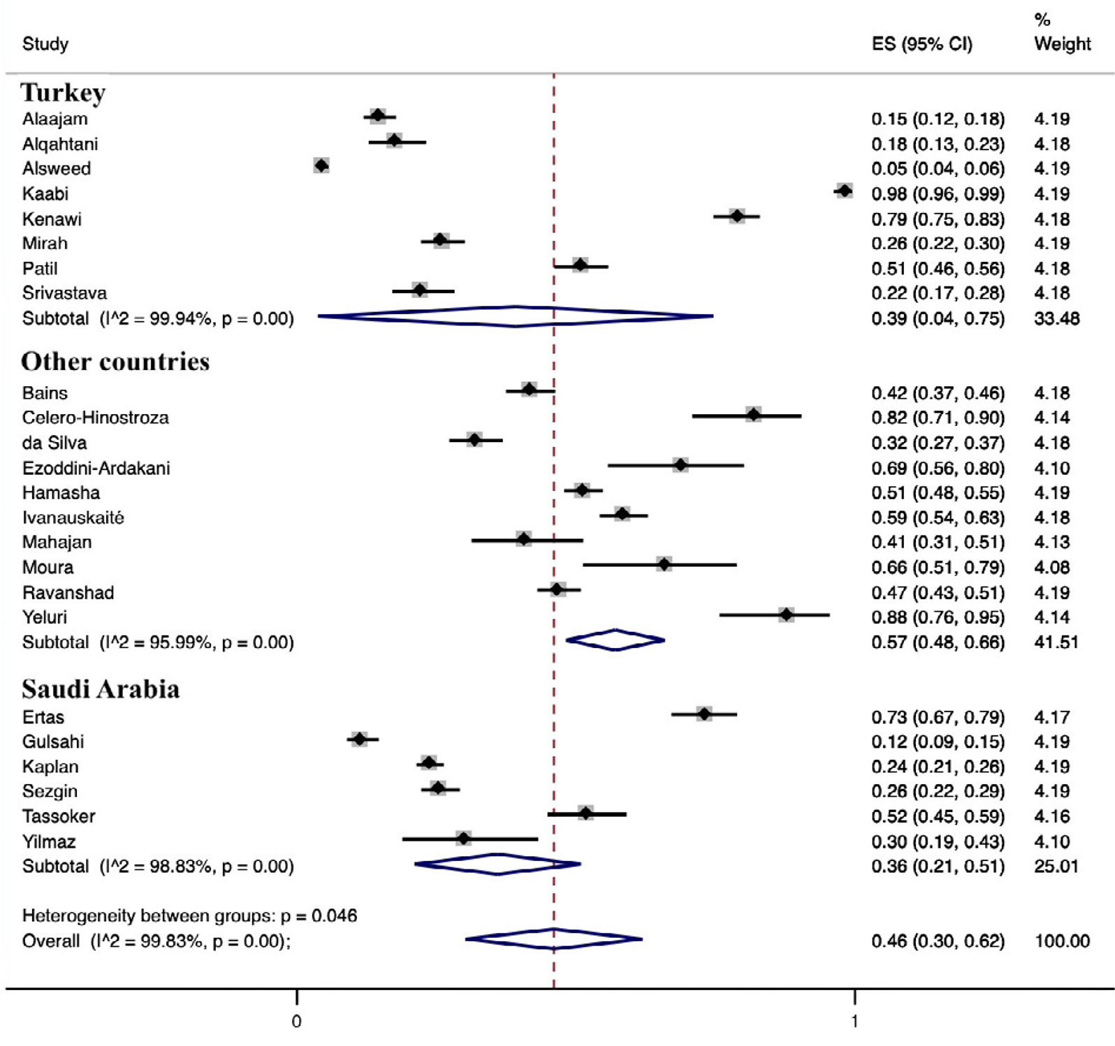
Forest plot of subgroup analysis by country, showing pooled prevalence of pulp stones across geographic categories.
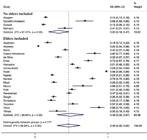
Forest plot of subgroup analysis by inclusion of older adults in the study sample, showing pooled prevalence of pulp stones at the individual level.
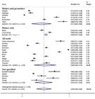
Forest plot of subgroup analysis by tooth type, showing pooled prevalence of pulp stones at the individual level.

Forest plot of subgroup analysis by diagnostic method (CBCT versus other imaging modalities), showing pooled prevalence of pulp stones at the individual level.
Larger and statistically significant differences were observed for systemic conditions and country. The prevalence of pulp stones was 46% in studies including systemically healthy individuals, compared with 31% in studies including individuals with cardiovascular diseases and 65% in those including individuals with both cardiovascular and renal diseases (p=0.007; Figure 9). Prevalence was also significantly higher in countries other than Turkey and Saudi Arabia (p=0.046; Figure 10).
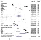
Forest plot of subgroup analysis by systemic conditions, showing pooled prevalence of pulp stones across categories of cardiovascular and renal disease status.
Meta-regression
Meta-regression models are presented in Table 4. Coefficients represent differences in prevalence relative to the reference category. The final model explained 26% of the observed variance in prevalence (R2 = 26.0) and retained systemic diseases and tooth type as significant variables. Studies including individuals with cardiovascular diseases showed a 34% lower prevalence than studies including systemically healthy individuals. In contrast, studies including individuals with both cardiovascular and renal diseases showed a 31% higher prevalence than those including systemic healthy individuals. Regarding tooth type, studies evaluating premolars and molars showed lower prevalence than studies that did not specify tooth types.
Strength of evidence
The results of the GRADE assessment are summarized in Table 5. The studies were rated as having very serious risk of bias, inconsistency, and imprecision; no serious concerns regarding indirectness; and were not upgraded in the “other considerations” domain. Consequently, the overall certainty of the evidence was very low.
Discussion
This systematic review synthesized data from 24 studies involving 10,738 participants and 94,416 teeth to evaluate the global prevalence of pulp stones and associated factors. We found an overall prevalence of 46% at the individual level and 20% at the tooth level. The included studies demonstrated substantial variability in prevalence estimates, ranging from 4.6% to 98.3% at the individual level and from 3.3% to 52.1% at the tooth level. Meta-regression was performed to investigate potential variables contributing to this high heterogeneity. Two variables showed statistically significant differences and partially explained the pooled results, suggesting a possible association with a higher pulp stone prevalence.
Meta-regression indicated that studies including individuals with cardiovascular diseases reported a lower prevalence than studies involving systemically healthy participants, whereas studies including individuals with both cardiovascular and renal diseases reported a higher prevalence. Previous systematic reviews have reported a positive association between cardiovascular diseases and pulp stones.8-10 Despite this strong association, our study found a lower prevalence among individuals with cardiovascular diseases than among systemically healthy individuals. This finding likely reflects our inability to retrieve separate participant-level data for individuals with cardiovascular diseases from studies that included participants with both cardiovascular and renal conditions. Clinicians should remain aware of this association, as the prevalence of pulp stones in this population may be higher than our estimates suggest. Future analyses should further clarify this relationship. A plausible explanation for the greater prevalence in individuals with both cardiovascular and renal diseases is that both conditions are associated with systemic calcification processes and metabolic disturbances that may predispose individuals to ectopic mineralization, including within the dental pulp, and may exert a synergistic effect, increasing the likelihood of pulp stones formation beyond that expected from either condition alone. Chronic kidney disease, in particular, has been strongly linked to vascular and soft tissue calcifications due to altered calcium–phosphate metabolism and secondary hyperparathyroidism, which may contribute to a higher tendency for pulp stone formation when combined with vascular changes observed in cardiovascular diseases.41 It is important to emphasize that this analysis estimates prevalence in these populations rather than establishing causal or associative relationships. Nevertheless, this finding suggests a potential relationship between pulp stones and systemic health status, underscoring the need for further investigation into their clinical relevance.
Variation was also observed in tooth-related factors. Meta-regression showed that studies evaluating premolars and molars showed lower prevalence than those including all types of teeth. It is important to note that studies assessing all teeth also included premolars and molars and generally had larger sample sizes than studies restricted to specific tooth groups. These larger samples may have influenced overall prevalence estimates and diluted the specific effect of tooth type. Premolars and molars are known to be more susceptible to functional stresses, such as bruxism and occlusal overload, as well as conditions associated with premature oral aging, all of which may increase pulp stone formation in these teeth.42 Therefore, their inclusion within broader samples may explain why studies evaluating premolars and molars separately did not show a higher prevalence. This finding underscores the need for future studies to stratify results according to tooth type.
Sex and age distributions were assessed across the included studies, but meta-regression did not identify them as significant predictors. Despite this lack of significance, some limitations must be highlighted. Biologically, sex-specific hormonal fluctuations across different life stages may contribute to dynamic alterations in the dental pulp microenvironment43 and could favor dystrophic calcification. However, the exact number of individuals per sex or age group could not be determined from the included studies. Furthermore, age was categorized broadly based on the presence or absence of older adults (≥60 years). Therefore, these findings should be interpreted only as indicating no significant difference in overall prevalence with the inclusion of older populations. Identifying specific age ranges associated with a greater prevalence of pulp stones remains challenging, largely due to substantial heterogeneity and lack of standardization in age-group stratification across the included studies.
Nine studies used CBCT6,20,22,23,30,32,34,36,37, one used both CBCT and panoramic radiographs38, while the remaining studies relied on panoramic, interproximal, or periapical radiographs. Although CBCT is generally considered a more accurate imaging modality for several diagnostic applications,44 our meta-regression did not demonstrate significant differences in prevalence estimates between CBCT and conventional radiographic techniques. This diagnostic equivalence likely stems from the inherent radiological characteristics of pulp stones, which present as distinct radiopaque calcifications that contrast sharply with the radiolucent background of the pulp space, rendering them readily detectable even on 2D images. However, it is important to emphasize that several technical aspects, such as overall CBCT image quality, may have influenced this finding. Specific parameters that determine CBCT accuracy,45 such as field of view, voxel size, and artifacts, were not evaluated in this systematic review.
The included studies represented populations from eight countries, with Saudi Arabia and Turkey accounting for more than half of the available evidence.5,6,1920,24,26,29,3032,34,36-38,40 Prevalence was higher in countries other than Turkey and Saudi Arabia. The country variable likely reflects methodological and sampling differences across studies rather than a true geographic effect. Turkey and Saudi Arabia accounted for more than half of the included studies, which may have produced more stable pooled estimates, whereas the “other countries” category grouped a smaller number of heterogeneous studies with diverse populations, diagnostic protocols, and sampling strategies. Differences in imaging modality, participant selection, age distribution, systemic health status, and tooth types evaluated may also have contributed to this variation. Therefore, these results do not support a causal relationship between geographic location and pulp stone prevalence but instead highlight the need for more standardized epidemiological studies across diverse populations.
The risk of bias assessment showed that most studies were classified as presenting a moderate risk of bias, while four studies had a high risk4,31,32,39 and five a low risk of bias.19,2627,30,36 Mainly due to these limitations and the high heterogeneity, the GRADE assessment demonstrated that the certainty of evidence was very low.
Despite providing a comprehensive synthesis of the global prevalence of pulp stones, this systematic review has limitations that must be acknowledged. First, the included literature exhibited substantial heterogeneity and methodological inconsistencies, which hinder the ability to draw definitive conclusions. Geographic representation was also uneven, with more than half of the analyzed evidence originating from populations in just two countries. In this review, only two systemic conditions (cardiovascular and renal diseases) could be evaluated in depth. However, emerging evidence suggests that other systemic conditions, such as diabetes, may also be associated with pulp stones. Further primary studies are necessary to rigorously investigate these potential associations. Moreover, variability in the inclusion criteria and sample sources across the primary literature was remarkably high. Consequently, we were unable to categorize these variables or quantitatively estimate their impact on prevalence within the meta-regression. While the total number of studies and individuals included in the overall meta-analysis was high, the study-level sample size for meta-regression may have been insufficient to reliably detect statistically significant associations, as reflected by several borderline p-values. Furthermore, Egger’s test applied for publication bias must be interpreted cautiously, given its known limitations. Finally, because the quantitative analysis was strictly limited to cross-sectional prevalence data, it is not possible to establish true causal relationships between pulp stone occurrence and systemic health conditions. This highlights the need for more rigorously designed studies to strengthen the evidence base regarding the prevalence and implications of pulp stones. Therefore, future investigations should prioritize standardized diagnostic criteria, stratification by tooth type and demographic factors, and inclusion of diverse populations to clarify the clinical significance of pulp stones and their potential role as indicators of systemic health.
In conclusion, this systematic review provides the most comprehensive synthesis to date of the global prevalence of pulp stones, showing that nearly half of individuals and approximately one-fifth of teeth included in the analysis present at least one pulp stone. Despite these high rates, substantial heterogeneity was observed across studies, partially influenced by systemic conditions and tooth-related factors. While the findings suggest potential links between pulp stone occurrence and systemic health, particularly in populations with cardiovascular and renal diseases, our analysis was limited to prevalence data and cannot establish causal associations. Nevertheless, clinicians should be aware of the potential implications of pulp stones as potential indicators of systemic conditions and should consider this in treatment planning and in the assessment of patients from high-risk populations.
Conclusion
Overall prevalence of pulp stones was 46% at the individual-level and 20% at the tooth level. Despite the very low certainty of evidence, prevalence varied substantially across populations, tooth types, and diagnostic methods, and was influenced by systemic conditions such as cardiovascular and renal diseases.
Supplementary File
Supplementary File – Excluded studies and reason for exclusion
References
-
1 - Ninomiya M, Ohishi M, Kido J, Ohsaki Y, Nagata T. Immunohistochemical localization of osteopontin in human pulp stones. J Endod. 2001;27(4):269-72. doi: 10.1097/00004770-200104000-00007
» https://doi.org/10.1097/00004770-200104000-00007 -
2 - Zeng J, Yang F, Zhang W, Gong Q, Du Y, Ling J. Association between dental pulp stones and calcifying nanoparticles. Int J Nanomedicine. 2011;6:109-18. doi: 10.2147/IJN.S13267
» https://doi.org/10.2147/IJN.S13267 -
3 - Goga R, Chandler NP, Oginni AO. Pulp stones: a review. Int Endod J. 2008;41(6):457-68. doi: 10.1111/j.1365-2591.2008.01374.x
» https://doi.org/10.1111/j.1365-2591.2008.01374.x -
4 - Siddiqui SH, Mohamed AN. Calcific metamorphosis: a review. Int J Health Sci (Qassim). 2016;10(3):437-42. doi: 10.12816/0048738
» https://doi.org/10.12816/0048738 -
5 - Alsweed A, Farah R, Satheeshkumar PS, Farah R. The prevalence and correlation of carotid artery calcifications and dental pulp stones in a Saudi Arabian population. Diseases. 2019;7(3):50. doi: 10.3390/diseases7030050
» https://doi.org/10.3390/diseases7030050 -
6 - Kaabi HH, Riyahi AM, Al-Maflehi NS, Alrumayyan SF, Bakrman AK, Almutaw YA. Three-dimensional analysis for detection of pulp stones in a Saudi population using cone-beam computed tomography. J Oral Sci. 2023;65(4):257-60. doi: 10.2334/josnusd.23-0091
» https://doi.org/10.2334/josnusd.23-0091 - 7 - Bergmann S. Simulated major trifacial neuralgia caused by pulp stones: report of a case. J Am Dent Assoc. 1943;30(9):701-3.
-
8 - Almadhoon HW, Al-Kafarna M, Asla MM, Gbreel MI, Abd Allah MA, Almotairy N. The association of dental pulp stones to cardiovascular and renal diseases: a systematic review and meta-analysis. J Endod. 2022;48(7):845-54. doi: 10.1016/j.joen.2022.02.010
» https://doi.org/10.1016/j.joen.2022.02.010 -
9 - Parashar SR, Kasabwala K, Ulaganathan S, Suresh M, Sirohi R, Patil S. Association of pulp calcifications and cardiovascular disease: a systematic review and meta-analysis. J Evid Based Dent Pract. 2022;22(2):101707. doi: 10.1016/j.jebdp.2022.101707
» https://doi.org/10.1016/j.jebdp.2022.101707 -
10 - Vieira WA, Paranhos LR, Meneses-Santos D, Pereira JF, Moura AP, Silva JC, et al. Association between the prevalence of pulp stones and calcified atherosclerotic plaques: a systematic review and meta-analysis. Dentomaxillofac Radiol. 2023;52(4):20230004. doi: 10.1259/dmfr.20230004
» https://doi.org/10.1259/dmfr.20230004 -
11 - Hasan S, Sharma T, Saeed S, Kaur M, Gombra V, Masood R. Prevalence of pulp stones on panoramic radiographs in patients with chronic systemic diseases: a cross-sectional study. Rambam Maimonides Med J. 2025;16(4):e0022. doi: 10.5041/RMMJ.10557
» https://doi.org/10.5041/RMMJ.10557 -
12 - Gabardo MC, Wambier LM, Rocha JS, Kuchler EC, de Lara RM, Leonardi DP, et al. Association between pulp stones and kidney stones: a systematic review and meta-analysis. J Endod. 2019;45(9):1099-105.e2. doi: 10.1016/j.joen.2019.06.006
» https://doi.org/10.1016/j.joen.2019.06.006 -
13 - Jannati R, Afshari M, Moosazadeh M, Allahgholipour SZ, Eidy M, Hajihoseini M. Prevalence of pulp stones: a systematic review and meta-analysis. J Evid Based Med. 2019;12(2):133-9. doi: 10.1111/jebm.12331
» https://doi.org/10.1111/jebm.12331 -
14 - Page MJ, McKenzie JE, Bossuyt PM, Boutron I, Hoffmann TC, Mulrow CD, et al. Mapping of reporting guidance for systematic reviews and meta-analyses generated a comprehensive item bank for future reporting guidelines. J Clin Epidemiol. 2020;118:60-8. doi: 10.1016/j.jclinepi.2019.11.010
» https://doi.org/10.1016/j.jclinepi.2019.11.010 -
15 - Munn Z, Stern C, Aromataris E, Lockwood C, Jordan Z. What kind of systematic review should I conduct? A proposed typology and guidance for systematic reviewers in the medical and health sciences. BMC Med Res Methodol. 2018;18(1):5. doi: 10.1186/s12874-017-0468-4
» https://doi.org/10.1186/s12874-017-0468-4 - 16 - Munn Z, Moola S, Lisy K, Riitano D, Tufanaru C. Chapter 5: systematic reviews of prevalence and incidence. In: Aromataris E, Munn Z, editors. JBI manual for evidence synthesis. Adelaide: JBI; 2020.
-
17 - Munn Z, Moola S, Lisy K, Riitano D, Tufanaru C. Methodological guidance for systematic reviews of observational studies reporting prevalence data. Int J Evid Based Healthc. 2015;13(3):147-53. doi: 10.1097/XEB.0000000000000054
» https://doi.org/10.1097/XEB.0000000000000054 -
18 - Guyatt G, Oxman AD, Akl EA, Kunz R, Vist G, Brozek J, et al. GRADE guidelines: 1. Introduction-GRADE evidence profiles and summary of findings tables. J Clin Epidemiol. 2011;64(4):383-94. doi: 10.1016/j.jclinepi.2010.04.026
» https://doi.org/10.1016/j.jclinepi.2010.04.026 -
19 - Alaajam WH, Saleh AA, Alghamdi NS, Ain TS, Algarni YA, Abubaker M. Incidence and distribution of pulp stones among Southern Saudi Arabian sub-population. SAGE Open Med. 2021;9:20503121211062796. doi: 10.1177/20503121211062796
» https://doi.org/10.1177/20503121211062796 -
20 - Alqahtani AS. Prevalence and association of calcified pulp stones with periodontitis: a cone-beam computed tomography study in Saudi Arabian population. J Pharm Bioallied Sci. 2024;16(Suppl 1):S644-50. doi: 10.4103/jpbs.jpbs_913_23
» https://doi.org/10.4103/jpbs.jpbs_913_23 -
21 - Bains SK, Bhatia A, Singh HP, Biswal SS, Kanth S, Nalla S. Prevalence of coronal pulp stones and its relation with systemic disorders in northern Indian central Punjabi population. ISRN Dent. 2014;2014:617590. doi: 10.1155/2014/617590
» https://doi.org/10.1155/2014/617590 -
22 - Calero-Hinostroza GG, Tinedo-López PL, García-Rupaya CR. Prevalencia y distribución de cálculos pulpares en un grupo de adultos peruanos: un estudio mediante tomografías de haz cónico. Odovtos Int J Dent Sci. 2021;23(2):161-70. Spanish. doi: 10.15517/ijds.2021.44468
» https://doi.org/10.15517/ijds.2021.44468 -
23 - Silva EJ, Prado MC, Queiroz PM, Nejaim Y, Brasil DM, Groppo FC, et al. Assessing pulp stones by cone-beam computed tomography. Clin Oral Investig. 2017;21(7):2327-33. doi: 10.1007/s00784-016-2027-5
» https://doi.org/10.1007/s00784-016-2027-5 -
24 - Tarim Ertas E, Inci M, Demirtas A, Ertas H, Yengil E, Sisman Y, et al. A radiographic correlation between renal and pulp stones. West Indian Med J. 2014;63(6):620-5. doi: 10.7727/wimj.2013.077
» https://doi.org/10.7727/wimj.2013.077 - 25 - Ezoddini-Ardakani F, Namayandeh SM, Sadr-Bafghi SM, Fatehi F, Mohammadi Z, Shahrabi-Farahani S, et al. Association of pulp stones with coronary artery stenosis. Community Dent Health. 2011;28(4):305-7.
-
26 - Gulsahi A, Cebeci AI, Ozden S. A radiographic assessment of the prevalence of pulp stones in a group of Turkish dental patients. Int Endod J. 2009;42(8):735-9. doi: 10.1111/j.1365-2591.2009.01580.x
» https://doi.org/10.1111/j.1365-2591.2009.01580.x -
27 - al-Hadi Hamasha A, Darwazeh A. Prevalence of pulp stones in Jordanian adults. Oral Surg Oral Med Oral Pathol Oral Radiol Endod. 1998;86(6):730-2. doi: 10.1016/S1079-2104(98)90212-8
» https://doi.org/10.1016/S1079-2104(98)90212-8 - 28 - Ivanauskaite D, Kubiliute D, Janaviciene D, Brukiene V. Prevalence of pulp stones in molars based on bitewing and periapical radiographs. Stomatologija. 2021;23(1):9-15.
-
29 - Sönmez Kaplan S, Kaplan T, Sezgin GP. Assessment of the frequency and correlation of carotid artery calcifications and pulp stones with idiopathic osteosclerosis using digital panoramic radiographs. Clin Exp Health Sci. 2021;11(2):342-7. doi: 10.33808/clinexphealthsci.841006
» https://doi.org/10.33808/clinexphealthsci.841006 -
30 - Kenawi LM, Jaha HS, Alzahrani MM, Alharbi JI, Alharbi SF, Almuqati TA, et al. Cone-beam computed tomography-based investigation of the prevalence and distribution of pulp stones and their relation to local and systemic factors in the Makkah population: a cross-sectional study. Cureus. 2024;16(1):e51633. doi: 10.7759/cureus.51633
» https://doi.org/10.7759/cureus.51633 -
31 - Mahajan B, Ettishree, Anjum R, Chhibber N. Presence of pulp stones and its relation with systemic disorders. J Cardiovasc Dis Res. 2023;14(5):2077-80. https://doi.org/10.48047/
» https://doi.org/10.48047/ -
32 - Mirah MA, Bafail A, Shaheen S, Baik A, Abu Zaid B, Alharbi A, et al. Assessment of pulp stones among western Saudi populations: a cross-sectional study. Cureus. 2023;15(9):e46056. doi: 10.7759/cureus.46056
» https://doi.org/10.7759/cureus.46056 -
33 - Maranhão de Moura AA, de Paiva JG. Pulpal calcifications in patients with coronary atherosclerosis. Endod Dent Traumatol. 1987;3(6):307-9. doi: 10.1111/j.1600-9657.1987.tb00640.x
» https://doi.org/10.1111/j.1600-9657.1987.tb00640.x -
34 - Patil SR, Ghani HA, Almuhaiza M, Al-Zoubi IA, Anil KN, Misra N, et al. Prevalence of pulp stones in a Saudi Arabian subpopulation: a cone-beam computed tomography study. Saudi Endod J. 2018;8(2):93-8. doi: 10.4103/sej.sej_32_17
» https://doi.org/10.4103/sej.sej_32_17 - 35 - Ravanshad S, Khayat S, Freidonpour N. The prevalence of pulp stones in adult patients of Shiraz Dental School: a radiographic assessment. J Dent (Shiraz). 2015;16(4):356-61.
-
36 - Sezgin GP, Sönmez Kaplan S, Kaplan T. Evaluation of the relation between the pulp stones and direct restorations using cone beam computed tomography in a Turkish subpopulation. Restor Dent Endod. 2021;46(3):e34. doi: 10.5395/rde.2021.46.e34
» https://doi.org/10.5395/rde.2021.46.e34 -
37 - Srivastava KC, Shrivastava D, Nagarajappa AK, Khan ZA, Alzoubi IA, Mousa MA, et al. Assessing the prevalence and association of pulp stones with cardiovascular diseases and diabetes mellitus in the Saudi Arabian population: a CBCT based study. Int J Environ Res Public Health. 2020;17(24):9293. doi: 10.3390/ijerph17249293
» https://doi.org/10.3390/ijerph17249293 -
38 - Tassoker M, Magat G, Sener S. A comparative study of cone-beam computed tomography and digital panoramic radiography for detecting pulp stones. Imaging Sci Dent. 2018;48(3):201-12. doi: 10.5624/isd.2018.48.3.201
» https://doi.org/10.5624/isd.2018.48.3.201 -
39 - Yeluri G, Kumar CA, Raghav N. Correlation of dental pulp stones, carotid artery and renal calcifications using digital panoramic radiography and ultrasonography. Contemp Clin Dent. 2015;6(Suppl 1):S147-51. doi: 10.4103/0976-237X.166837
» https://doi.org/10.4103/0976-237X.166837 -
40 - Gunen Yilmaz S, Yilmaz F, Bayrakdar IS, Harorli A. The relationship between carotid artery calcification and pulp stone among hemodialysis patients: a retrospective study. Saudi J Kidney Dis Transpl. 2019;30(4):755-63. doi: 10.4103/1319-2442.265449
» https://doi.org/10.4103/1319-2442.265449 -
41 - Moe SM, Chen NX. Pathophysiology of vascular calcification in chronic kidney disease. Circ Res. 2004;95(6):560-7. doi: 10.1161/01.RES.0000141775.67189.98
» https://doi.org/10.1161/01.RES.0000141775.67189.98 -
42 - Sener S, Cobankara FK, Akgünlü F. Calcifications of the pulp chamber: prevalence and implicated factors. Clin Oral Investig. 2009;13(2):209-15. doi: 10.1007/s00784-008-0219-x
» https://doi.org/10.1007/s00784-008-0219-x -
43 - Sangalli L, Souza LC, Letra A, Shaddox L, Ioannidou E. Sex as a biological variable in oral diseases: evidence and future prospects. J Dent Res. 2023;102(13):1395-416. doi: 10.1177/00220345231197143
» https://doi.org/10.1177/00220345231197143 -
44 - Dutra KL, Haas L, Porporatti AL, Flores-Mir C, Nascimento Santos J, Mezzomo LA, et al. Diagnostic accuracy of cone-beam computed tomography and conventional radiography on apical periodontitis: a systematic review and meta-analysis. J Endod. 2016;42(3):35-64. doi: 10.1016/j.joen.2015.12.015
» https://doi.org/10.1016/j.joen.2015.12.015 -
45 - Patel S, Brown J, Pimentel T, Kelly RD, Abella F, Durack C. Cone beam computed tomography in endodontics: a review of the literature. Int Endod J. 2019;52(8):1138-52. doi: 10.1111/iej.13115
» https://doi.org/10.1111/iej.13115