 Left Pulmonary Artery Aneurysm in an Adult Patient With Uncorrected Tetralogy of Fallot and Patent Ductus Arteriosus: A Case Report
Abstract
The authors describe a case of an infrequent triple association: left pulmonary artery aneurysm (PAA), uncorrected tetralogy of Fallot (TOF) and patent ductus arteriosus (PDA), diagnosed in a 45-year-old adult patient during a check-up. The patient refused to undergo surgery and is currently being followed clinically.
Keywords
Pulmonary Artery; Aneurysm; Tetralogy of Fallot
Introduction
Pulmonary artery aneurysm (PAA) is an infrequent abnormality of the pulmonary vasculature with an unclear natural history and pathogenesis.1 It represents around 1% of all aneurysms in the thoracic cavity, with an incidence of 1:14,000 cases, most of which diagnosed post-mortem.2 Although PAA was first reported over 100 years ago through autopsies, ante-mortem diagnosis is still a challenge.3
The etiology of PAA is varied and is mostly associated with congenital heart disease (CHD), mainly tetralogy of Fallot (TOF), where only 3% of patients reach the fourth decade of life.4,5 Its symptomatology, with severe chronic hypoxemia, is related to pulmonary valve stenosis, causing low blood flow to the pulmonary artery and a right-to-left shunt.5
Other causes of PAA include pulmonary hypertension, vasculitis, syphilis, tuberculosis, left-to-right shunts, pulmonary valve stenosis with post-stenotic dilatation, vasa vasorum dysfunction, trauma, Eisenmenger's syndrome and focal endovascular mycosis infection, which is highly related to patent ductus arteriosus (PDA).1-3
The diagnosis of PAA has been improved by imaging methods (echocardiography, computed tomography angiography and magnetic resonance imaging), following clinical suspicion.2,6 There is currently no consensus on the best approach for this infrequent condition; however, when the patient is asymptomatic, conservative treatment is considered, otherwise surgical treatment is recommended as early as possible.1
In this study, the authors describe a case of left PAA associated with non-surgically corrected TOF and PDA in a 45-year-old patient.
Clinical case
A 45-year-old patient with a history of unspecified CHD reported at birth. He had symptoms characterized by intolerance to moderate exertion, dyspnea and asthenia during his childhood, and history of multiple hospitalizations for diseases commonly found in the Angolan epidemiological scenario (malaria, typhoid fever, among others). He was diagnosed with TOF and PDA at the age of 30 during a check-up visit requested by his company, and had no follow-up for the management of these conditions.
At 39 years of age, he was diagnosed with hypertension treated with losartan + hydrochlorothiazide (100/25mg). The patient denies history of cyanosis, hypoxemia crisis, syncope or chest pain.
At 45 years of age, the patient presented clinical worsening characterized by dyspnea on mild exertion. An isolated measurement of oxygen saturation revealed an estimated value between 77-85%. The patient denied other symptoms, and thus was referred to cardiology consultation.
At examination, the patient was acyanotic, with no signs of respiratory distress, oxygen saturation 94-95% at rest and blood pressure 128/79mmHg.
Cardiac auscultation revealed a systolic ejection murmur grade III/VI at the left sternal border. Pulmonary auscultation was normal.
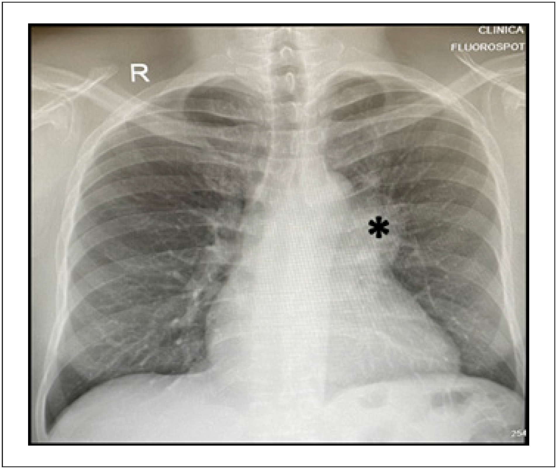
Chest X-ray showed no evidence of pathological images in both lung fields, enlargement of the left hilum and left parietal thickening. Enlarged cardiac silhouette at the expense of the left ventricle (Figure 1).

Chest X-ray showing enlargement of the left hilum (asterisk), parietal thickening to the left, enlarged cardiac silhouette to the left at the expense of the left ventricle
Electrocardiogram revealed sinus rhythm, QRS axis in the frontal plane deviated to the right between 150-120 degrees and signs of right atrial and right ventricular overload (Figure 2).

Twelve-lead electrocardiogram, sinus rhythm, HR: 68; axis deviated to the right (150º-120º), signs of overload of the right chambers and left atrium
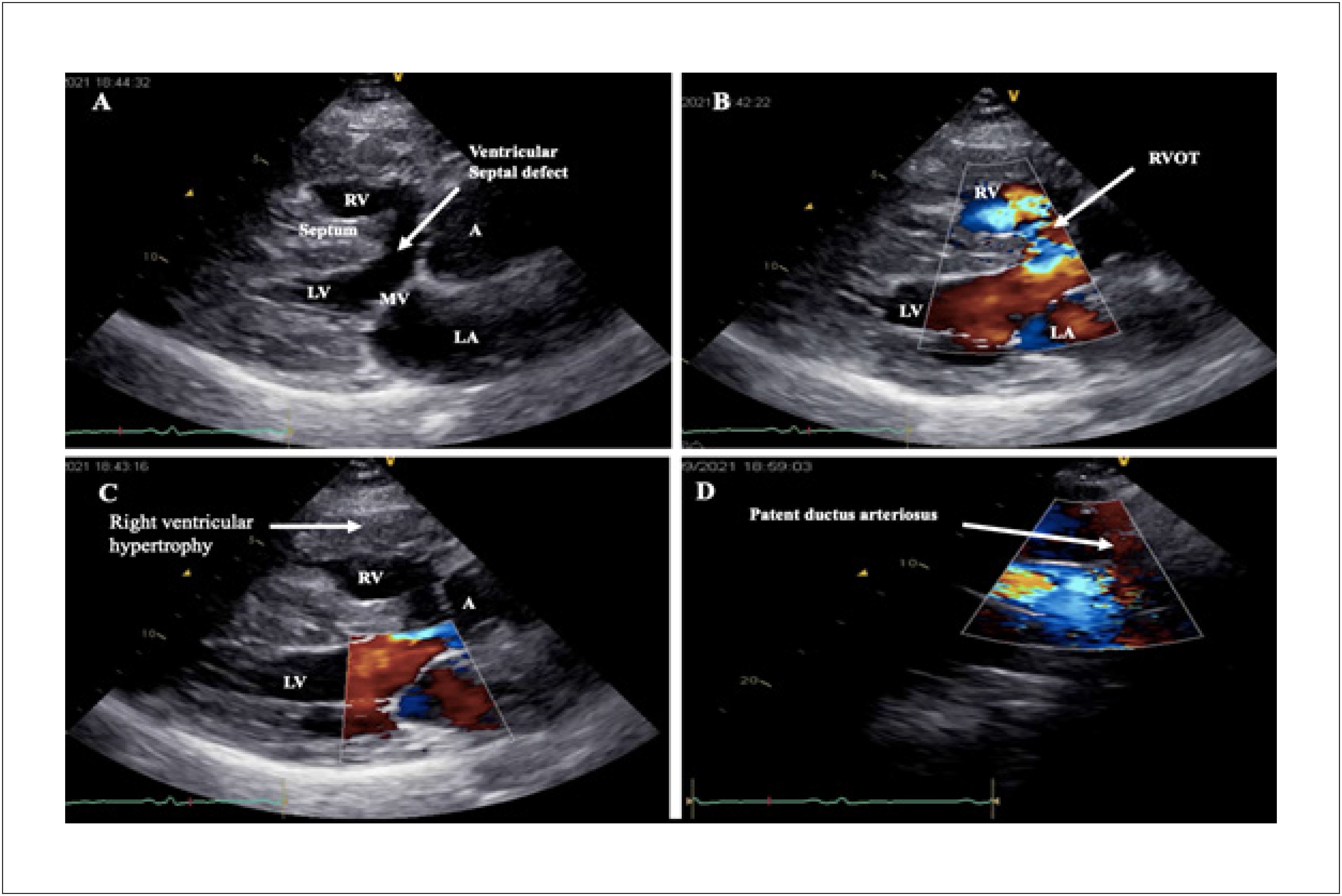
The echocardiogram showed a perimembranous ventricular septal defect (VSD) with a bidirectional shunt of left-right predominance, anterior deviation of the interventricular septum (IVS) of approximately 25%, IVS defect with aortic overlap, pulmonary valve stenosis with peak transvalvular gradient between 80-90 mmHg, right atrial indexed volume (4C): 44mL/m2 and left atrial 25mL/m2. Right ventricular hypertrophy (RVH) and PDA were evident on echocardiogram (Figure 3A-D).
Echocardiogram, A) Paraesternal long axis, interventricular septal defect with aortic overlap (white arrow); B) Bidirectional shunt of left-right predominance, right ventricular outflow tract (white arrow); C) RVH (white arrow); D) PDA (white arrow). RV: right ventricle; AA: aortic artery; LV: left ventricle; LA: left atrium; MV: mitral valve; RVOT: right ventricular outflow tract
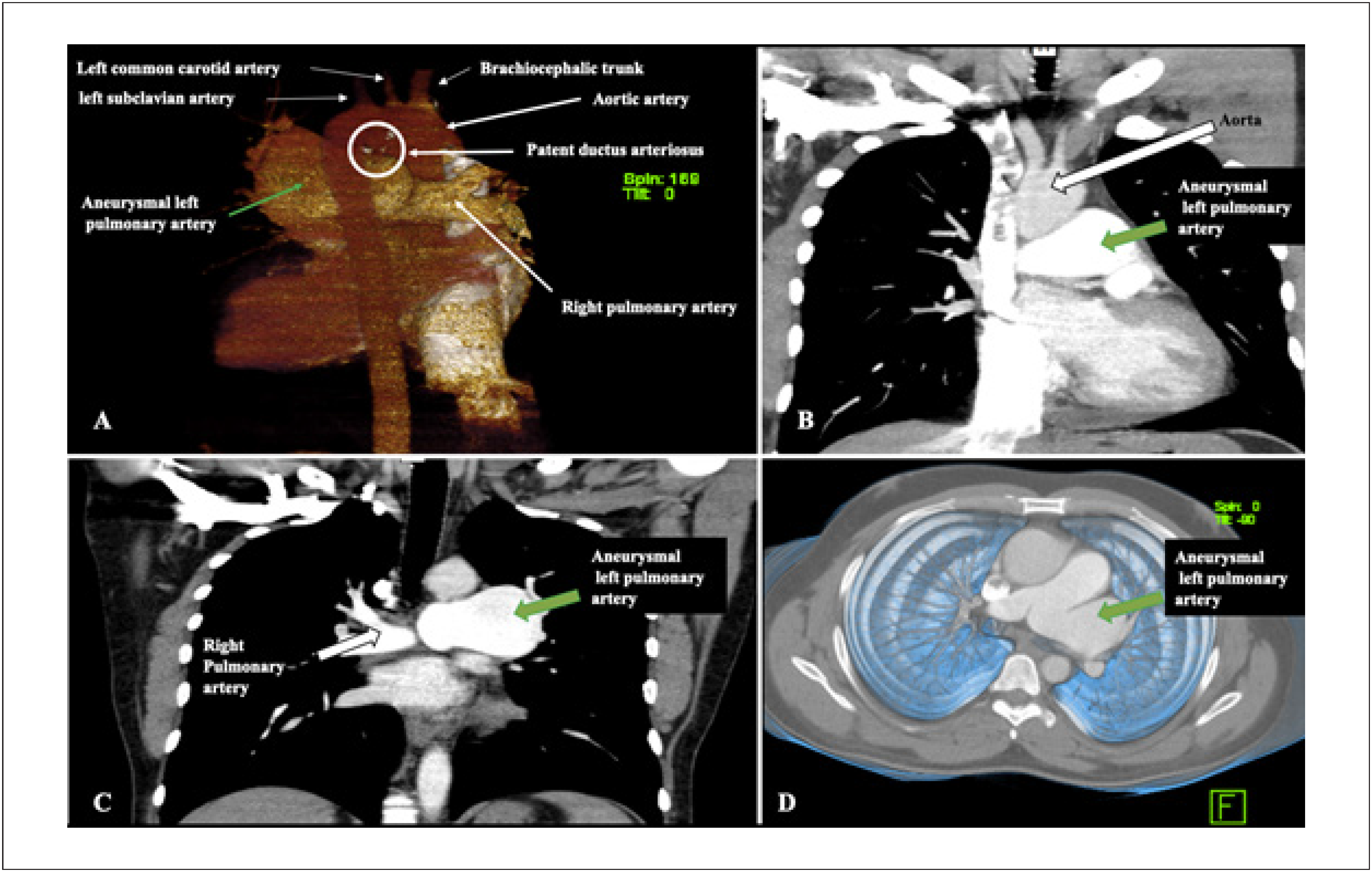
For better characterization of the pulmonary vasculature and exclusion of collateral circulation, chest computed tomography angiography was performed, which showed PDA (Figure 4A) with left-to-right flow, dilated pulmonary artery trunk measuring 37 mm, dilated right pulmonary artery measuring 22 mm and fusiform aneurysm of the left pulmonary artery with a diameter of 55×43 mm. No systemic-pulmonary collaterals were visualized (Figure 4B-C).

Chest computed tomography angiography, A) PDA (White circle); B-D) Aneurysmal left pulmonary artery (green arrow)
Surgery for total correction of the congenital defect was recommended, which the patient refused for religious/cultural reasons. For this same reason, right catheterization to assess pulmonary resistance was not performed either.
To date, the patient has not reported any relevant complaint, with an irregular follow-up in the Cardiology clinic.
Discussion
PAAs are usually treated early surgically due to the high risk of ruptured dissection and death, especially when symptomatic.3 The association between PAAs and TOF is infrequent; few cases have been reported in the medical literature.6-10
Anatomo-physiological conditions such as left-right shunt (increases flow to the pulmonary artery), systemic vascular resistance and PDA contributed to the longevity of the patient in question into adulthood without the severe symptoms characteristic of TOF, even without surgery.2,5,10,11 This clinical context predisposes to the occurrence of Eisenmenger's syndrome, which was not seen in the patient described.
Bearing in mind the etiological variety of this nosological entity, we can infer that the formation of PAA in our patient could be related to the compensatory/adaptive process of the vessel that has been chronically subjected to high pressures from the right chambers.1 Another hypothesis is infection by Treponema Pallidum and Koch's bacillus in a scenario dominated by infectious diseases, which could be the cause of vascular inflammatory changes and aneurysms.1,2,6
Furthermore, we cannot forget the possibility of a mycotic aneurysm secondary to bacterial endarteritis; however, the infection was not confirmed in our patient. Therefore, more studies are needed to better clarify the pathogenesis of this triple relationship between PAA, uncorrected TOF and PDA in adults.6,10 Probably, surgical treatment and histological study would help to clarify the underlying causes involved.12
Computed tomography angiography, cardiac catheterization, magnetic resonance imaging and echocardiography help with the diagnosis, but are limited to identifying the causative agent of these abnormalities. In this case, the diagnosis was made using echocardiography and CT angiography of the chest.
The prevalence of CHD and infectious diseases in the African continent is relatively high, and is associated with low adherence and accessibility to the few specialized centers available. This limits early detection and probable underdiagnosis of this condition.4,6
The present case was a case of an adult patient with CHD that was not surgically corrected. Although there is a need for correction due to the inherent short-term risks (intensifying hypoxic crises), there are also long-term risks including decompensation of the CHD, development of PAA, pulmonary hypertension and Eisenmenger's syndrome.7-10
Conclusion
The triple association of PAA, TOF and PDA is infrequent. Several factors may explain this rare nosological entity, mainly because of the high association between CHD and infectious diseases in the African continent. The real causes of this condition in our case have not been clarified. The patient refused to undergo surgery and has therefore been followed clinically and remained stable to date.
-
Sources of Funding
There were no external funding sources for this study.
-
Study Association
This study is not associated with any thesis or dissertation work.
-
Ethics Approval and Consent to Participate
This study was approved by the Ethics Committee of the Office of Postgraduate Education and Research Clínica Girassol under the protocol number GEPPCG-06/22LDA. All the procedures in this study were in accordance with the 1975 Helsinki Declaration, updated in 2013. Informed consent was obtained from all participants included in the study.
-
Use of Artificial Intelligence
The authors did not use any artificial intelligence tools in the development of this work.
Availability of Research Data and Other Materials
The underlying content of the research text is contained within the manuscript.
References
-
1 Theodoropoulos P, Ziganshin BA, Tranquilli M, Elefteriades JA. Pulmonary Artery Aneurysms: Four Case Reports and Literature Review. Int J Angiol. 2013;22(3):143-8. doi: 10.1055/s-0033-1347907.
» https://doi.org/10.1055/s-0033-1347907 -
2 Gupta M, Agrawal A, Iakovou A, Cohen S, Shah R, Talwar A. Pulmonary Artery Aneurysm: A Review. Pulm Circ. 2020;10(1):2045894020908780. doi: 10.1177/2045894020908780.
» https://doi.org/10.1177/2045894020908780 -
3 Santos-Martínez LE, Meza-López LR, Flores-García CA, Rodríguez-Almendros NA, Hernández-Meneses S, Lozano-Torres VM, et al. Aneurysm in the Main Pulmonary Artery in Patient with Pulmonary Hypertension: Successful Surgical Treatment. Arch Cardiol Mex. 2016;86(2):185-8. doi: 10.1016/j.acmx.2015.09.004.
» https://doi.org/10.1016/j.acmx.2015.09.004 - 4 Nwaneli CU, Onwubuya EI, Obichukwu ME, Ezike OL, Aduka PN. Uncorrected Tetralogy of Fallot in a 47-Year-Old Nigerian Man: A Case Report. Orient J Med. 2017;29(3):140-3.
- 5 Louro F, Gomes A, Almeida J, Abreu J, Agapito A, Magalhães MP, et al. Fallot's Tetralogy in an Adult. Rev Soc Port Med Interna. 1999;6(4):250-3.
-
6 Afrikh K, Hattach L, Fellat N, El Bakkali M, Benjelloun H. Aneurysm of the Pulmonary Artery in Fallot's Tetralogy. Case Rep Cardiol. 2017;2017:1384905. doi: 10.1155/2017/1384905.
» https://doi.org/10.1155/2017/1384905 -
7 Goebel N, Hess OM. Aneurysm of the Pulmonary Artery in Fallot's Tetralogy. Rofo. 1982;136(6):725-7. German. doi: 10.1055/s-2008-1056140.
» https://doi.org/10.1055/s-2008-1056140 - 8 Murayama H, Takahara Y, Nakada I, Sudo Y, Sezaki T, Nakamura T. A Case of Tetralogy of Fallot with Pulmonary Aneurysm. Kyobu Geka. 1989;42(3):229-31.
- 9 Levine OR, Antillon JR, Lauton B, Marquis JR. Fallot's Tetralogy with Pulmonary Arterial Aneurysm. J Med Soc N J. 1975;72(10): 839-42.
-
10 Yadav R, Yadav M. Pulmonary Artery Aneurysm with Tetralogy of Fallot: A Rare Entity. Int J Anat Radiol Surg. 2022;11(2): RC04-RC05. doi: 10.7860/IJARS/2022/53180.2774.
» https://doi.org/10.7860/IJARS/2022/53180.2774 -
11 Alkashkari W, Al-Husayni F, Almaqati A, AlRahimi J, Albugami S. An Adult Patient with a Tetralogy of Fallot Case. Cureus. 2020;12(11):e11658. doi: 10.7759/cureus.11658.
» https://doi.org/10.7759/cureus.11658 -
12 Shindo T, Kuroda T, Watanabe S, Hojo Y, Sekiguchi H, Shimada K. Aneurysmal Dilatation of the Pulmonary Trunk with Mild Pulmonic Stenosis. Intern Med. 1995;34(3):199-202. doi: 10.2169/internalmedicine.34.199.
» https://doi.org/10.2169/internalmedicine.34.199
Edited by
-
Editor responsible for the review:
Isabel Guimaraes