 Association Between Inflammatory Hematologic Indices and Acute Coronary Syndrome Phenotypes
Abstract
Background: The systemic immune-inflammation index (SII) and the systemic inflammation response index (SIRI) are inflammatory biomarkers derived from neutrophil (N), platelet (P), lymphocyte (L), and monocyte (M) counts. These indices have been associated with prognostic value in patients with coronary artery disease (CAD) and may contribute to risk stratification in acute coronary syndrome (ACS).
Objectives: To evaluate the independent association between inflammatory hematologic indices and ACS phenotypes and to explore their relationship with cardiovascular risk factors.
Methods: This retrospective study analyzed data collected from medical records between July 2022 and July 2023, including 333 patients aged ≥ 18 years of both sexes diagnosed with ACS and admitted to the emergency department of a public hospital in the state of São Paulo, Brazil. Sample was divided into two groups: i) ST-segment elevation myocardial infarction (STEMI) and ii) non-ST-segment elevation myocardial infarction plus unstable angina (NSTEMI+UA). SII was calculated as N × P / L and SIRI as N × M / L. The association between inflammatory indices and ACS phenotypes was assessed using multivariable logistic regression models. Statistical significance was set at 5%.
Results: Higher values of the inflammatory indices SII (odds ratio [OR], 2.27; 95%CI, 1.66-3.10) and SIRI (OR, 2.92; 95%CI, 2.15-3.96), as well as a history of smoking, were independently associated with a higher likelihood of STEMI diagnosis. Additionally, the STEMI group showed significantly higher values of SII, SIRI, MLR, NLR, and PLR compared with the NSTEMI+UA group.
Conclusions: Higher SII and SIRI values were independently associated with STEMI diagnosis, which suggests these indices may reflect differences in the inflammatory profile across ACS phenotypes.
Keywords:
Inflammation; Biomarcadores; Acute Coronary Syndrome; Coronary Artery Disease
Introduction
Coronary artery disease (CAD) can present in chronic form (stable angina) or as acute manifestations, including ST-segment elevation myocardial infarction (STEMI), non-ST-segment elevation myocardial infarction (NSTEMI), and unstable angina (UA).1 Such conditions are associated with an inflammatory profile. When an atherosclerotic plaque approaches rupture, it releases metabolites and necrotic products, resulting in increased expression of inflammatory markers.2,3 One approach to assessing inflammatory processes in CAD is through leukocyte counts obtained from a complete blood count, a widely accessible test.3 In addition to reflecting systemic inflammation, markers derived from these counts have been identified as independent predictors of mortality in myocardial infarction (MI).4
The systemic immune-inflammation index (SII) and the systemic inflammation response index (SIRI) reflect the interaction between inflammatory and immune status and the risk of cardiovascular disease.5 Both SII and SIRI have recently been investigated as novel prognostic markers for cardiovascular and all-cause mortality.6,7 Increased SII has been described as an independent predictor of adverse outcomes in CAD, including stable angina, STEMI, and NSTEMI.8 However, the relationship between SIRI and acute coronary syndrome (ACS) phenotypes remains insufficiently characterized. Notably, both indices are low-cost and easily applicable, and their combined use may provide complementary information with greater prognostic accuracy. In addition, most studies evaluating these markers have been conducted in non-Brazilian populations, which highlights the relevance of investigating their role in a Brazilian cohort.
Accordingly, the primary objective of this study was to evaluate the independent association between the systemic inflammatory indices SII and SIRI and different ACS presentations, comparing patients with STEMI and NSTEMI+UA. Secondary objectives included exploring differences in inflammatory indices according to the presence of cardiovascular risk factors and examining the distribution of these factors across ACS phenotypes.

Association Between Inflammatory Hematologic Indices and ACS Phenotypes. DM: diabetes mellitus; MI: myocardial infarction; NSTEMI: non-ST-segment elevation MI; OR: odds ratio; SII: systemic immune-inflammation index; SIRI: systemic inflammation response index; STEMI: ST-segment elevation MI; UA: unstable angina; ACS: acute coronary syndrome.
Methods
Study design and population
A retrospective, single-center study was conducted using data obtained from medical records between July 2022 and July 2023. The study included patients aged ≥ 18 years of both sexes with a diagnosis of ACS who were admitted to the emergency department of a public hospital in the state of São Paulo, Brazil.
Ethical approval
This study was submitted to the hospital's human research ethics committee, which approved the protocol and waived the requirement for informed consent.
Clinical and laboratory data collection
Data on patients’ clinical history, comorbidities (hypertension, hypercholesterolemia, diabetes mellitus [DM], smoking history, and prior MI), and laboratory parameters were collected. Hypertension was defined as blood pressure > 140/90 mmHg, and DM as fasting glucose > 125 mg/dL or glycated hemoglobin > 6.5%. The remaining variables, including hypercholesterolemia, smoking history, prior MI, and angina, were self-reported. There was no documentation in the medical records regarding prior medication use, including lipid-lowering therapies, before hospital admission. From the time of emergency care, all patients received simvastatin at a dose of 40 mg, which was maintained throughout hospitalization.
Laboratory data, including leukocyte, neutrophil (N), monocyte (M), platelet (P), and lymphocyte (L) counts obtained on the day of emergency admission, were used to calculate inflammatory hematologic indices. The monocyte-to-lymphocyte ratio (MLR) was calculated by dividing the M count by the L count, the neutrophil-to-lymphocyte ratio (NLR) by dividing the N count by the L count, and the platelet-to-lymphocyte ratio (PLR) by dividing the P count by the L count. Such cell counts were also used to calculate SII, which was calculated as N × P / L, and SIRI, which was calculated as N × M / L.
Statistical analysis
For the analyses, the sample was divided into two groups: STEMI and NSTEMI+UA, the latter comprising patients with NSTEMI or UA. Continuous variables were tested for normality using the Shapiro-Wilk test. Normally distributed data were presented as mean ± standard deviation and analyzed using the unpaired t test (independent samples). Nonparametric data were presented as median [interquartile range] and compared using the Mann-Whitney test. Categorical variables were presented as absolute values and percentages and analyzed using the chi-square (χ2) test.
The association between inflammatory hematologic indices (SII and SIRI) and ACS phenotypes was assessed using binary logistic regression models, with STEMI diagnosis as the outcome. Odds ratios (ORs) and corresponding 95%CIs were estimated. Models were adjusted for age, sex, hypertension, hypercholesterolemia, DM, smoking history, and prior MI. Due to skewed distribution and wide variability, SII and SIRI were log-transformed using the natural logarithm (lnSII and lnSIRI) before inclusion in the models.
Because of the high collinearity between SII and SIRI, two separate adjusted multivariable models were specified, each including only one inflammatory marker. A p-value < 0.05 was considered statistically significant in all analyses. All statistical analyses were performed using jamovi (version 2.6), a free, open-source statistical software (The jamovi project, 2025, https://www.jamovi.org).
Results
The sample included 333 patients, 66.7% male, with a mean age of 61.5 ± 11.6 years. Of these, 48.9% (163 patients) were diagnosed with STEMI, 41.7% (139 patients) with NSTEMI, and 9.3% (31 patients) with UA. Comorbidity analysis showed that 64% had hypertension, 29.7% DM, 28.5% dyslipidemia, 51.7% a history of smoking, and 18.3% a prior MI.
When patients were grouped according to ACS diagnosis (two groups: STEMI and NSTEMI+UA), a statistically significant difference in sex distribution was observed between groups. Women were more frequent in the NSTEMI+UA group, whereas men predominated in the STEMI group. Hypertension, dyslipidemia, and DM were significantly more frequent among patients with NSTEMI+UA. Smoking was more frequent in the STEMI group. Patients with 3 or more comorbidities were more prevalent in the NSTEMI+UA group. There was no significant difference between diagnostic groups regarding prior MI. Similarly, no significant age difference was observed between groups. The clinical characteristics of the sample are presented in Table 1.
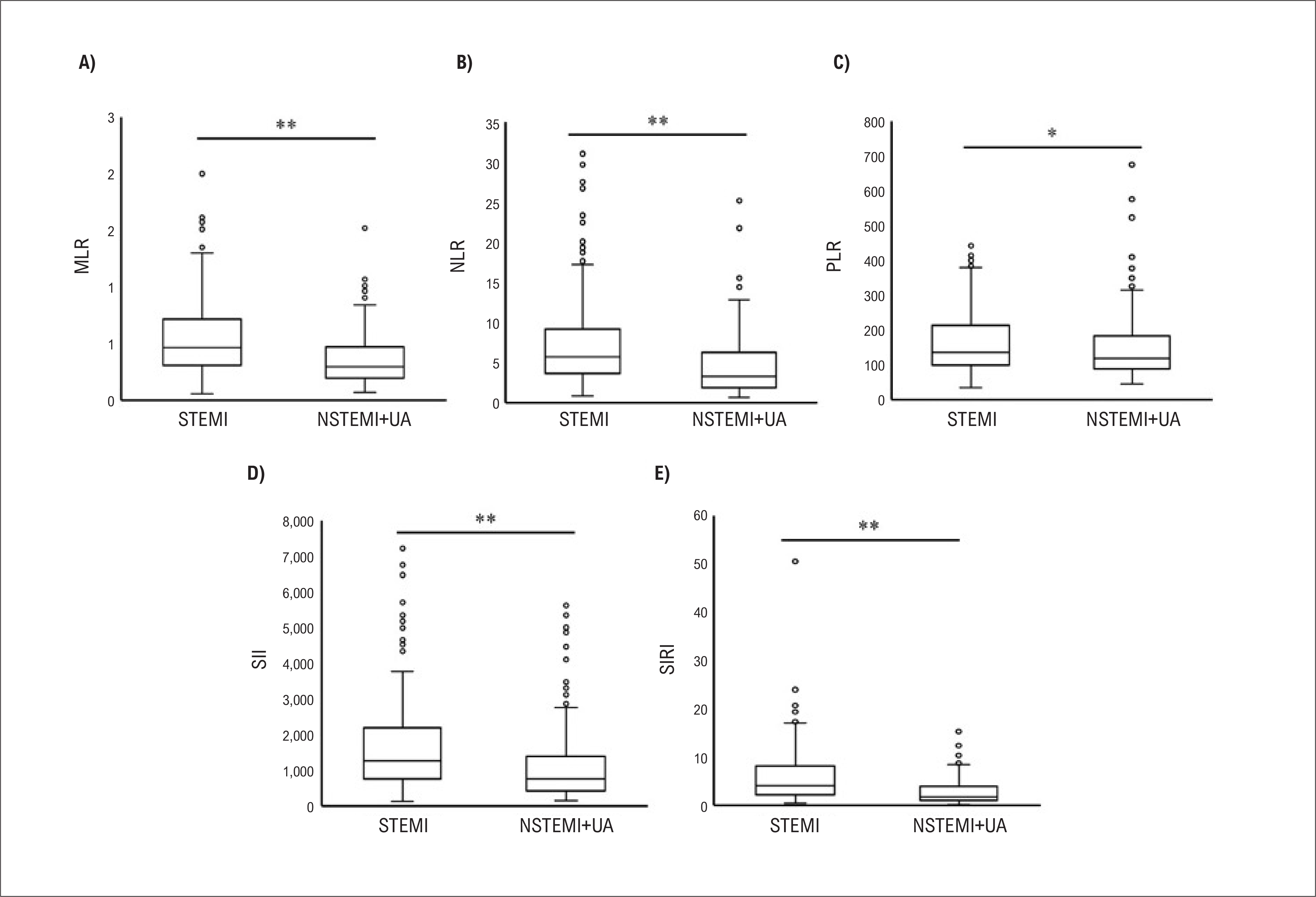
Analyses of inflammatory hematologic indices demonstrated significant differences between the groups. Patients in the STEMI group had significantly higher values of MLR (0.4 vs. 0.2), NLR (5.7 vs. 3.2), PLR (136 vs. 118), SII (1257 vs. 764), and SIRI (4.1 vs. 1.7) compared with those in the NSTEMI+UA group, reflecting higher levels of inflammation (Figure 1).

Inflammatory hematologic indices according to ACS diagnostic status. Source: Prepared by the authors. *p < 0.05; **p < 0.001. MLR: monocyte-to-lymphocyte ratio; NLR: neutrophil-to-lymphocyte ratio; NSTEMI: non-ST-segment elevation myocardial infarction; PLR: platelet-to-lymphocyte ratio; SII: systemic immune-inflammation index; SIRI: systemic inflammation response index; STEMI: ST-segment elevation myocardial infarction; UA: unstable angina.
Inflammatory hematologic indices were further compared according to the presence of cardiovascular risk factors, with analyses stratified by ACS phenotype. Among patients with STEMI, individuals with a history of smoking showed significantly lower values of MLR, NLR, SII, and SIRI compared with nonsmokers. In this group, SIRI values were also significantly lower in patients with hypercholesterolemia (Table 2). In the NSTEMI+UA group, patients with hypertension had significantly higher PLR values, whereas smokers had lower NLR values and a trend toward decreased SII (Table 3).
Comparison of inflammatory hematologic indices according to the presence of cardiovascular comorbidities in patients with STEMI
Comparison of inflammatory hematologic indices according to the presence of cardiovascular comorbidities in patients with NSTEMI+UA
In multivariable logistic regression models, higher values of SII and SIRI were independently associated with a greater likelihood of STEMI diagnosis after adjustment for age, sex, hypertension, hypercholesterolemia, DM, smoking history, and prior MI. In the model including SII, each unit increase in the lnSII was associated with a 2.27-fold higher odds of STEMI diagnosis (OR, 2.27; 95%CI, 1.66-3.10). Similarly, in the model including SIRI, each unit increase in the lnSIRI was associated with a 2.92-fold higher odds of STEMI diagnosis (OR, 2.92; 95%CI, 2.15-3.96). The full results of the adjusted models are presented in Table 4 and Table 5.
Discussion
In this ACS cohort, we identified a consistent association between the inflammatory hematologic indices SII and SIRI and the STEMI phenotype, even after adjustment for relevant clinical factors. These findings suggest that SII and SIRI could be used as prognostic markers in ACS. This reinforces the existing evidence for SII and adds to the support for the role of SIRI in CAD within a Brazilian cohort.
Patients with CAD are known to exhibit increased inflammatory markers, such as leukocyte count and high-sensitivity C-reactive protein (hs-CRP), which are associated with increased cardiovascular risk,8 impaired myocardial perfusion,9,10 atherosclerotic plaque instability,11 and mortality.12 These observations highlight the importance of monitoring and controlling inflammatory biomarkers as part of clinical assessment. Biomarkers derived primarily from three cellular lineages (i.e., N, L, and P) have been increasingly investigated as relevant prognostic markers. Among the most commonly used are the PLR and the NLR. Both NLR and PLR have been identified as strong independent predictors of major adverse cardiovascular events (MACE) in patients with STEMI.12 PLR has been reported as an effective predictor of severe atherosclerosis, whereas increased NLR has been associated with worse clinical outcomes in both ACS and stable CAD, particularly among patients undergoing percutaneous coronary intervention. In addition, NLR has been linked to the severity and complexity of CAD as assessed by the SYNTAX score.12
In the present study, patients with STEMI showed higher values of MLR, NLR, and PLR. Furthermore, when evaluating the inflammatory profile using SII and SIRI, indices that incorporate three distinct immune cell types and therefore reflect both immune response and inflammatory status, our findings reinforce the association of a more pronounced inflammatory state with the more severe ACS phenotype. These results support the use of low-cost, easily obtainable clinical markers with potential applicability in population-level settings.
In our analysis, nonsmoking patients with STEMI exhibited higher SII values. This association is likely more closely related to the STEMI phenotype itself than to smoking status prior to hospitalization. One possible explanation is that patients in the STEMI group were in a more pronounced inflammatory state due to the acute phase of MI, characterized by plaque instability and the release of proinflammatory mediators as well as the overall severity of the clinical condition.
Additionally, other paradoxical associations were observed, such as the presence of dyslipidemia being associated with lower SII and SIRI values. This finding may be related to the effects of lipid-lowering therapy prior to hospitalization, most commonly statin use. Lower SII values may reflect reduced systemic inflammation due to the anti-inflammatory effects of statins.13 Similarly, SIRI levels may also be influenced by statin therapy, given that both indices are derived from related cellular components.4
The anti-inflammatory effects of statin therapy are well established, including reductions in cardiovascular outcomes, as demonstrated in the JUPITER trial.14 Zhang et al. also conducted a meta-analysis evaluating C-reactive protein (CRP) concentrations across different types and doses of statins, showing beneficial effects, including long-term outcomes.15 The PRINCE study further demonstrated pravastatin 40 mg/day significantly decreased plasma levels of CRP, independently of any changes in LDL-C concentrations.16
Although statins are the most extensively studied agents, other lipid-lowering therapies have also been investigated and have shown similar effects. Morrone et al. reported greater CRP reduction with combination therapy using ezetimibe and statins compared with statin monotherapy.17 In the HOPE-3 trial, conducted in patients at intermediate cardiovascular risk, rosuvastatin decreased the levels of hs-CRP and MACE independently of lipid concentrations.18 Bempedoic acid, evaluated in the CLEAR Outcomes trial, reduced the levels of LDL-C and hs-CRP by approximately 20% in patients intolerant to statins.19 Lomitapide and mipomersen have also demonstrated potential anti-inflammatory effects in the setting of familial hypercholesterolemia.20 Overall, these findings support the observation of a lower inflammatory profile in patients with dyslipidemia, likely reflecting the effects of lipid-lowering therapies, predominantly statins.
Another relevant finding is the sex-based difference in ACS phenotypes. Women were more frequently associated with NSTEMI+UA (60.4% of women), whereas men were more frequently associated with STEMI (53.6% of men). CAD in women is more likely to present as less obstructive and more diffuse disease, with a greater contribution of microvascular and inflammatory components, and a higher likelihood of plaque disruption or instability compared with men.21
Attenuation of the inflammatory process is critical for the prevention of cardiovascular outcomes, particularly in higher-risk individuals22 Risk stratification using inflammatory biomarkers is therefore clinically relevant, as it enables early identification of patients with more severe disease.23,24 In this context, both SII and SIRI may allow early detection of the intensity of the inflammatory process, contributing to risk stratification and identification of patients with more severe ACS phenotypes. However, further investigation is needed, including larger, well-designed, randomized studies to better define the clinical utility of these indices.
In the present study, complete blood count parameters were collected within the first 24 h after hospital admission, reflecting the inflammatory profile of the early acute phase of ACS. The inflammatory response to ischemic injury is dynamic, with temporal variations in leukocyte subpopulations over the hours and days following symptom onset. The acute proinflammatory response is intense but transient, followed by a reparative anti-inflammatory phase associated with cardiac remodeling.25,26 Therefore, the timing of blood collection may influence the absolute values of hematologic inflammatory indices. However, the proinflammatory state appears to persist for up to 72 h after the event.27 Thus, sampling within the first 24 h captures the initial phase of inflammation. In addition, early sampling minimizes potential interference from therapies initiated during hospitalization, providing a more accurate representation of the initial inflammatory response associated with ACS phenotype. Although serial measurements of these indices over time may provide additional insights into the inflammatory trajectory, this aspect was not addressed in the present study and should be explored in future investigations.
Study limitations
We acknowledge several limitations of this study. Its retrospective and single-center design may limit generalizability. Data were obtained from hospital records, which restricted the availability of variables and the scope of analyses. The absence of clinical and laboratory outcome data limits the assessment of the predictive value of inflammatory indices for MACE. P, N, and L counts were measured only once at admission, with no serial follow-up during hospitalization.
In addition, some laboratory parameters, such as uric acid and hs-CRP, could not be evaluated due to the lack of available data. The individual impact of specific medications, including lipid-lowering therapies, could not be assessed despite their known influence on inflammatory status. The use of medical records may have introduced selection bias and led to the exclusion of patients with incomplete data, while also limiting sociodemographic diversity and reducing representativeness of the broader Brazilian population. Future studies, preferably prospective in design, are needed to further investigate these associations.
Conclusion
The inflammatory indices SII and SIRI were independently associated with the STEMI phenotype, suggesting their potential role as markers of inflammatory stratification in ACS. These findings support the use of hematologic inflammatory indices as potential tools for risk stratification during the acute phase of ACS. The observed association indicates a higher inflammatory burden and greater disease severity in patients with STEMI compared with those with NSTEMI+UA (Central Illustration). Early characterization of the inflammatory profile may be clinically relevant for risk stratification in this population.
-
Sources of Funding
There were no external funding sources for this study.
-
Study Association
This study is not associated with any thesis or dissertation work.
-
Ethics Approval and Consent to Participate
This study was approved by the Ethics Committee of the Santa Casa de Misericórdia de São Paulo under the protocol number 586426 228 00005476. All the procedures in this study were in accordance with the 1975 Helsinki Declaration, updated in 2013. Informed consent was obtained from all participants included in the study.
-
Use of Artificial Intelligence
The authors did not use any artificial intelligence tools in the development of this work.
Availability of Research Data
The underlying content of the research text is contained within the manuscript.
References
-
1 Nicolau JC, Feitosa GS Filho, Petriz JL, Furtado RHM, Précoma DB, Lemke W, et al. Brazilian Society of Cardiology Guidelines on Unstable Angina and Acute Myocardial Infarction without ST-Segment Elevation - 2021. Arq Bras Cardiol. 2021;117(1):181-264. doi: 10.36660/abc.20210180.
» https://doi.org/10.36660/abc.20210180 -
2 Thygesen K, Alpert JS, Jaffe AS, Simoons ML, Chaitman BR, White HD, et al. Third Universal Definition of Myocardial Infarction. Circulation. 2012;126(16):2020-35. doi: 10.1161/CIR.0b013e31826e1058.
» https://doi.org/10.1161/CIR.0b013e31826e1058 -
3 Libby P, Ridker PM, Hansson GK; Leducq Transatlantic Network on Atherothrombosis. Inflammation in Atherosclerosis: From Pathophysiology to Practice. J Am Coll Cardiol. 2009;54(23):2129-38. doi: 10.1016/j.jacc.2009.09.009.
» https://doi.org/10.1016/j.jacc.2009.09.009 -
4 Yang YL, Wu CH, Hsu PF, Chen SC, Huang SS, Chan WL, et al. Systemic Immune-Inflammation Index (SII) Predicted Clinical Outcome in Patients with Coronary Artery Disease. Eur J Clin Invest. 2020;50(5):e13230. doi: 10.1111/eci.13230.
» https://doi.org/10.1111/eci.13230 -
5 Xia Y, Xia C, Wu L, Li Z, Li H, Zhang J. Systemic Immune Inflammation Index (SII), System Inflammation Response Index (SIRI) and Risk of All-Cause Mortality and Cardiovascular Mortality: A 20-Year Follow-Up Cohort Study of 42,875 US Adults. J Clin Med. 2023;12(3):1128. doi: 10.3390/jcm12031128.
» https://doi.org/10.3390/jcm12031128 -
6 Horne BD, Anderson JL, John JM, Weaver A, Bair TL, Jensen KR, et al. Which White Blood Cell Subtypes Predict Increased Cardiovascular Risk? J Am Coll Cardiol. 2005;45(10):1638-43. doi: 10.1016/j.jacc.2005.02.054.
» https://doi.org/10.1016/j.jacc.2005.02.054 -
7 Sanchis-Gomar F, Perez-Quilis C, Leischik R, Lucia A. Epidemiology of Coronary Heart Disease and Acute Coronary Syndrome. Ann Transl Med. 2016;4(13):256. doi: 10.21037/atm.2016.06.33.
» https://doi.org/10.21037/atm.2016.06.33 -
8 Ye Z, Hu T, Wang J, Xiao R, Liao X, Liu M, et al. Systemic Immune-Inflammation Index as a Potential Biomarker of Cardiovascular Diseases: A Systematic Review and Meta-Analysis. Front Cardiovasc Med. 2022;9:933913. doi: 10.3389/fcvm.2022.933913.
» https://doi.org/10.3389/fcvm.2022.933913 -
9 Ikonomidis I, Michalakeas CA, Parissis J, Paraskevaidis I, Ntai K, Papadakis I, et al. Inflammatory Markers in Coronary Artery Disease. Biofactors. 2012;38(5):320-8. doi: 10.1002/biof.1024.
» https://doi.org/10.1002/biof.1024 -
10 Lemos JA, Morrow DA, Blazing MA, Jarolim P, Wiviott SD, Sabatine MS, et al. Serial Measurement of Monocyte Chemoattractant Protein-1 after Acute Coronary Syndromes: Results from the A to Z Trial. J Am Coll Cardiol. 2007;50(22):2117-24. doi: 10.1016/j.jacc.2007.06.057.
» https://doi.org/10.1016/j.jacc.2007.06.057 -
11 Núñez J, Núñez E, Bodí V, Sanchis J, Miñana G, Mainar L, et al. Usefulness of the Neutrophil to Lymphocyte Ratio in Predicting Long-Term Mortality in ST Segment Elevation Myocardial Infarction. Am J Cardiol. 2008;101(6):747-52. doi: 10.1016/j.amjcard.2007.11.004.
» https://doi.org/10.1016/j.amjcard.2007.11.004 -
12 Budzianowski J, Pieszko K, Burchardt P, Rzeźniczak J, Hiczkiewicz J. The Role of Hematological Indices in Patients with Acute Coronary Syndrome. Dis Markers. 2017;2017:3041565. doi: 10.1155/2017/3041565.
» https://doi.org/10.1155/2017/3041565 -
13 Proute MC, Kothur N, Georgiou P, Serhiyenia T, Shi W, Kerolos ME, et al. The Effect of Statin Therapy on Inflammatory Biomarkers: A Systematic Review. Cureus. 2021;13(9):e18273. doi: 10.7759/cureus.18273.
» https://doi.org/10.7759/cureus.18273 -
14 Ridker PM, Danielson E, Fonseca FA, Genest J, Gotto AM Jr, Kastelein JJ, et al. Rosuvastatin to Prevent Vascular Events in Men and Women with Elevated C-Reactive Protein. N Engl J Med. 2008;359(21):2195-207. doi: 10.1056/NEJMoa0807646.
» https://doi.org/10.1056/NEJMoa0807646 -
15 Zhang J, Wang X, Tian W, Wang T, Jia J, Lai R, et al. The Effect of Various Types and Doses of Statins on C-Reactive Protein Levels in Patients with Dyslipidemia or Coronary Heart Disease: A Systematic Review and Network Meta-Analysis. Front Cardiovasc Med. 2022;9:936817. doi: 10.3389/fcvm.2022.936817.
» https://doi.org/10.3389/fcvm.2022.936817 -
16 Albert MA, Staggers J, Chew P, Ridker PM; PRINCE Investigators. The Pravastatin Inflammation CRP Evaluation (PRINCE): Rationale and Design. Am Heart J. 2001;141(6):893-8. doi: 10.1067/mhj.2001.115297.
» https://doi.org/10.1067/mhj.2001.115297 -
17 Morrone D, Weintraub WS, Toth PP, Hanson ME, Lowe RS, Lin J, et al. Lipid-Altering Efficacy of Ezetimibe Plus Statin and Statin Monotherapy and Identification of Factors Associated with Treatment Response: A Pooled Analysis of Over 21,000 Subjects from 27 Clinical Trials. Atherosclerosis. 2012;223(2):251-61. doi: 10.1016/j.atherosclerosis.2012.02.016.
» https://doi.org/10.1016/j.atherosclerosis.2012.02.016 -
18 Katsiki N, Mikhailidis DP, Tsioufis C. The Heart Outcomes Prevention Evaluation (HOPE)-3 Trial: Where do We Stand? Curr Opin Lipidol. 2017;28(1):88-9. doi: 10.1097/MOL.0000000000000378.
» https://doi.org/10.1097/MOL.0000000000000378 -
19 Nissen SE, Lincoff AM, Brennan D, Ray KK, Mason D, Kastelein JJP, et al. Bempedoic Acid and Cardiovascular Outcomes in Statin-Intolerant Patients. N Engl J Med. 2023;388(15):1353-64. doi: 10.1056/NEJMoa2215024.
» https://doi.org/10.1056/NEJMoa2215024 -
20 Oliveira GMM, Almeida MCC, Arcelus CMA, Neto Espíndola L, Rivera MAM, Silva-Filho ALD, et al. Brazilian Guideline on Menopausal Cardiovascular Health - 2024. Arq Bras Cardiol. 2024;121(7):e20240478. doi: 10.36660/abc.20240478.
» https://doi.org/10.36660/abc.20240478 -
21 Weintraub WS, Daniels SR, Burke LE, Franklin BA, Goff DC Jr, Hayman LL, et al. Value of Primordial and Primary Prevention for Cardiovascular Disease: A Policy Statement from the American Heart Association. Circulation. 2011;124(8):967-90. doi: 10.1161/CIR.0b013e3182285a81.
» https://doi.org/10.1161/CIR.0b013e3182285a81 -
22 Montecucco F, Liberale L, Bonaventura A, Vecchiè A, Dallegri F, Carbone F. The Role of Inflammation in Cardiovascular Outcome. Curr Atheroscler Rep. 2017;19(3):11. doi: 10.1007/s11883-017-0646-1.
» https://doi.org/10.1007/s11883-017-0646-1 -
23 Möckel M, Danne O, Müller R, Vollert JO, Müller C, Lueders C, et al. Development of an Optimized Multimarker Strategy for Early Risk Assessment of Patients with Acute Coronary Syndromes. Clin Chim Acta. 2008;393(2):103-9. doi: 10.1016/j.cca.2008.03.022.
» https://doi.org/10.1016/j.cca.2008.03.022 -
24 Frangogiannis NG. The Inflammatory Response in Myocardial Injury, Repair, and Remodelling. Nat Rev Cardiol. 2014;11(5):255-65. doi: 10.1038/nrcardio.2014.28.
» https://doi.org/10.1038/nrcardio.2014.28 -
25 Brie DM, Mornoș C, Adam O, Tîrziu A, Popescu R, Brie AD. Inflammatory Mechanisms in Acute Coronary Syndromes: From Pathophysiology to Therapeutic Targets. Cells. 2025;15(1):72. doi: 10.3390/cells15010072.
» https://doi.org/10.3390/cells15010072 -
26 Ong SB, Hernández-Reséndiz S, Crespo-Avilan GE, Mukhametshina RT, Kwek XY, Cabrera-Fuentes HA, et al. Inflammation Following Acute Myocardial Infarction: Multiple Players, Dynamic Roles, and Novel Therapeutic Opportunities. Pharmacol Ther. 2018;186:73-87. doi: 10.1016/j.pharmthera.2018.01.001.
» https://doi.org/10.1016/j.pharmthera.2018.01.001 -
27 Ong SB, Hernández-Reséndiz S, Crespo-Avilan GE, Mukhametshina RT, Kwek XY, Cabrera-Fuentes HA, et al. Inflammation Following Acute Myocardial Infarction: Multiple Players, Dynamic Roles, and Novel Therapeutic Opportunities. Pharmacol Ther. 2018;186:73-87. doi: 10.1016/j.pharmthera.2018.01.001.
» https://doi.org/10.1016/j.pharmthera.2018.01.001
Edited by
-
Editor responsible for the review:
Glaucia Maria Moraes de Oliveira