 PET-CT in Infectious Endocarditis Diagnosis
Keywords
Endocarditis; Positron Emission Tomography Computed Tomography; endocarditis
Keywords
Endocarditis; Positron Emission Tomography Computed Tomography; endocarditis
Introduction
A 63-year-old male patient, with a history of mitral valve replacement with a bioprosthesis in 2019 due to native valve dysfunction secondary to bacterial endocarditis, and under regular medical follow-up since the procedure, presented to the cardiology emergency department with a complex clinical case. The patient reported a two-week history of fever (with temperature peaks of 38°C), night sweats, dry cough, weight loss, cervical pain when moving the neck, increased urinary frequency and urinary incontinence. At presentation, there were no classic cardiovascular symptoms, signs of valve dysfunction, or embolic phenomena. Notably, he had recently undergone a dental procedure without antibiotic prophylaxis. He denied symptoms such as rhinorrhea or pharyngitis. His physical and cardiovascular examinations were unremarkable.
Diagnostic Investigation
Initial laboratory results showed leukocytosis (11,100/µL) with lymphopenia (680/µL). Inflammatory markers were elevated, with an erythrocyte sedimentation rate of 116 mm/h and C-reactive protein level of 12 mg/L. The electrocardiogram revealed sinus rhythm with occasional extrasystoles and signs of left atrial overload.
A transthoracic echocardiogram showed thickening of the mitral bioprosthesis, which prompted additional imaging studies. Abdominal computed tomography (CT) suggested bilateral pyelonephritis, while thoracic CT showed no significant findings.
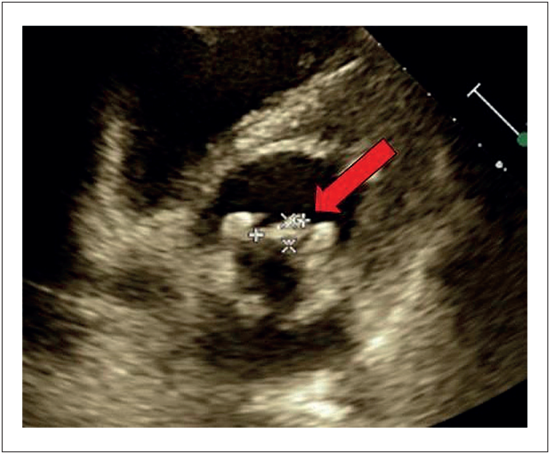
A transesophageal echocardiogram was subsequently performed, showing the biological prosthesis in the mitral position with good mobility of the leaflets, no significant valvular regurgitation or leak, and thickening of one leaflet located topographically close to the aorta, with an image suggestive of vegetation (Figure 1).
Transesophageal echocardiogram demonstrated a biological prosthesis with thickening of one of the leaflets located topographically close to the aorta, with an image suggestive of vegetation.
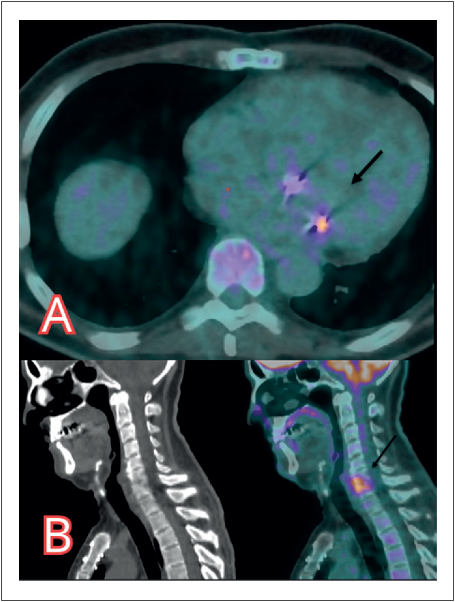
With the possibility of endocarditis raised, and according to the revised Duke Criteria (1 major criteria and 2 minor criteria), positron emission tomography combined with CT (PET-CT) was performed to confirm the suspected diagnosis and clarify the extent and location of the infection. The scan demonstrated increased glycolytic metabolism in the mitral bioprosthesis (SUV max 5.7), suggesting an infectious process (Figure 2A). Additionally, significant hypermetabolism was observed in the cervical spine at the C6-C7 level (SUV 7.2), suggestive of spondylodiscitis (Figure 2B). Subsequently, blood cultures were positive for Streptococcus sanguinis.

A: Positron emission tomography computed tomography –(PET-CT) demonstrates increased glycolytic metabolism (arrow) in the mitral bioprosthesis (SUV max 5.7). B: PET-CT demonstrates significant hypermetabolism in the cervical spine (arrow) at the C6-C7 level (SUV max 7.2)
Diagnosis
The patient was diagnosed with mitral endocarditis due to Streptococcus sanguinis, complicated by cervical spondylodiscitis, probably secondary to septic embolization.
Treatment and outcome
The patient was started on intravenous ceftriaxone. In light of the PET-CT findings, a conservative approach to cervical spondylodiscitis was preferred. Regular follow-up MRI of the cervical spine was planned to monitor treatment response. At the last evaluation, the patient reported feeling well, with rare episodes of neck pain and no neurological deficits.
Conclusion
This case highlights the fundamental role of PET-CT in diagnosing and confirming endocarditis, as well as in evaluating its extent, especially in patients with prosthetic valves, where traditional imaging can be inconclusive.1 The ability of PET-CT to detect associated complications, such as spondylodiscitis, further reinforces its value in guiding comprehensive treatment strategies.
PET-CT with fluorodeoxyglucose labeled with Fluorine-18 (F18-FDG) has become an important tool in the diagnosis and complementary evaluation of infectious endocarditis. The images are obtained through a combination of Positron Emission Tomography and Computed Tomography to provide detailed information on both metabolic function and anatomical structures. The patient receives an injection of F18-FDG, a radioactive tracer that accumulates in cells with high metabolic activity, such as those involved in infectious activity. PET images detect the radiation emitted by F18-FDG, highlighting areas of increased metabolic activity, while CT images provide precise anatomical details. The combination of these modalities allows for accurate localization and assessment of infectious foci.1,2
Advanced imaging techniques provide valuable additional information that can improve diagnostic accuracy, assess complications, and optimize treatment strategies. As a whole-body scan, PET-CT has the ability to detect distant embolic lesions in up to 35% of patients, which can be crucial for guiding treatment decisions. Furthermore, it is a useful tool for detecting the early development of periprosthetic complications and for identifying the extension of infection beyond the valve, thereby overcoming the technical limitations of echocardiographic imaging in suspected prosthetic valve endocarditis.1,2
In cases where the initial echocardiogram image is inconclusive, PET-CT becomes even more useful. The most recent guideline from the European Society of Cardiology recommends the use of PET-CT for the diagnostic evaluation of endocarditis in prosthetic valves with an inconclusive initial imaging (Class 1, level of evidence B). PET-CT demonstrates a sensitivity of 86% and a specificity of 84% for diagnosing endocarditis in patients with prosthetic valves.1,3
In patients with suspected native valve endocarditis, the sensitivity of PET-CT is lower (around 31%), due to the reduced incidence of paravalvular infectious involvement in native valve endocarditis, but it maintains its high specificity (up to 98%). In this context, a positive test confirms the diagnosis, but a negative test cannot exclude it. Despite its low sensitivity, PET-may still be considered, especially when septic embolism is suspected.3-6
In cases where septic embolism is suspected, PET-CT becomes particularly valuable, as it is a full-body examination. Its ability to detect inflammatory and infectious processes at distant sites is unparalleled. It is still again recommended by the latest guideline (Class 1, level of evidence B) and has also been classified as appropriate for diagnosing distant infections by the most recent expert consensus on PET-CT for cardiovascular infections. The organs most affected include the spleen, lungs, kidneys, intervertebral discs, joints, and liver, in both patients with native and prosthetic valves.1,2,7,8
The robustness and reproducibility of these results led to changes in the diagnostic Duke criteria for endocarditis in 2023, with a positive PET-CT scan now being considered a Major Criteria for diagnosing infective endocarditis.9
In this case the use of PET-CT not only confirmed the diagnosis of endocarditis but also identified a serious complication – cervical spondylodiscitis – which might otherwise have gone unnoticed. This underscores the importance of advanced imaging in the management of complex infectious diseases, particularly in patients with prosthetic devices. Early and accurate identification of infectious foci using advanced imaging techniques can significantly influence patient management and prognosis.
-
Sources of funding
There were no external funding sources for this study.
-
Study association
This study is not associated with any thesis or dissertation work.
-
Ethics approval and consent to participate
This study was approved by the Ethics Committee of the Hospital Moinhos de Vento - HMV under the protocol number 6.939.519. All the procedures in this study were in accordance with the 1975 Helsinki Declaration, updated in 2013. Informed consent was obtained from all participants included in the study.
-
Use of Artificial Intelligence
The authors did not use any artificial intelligence tools in the development of this work.
Data Availability Statement
The underlying content of the research text is contained within the manuscript.
References
-
1 Erba PA, Lancellotti P, Vilacosta I, Gaemperli O, Rouzet F, Hacker M, et al. Recommendations on Nuclear and Multimodality Imaging in IE and CIED Infections. Eur J Nucl Med Mol Imaging. 2018;45(10):1795-815. doi: 10.1007/s00259-018-4025-0.
» https://doi.org/10.1007/s00259-018-4025-0 -
2 Bourque JM, Birgersdotter-Green U, Bravo PE, Budde RPJ, Chen W, Chu VH, et al. 18F-FDG PET/CT and Radiolabeled Leukocyte SPECT/CT Imaging for the Evaluation of Cardiovascular Infection in the Multimodality Context: ASNC Imaging Indications (ASNC I2) Series Expert Consensus Recommendations from ASNC, AATS, ACC, AHA, ASE, EANM, HRS, IDSA, SCCT, SNMMI, and STS. J Nucl Cardiol. 2024;34:101786. doi: 10.1016/j.nuclcard.2023.101786.
» https://doi.org/10.1016/j.nuclcard.2023.101786 -
3 Wang TKM, Sánchez-Nadales A, Igbinomwanhia E, Cremer P, Griffin B, Xu B. Diagnosis of Infective Endocarditis by Subtype Using 18F-Fluorodeoxyglucose Positron Emission Tomography/Computed Tomography: A Contemporary Meta-Analysis. Circ Cardiovasc Imaging. 2020;13(6):e010600. doi: 10.1161/CIRCIMAGING.120.010600.
» https://doi.org/10.1161/CIRCIMAGING.120.010600 -
4 Duval X, Le Moing V, Tubiana S, Esposito-Farèse M, Ilic-Habensus E, Leclercq F, et al. Impact of Systematic Whole-Body 18F-Fluorodeoxyglucose PET/CT on the Management of Patients Suspected of Infective Endocarditis: The Prospective Multicenter TEPvENDO Study. Clin Infect Dis. 2021;73(3):393-403. doi: 10.1093/cid/ciaa666.
» https://doi.org/10.1093/cid/ciaa666 -
5 Albano D, Dondi F, Gazzilli M, Giubbini R, Bertagna F. Meta-Analysis of the Diagnostic Performance of 18F-FDG-PET/CT Imaging in Native Valve Endocarditis. JACC Cardiovasc Imaging. 2021;14(5):1063-5. doi: 10.1016/j.jcmg.2020.09.021.
» https://doi.org/10.1016/j.jcmg.2020.09.021 -
6 Camargo RA, Bitencourt MS, Meneghetti JC, Soares J, Gonçalves LFT, Buchpiguel CA, et al. The Role of 18F-Fluorodeoxyglucose Positron Emission Tomography/Computed Tomography in the Diagnosis of Left-Sided Endocarditis: Native vs Prosthetic Valves Endocarditis. Clin Infect Dis. 2020;70(4):583-94. doi: 10.1093/cid/ciz267.
» https://doi.org/10.1093/cid/ciz267 -
7 Mikail N, Benali K, Mahida B, Vigne J, Hyafil F, Rouzet F, et al. 18F-FDG-PET/CT Imaging to Diagnose Septic Emboli and Mycotic Aneurysms in Patients with Endocarditis and Cardiac Device Infections. Curr Cardiol Rep. 2018;20(3):14. doi: 10.1007/s11886-018-0956-0.
» https://doi.org/10.1007/s11886-018-0956-0 -
8 Kouijzer IJ, Bleeker-Rovers CP, Oyen WJ. 18F-FDG PET/CT for the Detection of Septic Embolisms in Patients with Infectious Endocarditis. J Nucl Med. 2014;55(7):1045-6. doi: 10.2967/jnumed.114.140707.
» https://doi.org/10.2967/jnumed.114.140707 -
9 Fowler VG, Durack DT, Selton-Suty C, Athan E, Bayer AS, Chamis AL, et al. The 2023 Duke-International Society for Cardiovascular Infectious Diseases Criteria for Infective Endocarditis: Updating the Modified Duke Criteria. Clin Infect Dis. 2023;77(4):518-26. doi: 10.1093/cid/ciad271.
» https://doi.org/10.1093/cid/ciad271
Edited by
-
Editor responsible for the review:
Ana Carolina do Amaral Henrique de Souza