 Cerebral Thromboembolism as a Primary Manifestation of Severe Rheumatic Mitral Stenosis: A Clinical Case from Guayaquil, Ecuador
Abstract
Mitral stenosis (MS) is a valvular disease that predominantly affects women, with rheumatic fever being the most prevalent etiological cause. The hemodynamic consequences of rheumatic MS may include left atrial (LA) pressure overload and dilation, atrial fibrillation (AF), secondary pulmonary hypertension, heart failure, atrial thrombosis, and systemic thromboembolism. Echocardiography is the gold standard for diagnosing, quantifying severity, and evaluating the hemodynamic impact of MS.
We present the case of a 41-year-old female patient who arrived at the emergency department with a 6-hour history of dysarthria and right hemiparesis. Cardiovascular examination revealed sinus rhythm and a diastolic murmur with an opening snap at the mitral focus. Imaging studies revealed severe MS with LA dilation and a patent foramen ovale without hemodynamic impact. The patient underwent percutaneous balloon mitral valvuloplasty, which resulted in an increase in mitral valve area (MVA), a reduction in pulmonary pressure, and significant functional improvement.
Cerebral thromboembolism can be the first manifestation of severe MS, even in patients with sinus rhythm. Anticoagulation is crucial for those with severe atrial dilation to reduce the risk of embolic events. This case highlights the importance of early diagnosis and appropriate treatment based on clinical guidelines, as they improve patient quality of life and reduce the risk of future complications. It also serves as a reminder that stroke may be the sentinel event in silent but severe mitral valve pathology.
Keywords
Mitral Valve Stenosis; Rheumatic Heart Disease; Ischemic Stroke
Introduction
Mitral stenosis (MS) is a valvular heart disease, which is predominant in female patients, with rheumatic fever being the most common underlying cause.1 This pathology leads to fusion of the commissures, leaflets, and chordae tendineae, resulting in restricted and altered rapid and biphasic motion of the mitral valve. As a result, an obstruction of blood flow from the left atrium to the left ventricle (LV) occurs. The hemodynamic consequences of MS may include left atrial (LA) pressure overload and dilation, atrial fibrillation (AF), secondary pulmonary hypertension, heart failure, atrial thrombosis, and systemic thromboembolism.2 Early diagnosis is essential, and echocardiography is the gold standard for diagnosis, severity quantification, and evaluation of the hemodynamic impact of MS. The Wilkins score is a tool used to establish the viability of performing percutaneous mitral balloon valvuloplasty (PMBV) instead of surgical valve replacement, based on the anatomical characteristics of the rheumatic valve damage. In patients with a Wilkins score of 8 or less, PMBV has a better prognosis with favorable outcomes.3-6
This article presents a clinical case in which cerebral thromboembolism was the first manifestation of severe MS without documented AF, highlighting the importance of early diagnosis and appropriate treatment.
Case presentation
We present the case of a 41-year-old female patient who presented to the emergency department with a clinical history of 6 hours of dysarthria and right brachiocrural hemiparesis. The cardiovascular exam revealed sinus rhythm and a diastolic murmur with an opening snap at the mitral focus. The neurological exam showed a confused verbal response, Glasgow 14/15, right-sided weakness and muscle tone with mild right hemiparesis, right Babinski sign, and hypoesthesia.
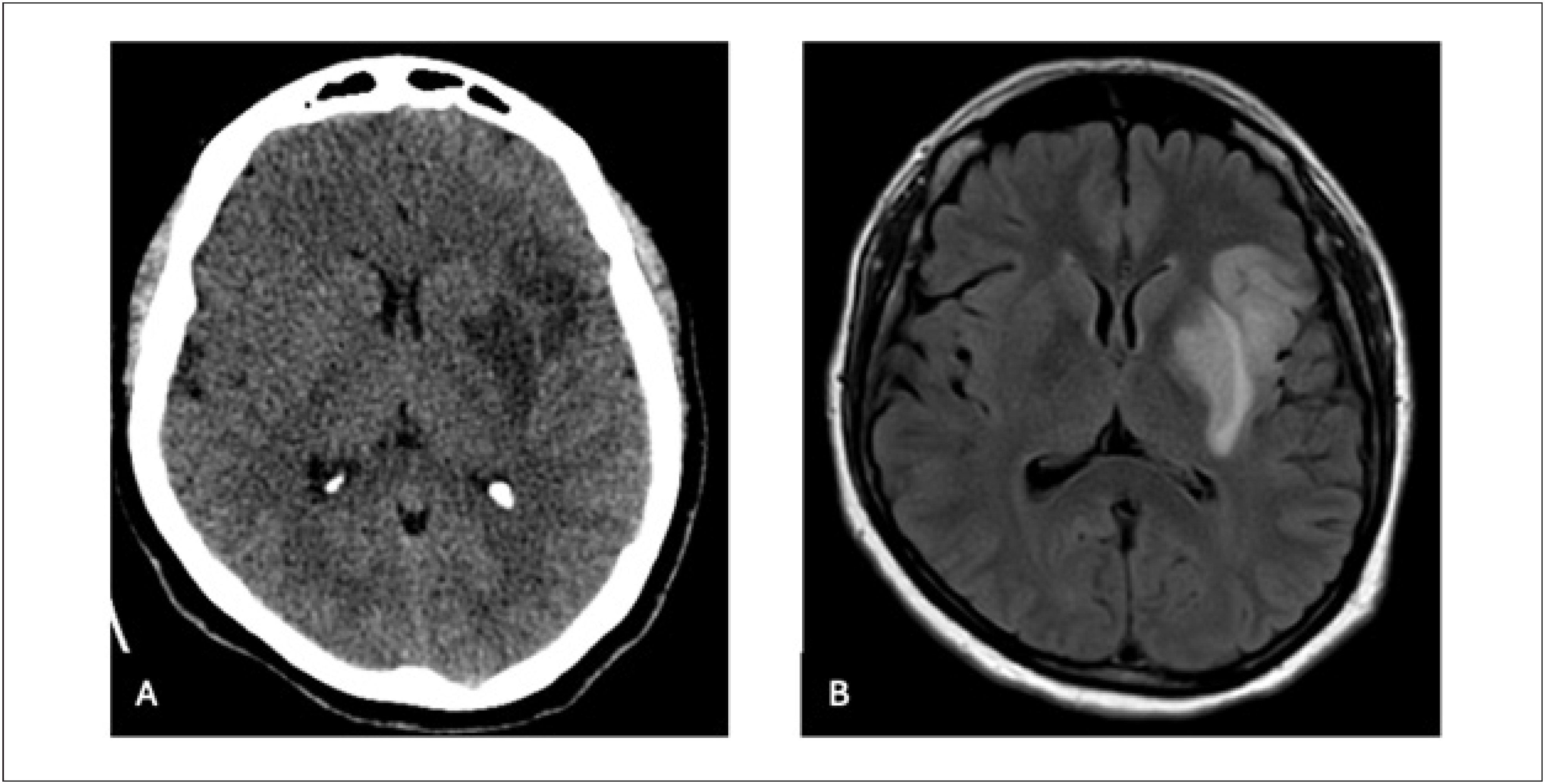
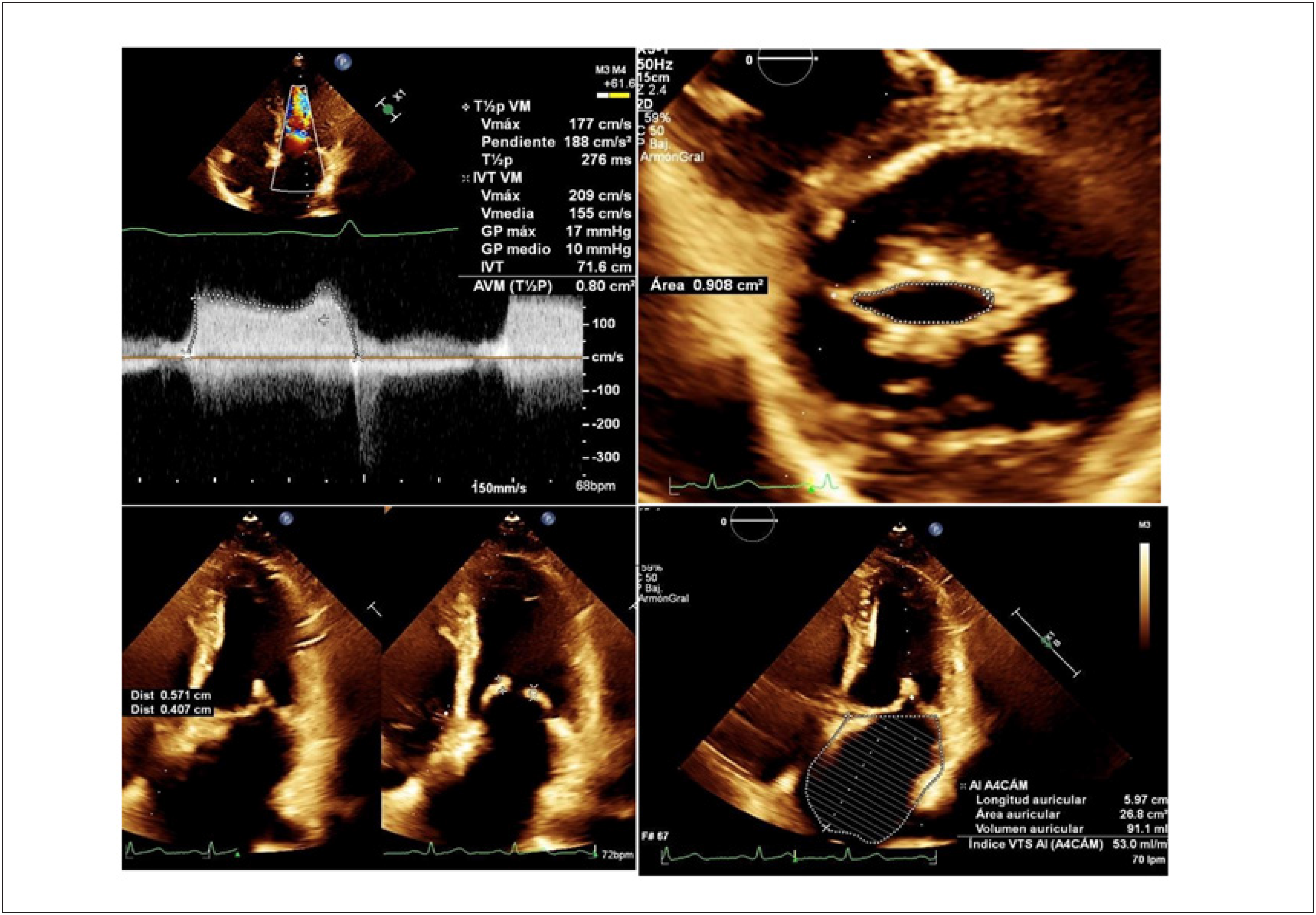
Given the clinical presentation and cardiovascular exam findings, MS and cardioembolic thromboembolism were suspected, and non-contrast head computed tomography was performed, revealing hypodensity in the basal ganglia and insular cortex in the territory of the middle cerebral artery and left lenticulostriate perforating arteries, indicating an ischemic cerebrovascular event in the territory of the middle cerebral artery (Figure 1). A transthoracic echocardiogram (TTE) showed inactive rheumatic heart disease (RHD) with double mitral valve lesions, predominantly severe MS with mild mitral regurgitation (Wilkins score 7), severe LA dilation of 54 mm with no thrombi, normal systolic function, prolonged diastasis due to extreme bradycardia down to 33 bpm, normal filling pressures, and pulmonary systolic pressure of 37 mmHg (Figure 2).

Non-contrast brain computed tomography reveals an ischemic stroke in the middle cerebral artery territory (Image A). Non-contrast brain magnetic resonance imaging in FLAIR sequence confirms a hyperacute ischemic stroke (Image B).

TTE with mitral Doppler shows a pressure half-time of 276 ms and an estimated valve area of 0.8 cm² (Image A). Planimetry measures a valve area of 0.9 cm² (Image B). Thickened mitral valve with rheumatic changes (Image C). Severely dilated left atrium with a volume of 91 ml (Image D).
The patient was started on prophylactic enoxaparin. After 72 hours, there was significant recovery of right-side mobility and normalization of speech, with mild bradyphrenia remaining. The patient denied any clinical history suggestive of rheumatic fever during childhood, although she did recall a previous episode of tonsillitis. She also reported no use of oral contraceptives at any time. A preoperative evaluation for ovarian cyst removal in May 2021 revealed a mitral murmur, leading to a recommendation for an echocardiogram that was not performed. During hospitalization, a transesophageal echocardiogram (TEE) was performed, confirming LA enlargement without thrombi (54 mm, volume 57 ml/m²), patent foramen ovale without hemodynamic impact, inactive RHD with double mitral valve lesions, predominantly severe MS with a mitral valve area (MVA) of 0.9 cm² by planimetry, anterior leaflet thickness of 4.8 mm, posterior leaflet thickness of 4.4 mm, and restricted diastolic motion of the valve, with the posterior leaflet showing restricted dynamics (Wilkins score 8). In the esophageal plane at 90 degrees, an auricle with a hockey stick morphology was observed, with low transappendageal systolic velocities of 12 cm/s. Color Doppler of the mitral valve showed evidence of turbulent flow with a filling velocity of 229 cm/s, A-wave velocity of 123 cm/s, and deceleration time of 231 ms. The patient was discharged on warfarin and bisoprolol for rhythm control.
Upon resuming routine physical activity, the patient reported limitations with moderate exertion (New York Heart Association [NYHA] class II/IV). Neurology recommended a waiting period of 3 to 5 months before performing PMBV. Prior to the procedure, a new TTE was performed, showing an MVA of 0.54 cm² and posterior thickness of 0.44 cm, with severely restricted diastolic motion, and MVA by planimetry of 0.89 cm². The Wilkins score was 7. Additionally, patent foramen oval was seen without right-to-left saline passage, normal systolic and diastolic function (type 1), and pulmonary systolic pressure of 29 mmHg.
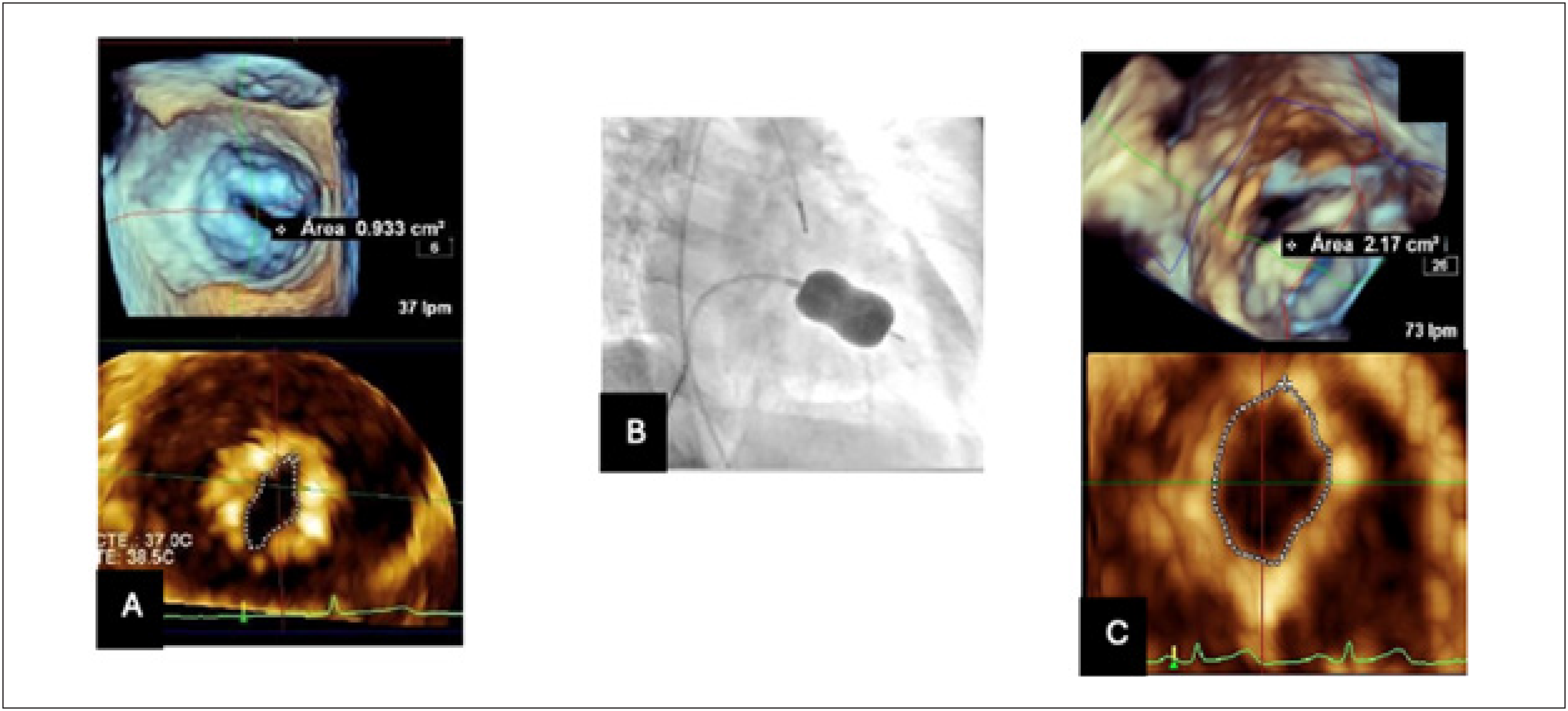
The patient underwent PMBV, using an Inoue balloon inflated to 28 mmHg. The result was MVA of 2.17 cm², with partial rupture of the anterolateral and posteromedial commissures. Post-intervention echocardiographic control revealed normal LV mass, normal systolic function, and type 1 diastolic function, with pulmonary systolic pressure of 23 mmHg (Figure 3). One month after the intervention, the patient attended a follow-up visit, reporting functional improvement (NYHA class I/IV). Subsequently, the patient resumed dance therapy and normal physical and professional activities.
Three-dimensional transesophageal echocardiography shows the MVA before valvuloplasty: 0.9 cm² (Image A). Ballon inflation during PMBV (Image B). Postprocedural 3-dimensional transesophageal echocardiography demonstrates an increased MVA of 2.17 cm², with significant improvement (Image C).
Discussion
MS, generally of rheumatic origin, is a progressive disease with significant hemodynamic and clinical implications. Its prevalence is lower in developed countries, but it remains high in resource-limited and developing countries, where incidence rates can exceed 100 cases per 100,000 population, contrasting sharply with less than 0.5 per 100,000 in high-income nations.6,7 Each year, approximately 500,000 new cases of acute rheumatic fever are diagnosed globally, leading to an estimated 230,000 deaths. One of the most severe complications of rheumatic fever is RHD, a major cause of cardiovascular mortality worldwide. RHD is characterized by progressive valvular damage, particularly affecting the mitral valve, which can lead to congestive heart failure, pulmonary hypertension, AF, stroke, systemic embolism, and ultimately premature death. The severity of valvular dysfunction correlates strongly with mortality.7-10
MS is more common in women, accounting for about 70% to 80% of cases. Diagnosis is usually made in early adulthood, around the age of 30. The initial diagnostic test of choice for assessing the severity of stenosis is TTE, whereas TEE is useful for excluding LA thrombi and for comprehensive evaluation of the mitral valve apparatus (chordae and commissures). The 2020 American College of Cardiology guidelines classify MS into four stages: Stage A: mitral valve prolapse without hemodynamic impact, Stage B: progressive MS with early valve changes, Stage C: severe asymptomatic MS, and Stage D: severe symptomatic MS.2,5
Severe MS is defined when MVA is ≤ 1.5 cm², corresponding to a mean transmitral gradient of 5 to 10 mmHg at normal heart rates. In symptomatic patients with NYHA class II/IV or Stage D disease, PMBV also known as percutaneous mitral commissurotomy, is a recommended therapeutic option. Long-term oral anticoagulation in patients with rheumatic MS and sinus rhythm remains controversial.5-6
However, emerging evidence suggests that the thromboembolic risk in patients with rheumatic MS in sinus rhythm may be underestimated. Several observational studies have shown an annual embolic event rate ranging from 1% to 2% in this population. Even in the absence of documented AF, LA stasis resulting from severe dilation or mechanical dysfunction can predispose to thrombus formation. Paroxysmal AF may go undetected, and phenomena such as "atrial stunning" or mechanical silence further compound the risk. Recurrence of stroke has also been reported in non-anticoagulated patients or those with subtherapeutic international normalized ratio control.5,11 Although AF is a well-known risk factor for thromboembolism in MS, this is thought to be related to LA stasis resulting from severe dilation, mechanical dysfunction, and atrial fibrosis, which may impair contractility and promote thrombus formations.11,12 In addition, paroxysmal AF may go undetected, and conditions such as "atrial stunning" or mechanical atrial silence have also been associated with thromboembolic events in this populations. Therefore, although routine anticoagulation in sinus rhythm remains debated, its use is supported in patients with a prior embolic event or marked atrial enlargement, particularly following PBMV. In such cases, structural and functional atrial remodeling may take weeks to months, necessitating continued anticoagulation despite mechanical improvement of valvular obstruction.11,12
Another parameter that determines the risk of thrombus formation on TEE is the blood flow velocity through the left atrial appendage (LAA) during systole, known as trans appendageal systolic velocity. In the setting of arrhythmia, this measurement is evaluated to determine or estimate the risk of thrombosis. A peak systolic velocity > 40 cm/s is considered normal and suggests good contractility of the LAA. On the other hand, a peak systolic velocity < 20 cm/s is associated with a high risk of thrombosis due to stagnant flow, which predisposes to thrombus formation within the LAA. An intermediate velocity range between 20 and 40 cm/s is not classified as moderate risk per se, but in such cases, the presence of thrombi must be ruled out.13
Management guidelines from the European Society of Cardiology recommend oral anticoagulation with a vitamin K antagonist in patients with AF, as most trials exclude patients with rheumatic valve disease. Anticoagulation is also recommended for patients with moderate to severe MS in sinus rhythm, a history of systemic embolism, LA thrombi, or significant LA dilation (diameter > 50 mm or volume > 60 ml/m²).12,14,15 The INVICTUS study showed that a vitamin K antagonist may offer better efficacy in reducing cardiovascular events compared to rivaroxaban in patients with AF in the context of RHD. In the case presented, the patient had severe LA dilation (54 mm) and an embolic event, which justified the use of warfarin as anticoagulant therapy. This approach follows current recommendations to reduce thromboembolic risk, given the limited evidence available in patients with rheumatic MS.14-16
It is also essential to verify previous diagnosis of rheumatic fever as the underlying etiology of MS, through a combination of clinical history (e.g., migratory arthritis, carditis, fever, family history) and laboratory markers such as elevated antistreptolysin O titers or a record of penicillin prophylaxis. In this patient, although no formal diagnosis of rheumatic fever was made, a childhood history of recurrent tonsillitis raises the possibility of subclinical or undiagnosed streptococcal pharyngitis as a trigger. Furthermore, she reported no use of oral contraceptives, which excludes an additional prothrombotic factor. This is relevant, as oral contraceptives may further increase thrombotic risk, particularly when combined with other risk factors such as valvular heart disease, immobility, or inherited thrombophilia.17
Conclusion
MS remains a globally relevant valvular disease, with a notable predominance in women and a persistent burden in low-resource settings. This case illustrates how a cerebral thromboembolic event may be the first clinical manifestation of severe rheumatic MS, even in the absence of AF, highlighting the importance of high clinical suspicion and early echocardiographic evaluation. It also reinforces the role of integrated, multimodal assessment combining imaging and clinical risk stratification tools such as the Wilkins score to guide timely therapeutic decisions. PMBV proved to be a safe and effective strategy, significantly improving MVA, reducing pulmonary pressures, and restoring functional capacity. In addition, the choice of warfarin over novel oral anticoagulants followed evidence-based recommendations from international guidelines and was justified by the presence of marked LA dilation and embolic risk.
The use of vitamin K antagonists in patients with MS is controversial; however, in this case, their recommendation was based on three substantial and relevant reasons. First, the patient had a history of a prior embolic event. Second, there was a high risk of thrombosis, as evidenced by a decreased transappendageal flow velocity of < 40 cm/s on TEE. Additionally, the presence of a patent foramen ovale was identified as an associated comorbidity.
This case underscores the need for personalized, guideline-driven management of RHD and serves as a reminder that stroke may be the sentinel event in silent but severe mitral valve pathology.
-
Sources of Funding
There were no external funding sources for this study.
-
Study Association
This study is not associated with any thesis or dissertation work.
-
Ethics Approval and Consent to Participate
This study was approved by the Ethics Committee of the Universidad Internacional del Ecuador (UIDE) under the protocol number EX_01_2025_UIDE_PD. All the procedures in this study were in accordance with the 1975 Helsinki Declaration, updated in 2013. Informed consent was obtained from all participants included in the study.
-
Use of Artificial Intelligence
The authors did not use any artificial intelligence tools in the development of this work.
Availability of Research Data
The underlying content of the research text is contained within the manuscript.
References
-
1 Soliman OI, Anwar AM, Metawei AK, McGhie JS, Geleijnse ML, Ten Cate FJ. New Scores for the Assessment of Mitral Stenosis Using Real-Time Three-Dimensional Echocardiography. Curr Cardiovasc Imaging Rep. 2011;4(5):370-7. doi: 10.1007/s12410-011-9099-z.
» https://doi.org/10.1007/s12410-011-9099-z -
2 Carabello BA. Modern Management of Mitral Stenosis. Circulation. 2005;112(3):432-7. doi: 10.1161/CIRCULATIONAHA.104.532498.
» https://doi.org/10.1161/CIRCULATIONAHA.104.532498 -
3 Prendergast BD, Shaw TR, Iung B, Vahanian A, Northridge DB. Contemporary Criteria for the Selection of Patients for Percutaneous Balloon Mitral Valvuloplasty. Heart. 2002;87(5):401-4. doi: 10.1136/heart.87.5.401.
» https://doi.org/10.1136/heart.87.5.401 -
4 Wilkins GT, Weyman AE, Abascal VM, Block PC, Palacios IF. Percutaneous Balloon Dilatation of the Mitral Valve: An Analysis of Echocardiographic Variables Related to Outcome and the Mechanism of Dilatation. Br Heart J. 1988;60(4):299-308. doi: 10.1136/hrt.60.4.299.
» https://doi.org/10.1136/hrt.60.4.299 -
5 Otto CM, Nishimura RA, Bonow RO, Carabello BA, Erwin JP 3rd, Gentile F, et al. 2020 ACC/AHA Guideline for the Management of Patients with Valvular Heart Disease: Executive Summary: A Report of the American College of Cardiology/American Heart Association Joint Committee on Clinical Practice Guidelines. Circulation. 2021;143(5):e35-e71. doi: 10.1161/CIR.0000000000000932.
» https://doi.org/10.1161/CIR.0000000000000932 -
6 Coyle M, McGreal A, McCormick J, Galvin O, McInerney A, Holmes A, et al. Acute Rheumatic Fever in a Low Prevalence Setting. JACC Case Rep. 2025;30(5):103178. doi: 10.1016/j.jaccas.2024.103178.
» https://doi.org/10.1016/j.jaccas.2024.103178 -
7 Artola CB, Solorzano NG, Matamoros DO. Fiebre Reumática: Actualización de los Criterios Diagnósticos. Rev Medica Sinerg. 2020;5(2):e357. doi: 10.31434/rms.v5i2.357.
» https://doi.org/10.31434/rms.v5i2.357 -
8 Benger J, Constantin I, Carrero MC, Makhoul S, Baratta S, Cintora F, et al. Argentine Registry of Valvular Diseases (ARGENVAL). Rev Argent Cardiol. 2021;89(4):290-7. doi: 10.7775/rac.es.v89.i4.10418.
» https://doi.org/10.7775/rac.es.v89.i4.10418 -
9 Meneses-Silvera K, Castro-Monsalve J, Flórez-Rodríguez C, Donis I, Crespo SEM. Rheumatic Heart Disease: Are we Really Doing Enough? Rev Colomb Cardiol. 2020;27(3):189-92. doi: 10.1016/j.rccar.2019.10.003.
» https://doi.org/10.1016/j.rccar.2019.10.003 -
10 Rwebembera J, Beaton AZ, de Loizaga SR, Rocha RTL, Doreen N, Ssinabulya I, et al. The Global Impact of Rheumatic Heart Disease. Curr Cardiol Rep. 2021;23(11):160. doi: 10.1007/s11886-021-01592-2.
» https://doi.org/10.1007/s11886-021-01592-2 - 11 Kumar D, Bhat S. Incidence of Left Atrial Thrombus in Patients with Rheumatic Mitral Stenosis in Sinus Rhythm & to Find out the Factor Responsible for Clot Formation : A Transesophageal Echocardiographic Study. JK Science. 2017;19(1):13-6.
-
12 Vahanian A, Beyersdorf F, Praz F, Milojevic M, Baldus S, Bauersachs J, et al. 2021 ESC/EACTS Guidelines for the Management of Valvular Heart Disease. Eur Heart J. 2022;43(7):561-632. doi: 10.1093/eurheartj/ehab395.
» https://doi.org/10.1093/eurheartj/ehab395 -
13 Lang RM, Badano LP, Mor-Avi V, Afilalo J, Armstrong A, Ernande L, et al. Recommendations for Cardiac Chamber Quantification by Echocardiography in Adults: An Update from the American Society of Echocardiography and the European Association of Cardiovascular Imaging. J Am Soc Echocardiogr. 2015;28(1):1-39.e14. doi: 10.1016/j.echo.2014.10.003.
» https://doi.org/10.1016/j.echo.2014.10.003 -
14 Connolly SJ, Karthikeyan G, Ntsekhe M, Haileamlak A, El Sayed A, El Ghamrawy A, et al. Rivaroxaban in Rheumatic Heart Disease-Associated Atrial Fibrillation. N Engl J Med. 2022;387(11):978-88. doi: 10.1056/NEJMoa2209051.
» https://doi.org/10.1056/NEJMoa2209051 -
15 Roberts WC, Virmani R. Aschoff Bodies at Necropsy in Valvular Heart Disease. Evidence from an Analysis of 543 Patients Over 14 Years of Age that Rheumatic Heart Disease, at Least Anatomically, is a Disease of the Mitral Valve. Circulation. 1978;57(4):803-7. doi: 10.1161/01.cir.57.4.803.
» https://doi.org/10.1161/01.cir.57.4.803 -
16 Connolly SJ, Karthikeyan G, Ntsekhe M, Haileamlak A, El Sayed A, El Ghamrawy A, et al. Rivaroxaban in Rheumatic Heart Disease-Associated Atrial Fibrillation. N Engl J Med. 2022;387(11):978-88. doi: 10.1056/NEJMoa2209051.
» https://doi.org/10.1056/NEJMoa2209051 - 17 Shah SN, Sharma S. Mitral Stenosis. Treasure Island: StatPearls Publishing; 2025.
Edited by
-
Editor responsible for the review:
Glaucia Maria Moraes de Oliveira